. . . .
. . . .
T-cadherin attenuates insulin-dependent
signalling, eNOS activation, and angiogenesis
in vascular endothelial cells
Maria Philippova
1†, Manjunath B. Joshi
1†, Dennis Pfaff
1, Emmanouil Kyriakakis
1,
Kseniya Maslova
1, Paul Erne
2, and The´re`se J. Resink
1*
1
Laboratory for Signal Transduction, Department of Biomedicine, Basel University Hospital, Hebelstrasse 20, CH 4031 Basel, Switzerland; and2
Division of Cardiology, Kantonsspital Luzern, Luzern, Switzerland
Received 17 May 2011; revised 4 January 2012; accepted 5 January 2012; online publish-ahead-of-print 10 January 2012
Time for primary review: 33 days
Aims T-cadherin (T-cad) is a glycosylphosphatidylinositol-anchored cadherin family member. Experimental, clinical, and genomic studies suggest a role for T-cad in vascular disorders such as atherosclerosis and hypertension, which are associated with endothelial dysfunction and insulin resistance (InsRes). In endothelial cells (EC), T-cad and insulin ac-tivate similar signalling pathways [e.g. PI3-kinase (PI3K)/Akt/mammalian target of rapamycin (mTOR)] and processes (e.g. angiogenesis). We hypothesize that T-cad is a regulatory component of insulin signalling in EC and therefore a determinant of the development of endothelial InsRes.
Methods and results
We investigated T-cad-dependent effects on insulin sensitivity using human EC stably transduced with respect to T-cad overexpression or T-cad silencing. Responsiveness to insulin was examined at the level of effectors of the insulin signalling cascade, EC nitric oxide synthase (eNOS) activation, and angiogenic behaviour. Overexpression and ligation of T-cad on EC attenuates insulin-dependent activation of the PI3K/Akt/mTOR signalling axis, eNOS, EC migration, and angiogenesis. Conversely, T-cad silencing enhances these actions of insulin. Attenuation of EC re-sponsiveness to insulin results from T-cad-mediated chronic activation of the Akt/mTOR-dependent negative feed-back loop of the insulin cascade and enhanced degradation of the insulin receptor (IR) substrate. Co-immunoprecipitation experiments revealed an association between T-cad and IR. Filipin abrogated inhibitory effects of T-cad on insulin signalling, demonstrating localization of T-cad-insulin cross-talk to lipid raft plasma mem-brane domains. Hyperinsulinaemia up-regulates T-cad mRNA and protein levels in EC.
Conclusion T-cad expression modulates signalling and functional responses of EC to insulin. We have identified a novel signalling mechanism regulating insulin function in the endothelium and attribute a role for T-cad up-regulation in the patho-genesis of endothelial InsRes.
-Keywords T-cadherin † Endothelial cell † Insulin resistance † Signal transduction † Angiogenic behaviour
1. Introduction
Insulin resistance (InsRes) is typically defined as a decreased sensitiv-ity/responsiveness to the metabolic actions of insulin that promote glucose disposal in traditional target tissues (muscle, liver, and adipose tissue) and is a hallmark of metabolic disorders including type 2 diabetes and obesity. Recognition of the pathophysiological
importance of InsRes in non-traditional target tissues such as the endothelium is more recent. Associated micro- and macrovascular complications of metabolic disorders (e.g. retinopathy, nephropathy, hypertension, atherosclerosis, coronary artery disease) are preceded by a state of endothelial dysfunction (ED) which is characterized by impaired nitric oxide (NO) bioavailability and vasorelaxation. Diverse molecular and cellular reciprocal relationships between
†These authors contributed equally to this work.
*Corresponding author. Tel:+41 612652422; fax: +41 612652350, Email: [email protected]
InsRes and ED in metabolic and vascular tissues are considered to govern the frequent association between metabolic and cardiovascu-lar disorders.1–5
Because endothelial cells (EC) do not possess the insulin-stimulated glucose carrier GLUT4, insulin stimulation of the cognate insulin receptor (IR) in EC has physiological consequences that do not directly involve glucose intake. Insulin induces pleiotrop-ic responses in the endothelium. It promotes vasorelaxation and ca-pillary recruitment in peripheral tissues5 and is also a potent pro-angiogenic molecule regulating neovascularization, EC migration, and wound healing.6 These functions are mediated by a signalling cascade involving IR substrate-1 (IRS-1), PI3-kinase (PI3K), Akt, endothelial NO synthase (eNOS), and NO generation. Additionally, insulin has vasoconstrictor and growth promoting functions that are mediated through a signal cascade involving Ras, Raf, MAPK/extracel-lular signal-related kinases and endothelin-1 synthesis and secretion. Pathological outcomes of endothelial InsRes are complex and include ED, impaired vasodilation, microvessel disease, enhanced vascular inflammation, atherosclerosis, and hypertension.3 How insulin signalling in the endothelium becomes impaired remains unclear.
A striking feature of InsRes is that insulin activation of PI3K/Akt signalling branch is selectively impaired.5,7However, also chronic ac-tivation of Akt signalling by hyperinsulinaemia or other factors may contribute to the development of InsRes. Chronic insulin exposure results in serine phosphorylation and degradation of IRS-1 and IRS-2 with consequential down-regulation of insulin signalling in the muscle, liver, and adipocytes.8,9 Insulin-induced glucose disposal in these systems may be rescued by inhibition of mammalian target of rapamycin (mTOR),8,9 while mTOR activation acutely inhibits insulin-dependent Akt phosphorylation and glucose transport in human adipocytes.10 In the heart, chronic Akt activation increases basal glucose uptake but inhibits responses to insulin.11Thus, hyper-activation of the Akt/mTOR-dependent negative feedback pathway of insulin signalling can render a state of insulin insensitivity/ resistance.
T-cadherin (T-cad), an atypical glycosylphosphatidylinositol (GPI)-anchored member of the cadherin superfamily, is gaining rec-ognition as a regulator of EC function.12 T-cad expression is increased in vivo in human atherosclerotic lesions13and experimental restenosis,14 and in vitro on proliferating EC and smooth muscle cells15 and on EC during oxidative and endoplasmic reticulum stress.16,17 Overexpression and ligation of T-cad activate the PI3K/ Akt/mTOR signalling pathway and promote proliferation and angio-genesis.16,18Elevation of T-cad in human plasma correlates with clin-ical progression of atherosclerosis and ED.19 Notably, in EC, T-cad and insulin stimulate common signalling pathways (PI3K/Akt/ mTOR) and control similar (patho)physiological processes (angio-genesis and ED). Additionally, T-cad acts as a heterophilic receptor for adiponectin, an adipokine that regulates glucose and fatty acid metabolism and mimics some effects of insulin.20T-cad plays a crit-ical role in binding of adiponectin to the vascular wall and mediates its cardioprotective functions.21–23 These data prompted us to hypothesize a cross-talk between insulin and T-cad signalling. We demonstrate that T-cad overexpression in EC attenuates insulin-induced activation of Akt pathway with concomitantly reduced insulin-stimulated eNOS activation, migration, and angiogen-esis, suggesting a role for T-cad in the pathogenesis of endothelial InsRes and ED.
2. Methods
2.1 Cells and lentivector transduction
The investigation conforms with the principles outlined in the Declaration of Helsinki for the use of human tissues. Human microvascular EC line HMEC-124was stably transduced with respect to T-cad overexpression or T-cad-silencing (siTcad) and respective empty vector- (E) or non-target shRNA (siC) controls using lentiviral vectors.25 Full-length human c-myc-tagged T-cad was excised from adenovector26 and cloned into pLVX-puro lentivector for stable transduction of HMEC-1. Transient overexpression of T-cad in primary EC cultures from the human umbilical vein (HUVEC; Promocell GmbH, Allschwil, Switzerland) and human aorta (HAEC; Promocell) was achieved using adenovector-mediated transfec-tion procedures.27The application of viral vector-mediated transfection was approved by the Swiss Federal Office for the Environment. Supple-mentary material online provides details on vectors and culture condi-tions, data on T-cad protein and transcript expression levels in the transductants (see Supplementary material online, Figure S1), and proof of surface localization of both endogenous and overexpressed T-cad protein in HMEC-1 (see Supplementary material online, Figure S2).
2.2 Isolation of microparticles from
cultured EC
Procedures for harvesting microparticles (MP) from tumour necrosis factor-a (TNFa)-stimulated E and Tcad transductants (respectively, MP-E and MP-Tcad) and the preparation of controls to exclude any trace of foetal calf serum (FCS) or cytokine have been described before19and are detailed in Supplementary material online. Briefly, the medium was collected after culture in the presence of TNFa (50 ng/mL, 48 h) and clarified from dead cells and cell fragments (centrifugation at 3000 g, 15 min, 48C). Thereafter, MP were pelleted by ultracentrifugation (100 000 g, 1 h, 48C), washed twice in phosphate-buffered saline (PBS), resuspended in PBS, and used for stimulation of target EC cultures.
2.3 Cell activation and immunoblotting
Subconfluent cultures were incubated in low serum-containing medium [DMEM/0.1% bovine serum albumin (BSA)/0.5% FCS] for 3 h before stimulation with insulin or MP. Whole-cell lysates were prepared and ana-lysed by immunoblotting as described.16Primary antibodies against follow-ing proteins/epitopes were used: T-cad (R&D Systems Europe Ltd, Abingdon, UK), Akt, phospho (p)-AktSer473, p-AktThr308, mTOR, p- mTORSer2448, S6RP, p-S6RPSer240/244, p70S6K, p-p70S6KThr389, IRS-1, p-IRS-1Ser636/639, p-eNOSSer1177, insulin receptor-b (IRb) (Cell Signaling, New England Biolabs GMBH, Frankfurt, Germany), c-myc (Clontech/ Takara Bio Europe, Saint-Germain-en-Laye, France), GAPDH (Abcam, Cambridge, UK), and p-Tyr clone 4G10 (Millipore AG, Zug, Switzerland). Secondary horseradish peroxidase-conjugated anti-species IgG were from Southern Biotechnology (BioReba AG, Reinach, Switzerland).
2.4 Immunoprecipitation
HMEC-1 transduced with native T-cad or c-myc-tagged T-cad were serum-deprived (3 h) in Cloneticsw
Endothelial Cell Growth Medium (ECGM) containing 1% BSA/0.5% FCS, then stimulated with insulin. Immu-noprecipitation (IP) was performed26 with two different lysis buffers: Triton X-114 buffer [Tris – HCl 50 mM (pH 8.0), 100 mM NaCl, 5 mM CaCl2, 0.2% SDS, 1% Triton X-114] or NP-40 buffer [150 mM NaCl,
1% NP-40, and 50 mM Tris – HCl, pH 8.0]. Buffers were supplemented with Complete Mini protease inhibitor cocktail. Anti-c-myc or anti-IRb subunit antibodies were used for IP, with subsequent detection of T-cad and IRb by immunoblotting. IP for IRS-1 was performed using NP-40 buffer and anti-IRS-1 antibodies, with subsequent immunoblot analysis for IRS-1 and p-Tyr. Total amount of IgG in the pellets served as an intern-al loading control.
2.5 Wound assay and time-lapse
videomicroscopy
Assay for migration by time-lapse videomicroscopy of wound healing was performed as detailed before.24Confluent cultures were scrape-wounded and serum-deprived (3 h) in DMEM/1% FCS/0.1% BSA and then exposed to insulin. Wound closure was filmed (48 h, 1 frame/h) under an Olympus IX-81 inverted time-lapse videomicroscope (Olympus Optical Co., Geneva, Switzerland). Acquired images were processed and analysed for distance of cell migration into the wound area using CellRsoftware (Soft Imaging System GmbH, Muenster, Germany). Experiments con-tained two parallel wells for every experimental condition, and in each well, three different fields of observation at the initial wound front (time 0) were randomly selected.
2.6 Endothelial tube-formation assay
The EC spheroid assay in 3D fibrin gels was performed as detailed18 except that spheroids (500 cells/spheroid) were prepared using the ‘hanging drop’ method28and gels were overlaid with ECGM supplemen-ted with 2% FCS without or with inclusion of 100 nM insulin and signalling inhibitors. After 24 h incubation, spheroids were stained with TRITC-conjugated phalloidin and sprout outgrowth was analysed morphometric-ally (CellRsoftware).18
2.7 Real-time polymerase chain reaction
Subconfluent cultures were serum-deprived (3 h) in DMEM/0.5% FCS/ 0.1% BSA before insulin stimulation. T-cad transcript expression was mea-sured by real-time polymerase chain reaction.29Primer details are given in Supplementary material online.
2.8 Statistical analysis
Unless otherwise stated, experiments were performed on at least three independent occasions and results are given as mean + SD. Differences were determined using one-way repeated-measures ANOVA with Tukey’s multiple comparison using GraphPad Prism 5.0 software (Graph-Pad Software, San Diego, CA, USA). A P-value of ,0.05 was considered significant.
3. Results
3.1 T-cad attenuates activation
of Akt/mTOR pathway by insulin
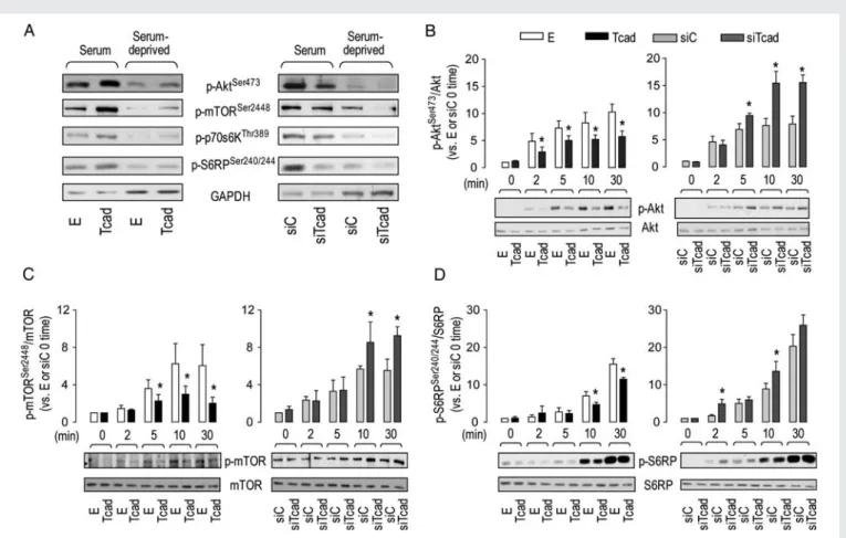
PI3K/Akt/mTOR signalling pathway is one of the main targets of both T-cad and insulin in EC (see pathway depiction in Figure 6). To investigate whether T-cad modulates signalling responses to insulin, we compared Tcad and siTcad HMEC-1 with respect to baseline and insulin-stimulated phosphorylation of key signalling effectors within the Akt/mTOR cascade. Under baseline serum-containing or serum-deprivation culture conditions, levels of p-AktSer473, p-mTORSer2448, p-p70S6KThr389, and p-S6RPSer240/244 were higher in Tcad (vs. E) and lower in siTcad (vs. siC); as expected, phosphorylation levels of all effectors were reduced under serum-deprivation conditions (Figure1A). These data agree with our previous findings in HUVEC.16 Insulin (100 nM) induced a rapid phosphorylation of Akt on Ser473which was attenuated in Tcad and enhanced in siTcad cells (Figure 1B). Accordingly, T-cad overexpression attenuated and silencing promoted insulin-induced phosphorylation of mTOR, an immediate downstream target of Akt (Figure1C), and of S6RP (Figure1D) and p70S6 kinase (see Sup-plementary material online, Figure S3A) which are downstream targets of mTOR. Insulin-induced phosphorylation of Akt on
Thr308was attenuated in Tcad cells, but not significantly modulated by T-cad silencing (see Supplementary material online, Figure S3B). The differential insulin responsiveness between Tcad and siTcad at the level of p-AktSer473was evident also at lower insulin concentra-tions (1 and 10 nM; see Supplementary material online, Figure S4). Attenuating effects of T-cad overexpression on insulin-stimulated phosphorylation of Akt on Ser473 were true too for primary HUVEC and HAEC cultures [see Supplementary material online, Figures S4 (dose) and S5 (kinetics)].
3.2 T-cad expression modulates
phosphorylation and total protein
levels of IRS-1
A crucial initiating event in the insulin-dependent signalling cascade is IR-induced phosphorylation of IRS-1 on Tyr1101 which recruits the regulatory p85 subunit of PI3K, enabling downstream activation of the Akt signalling axis.30 Attenuation of insulin signalling is achieved by a negative feedback loop involving Akt-dependent stimulation of mTOR complex with Raptor (mTORC1) and the ac-tivation of S6K1 kinase which phosphorylates IRS-1 on Ser636/639, causing IRS-1 dissociation from p85, inactivation, and degradation.31 To determine whether T-cad modulates IRS-1 expression and/or activity, we analysed total and p-IRS-1 levels in transduced HMEC-1. Levels of tyrosine phosphorylated IRS-1, analysed by IP of total IRS-1 protein from insulin-stimulated cells followed by im-munoblotting with anti-p-Tyr antibodies, were reduced in Tcad but increased in siTcad relative to total IRS-1 (Figure2A). Phosphoryl-ation of IRS-1Ser636/639was measured by direct immunoblotting of lysates from untreated and insulin-treated cells and found to be ele-vated in Tcad and decreased in siTcad, respectively (Figure2B, upper histogram). Importantly, both methods also revealed a reduction in levels of total IRS-1 protein in Tcad and an increase in siTcad cells under basal and insulin-stimulated conditions (Figure 2B, lower histogram).
3.3 Tcad causes PI3K- and
mTOR-dependent degradation
of IRS-1 protein
To investigate whether T-cad modulates the IRS-1 protein level by affecting its stability, we analysed kinetics of IRS-1 degradation in transduced HMEC-1 cultured under normal conditions after treat-ment with cycloheximide, which prevents protein synthesis de novo. In the presence of cycloheximide, the decline in IRS-1 expression oc-curred more rapidly in Tcad but was unaffected in siTcad cells (Figure2C). Inclusion of PI3K inhibitor LY-294002 or mTOR inhibitor rapamycin under normal culture conditions rescued IRS-1 levels in Tcad cells (Figure2D). Thus, IRS-1 degradation induced by T-cad over-expression is dependent on PI3K and mTOR activation.
3.4 T-cad ligation activates insulin
signalling pathway
Our previous investigations have demonstrated that T-cad-dependent responses (including Akt phosphorylation) may be triggered by either T-cad up-regulation on the cell surface (presumably via enhanced clustering, lateral cis-ligation and recruitment adapter molecules) or trans-ligation (via homophilic binding to T-cad molecules on neigh-bouring cells or endothelial-derived MP, with recombinant T-cad protein or agonistic antibodies).16,19,27 Here, we investigated
whether T-cad ligation affects insulin signalling. Exposure of parental HMEC-1 to MP harvested from E or Tcad HMEC-1 transductants caused tyrosine phosphorylation of IRS-1 (Figure 3A) as well as rapid and reversible elevation of p-IRS-1Ser636/639 relative to total IRS-1 expression (Figure3B), and these responses were more prom-inent for MP-Tcad. While levels of total IRS-1 in parental HMEC-1 remained unchanged during short-term (up to 30 min) exposure to MP (Figure3B), they were reduced following longer term (4 h) incuba-tion with MP, this effect being more prominent in the case of MP-Tcad (Figure3C).
3.5 T-cad interacts with IR
In addition to the ability of T-cad to regulate insulin signalling pathway by cross-talk with signalling networks downstream of IR, direct inter-actions between IR and T-cad in the plasma membrane might repre-sent an alternative mode of regulation. To explore the latter possibility, we performed co-IP experiments. Two different lysis buffers were used: standard NP-40 buffer and a raft-solubilizing Triton X-114 buffer that has been successfully used for identification of molecules specifically associating with GPI-anchored proteins.26 Using either of these extraction approaches and anti-IRb antibodies for IP of lysates from Tcad cells, we found that T-cad co-precipitated
with IR (Figure4A). Due to lack of commercially available anti-T-cad antibodies suitable for IP, we performed reverse co-IP using HMEC-1 expressing T-cad protein with c-myc tag and c-myc anti-body.26 This approach demonstrated co-precipitation of IR with T-cad, confirming physical association of the two proteins in the plasma membrane (Figure4B).
3.6 T-cad effects on insulin signalling
depend on lipid raft localization
To assess the importance of plasma membrane lipid raft domains for a cross-talk between T-cad and insulin signalling, we measured insulin-induced Akt phosphorylation after pre-treating EC with filipin, a cholesterol-binding compound that disrupts lipid rafts. Filipin (3 mg/mL) inhibited insulin-dependent activation of AktSer473 phosphorylation in Tcad, E, and siC transductants but not in the siTcad cells (Figure 4C). This suggests that the presence of T-cad and its interaction with IR is needed for localization of IR to lipid rafts, whereas siTcad may enable redistribution of IR away from this compartment and thereby render the pathway insensitive to lipid raft disruption.
Figure 1 T-cad modulates activation of Akt/mTOR pathway by insulin. (A) Immunoblots showing phosphorylation status of Akt/mTOR signalling cascade components in HMEC-1 under normal culture conditions or following serum deprivation (whole-cell lysates loaded at 5 and 10 mg, respect-ively). Insulin (100 nm)-stimulated transductants were analysed by immunoblotting for total and phosphorylated AktSer473(B), mTOR (C ), and S6RP (D). Changes in phosphorylated/total effector ratios in Tcad and siTcad are expressed relative to those in their respective E and siC 0 time controls. *P at least ,0.05 for Tcad vs. E or siTcad vs. siC. Total effector levels did not differ between the transductants and remained constant regardless of the experimental condition (i.e. serum-containing, serum-deprivation insulin stimulation).
3.7 T-cad overexpression attenuates
and silencing promotes insulin-induced
phosphorylation of eNOS, cell migration,
and angiogenesis in vitro
Tyrosine phosphorylation of IRS-1 triggers PI3K-dependent activation of Akt which in its turn directly phosphorylates eNOS on Ser1177 leading to NO production and vasorelaxation.32 Therefore, we ana-lysed whether T-cad-dependent modulation of insulin signalling also changes eNOS activity. Insulin-induced phosphorylation of eNOS was decreased in Tcad and enhanced in siTcad cells (Figure5A). Inter-estingly, under basal conditions, siTcad exhibited a moderate but re-producible down-regulation of p-eNOSSer1177levels when compared with siC, yielding a greatly augmented insulin response (calculated as increase in p-eNOSSer1177/eNOS vs. siC 0 time).
Insulin-dependent stimulation of cell migration is an important com-ponent of wound healing and angiogenesis. We studied the role for T-cad in modulation of insulin effects on EC migration by measuring wound closure rates using time-lapse videomicroscopy. As demon-strated previously,27 T-cad overexpression per se increased and T-cad silencing decreased migration of EC into the wound area
under basal non-stimulated conditions (basal data as obtained in this study are not shown). However, insulin-dependent migration was less in Tcad cells and more pronounced in siTcad cells (Figure5B).
Stimulation of the PI3K/Akt pathway in EC by insulin results in ac-tivation of angiogenesis. We measured the influence of T-cad expres-sion on insulin-induced angiogenesis using the 3D-spheroid in vitro assay. Insulin increased (vs. unstimulated) total sprout outgrowth from E and siC spheroids (Figure 5C). Insulin-induced angiogenesis was abrogated in Tcad spheroids and enhanced in siTcad spheroids (Figure 5C). Inclusion of either rapamycin or LY-294002 normalized effects of T-cad on insulin-induced sprout outgrowth (Figure 5D), demonstrating involvement of PI3K and mTOR in T-cad-dependent modulation of the proangiogenic actions of insulin.
3.8 Insulin up-regulates T-cad expression in
EC via reactive oxygen species-dependent
mechanism
To determine whether insulin might modulate T-cad expression, we treated parental HMEC-1 with 100 nM insulin. Insulin induced a time-dependent increase in T-cad protein and mRNA (see Supplementary Figure 2 T-cad modulates phosphorylation and total protein levels of IRS-1. (A) Following insulin stimulation (100 nM, 15 min), IRS-1 was immu-noprecipitated using anti-total IRS-1 antibody and Tyr phosphorylation determined by immunoblotting with anti-p-Tyr antibody. (B) Immunoblot ana-lysis for p-IRS-1Ser636/639and total IRS-1 after insulin (100 nM) stimulation. Ratios of total IRS-1/GAPDH or p-IRS-1Ser636/639/total IRS-1 in Tcad and siTcad are expressed relative to those in their respective unstimulated E and siC controls. *P at least ,0.05 for Tcad vs. E or siTcad vs. siC. (C ) Immunoblot analysis for total IRS-1 following incubation with 5 mg/mL cycloheximide under normal culture conditions. IRS-1 is expressed relative to levels (arbitrarily taken as 100%) in the absence of cycloheximide. *P at least ,0.05 for Tcad vs. E, siTcad, or siC. (D) E and Tcad were incubated (6 h) without (Ctrl) or with rapamycin (Rapa, 10 nM) or LY-294002 (LY, 20 mM). IRS-1, p-AktSer473, and p-S6RPSer240/244were measured (inhibited phosphorylation of AktSer473and S6RPSer240/244validates the efficacy of LY-294002 and rapamycin, respectively). IRS-1 is expressed relative to the level
material online, Figure S6). Inclusion of reactive oxygen species (ROS) scavenger N-acetylcysteine inhibited insulin-induced up-regulation of T-cad transcription (see Supplementary material online, Figure S6).
4. Discussion
This study has identified a novel mechanism for the regulation of insulin sensitivity in the endothelium. Levels of T-cad expression in EC profoundly modulate insulin responsiveness as manifested by altered insulin-induced stimulation of PI3K/Akt/mTOR signalling and accordingly altered vasorelaxant, promigratory, and proangiogenic actions of insulin. Up-regulation of T-cad promotes InsRes, while down-regulation of T-cad favours insulin sensitivity.
The most obvious explanation for T-cad-associated promotion of InsRes in EC is that T-cad overexpression causes chronic activation of the PI3K/Akt pathway,16–18resulting in IRS-1 degradation and con-sequential EC insensitivity to insulin stimulation. To understand the sequence of events leading to T-cad-dependent InsRes in EC, one must distinguish between effects of T-cad on components of the insulin cascade in either the absence (basal) or the presence of
insulin. Our previous investigations in EC demonstrated that T-cad overexpression and ligation increase AktSer473and GSK3b phosphor-ylation and nuclear translocation of b-catenin in a PI3K/mTOR-dependent manner to promote proliferation, survival, and angiogen-esis.16–18 Here, and in accordance with the basal status of Akt/ mTOR signalling in Tcad (enhanced) and siTcad (decreased) cells (Figure1A), in the absence of insulin, Tcad exhibited faster degradation rates and reduced levels of IRS-1, while siTcad displayed some stabil-ization of IRS-1 protein (Figure2). Consistent with IRS-1 being an es-sential component of insulin signalling,30,31Tcad (with pre-degraded IRS-1) responded poorly to insulin, whereas siTcad (with constitutive-ly higher IRS-1) exhibited increased responsiveness. This differential was manifest with respect to PI3K/Akt/mTOR signalling (Figure 1), IRS-1 phosphorylation on tyrosine and serine residues (Figure 2), eNOS phosphorylation, migration, and sprouting (Figure5).
How T-cad activates basal Akt signalling and the negative feedback loop in the insulin signalling cascade is unclear. GPI-anchorage of T-cad implies a requirement for transmembrane adaptors that interact with T-cad on the outer plasma membrane surface and enable signal transmission to cytoplasmic downstream targets. We previously Figure 3 T-cad ligation affects insulin signalling. Parental HMEC-1 was stimulated or not (Ctrl) with MP collected from Tcad (MP-Tcad) or E (MP-E) HMEC-1. (A) Tyr phosphorylation of IRS-1 after 15 min stimulation was determined by IRS-1 IP and immunoblotting with anti-p-Tyr antibody. (B) p-IRS-1Ser636/639and total IRS-1 were measured by immunoblot analysis of whole-cell lysates after up to 30 exposure to MP. Levels of total IRS-1 did not change. Ratios of p-IRS-1Ser636/639/total IRS-1 are expressed relative to that of Ctrl/0 time. *P at least ,0.05 for MP-Tcad vs. MP-E. (C ) Whole-cell lysates were probed for total IRS-1 levels by immunoblotting after 4 h stimulation.
demonstrated lipid raft domain localization of T-cad in EC and smooth muscle cells33 and identified several membrane partners, including Grp78/BiP, integrin-linked kinase, and integrin b3.26,34The first two participate in T-cad-dependent activation of Akt.26,34Thus, one ex-planation for the ability of T-cad to impact insulin signalling is that its adaptor recruitment activates signalling responses (e.g. Akt activa-tion) that converge with the insulin-IR-dependent pathway at the level of common intracellular targets. Another possibility is that T-cad increases basal Akt activity via direct activation of IR. Like T-cad, IR is present in plasma membrane lipid raft domains.33,35Correlations between lipid membrane composition, membrane viscosity, and IR
activity suggest some link between lipid raft localization of IR and InsRes.35 Our IP experiments demonstrated that T-cad and IR co-precipitate, and abrogation of inhibitory effects of T-cad on the insulin cascade by filipin supports lipid raft localization of insulin-T-cad cross-talk (Figure 4). Functional relevance of this interaction is sup-ported through the use of EC-derived MP (which harbour T-cad) to mimic surface homophilic ligation.19EC-derived MP stimulate Akt activation and angiogenic behaviour in target EC19 and, as shown herein, also stimulate tyrosine and serine phosphorylation of IRS-1, with amplification of these responses for Tcad cell-derived MP (Figure 3). Both T-cad/IR co-precipitation and effects of T-cad Figure 4 T-cad interacts with IR. (A) IR was immunoprecipitated (IP) from Tcad transductants using anti-IRb antibody or non-immune IgG (IP n/i). Two different lysis buffers containing NP-40 or Triton X-114 were used. (B) T-cad was IP from HMEC-1 transduced with c-myc-tagged-T-cad protein after incubation without or with insulin (100 nM, 15 min) using anti-c-myc antibody or n/i IgG. (A and B) The presence of IR and T-cad in immuno-precipitates was checked by immunoblotting (WB). (C ) Filipin-pre-treated (30 min, 3 mg/mL) cells were stimulated with insulin (100 nM, 15 min) and immunoblotted for p-AktSer473. Changes in p-AktSer473levels in E or Tcad and siC or siTcad are expressed relative to levels in E and siC controls, respectively. *P at least ,0.05 between filipin-treated cells vs. the same non-treated cell type.
harbouring MP on insulin-related signal pathway activity occur even in the absence of insulin, suggesting that T-cad per se can utilize compo-nents of the insulin signalling cascade to exert its effects on EC.
Insulin-like growth factor-1 receptor (IGF-1R) is structurally similar to IR and can also bind insulin, albeit at≈100 times lower affinity.36 Additionally, insulin can also bind with low affinity to hybrid receptors composed of subunits from the different receptor types, although it has been questioned whether signalling of insulin through hybrid receptors is physiologically relevant.37 We have not yet addressed whether T-cad effects on insulin signal pathway activity in EC also involve interaction with IGF-1R. However, T-cad expression in EC clearly affects insulin-induced Akt pathway activation at doses relevant for both IR (1 – 10 nM) and IGF-1R (100 nM).
This study also offers new insight on roles for T-cad during condi-tions associated with EC activation/dysfunction. Previous publicacondi-tions demonstrated beneficial prosurvival functions for T-cad in vascular EC.16,17 Since basal T-cad-dependent Akt activation promotes cell survival during oxidative and endoplasmic reticulum stress16,17 and facilitates angiogenesis,18,27 T-cad-up-regulation on vascular cells in atherosclerosis and restenosis was interpreted as a protective and re-generative cell response to unfavourable proinflammatory and dam-aging conditions. Positive effects of T-cad-up-regulation in the vessel
are also linked to actions of adiponectin, identified as a heterophilic ligand for T-cad.23 Adiponectin mimics some effects of insulin and, in EC, through adiponectin receptors AdipoR1/AdipoR2, stimulates NO production and angiogenesis by promoting a cross-talk between AMPK and Akt. Since T-cad expression is critical for adipo-nectin interactions with the vessel and its cardioprotective effects during ischaemic injury,21,22 one might expect T-cad up-regulation to promote beneficial metabolic and protective influences of adipo-nectin in the cardiovasculature. The role for T-cad per se in regulation of vascular tone is unknown.
This study provides the first direct demonstration of an important deleterious consequence of T-cad up-regulation in EC, namely pro-gression of endothelial InsRes. Moreover, our data may offer a mech-anistic link between elevation of T-cad in the vessel and blood and progression of cardiovascular disease. T-cad up-regulation on acti-vated EC at early disease stages is likely due to thioredoxin-mediated modulation of T-cad gene expression29 and aims at overriding damaging effects of oxidative stress caused by inflammatory events within the vessel. Based on our current finding that unstimu-lated T-cad-silenced cells exhibit slightly lower p-eNOS levels, it cannot be excluded that T-cad, via Akt pathway signalling, might par-ticipate in maintenance of basal eNOS activity/vasorelaxation. Yet, Figure 5 T-cad regulates functional effects of insulin on eNOS activation, cell motility, and angiogenesis. (A) Insulin (100 nM)-stimulated transduc-tants were analysed by immunoblotting for total and p-eNOS. Total eNOS levels were neither different between the transductransduc-tants nor altered by insulin. p-eNOSSer1177/eNOS ratios in Tcad or siTcad are expressed relative to levels in their respective 0 time E and siC controls. *P at least ,0.05
for Tcad vs. E or siTcad vs. siC. (B) Scrape-wounded monolayer cultures were stimulated with 100 nM insulin, and wound closure filmed for 48 h. Insulin-stimulated migration for each transductant is expressed relative to their migration in the absence of insulin. *P at least ,0.05. (C ) Morpho-metric analysis of the total length of sprout outgrowth per spheroid (at least 20 for each experimental point) after 24 h incubation without (control) or with inclusion of insulin (100 nM) in the medium overlay. *P at least ,0.05 for insulin-stimulated vs. non-stimulated cells;§P at least
,0.05 for Tcad vs. E or siTcad vs. siC. (D) Rapamycin (10 nM, Rapa) or LY-294002 (20 mM, LY) was included in the medium overlay 1 h before and throughout the 24 h incubation. *P at least ,0.05 for insulin-stimulated vs. non-stimulated cells.
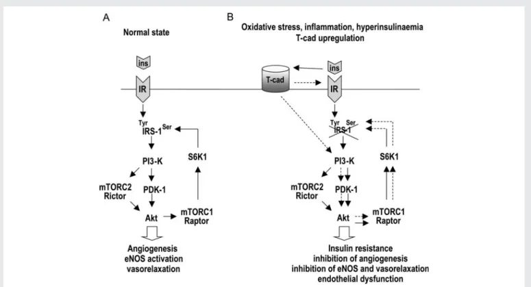
sustained T-cad up-regulation would result in attenuation of insulin signalling. Subsequent enhanced ligation of T-cad molecules on the endothelium by circulating MP harbouring increased levels of T-cad,19 although initially able to activate insulin signalling pathway components, would eventually, upon chronic exposure, also cause inactivation of this pathway. Importantly, as we demonstrate here in vitro, an increase in T-cad protein and transcript expression in EC might be caused not only by oxidative stress, but also by pro-longed exposure of cells to insulin. Furthermore, transcriptional up-regulation of T-cad in EC by insulin can be prevented by ROS scavenger N-acetylcysteine, which is in accordance with the reported role for NADPH oxidase-produced ROS in insulin signal-ling38 and the presence of ROS-sensitive elements in T-cad pro-moter.29 Therefore, in vivo, hyperinsulinaemia initiated as a compensatory response to loss of insulin sensitivity might also lead to T-cad up-regulation and T-cad-dependent inactivation of insulin signalling, further promoting the vicious cycle of InsRes pro-gression. Exacerbation of endothelial InsRes would in its turn result in ED manifest as reduced insulin-dependent release of relaxing factors (e.g. NO) and impaired endothelium-dependent vasodilation which plays an important role in the pathophysiology of essential hypertension.39 Our current view on a cross-talk between T-cad, insulin, eNOS, and angiogenesis in EC and the putative pathophysio-logical consequences for vessel function is schematically depicted in Figure6.
There is mounting interest in the role for T-cad in pathogenesis of metabolic disorders. Recent genome-wide association studies suggest that apart from mediating interactions of adiponectin with the vascular wall, T-cad might also modulate plasma levels of adiponectin.40,41 Tyrberg et al.42demonstrated that T-cad-KO mice display progressive glucose intolerance, attributable to a necessary requirement of T-cad for insulin secretion from pancreatic b-cells. We have identified another aspect of T-cad involvement in regulation of insulin function, namely, a direct ability of T-cad to modulate activity of the insulin sig-nalling cascade in the endothelium. Clinical and experimental data suggest a tight reciprocal relationship between ED and InsRes. ED is a characteristic feature of diabetes and obesity and, together with endothelial InsRes, has been suggested to precede the development of metabolic InsRes. Disclosure of T-cad-dependent control of endo-thelial insulin sensitivity as a novel signalling pathway at the cross-roads of vascular and metabolic disorders advances our understanding of the complicated network of cellular mechanisms responsible for vascular InsRes and associated vascular dysfunction.
Supplementary material
Supplementary material is available at Cardiovascular Research online. Conflict of interest: none declared.
Figure 6 Proposed model for a cross-talk between T-cad and insulin signalling in EC and its pathophysiological consequences. (A) Under normal physiological conditions, there is a balance between the activation of the insulin (ins) signalling cascade flowing downstream from IR to Akt and mediated by tyrosine phosphorylation of IRS-1, and the negative regulatory loop running from Akt to S6K1 and mediated by serine phosphorylation of IRS-1. Functionally, it sustains appropriate angiogenic and vasorelaxant responses of the endothelium to insulin. (B) Increased expression and ligation of T-cad on the cell surface caused by oxidative stress, inflammation, or prolonged exposure to insulin results in chronic stimulation of the Akt cascade, which in its turn induces compensatory hyperactivation of the negative feedback loop, increased serine phosphorylation of IRS-1, its degrad-ation, and shutdown of the signalling. This leads to loss of insulin sensitivity in EC, attenuation of insulin-dependent angiogenesis and vasorelaxdegrad-ation, and the progression of ED. Solid lines, insulin-induced signalling; dashed lines, T-cad-dependent signalling.
Funding
This work was supported by the Swiss National Science Foundation (grant no. 310030_132469), the Herzkreislauf Stiftung, the Swiss Heart Founda-tion, Stiftung der Diabetes-Gesellschaft Region Basel, and an Astra Zeneca Grant-in-Aid award.
References
1. Bakker W, Eringa EC, Sipkema P, van Hinsbergh VW. Endothelial dysfunction and dia-betes: roles of hyperglycemia, impaired insulin signaling and obesity. Cell Tissue Res 2009;335:165 – 189.
2. Kim JA, Montagnani M, Koh KK, Quon MJ. Reciprocal relationships between insulin resistance and endothelial dysfunction: molecular and pathophysiological mechanisms. Circulation 2006;113:1888 – 1904.
3. Muniyappa R, Iantorno M, Quon MJ. An integrated view of insulin resistance and endothelial dysfunction. Endocrinol Metab Clin North Am 2008;37:685 – 711, ix – x. 4. Caballero AE. Endothelial dysfunction in obesity and insulin resistance: a road to
dia-betes and heart disease. Obes Res 2003;11:1278 – 1289.
5. Muniyappa R, Montagnani M, Koh KK, Quon MJ. Cardiovascular actions of insulin. Endocr Rev 2007;28:463 – 491.
6. Liu Y, Petreaca M, Martins-Green M. Cell and molecular mechanisms of insulin-induced angiogenesis. J Cell Mol Med 2009;13:4492 – 4504.
7. Zdychova J, Komers R. Emerging role of Akt kinase/protein kinase B signaling in pathophysiology of diabetes and its complications. Physiol Res 2005;54:1 – 16. 8. Berg CE, Lavan BE, Rondinone CM. Rapamycin partially prevents insulin resistance
induced by chronic insulin treatment. Biochem Biophys Res Commun 2002;293: 1021 – 1027.
9. Ueno M, Carvalheira JB, Tambascia RC, Bezerra RM, Amaral ME, Carneiro EM et al. Regulation of insulin signalling by hyperinsulinaemia: role of IRS-1/2 serine phosphor-ylation and the mTOR/p70 S6K pathway. Diabetologia 2005;48:506 – 518. 10. Tremblay F, Gagnon A, Veilleux A, Sorisky A, Marette A. Activation of the mammalian
target of rapamycin pathway acutely inhibits insulin signaling to Akt and glucose trans-port in 3T3-L1 and human adipocytes. Endocrinology 2005;146:1328 – 1337. 11. Matsui T, Nagoshi T, Hong EG, Luptak I, Hartil K, Li L et al. Effects of chronic Akt
activation on glucose uptake in the heart. Am J Physiol Endocrinol Metab 2006;290: E789 – E797.
12. Philippova M, Joshi MB, Kyriakakis E, Pfaff D, Erne P, Resink TJ. A guide and guard: the many faces of T-cadherin. Cell Signal 2009;21:1035 – 1044.
13. Ivanov D, Philippova M, Antropova J, Gubaeva F, Iljinskaya O, Tararak E et al. Expres-sion of cell adheExpres-sion molecule T-cadherin in the human vasculature. Histochem Cell Biol 2001;115:231 – 242.
14. Kudrjashova E, Bashtrikov P, Bochkov V, Parfyonova Y, Tkachuk V, Antropova J et al. Expression of adhesion molecule T-cadherin is increased during neointima formation in experimental restenosis. Histochem Cell Biol 2002;118:281 – 290.
15. Ivanov D, Philippova M, Allenspach R, Erne P, Resink T. T-cadherin upregulation cor-relates with cell-cycle progression and promotes proliferation of vascular cells. Cardi-ovasc Res 2004;64:132 – 143.
16. Joshi MB, Philippova M, Ivanov D, Allenspach R, Erne P, Resink TJ. T-cadherin protects endothelial cells from oxidative stress-induced apoptosis. FASEB J 2005;19:1737–1739. 17. Kyriakakis E, Philippova M, Joshi MB, Pfaff D, Bochkov V, Afonyushkin T et al. T-cadherin modulates UPR activation and cell survival during endoplasmic reticulum stress in endothelial cells. Cell Signal 2010;22:1308 – 1316.
18. Philippova M, Banfi A, Ivanov D, Gianni-Barrera R, Allenspach R, Erne P et al. Atypical GPI-anchored T-cadherin stimulates angiogenesis in vitro and in vivo. Arterioscler Thromb Vasc Biol 2006;26:2222 – 2230.
19. Philippova M, Suter Y, Toggweiler S, Schoenenberger AW, Joshi MB, Kyriakakis E et al. T-cadherin is present on endothelial microparticles and is elevated in plasma in early atherosclerosis. Eur Heart J 2011;32:760 – 771.
20. Goldstein BJ, Scalia RG, Ma XL. Protective vascular and myocardial effects of adipo-nectin. Nat Clin Pract Cardiovasc Med 2009;6:27 – 35.
21. Denzel MS, Scimia MC, Zumstein PM, Walsh K, Ruiz-Lozano P, Ranscht B. T-cadherin is critical for adiponectin-mediated cardioprotection in mice. J Clin Invest 2010;120: 4342 – 4352.
22. Hebbard LW, Garlatti M, Young LJ, Cardiff RD, Oshima RG, Ranscht B. T-cadherin supports angiogenesis and adiponectin association with the vasculature in a mouse mammary tumor model. Cancer Res 2008;68:1407 – 1416.
23. Hug C, Wang J, Ahmad NS, Bogan JS, Tsao TS, Lodish HF. T-cadherin is a receptor for hexameric and high-molecular-weight forms of Acrp30/adiponectin. Proc Natl Acad Sci USA 2004;101:10308 – 10313.
24. Kyriakakis E, Cavallari M, Andert J, Philippova M, Koella C, Bochkov V et al. Invariant natural killer T cells: linking inflammation and neovascularization in human athero-sclerosis. Eur J Immunol 2010;40:3268 – 3279.
25. Pfaff D, Philippova M, Buechner SA, Maslova K, Mathys T, Erne P et al. T-cadherin loss induces an invasive phenotype in human keratinocytes and squamous cell carcinoma (SCC) cells in vitro and is associated with malignant transformation of cutaneous SCC in vivo. Br J Dermatol 2010;163:353 – 363.
26. Philippova M, Ivanov D, Joshi MB, Kyriakakis E, Rupp K, Afonyushkin T et al. Identifi-cation of proteins associating with glycosylphosphatidylinositol-anchored T-cadherin on the surface of vascular endothelial cells: role for Grp78/BiP in T-cadherin-dependent cell survival. Mol Cell Biol 2008;28:4004 – 4017.
27. Ivanov D, Philippova M, Tkachuk V, Erne P, Resink T. Cell adhesion molecule T-cadherin regulates vascular cell adhesion, phenotype and motility. Exp Cell Res 2004;293:207 – 218.
28. Timmins NE, Nielsen LK. Generation of multicellular tumor spheroids by the hanging-drop method. Methods Mol Med 2007;140:141 – 151.
29. Joshi MB, Ivanov D, Philippova M, Kyriakakis E, Erne P, Resink TJ. A requirement for thioredoxin in redox-sensitive modulation of T-cadherin expression in endothelial cells. Biochem J 2008;416:271 – 280.
30. Choi K, Kim YB. Molecular mechanism of insulin resistance in obesity and type 2 dia-betes. Korean J Intern Med 2010;25:119 – 129.
31. Zhande R, Mitchell JJ, Wu J, Sun XJ. Molecular mechanism of insulin-induced degrad-ation of insulin receptor substrate 1. Mol Cell Biol 2002;22:1016 – 1026.
32. Zeng G, Nystrom FH, Ravichandran LV, Cong LN, Kirby M, Mostowski H et al. Roles for insulin receptor, PI3-kinase, and Akt in insulin-signaling pathways related to pro-duction of nitric oxide in human vascular endothelial cells. Circulation 2000;101: 1539 – 1545.
33. Philippova MP, Bochkov VN, Stambolsky DV, Tkachuk VA, Resink TJ. T-cadherin and signal-transducing molecules co-localize in caveolin-rich membrane domains of vascu-lar smooth muscle cells. FEBS Lett 1998;429:207 – 210.
34. Joshi MB, Ivanov D, Philippova M, Erne P, Resink TJ. Integrin-linked kinase is an essen-tial mediator for T-cadherin-dependent signaling via Akt and GSK3{beta} in endothe-lial cells. FASEB J 2007;21:3083 – 3095.
35. Saltiel AR, Pessin JE. Insulin signaling in microdomains of the plasma membrane. Traffic 2003;4:711 – 716.
36. Andersen AS, Kjeldsen T, Wiberg FC, Vissing H, Schaffer L, Rasmussen JS et al. Iden-tification of determinants that confer ligand specificity on the insulin receptor. J Biol Chem 1992;267:13681 – 13686.
37. Slaaby R, Schaffer L, Lautrup-Larsen I, Andersen AS, Shaw AC, Mathiasen IS et al. Hybrid receptors formed by insulin receptor (IR) and insulin-like growth factor I re-ceptor (IGF-IR) have low insulin and high IGF-1 affinity irrespective of the IR splice variant. J Biol Chem 2006;281:25869 – 25874.
38. Goldstein BJ, Mahadev K, Wu X, Zhu L, Motoshima H. Role of insulin-induced react-ive oxygen species in the insulin signaling pathway. Antioxid Redox Signal 2005;7: 1021 – 1031.
39. Puddu P, Puddu GM, Zaca F, Muscari A. Endothelial dysfunction in hypertension. Acta Cardiol 2000;55:221 – 232.
40. Chung CM, Lin TH, Chen JW, Leu HB, Yang HC, Ho HY et al. A genome-wide asso-ciation study reveals a quantitative trait locus of adiponectin on CDH13 that predicts cardiometabolic outcomes. Diabetes 2011;60:2417 – 2423.
41. Morisaki H, Yamanaka I, Iwai N, Miyamoto Y, Kokubo Y, Okamura T et al. CDH13 gene coding T-cadherin influences variations in plasma adiponectin levels in Japanese population. Hum Mutat 2011; doi:10.1002/humu.21652.
42. Tyrberg B, Miles P, Azizian KT, Denzel MS, Nieves ML, Monosov EZ et al. T-cadherin (Cdh13) in association with pancreatic beta-cell granules contributes to second phase insulin secretion. Islets 2011;3:327 – 337.