Advance Access publication 15 October 2013
Systematic Review
Indication and timing of soft tissue augmentation at
maxillary and mandibular incisors in orthodontic patients.
A systematic review
Dimitrios Kloukos
*
, Theodore Eliades
**
, Anton Sculean
***
, and Christos Katsaros
*
*Department of Orthodontics and Dentofacial Orthopedics, University of Bern, **Department of Orthodontics and Paediatric Dentistry, University of Zurich, and ***Department of Periodontology, University of Bern, Switzerland Correspondence to: Theodore Eliades, Department of Orthodontics and Paediatric Dentistry, Center of Dental Medicine, University of Zurich, Plattenstrasse 11, CH-8032 Zurich, Switzerland. E-mail: [email protected]SUMMARy
OBjECTIvE: To assess the indication and timing of soft tissue augmentation for prevention or treatment of
gingival recession when a change in the inclination of the incisors is planned during orthodontic treatment.
MATERIAlS AnD METHODS: Electronic database searches of literature were performed. The following
elec-tronic databases with no restrictions were searched: MEDlInE, EMBASE, Cochrane, and CEnTRAl. Two authors performed data extraction independently using data collection forms.
RESUlTS: no randomized controlled trial was identified. Two studies of low-to-moderate level of evidence
were included: one of prospective and retrospective data collection and one retrospective study. Both implemented a periodontal intervention before orthodontics. Thus, best timing of soft tissue augmenta-tion could not be assessed. The limited available data from these studies appear to suggest that soft tissue augmentation of bucco-lingual gingival dimensions before orthodontics may yield satisfactory results with respect to the development or progression of gingival recessions. However, the strength of the avail-able evidence is not adequate in order to change or suggest a possible treatment approach in the daily practice based on solid scientific evidence.
COnClUSIOnS: Despite the clinical experience that soft tissue augmentation of bucco-lingual gingival
dimensions before orthodontic treatment may be a clinically viable treatment option in patients consid-ered at risk, this treatment approach is not based on solid scientific evidence. Moreover, the present data do not allow to draw conclusions on the best timing of soft tissue augmentation when a change in the inclination of the incisors is planned during orthodontic treatment and thus, there is a stringent need for randomized controlled trials to clarify these open issues.
Introduction
Gingival recession refers to the apical displacement of the gingival margin from the cemento-enamel junction (Kassab
and Cohen, 2003). Recessions can be localized or may
involve more teeth or tooth surfaces. The resulting root sur-face exposure often causes aesthetic concerns (Smith, 1997), dentin hypersensitivity (Lawrence et al., 1995), and increased susceptibility to root caries (Al-Wahadni and Linden, 2002). Gingival recessions have been found to be more frequent in mandibular than maxillary teeth, and on facial than lingual surfaces, especially with increasing age (Khocht et al., 1993).
As early as 1976, a justification regarding the pathogen-esis of gingival recession was brought forward (Baker and Seymour, 1976). Localized inflammation in a ‘thin type’ gingiva might involve the entire volume of gingival tissue and the consequent remodelling could lead to recession of
gingival margin. On the other hand, in a ‘thick type’ gingiva, this inflammatory lesion could be confined to only a part of the sulcus leaving the ‘outer gingival tissue’ unaffected. This could probably predispose to pocket formation rather than recession. Although the proposal of this mechanism was based on a rat model, it may be considered as the primary concept, which recognized that the thin gingival biotype can be a risk factor for recession. Additionally, many other factors may as well play a role in the development of gingival reces-sion, not necessarily simultaneously or equally. Periodontal diseases and mechanical trauma are the two primary etiologic factors in the pathogenesis of gingival recessions (Löe et al., 1992; Smith, 1997; Kassab and Cohen, 2003; Litonjua et al., 2003; Rawal et al., 2004; Levin et al., 2005). Traumatic tooth brushing appears to be one of the important factors associ-ated with gingival recessions (Khocht et al., 1993). Other,
secondary etiologic factors might include existing bone dehiscences, smoking, and intraoral and perioral piercings (Albandar et al., 2000; Susin et al., 2004; Levin et al., 2005).
A possible etiological factor for gingival recession is the orthodontic movement of teeth, especially the movement of teeth to positions outside the labial or lingual alveolar plate, which could lead to development of dehiscence (Wennström
et al., 1987). A systematic review indicated that proclina-tion and movement of the mandibular incisors out of the osseous envelope of the alveolar process may be associated with a higher tendency for developing gingival recessions. On the other hand, the amount of recession found in the included studies, which showed statistically significant dif-ferences between proclined and not proclined incisors, was small and the clinical consequence regarded as questionable (Joss-Vassalli et al., 2010). The precise mechanism by which orthodontic treatment influences the occurrence of reces-sions remains unclear. It has been assumed that the presence of bony dehiscence before the beginning of orthodontic ther-apy is a prerequisite for the development of gingival reces-sion (Wennström, 1996). Because a bony dehiscence does not always lead to recession (Thilander et al., 1983), other factors, as the ones described above, must be present.
Among the numerous orthodontic treatment procedures and modalities, the issue of incisor proclination outside their den-toalveolar envelope as a source of gingival recession has been considered for years. Although it is broadly believed that man-dibular incisor proclination leads to gingival recession, there are very few clinical studies that have actually investigated this. Some of them have shown gingival recession to be asso-ciated with proclination of the mandibular incisors (Årtun and Krogstad, 1987; Yared et al., 2006), whereas others have found no such correlation (Ruf et al., 1998; Djeu et al., 2002; Allais and Melsen, 2003). A series of recent retrospective studies has shown that although the change of inclination of lower inci-sors during orthodontic treatment may not affect the develop-ment of labial recessions (Renkema et al., 2012), nevertheless, these teeth are the most vulnerable to development of reces-sions (Renkema et al., 2013a), with their prevalence being age dependent and increasing steadily in the period between before and 5 years after therapy (Renkema et al., 2013b).
Periodontal prevention of gingival recessions in orthodon-tic patients remains, likewise, contradictory. Historically, periodontists have indicated gingival augmentation to rec-reate the zone of attached gingiva. The early concept for this approach was that attached gingiva is important to dissipate the force of muscle pull and unattached mucosa. Many still believe that attached gingiva is more suitable to withstand the trauma of mastication and tooth brushing (Corn, 1962;
Carranza and Carraro, 1970; Lang and Löe, 1972).
Such periodontal augmentation procedures include free gingival grafts, coronally positioned flap, subepithelial con-nective grafts, acellular dermal grafts, and enamel matrix proteins. Among them, subepithelial connective tissue grafts are generally considered as a ‘gold standard’ in gingival
augmentation (Roccuzzo et al., 2002; Chambrone et al., 2008). Opposite to this preventive concept, however, some clinicians consider this approach as overtreatment and pre-fer to wait until the potential gingival recession becomes a pathological and clinical entity. Subsequently, the developed recession may be treated during or after orthodontic therapy.
Although some guidelines exist about how much is ade-quate when it comes to the thickness of the attached gingi-val (Lang and Löe, 1972), the decision about its adequacy to withstand the stress and adverse effects related to mandibu-lar incisor increase in inclination as well as the timing of the proposed periodontal intervention remains a highly subjec-tive issue. Despite of the presence of some scarce evidence in the literature on this subject, a synthesis of their results has not been published yet.
The aim of this systematic review was, therefore, to search and assess the available literature in order to appraise if, first of all, soft tissue augmentation is indicated for prevention or treatment of gingival recession, and at which point of time related to orthodontic treatment, when a change in the inclination of the incisors is anticipated.
Materials and methods
Selection criteria applied for the review
• Study design: prospective and retrospective studies were considered in this review, including randomized clinical trials, controlled clinical trials, and other observational studies in the absence of the first. Animal studies were not considered eligible for inclusion in this review. Case reports were also excluded.
• Types of participants: patients referred for orthodontic treatment. Any age of patients was accepted.
• Types of intervention: periodontal treatment for the pre-vention or treatment of gingival recessions including gin-gival or epithelial grafting for soft tissue augmentation. Fixed orthodontic appliances that were designed to alter the inclination of the mandibular and/or maxillary incisors. • Outcome: success of periodontal therapy of gingival
recessions adjunct to alteration of incisors’ inclination. Timing of periodontal treatment was associated with the main outcome.
• Timing of periodontal treatment: no restriction was applied regarding the points of time that periodontal therapy may have taken place (before/during/after orthodontic treatment). • Exclusion criteria: orthodontic translative (bodily) tooth
movement through grafts, orthodontic tooth movement other than inclination, teeth other than mandibular/maxil-lary incisors.
Search strategy for identification of studies
For the identification of studies included or considered for this review, detailed search strategies were developed for each database searched. They were based on the search strategy
developed for MEDLINE but revised appropriately for each database to take account of differences in controlled vocabu-lary and syntax rules. The following electronic databases were searched: MEDLINE (via Ovid and PubMed, Appendix 1; 1946 to April week 4, 2013), EMBASE (via Ovid), the Cochrane Oral Health Group’s Trials Register, and CENTRAL.
Unpublished literature was searched on ClinicalTrials. gov (date last accessed, September 26, 2013), the National Research Register, and Pro-Quest Dissertation Abstracts and Thesis database.
The search attempted to identify all relevant studies irrespective of language. There were no restrictions on date of publication. The reference lists of all eligible stud-ies were handsearched for additional studstud-ies.
Selection of studies
Assessment of research for including studies in the review and extraction of data were performed independently and in duplicate by the first two authors who were not blinded to identity of the authors, their institution, or the results of the research. The full report of publications considered by either author to meet the inclusion criteria was obtained and assessed independently. Disagreements were resolved by discussion and consultation with the third author. A record of all decisions on study identification was kept.
Data extraction and management
The first two authors performed data extraction indepen-dently and in duplicate. Disagreements were resolved by discussion or the involvement of a collaborator (third author). Data collection forms were used to record the desired information. The following data were collected on a customized data collection form.
• Author/title of study • Design of the study
• Human race of study participants • Number/age/gender of patients recruited
• Type of orthodontic therapy and identification of the teeth that were moved
• Force applied to the teeth under treatment • Orthodontic treatment duration
• Timing of periodontal intervention
• Periodontal procedure and type of grafting material • Time points of outcome assessment and method of
meas-uring the outcome
• Attainment of desirable tooth movement
• Increase or decrease of gingival recession and possible factors related
Measures of treatment effect
For continuous outcomes, mean differences and standard deviation were used to summarize the data for each study.
Unit of analysis issues
In all cases, the unit of analysis was primarily the patient. In addition, a unit of analysis issue arose due to the different periodontal approaches. To resolve this, a separate analy-sis for each periodontal technique was planned to be used, where applicable.
Data synthesis
A meta-analysis was planned to be conducted only if there were studies of similar comparisons, reporting the same outcome measures at the same time points.
Quality assessment
The quality of methodology, performance, and statistics of each study were assessed, and the studies were graded with a score of A, B, or C (grade A: high value of evidence, grade C: low value of evidence) according to predetermined criteria using the system of Bondemark et al. (2007). This, validated also in other studies, system describes the criteria for grading the studies as follows:
• Grade A: high value of evidence (all criteria should be met):
– Randomized clinical study or a prospective study with a well-defined control group.
– Defined diagnosis and end points.
– Diagnostic reliability tests and reproducibility tests described.
– Blinded outcome assessment.
• Grade B: moderate value of evidence (all criteria should be met):
– Cohort study or retrospective case series with defined control or reference group.
– Defined diagnosis and end points.
– Diagnostic reliability tests and reproducibility tests described.
• Grade C: low value of evidence (one or more of the fol-lowing conditions):
– Large attrition.
– Unclear diagnosis and end points. – Poorly defined patient material.
Results
Description of studies
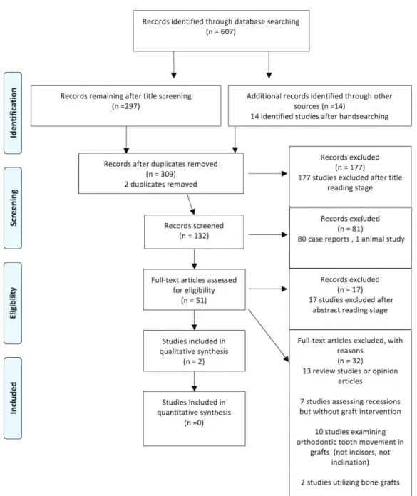
Studies that were initially deemed potentially relevant for the review were retrieved and inclusion criteria were applied. Tracking the eligible for inclusion studies appeared to be a difficult task. Many case reports, several studies examin-ing orthodontic tooth movement through grafts, or studies
examining other than inclination types of movement exist in the field, which were not relevant for this review. After removal of the duplicates, abstract and full text reading stage, two studies were finally regarded as eligible for inclu-sion (Figure 1). Both studies were included in the qualitative analysis but a quantitative synthesis was not appropriate. No randomized controlled trial was identified. One study had both prospective and retrospective data collection (Maynard and Ochsenbein, 1975), and the other had a retrospective design (Ngan et al., 1991; Tables 1 and 2). Both implemented a periodontal intervention before orthodontic treatment.
Quality assessment
One study was graded as moderate (grade B) value of evi-dence and this was the retrospective one (Ngan et al., 1991). The second was graded as low value of evidence (grade C). The reason was the poorly defined patient material (Maynard and Ochsenbein, 1975).
Qualitative synthesis of the included studies
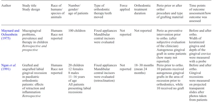
Study settings. An overview of the experimental
set-up of the included studies is given in Table 1. In the first study (Maynard and Ochsenbein, 1975), autogenous free gingival graft was implemented as a preventive measure, before orthodontics, in some young patients (exact num-ber not given). These consisted a part of a sample of 100 children. Mandibular central incisors were evaluated. In the second study (Ngan et al., 1991), the authors divided their 20 patients with more than 1 mm labial recession on one or more mandibular central incisors before treat-ment in two groups: one group received autogenous free gingival graft in the area of recession prior orthodontics and the second group (control group) had no graft before orthodontics. In both groups, the incisors were retroclined during treatment.
Clinical findings. Table 2 gives an overview of the results of the included studies regarding clinical parameters. Both studies have used periodontal surgery (e.g. soft tissue graft-ing) before orthodontic treatment. Consequently, the issue on the best timing (before, during, or after orthodontic treat-ment) could not be assessed.
Maynard and Ochsenbein (1975) stated that autogenous free gingival graft can be recommended as an acceptable procedure prior to tooth movement, where orthodontic ther-apy is anticipated and insufficient keratinized tissue exists. According to the authors, grafts can be recommended in patients with 1 mm or less of keratinized tissue.
Ngan et al. (1991) found out that teeth presenting true gingival recession had statistically less gingival recession after being retroclined, with no difference detected between grafted and ungrafted recessions. It was, however, a study, during which teeth were retroclined and not proclined.
Quantitative synthesis of the included studies
The lack of standardized protocols precluded a valid inter-pretation of the actual results of the studies. Methodological heterogeneity refers to important differences in the inter-ventions, participants, and outcomes of the included stud-ies and similarly for studstud-ies other than randomized clinical trials. Although both studies implemented surgical peri-odontal therapy before orthodontic treatment, the analysis of the indication and methodology revealed substantial dif-ferences with respect to the sample size, the type of peri-odontal procedure or regenerative material, and the time points of outcome assessment. Therefore, a meta-analysis was not possible.
Discussion
The inclination and the projected (empirically or through Visual Treatment Objective) post-treatment position of the mandibular or maxillary incisors play an important role in the diagnostic process and orthodontic treatment planning. It is frequently necessary to first establish proclination tol-erance limits before treatment, especially in patients with severe skeletal discrepancies, with arches that can accom-modate only a limited number of teeth, or in patients with inadequate attached gingiva. These limits to estimated pro-clination refer to biological factors, such as the character-istics and quality of the periodontal tissues in the area and thus, patients who already have thin soft tissue margins before treatment should be treated with caution.
Recession is not probably a direct consequence of incisor proclination. A relevant systematic review found no association between appliance-induced labial move-ment of mandibular incisors and gingival recession (Aziz
and Flores-Mir, 2011). The authors recommended to
also focus on other predisposing conditions of the man-dibular anatomy before orthodontic planning, as far as it concerns recessions. On the other hand, some studies have shown that excessive final inclination of incisors, in addition to individual characteristics of thin gingival margin and other local or even systemic factors, can ren-der it susceptible to the development of recession defects (Årtun and Krogstad, 1987; Yared et al., 2006). It can be anticipated that if the gingival margin maintains an appropriate thickness after orthodontic treatment, the tissue would be more resistant and less affected by ten-sion from excessive proclination. Consequently, the risk for developing gingival recession could be significantly reduced.
The issue of preventive periodontal intervention before orthodontic treatment, and especially before inclination of the incisors, has been long discussed in the scientific community (Maynard and Ochsenbein, 1975; Mehta and Lim, 2010). The objective of this systematic review was to include the results of as many studies as possible to obtain
information on the development or prevention of gingi-val recessions after combined periodontal–orthodontic treatment.
The low-to-moderate level of evidence of the included studies and the application of different periodontal proce-dures, analysed at different time intervals, made the analy-sis of the results impossible. Treatment duration, control groups, force applied, and grafting materials varied substan-tially, making the calculation of pooled estimates unfeasible. Despite the lack of consistency in methodological approaches, and taking into account that the available evidence derived from studies, which command a low to
moderate level of evidence, the qualitative analysis of the included studies revealed that:
• Periodontal soft tissue augmentation of bucco-lingual gingival dimensions before orthodontic treatment may yield satisfactory results, as far as it concerns the devel-opment or progress of gingival recessions. The lack of high level of evidence, though, cannot render these results generalizable.
• The evaluation of the attached gingiva as adequate or inad-equate, and in turn, the need of periodontal intervention before incisor inclination, still remains highly subjective.
Figure 1 Study Flow Diagram. From Moher D, Liberati A, Tetzlaff J, Altman D G, The PRISMA Group (2009). Preferred Reporting Items for Systematic Reviews and Meta-Analyses: The PRISMA Statement. PLoS Med 6: e1000097. doi:10.1371/journal.pmed1000097. For more information, visit www. prisma-statement.org (date last accessed, September 26, 2013).
• The final analysis in the included studies took place immediately after orthodontic therapy. Long-term results are clearly missing on this topic.
• In patients with a ‘thin’ type of gingiva, soft tissue graft-ing might be beneficial before orthodontic tooth move-ment to prevent the developmove-ment of a gingival recession. Whether this clinical decision can be considered as over-treatment, still remains an open question and should be evaluated in further studies.
The issue of proclination and its potential effect on the per-iodontal support of the root of mandibular incisors must
be considered within the broader context of current treat-ment trends and practices (Johal et al., 2013). A number of studies supporting the lack of definitive evidence link-ing proclination with dehiscence, recession, or other unfa-vourable effect on the periodontal condition of mandibular incisors have indicated minute differences of proclined relative to non-proclined teeth. However, two central argu-ments relating to the correlation of clinical examination of recessions with the actual status of periodontium and the implication of long-term recession are worth mentioning on this aspect.
Table 1 Design (materials and methods) of included studies.
Author Study title Study design Race of humans/ species of animals Number/ gender/ age of patients Type of orthodontic therapy/teeth moved Force applied Orthodontic treatment duration
Perio prior or after ortho/
procedure and type of grafting material Time points of outcome assessment/how outcome was assessed Maynard and Ochsenbein (1975) Mucogingival problems, prevalence and therapy in children Retrospective and prospective Humans Race not reported
100 children Fixed appliances Mandibular central incisors were evaluated
Not reported
Not reported Perio as preventive intervention prior to ortho. (after subjective evaluation of the clinician) Autogenous gingival graft in some patients (how many not reported)
Before and after ortho. Width of keratinized gingiva and depth of the gingival crevice were measured with a probe Ngan et al. (1991) Grafted and ungrafted labial gingival recession in paediatric orthodontic patients: effects of retraction and inflammation Retrospective Humans Race not reported 20 children 12 females 8 males 11–16 years of age All patients presenting labial recessions Fixed appliances Mandibular central incisors were evaluated (retroclination) Not reported 18–30 months (mean 24 months)
Perio prior to ortho. 10 patients received autogenous gingival grafts in the area of recession prior to orthodontics, while 10 received no graft
Before and after ortho. Gingival recessions were measured on projected transparent slides after photos taken from patients
Table 2 Results of included studies.
Study Desirable tooth movement and
achieved/not achieved
Gingival recession (increase/decrease) Gingival recession related/not related to:
Maynard and
Ochsenbein (1975) Not reported No additional gingival recession was observed over the grafted teeth. Where orthodontic therapy is anticipated and coincidentally insufficient keratinized tissue exists, a free gingival graft should be performed prior to tooth movement. Grafts would be recommended in children with 1 mm or less of keratinized tissue.
Ngan et al. (1991) Retroclination of mandibular incisors (from a pre-orthodontically prominent arch position)
Comparison of the pre-treatment and post-treatment gingival recessions between the control and grafted groups showed no statistically significant differences.
Pre-orthodontic gingival grafting did not further decrease the post- orthodontic gingival recession. It was postulated that the eruption and maturation of incisors during the treatment contributed more to the decreased gingival recession than the graft did.
First, the examination of the periodontal condition of the affected teeth was performed essentially only clini-cally without standardized radiographic evidence, thus limiting the identification of the unfavourable sequelae to the clinically detectable signs of gingival recession, with almost no information about the accompanying bone lev-els. Evidence from autopsy material of an individual who underwent orthodontic treatment and presented no signs of recession clinically, while she showed severe frontal periodontal destruction as evaluated histologically, sug-gests that this examination (and conventional radiographic antero-posterior assessment of bone levels) might under-estimate the impact of orthodontic proclination on tissue damage (Wehrbein et al., 1996). The introduction of cone beam computed tomography could provide clinically rele-vant information on this issue (Enhos et al., 2012), despite its limitations regarding overestimation of bone fenestra-tions and dehiscences (Leung et al., 2010; Patcas et al., 2012).
Secondly, with recent suggestions on long-term fixed retention of mandibular teeth (Littlewood et al., 2006), the issue of proclination must be viewed under the per-spective of potential induction of effects on the periodon-tal condition of mandibular teeth after the termination of active treatment. This factor has not been assessed in studies examining the effect of proclination after orthodontic tooth movement. Nevertheless, orthodontic treatment in general, and the following retention phase, may be considered as a risk factor for the development of labial gingival recessions (Renkema et al., 2013a). Furthermore, related evidence on this issue suggests that the clinical condition of periodontium of patients who had received orthodontic treatment with at least 2 mm advancement of their incisal edge, 7.8–9.4 years after treatment, was comparable to patients without such an advancement (Årtun and Grobéty, 2001). However, no information is available on the retention practices in this sample.
Conclusion
Despite the clinical experience that soft tissue augmenta-tion of bucco-lingual gingival dimensions before orthodon-tic treatment may be a clinically viable treatment option in patients considered at risk, this treatment approach is not based on solid scientific evidence. Moreover, the present data do not allow to draw any conclusion on the best timing of soft tissue augmentation when a change in the inclination of the incisors is planned during orthodontic treatment and thus, there is a stringent need for randomized controlled tri-als to clarify these open issues.
Funding
No funding was obtained for this review.
References
Albandar J M, Streckfus C F, Adesanya M R, Winn D M 2000 Cigar, pipe, and cigarette smoking as risk factors for periodontal disease and tooth loss. Journal of Periodontology 71: 1874–1881
Allais D, Melsen B 2003 Does labial movement of lower incisors influence the level of the gingival margin? A case-control study of adult orthodon-tic patients. European Journal of Orthodonorthodon-tics 25: 343–352
Al-Wahadni A, Linden G J 2002 Dentine hypersensitivity in Jordanian den-tal attenders. A case control study. Journal of Clinical Periodontology 29: 688–693
Årtun J, Grobéty D 2001 Periodontal status of mandibular incisors after pronounced orthodontic advancement during adolescence: a follow-up evaluation. American Journal of Orthodontics and Dentofacial Orthopedics 119: 2–10
Årtun J, Krogstad O 1987 Periodontal status of mandibular incisors follow-ing excessive proclination. A study in adults with surgically treated man-dibular prognathism. American Journal of Orthodontics and Dentofacial Orthopedics 91: 225–232
Aziz T, Flores-Mir C 2011 A systematic review of the association between appliance-induced labial movement of mandibular incisors and gingival recession. Australian Orthodontic Journal 27: 33–39
Baker D L, Seymour G J 1976 The possible pathogenesis of gingival reces-sion. A histological study of induced recession in the rat. Journal of Clinical Periodontology 3: 208–219
Bondemark L et al. 2007 Long-term stability of orthodontic treatment and patient satisfaction. A systematic review. The Angle Orthodontist 77: 181–191
Carranza F A Jr, Carraro J J 1970 Mucogingival techniques in periodontal surgery. Journal of Periodontology 41: 294–299
Chambrone L, Chambrone D, Pustiglioni F E, Chambrone L A, Lima L A 2008 Can subepithelial connective tissue grafts be considered the gold standard procedure in the treatment of Miller class I and II recession-type defects? Journal of Dentistry 36: 659–671
Corn H 1962 Periosteal separation: its clinical significance. Journal of Periodontology 33: 140–153
Djeu G, Hayes C, Zawaideh S 2002 Correlation between mandibular cen-tral incisor proclination and gingival recession during fixed appliance therapy. The Angle Orthodontist 72: 238–245
Enhos S, Uysal T, Yagci A, Veli İ, Ucar F I, Ozer T 2012 Dehiscence and fenestration in patients with different vertical growth patterns assessed with cone-beam computed tomography. The Angle Orthodontist 82: 868–874
Johal A et al. 2013 State of the science on controversial topics: orthodontic therapy and gingival recession (a report of the Angle Society of Europe 2013 meeting). Progress in Orthodontics 14: 16
Joss-Vassalli I, Grebenstein C, Topouzelis N, Sculean A, Katsaros C 2010 Orthodontic therapy and gingival recession: a systematic review. Orthodontics & Craniofacial Research 13: 127–141
Kassab M M, Cohen R E 2003 The etiology and prevalence of gingival recession. Journal of the American Dental Association 134: 220–225 Khocht A, Simon G, Person P, Denepitiya J L 1993 Gingival recession in
relation to history of hard toothbrush use. Journal of Periodontology 64: 900–905
Lang N P, Löe H 1972 The relationship between the width of keratinized gingiva and gingival health. Journal of Periodontology 43: 623–627 Lawrence H P, Hunt R J, Beck J D 1995 Three-year root caries incidence
and risk modeling in older adults in North Carolina. Journal of Public Health Dentistry 55: 69–78
Leung C C, Palomo L, Griffith R, Hans M G 2010 Accuracy and reli-ability of cone-beam computed tomography for measuring alveo-lar bone height and detecting bony dehiscences and fenestrations. American Journal of Orthodontics and Dentofacial Orthopedics 137: S109–S119
Levin L, Zadik Y, Becker T 2005 Oral and dental complications of intra-oral piercing. Dental Traumatology 21: 341–343
Litonjua L A, Andreana S, Bush P J, Cohen R E 2003 Toothbrushing and gingival recession. International Dental Journal 53: 67–72
Littlewood S J, Millett D T, Doubleday B, Bearn D R, Worthington H V 2006 Retention procedures for stabilising tooth position after treatment with orthodontic braces. Cochrane Database of Systematic Reviews 25: CD002283
Löe H, Anerud A, Boysen H 1992 The natural history of periodontal disease in man: prevalence, severity, and extent of gingival recession. Journal of Periodontology 63: 489–495
Maynard J G Jr, Ochsenbein C 1975 Mucogingival problems, prevalence and therapy in children. Journal of Periodontology 46: 543–552 Mehta P, Lim L P 2010 The width of the attached gingiva–much ado about
nothing? Journal of Dentistry 38: 517–525
Ngan P W, Burch J G, Wei S H 1991 Grafted and ungrafted labial gingi-val recession in pediatric orthodontic patients: effects of retraction and inflammation. Quintessence International 22: 103–111
Patcas R, Müller L, Ullrich O, Peltomäki T 2012 Accuracy of cone-beam computed tomography at different resolutions assessed on the bony cov-ering of the mandibular anterior teeth. American Journal of Orthodontics and Dentofacial Orthopedics 141: 41–50
Rawal S Y, Claman L J, Kalmar J R, Tatakis D N 2004 Traumatic lesions of the gingiva: a case series. Journal of Periodontology 75: 762–769 Renkema A M, Fudalej P S, Renkema A, Bronkhorst E, Katsaros C 2012
Gingival recessions and the change of inclination of mandibular incisors dur-ing orthodontic treatment. European Journal of Orthodontics 35: 249–255 Renkema A M, Fudalej P S, Renkema A A, Abbas F, Bronkhorst E,
Katsaros C 2013a Gingival labial recessions in orthodontically treated and untreated individuals: a case - control study. Journal of Clinical Periodontology 40: 631–637
Renkema A M, Fudalej P S, Renkema A, Kiekens R, Katsaros C 2013b Development of labial gingival recessions in orthodontically treated
patients. American Journal of Orthodontics and Dentofacial Orthopedics 143: 206–212
Roccuzzo M, Bunino M, Needleman I, Sanz M 2002 Periodontal plas-tic surgery for treatment of localized gingival recessions: a systemaplas-tic review. Journal of Clinical Periodontology 29: 178–194
Ruf S, Hansen K, Pancherz H 1998 Does orthodontic proclination of lower incisors in children and adolescents cause gingival recession? American Journal of Orthodontics and Dentofacial Orthopedics 114: 100–106
Smith R G 1997 Gingival recession. Reappraisal of an enigmatic condition and a new index for monitoring. Journal of Clinical Periodontology 24: 201–205
Susin C, Haas A N, Oppermann R V, Haugejorden O, Albandar J M 2004 Gingival recession: epidemiology and risk indicators in a representative urban Brazilian population. Journal of Periodontology 75: 1377–1386 Thilander B, Nyman S, Karring T, Magnusson I 1983 Bone regeneration
in alveolar bone dehiscences related to orthodontic tooth movements. European Journal of Orthodontics 5: 105–114
Wehrbein H, Bauer W, Diedrich P 1996 Mandibular incisors, alveolar bone, and symphysis after orthodontic treatment. A retrospective study. American Journal of Orthodontics and Dentofacial Orthopedics 110: 239–246
Wennström J L 1996 Mucogingival considerations in orthodontic treat-ment. Seminars in Orthodontics 2: 46–54
Wennström J L, Lindhe J, Sinclair F, Thilander B 1987 Some periodontal tissue reactions to orthodontic tooth movement in monkeys. Journal of Clinical Periodontology 14: 121–129
Yared K F, Zenobio E G, Pacheco W 2006 Periodontal status of man-dibular central incisors after orthodontic proclination in adults. American Journal of Orthodontics and Dentofacial Orthopedics 130: 6.e1–e8
Appendix 1: Search strategy PubMed, 29 April 2013
#1 (graft*[Title/Abstract]) AND orthodont*[Title/Abstract] 427
#2 (graft*[Title/Abstract]) AND incisor*[Title/Abstract] 345
#3 (graft*[Title/Abstract]) AND inclin*[Title/Abstract] 166
#4 (#1) NOT cleft*[Title/Abstract] 197
#5 (transplant*[Title/Abstract]) AND orthodont*[Title/Abstract] 180
#6 (#5) OR #4 360
#7 (#6) AND timing[Title/Abstract] 7
#8 (#6) AND time[Title/Abstract] 72
#9 “Tissue Transplantation”[Mesh] AND orthodont* 48
#10 “transplantation” [Subheading] AND orthodont* 430
#11 (periodont*[Title/Abstract]) AND orthodont*[Title/Abstract] 2478
#12 Add Search (#11) AND graft*[Title/Abstract] 83
#13 (#11) AND recession[Title/Abstract] 88
#14 (#13) AND graft*[Title/Abstract] 13