Persistence of
Bartonella spp. stealth pathogens: from
subclinical infections to vasoproliferative tumor formation
Arto T. Pulliainen1& Christoph Dehio21Institute of Biomedicine, University of Turku, Turku, Finland; and2Biozentrum, University of Basel, Basel, Switzerland
Correspondence: Christoph Dehio, Biozentrum, University of Basel, Klingelbergstrasse 50/70, CH-4056 Basel, Switzerland. Tel.: +41 61 267 21 40; fax: +41 61 267 21 18;
e-mail: [email protected]
Received 18 August 2011; revised 13 December 2011; accepted 13 December 2011. Final version published online 7 February 2012.
DOI: 10.1111/j.1574-6976.2012.00324.x
Editor: Neil Fairweather
Keywords
Bartonella; bacteremia; immunity; zoonosis; angiogenesis; cancer.
Abstract
Bartonella spp. are facultative intracellular bacteria that typically cause a long-lasting intraerythrocytic bacteremia in their mammalian reservoir hosts, thereby favoring transmission by blood-sucking arthropods. In most cases, nat-ural reservoir host infections are subclinical and the relapsing intraerythrocytic bacteremia may last weeks, months, or even years. In this review, we will fol-low the infection cycle of Bartonella spp. in a reservoir host, which typically starts with an intradermal inoculation of bacteria that are superficially scratched into the skin from arthropod feces and terminates with the pathogen exit by the blood-sucking arthropod. The current knowledge of bacterial coun-termeasures against mammalian immune response will be presented for each critical step of the pathogenesis. The prevailing models of the still-enigmatic primary niche and the anatomical location where bacteria reside, persist, and are periodically seeded into the bloodstream to cause the typical relapsing Bartonella spp. bacteremia will also be critically discussed. The review will end up with a discussion of the ability of Bartonella spp., namely Bartonella hense-lae, Bartonella quintana, and Bartonella bacilliformis, to induce tumor-like vas-cular deformations in humans having compromised immune response such as in patients with AIDS.
Introduction
Bartonella spp. are Gram-negative facultative intracellular bacteria that taxonomically belong to thea2-subgroup of the proteobacteria. Bartonella spp. have peculiar and demanding nutritional requirements such as the apparent inability to utilize glucose as the carbon source (Cheno-weth et al., 2004). Typically, the bacteria are grown in vitro on sheep blood or chocolate agar plates under 5% CO2atmosphere, and this may easily take several days
or even weeks with primary isolates for visible colonies to appear. This creates challenges in clinical settings despite complementary serological and molecular approaches (Agan & Dolan, 2002). There have been attempts to establish liquid growth media and growth conditions (Schwartzman et al., 1993; Wong et al., 1995a; Cheno-weth et al., 2004; Maggi et al., 2005; Riess et al., 2008), which improve isolation and also have great value for basic research, for example, in the analysis of Bartonella spp. pathogenic mechanisms.
Figure 1 outlines the phylogeny of Bartonella spp., which is based on nucleotide sequence information of 478 core genome genes of the ten currently available Bartonella spp. genomes and four house-keeping genes (rpoB, gltA, ribC, and groEL) of nonsequenced Bartonella spp. (Engel et al., 2011). Based on the work by Engel and co-workers, Bartonella spp. can be separated into four phylogenetic clades. First clade is represented by a single species, Bartonella bacilliformis, which is highly patho-genic in its human reservoir host. Clades 2, 3, and 4 con-tain species that cause more benign infections in their reservoir hosts as well as species such as Bartonella hense-lae, which represents a significant zoonotic threat to humans. At least one mammalian reservoir host is known for each of the described member of the genus Bartonella, that is, the bacterium has been cultivated from the blood of the corresponding mammal. Bartonella spp.–arthropod interactions remain poorly defined. However, Bartonella spp. have been detected from a number of blood-sucking arthropods either directly by molecular diagnostics or
MICR
after conventional culture recovery (Fig. 1) (Chomel et al., 2009b). Moreover, infection studies under labora-tory conditions indicate that blood-sucking arthropods act as vectors of Bartonella spp. As an example, cat fleas (Ctenocephalides felis) that had been feeding on B. hense-lae-infected cats efficiently transmitted the bacterium into pathogen-free cats (Chomel et al., 1996). Fleas (Ctenoph-thalmus nobilis nobilis) that were harvested from wild voles have also been reported to transmit Bartonella grah-amii and Bartonella taylorii into pathogen-free voles under laboratory conditions (Bown et al., 2004). Most likely flea feces and superficial scratching of the skin
mediate the actual transmission. It has been shown that intradermal inoculation of cats with B. henselae-contain-ing flea feces causes bacteremia (Foil et al., 1998).
The common theme in Bartonella spp. infection of the reservoir mammalian host is a chronic intraerythrocytic bacteremia (Abbott et al., 1997; Schu¨lein et al., 2001), which appears to be a specific adaptation to the mode of transmission by blood-sucking arthropods. In incidental hosts, Bartonella spp. do not appear to establish the intra-erythrocytic bacteremia. However, endothelial cells (ECs) appear to be targeted by Bartonella spp. both in the inci-dental host and in the reservoir host. Infections of the Fig. 1. Phylogeny and epidemiology of Bartonella spp. Bartonella spp. typically cause persistent and relapsing intraerythrocytic bacteremia in their mammalian reservoir hosts, thereby favoring transmission by blood-sucking arthropods. Incidental mammalian hosts may get infected via direct contact with the infected arthropod, with their feces, or with the infected animal. Phylogeny is modified from Engel et al. (2011), and it contains most of the currently known Bartonella species. Epidemiological data are based on (Engbaek & Lawson, 2004; Reeves et al., 2007) Bartonella vinsonii ssp. vinsoni, (Breitschwerdt et al., 1995; Chang et al., 2000; Roux et al., 2000; Breitschwerdt et al., 2009; Schaefer et al., 2011) Bartonella vinsonii ssp. berkhoffii, (Welch et al., 1999; Fenollar et al., 2005; Bai et al., 2010, 2011b) Bartonella vinsonii ssp. arupensis, (Heller et al., 1999; Raoult et al., 2006; Angelakis et al., 2008; Kernif et al., 2010) Bartonella alsatica, (Birtles et al., 1995; Bown et al., 2004; Engbaek & Lawson, 2004; Marie´ et al., 2006) Bartonella taylorii, (Birtles et al., 1995; Kerkhoff et al., 1999; Koesling et al., 2001; Bown et al., 2004; Engbaek & Lawson, 2004; Berglund et al., 2010) Bartonella grahamii, (Daly et al., 1993; O’Halloran et al., 1998; Ellis et al., 1999; Ying et al., 2002; Inoue et al., 2009; Tsai et al., 2010) Bartonella elizabethae, (Heller et al., 1998; Engbaek & Lawson, 2004; Li et al., 2007; Reeves et al., 2007; Tsai et al., 2010; Billeter et al., 2011) Bartonella tribocorum, (Droz et al., 1999; Rolain et al., 2003a; Avidor et al., 2004; Marie´ et al., 2006; Ohad et al., 2010; Mascarelli et al., 2011; Pe´rez et al., 2011) Bartonella koehlerae, (English et al., 1988; Welch et al., 1992; Wong et al., 1995b, Chomel et al., 1996; Abbott et al., 1997; Rolain et al., 2003a; Ohad et al., 2010; Mascarelli et al., 2011; Pe´rez et al., 2011; Regnery et al., 1992; Tsai et al., 2011) Bartonella henselae, (Fournier et al., 2001; Foucault et al., 2002; Safdar, 2002; George et al., 2006; Marie´ et al., 2006; Breitschwerdt et al., 2007; Vitale et al., 2009; Holmes et al., 2011; Huang et al., 2011; Yamada et al., 2011) Bartonella quintana, (Birtles et al., 1995; Marie´ et al., 2006; Telfer et al., 2007) Bartonella doshiae, (Bermond et al., 2000; Engbaek & Lawson, 2004; Reis et al., 2011) Bartonella birtlesii, (Chomel et al., 2009b; Gabriel et al., 2009; Henn et al., 2009; Schaefer et al., 2011) Bartonella rochalimae, (Lin et al., 2008; Engel et al., 2011) Bartonella sp. 1-1C, (Inoue et al., 2009) Bartonella sp. AR15-3, (Heller et al., 1997; Kordick et al., 1997; Sander et al., 2000b; Rolain et al., 2003a; Tsai et al., 2011) Bartonella clarridgeiae, (Bermond et al., 2002; Chung et al., 2004; Pe´rez et al., 2011) Bartonella bovis, (Maillard et al., 2004) Bartonella chomelii, (Bermond et al., 2002; Bai et al., 2011a) Bartonella capreoli, (Dehio et al., 2001; Rolain et al., 2003b; Dehio et al., 2004) Bartonella schoenbuchensis, and (Schultz, 2010) Bartonella bacilliformis.
reservoir hosts range from an apparently asymptomatic to subclinical (most animal-specific species), low morbidity to limited mortality (such as human-specific Bartonella quintana infections), and even to life-threatening, such as the severe hemolytic anemia associated with the human-specific infection by B. bacilliformis (Maguin˜a et al., 2009). In most cases, infections of the reservoir hosts do not lead to severe disease symptoms (Chomel et al., 1996; Regnery et al., 1996; Abbott et al., 1997; Guptill et al., 1997; O’Reilly et al., 1999; Pappalardo et al., 2000; Bou-louis et al., 2001; Koesling et al., 2001; Pappalardo et al., 2001; Schu¨lein et al., 2001; Zhang et al., 2004; Marignac et al., 2010), suggesting a highly specific adaptation to the corresponding host and circumvention of its immune responses.
A significant progress in our understanding of the molecular and cellular basis of Bartonella spp. pathogene-sis (Tables 1 and 2) has been achieved in recent years because of the establishment of bacterial genetics as well as animal and cell culture infection models. This review will focus on the current knowledge of mammalian host– Bartonella spp. interaction and excludes the arthropod host–Bartonella spp. interaction, which has recently been reviewed (Chomel et al., 2009b). The main emphasis is laid on the description of Bartonella spp. tool-box to
effi-ciently circumvent and subvert host antimicrobial func-tions and to establish the typical chronic and relapsing infection. In the end, the most significant Bartonella spp. human infections and their vasoproliferative tumor-like manifestations bacillary angiomatosis (BA), bacillary peli-osis (BP), and verruga peruana of the Carrion’s disease will be discussed in light of the proposed molecular mechanisms of pathogenesis. Bartonella spp.-triggered tumorigenesis has attracted considerable interest from both clinicians and basic scientists in the fields of infec-tion and cancer biology, and it represents a paradigm for pathogen-triggered tumorigenesis.
Progression ofBartonella spp. infection in the reservoir mammalian host
One of the strengths of the Bartonella spp. research field is the ability to conduct infection studies in natural reser-voir hosts such as a mouse, cat, rat, or a dog, with the most detailed information available for the rat model of Bartonella tribocorum infection (Schu¨lein et al., 2001). The first reservoir host models, however, were established for B. henselae in domestic cats (Chomel et al., 1996; Regnery et al., 1996; Abbott et al., 1997; Guptill et al., 1997; O’Reilly et al., 1999). Bartonella henselae infection Table 1. Synopsis of the proposed pathogenicity factors of Bartonella spp.
Factor Description with key reference(s) LPS Weak TLR4 agonist (Za¨hringer et al., 2004)
Flagella Weak TLR5 agonist, important in bacterial motility and possibly in bacterial adhesion to the erythrocytes (Andersen-Nissen et al., 2005)
BadA, BrpA, VompA, VompB, VompC, VompD
TAAs induce auto-aggregation, antiphagocytic properties, bind multiple ECM components, mediate cell adhesion, essential for intraerythrocytic bacteremia by B. tribocorum (BadA), B. quintana (Vomps), and B. birtlesii (BrpA), required in colonization of the primary niche and/or in seeding of the bacteria from the primary niche into the bloodstream (Riess et al., 2004; Zhang et al., 2004; Gilmore et al., 2005) Pap31 Fibronectin-binding adhesin, also known as hemin-binding protein A (HbpA) (Dabo et al., 2006a, b) Hemolysin Hemolysin of B. bacilliformis, proposed to mediate erythrocyte lysis in the end of Oroya fever (Hendrix,
2000)
VirB/VirD4-T4SS Type IV secretion system that mediates by its BepB effector proteins (BepA-G) the invasome-mediate uptake of B. henselae into ECs and inhibition of EC apoptosis, essential for intraerythrocytic bacteremia by B. tribocorum and B. birtlesii, required in the colonization of the primary niche and/or in seeding of the bacteria from the primary niche into the bloodstream (Schulein & Dehio, 2002)
Trw–T4SS Type IV secretion system that mediates the erythrocyte adhesion of Bartonella spp., essential for intraerythrocytic bacteremia by B. tribocorum and B. birtlesii (Seubert et al., 2003)
Deformin Induces deeply invaginated pits and trenches in the erythrocytes, presumably involved in the invasion of the erythrocytes, small molecule (~1.4 kDa) that binds albumin (Derrick & Ihler, 2001)
IalAB Involved in the invasion of the erythrocytes, IalB is similar to Ail invasion of Y. enterocolitica, IalA is a nucleoside polyphosphate hydrolase of the MutT motif family, both proteins are required for the erythrocyte invasion, essential for intraerythrocytic bacteremia by B. tribocorum and B. birtlesii (Mitchell & Minnick, 1995)
HbpB–HbpE A family of hemin-binding proteins, some members are essential for intraerythrocytic bacteremia by B. tribocorum and B. birtlesii (Carroll et al., 2000; Minnick et al., 2003b)
HutA Hemin receptor, essential for intraerythrocytic bacteremia by B. tribocorum and B. birtlesii (Parrow et al., 2009)
in cats appears mainly subclinical, may last for several months or even years, and is manifested by the presence of bacteria in the blood within the erythrocytes (Kordick & Breitschwerdt, 1995). As an example, one naturally B. henselae-infected cat had> 10 000 CFU of B. henselae per milliliter of blood at day 0 of a longitudinal 2-year study of infection under laboratory conditions (Abbott et al., 1997). The bacteremia gradually declined and the cat became culture negative 5 months later. However, bacteremia was again detected 2 months later. For the rest of the longitudinal infection study, the cat became cyclically culture negative at 2-month intervals with the abacteremic periods lasting, on average, for 2 months. The anti-B. henselae antibody titer of the cat remained stable throughout the whole 2-year follow-up (Abbott et al., 1997). The cyclical nature of bacteremia appears common for Bartonella spp. infections in their natural reservoir hosts (Fig. 2). The location, designated as the primary niche in the B. tribocorum-rat infection model (Schu¨lein et al., 2001), where the bacteria reside during the nonbacteremic state remains unknown and is one of the most significant open questions in the field (see Iden-tity of the primary niche). It has been shown that cats do not become bacteremic during housing with highly bac-teremic cats, but fleas that have been feeding on highly bacteremic cats efficiently transmit B. henselae infection to uninfected cats (Chomel et al., 1996; Abbott et al., 1997). These findings clearly indicate the importance of arthropod vector for the transmission of B. henselae, and this most likely applies to most species of Bartonella spp. (Fig. 1). In support of the natural transmission route of B. henselae, five pathogen-free cats that were inoculated intradermally with 19 106CFU of plate-grown B. henselae Table 2. The most significant open questions of the Bartonella spp. research field
(1) Adaptation to specific arthropod vectors and mammalian reservoir hosts seems to be a common strategy of Bartonella spp. pathogenesis. What determines the arthropod specificity and the mode of transmission, and what are the bacterial factors involved in the arthropod infection and transmission?
(2) Which cellular or acellular habitat constitute the primary niche of the reservoir host? During passage through the primary niche, how are Bartonella spp. programmed to become competent for the subsequent erythrocyte invasion? What determines the peculiar periodicity of bacterial release from the primary niche into the bloodstream?
(3) Immunosuppression appears as a prerequisite for vascular tumor formation by B. henselae, B. quintana, and B. bacilliformis in human. What does this commonly used phrase ‘immunosuppression’ actually mean at the molecular level at the interface of host–pathogen interaction?
(4) What is the nature of bacterial factor(s) that mediate the angiogenic activation of ECs? In contrast to the autocrine and paracrine route of pro-angiogenic factor release, what is the contribution of the direct bacterial activation of EC proliferation in vascular deformations? Can we establish an animal model for Bartonella spp.-triggered vascular deformations?
(5) What is the exact contribution of VirB/VirD4-T4SS in Bartonella spp.-triggered vascular deformations? What is the exact contribution of VirB/VirD4-T4SS in the reservoir host infections? How do the individual Bartonella-translocated effector proteins (Beps) contribute to the subversion of EC functions? Are there other cell types that are targeted by VirB/VirD4-T4SS and its effectors? How are Bep expression, timing of Bep translocation, quantities of the translocated Beps, and composition of the Bep pool that is received by a given cell type regulated? (6) Are Bartonella spp. infections an emerging zoonotic threat to humans? How well Bartonella spp. respond to commonly used antibiotics and can it be envisioned that the efficient capacity of these bacteria for conjugative spread of plasmids will create significant clinical problems in the future? How can we transfer the basic research findings into clinical practice?
Fig. 2. Progression of the chronic and relapsing Bartonella spp. intraerythrocytic bacteremia in a mammalian reservoir host. After initial inoculation, for example, of bacteria in arthropod feces that are superficially scratched into the skin, the bacteria reside and persist in the still-enigmatic primary niche (lag phase). Bacteremia is initiated several days postinoculation by a rapid appearance of high numbers of bacteria in the bloodstream (arrow 1), with bacteria binding to and subsequently invading the erythrocytes. Intraerythrocytic bacteria replicate until reaching a steady number, which is maintained for the remaining life span of the infected erythrocytes. The primary niche is believed to seed additional erythrocyte infection waves at regular intervals (arrows 2–4) until a specific antibody response clears the infection by blocking the erythrocyte invasion. However, bacteremia may peak (arrow 5) after prolonged periods (weeks to months) of abacteremia (dormant phase) presumably by clonal expansion of antigenic and/or phase variants, which are seeded into the bloodstream from the primary niche. During the long-lasting intraerythrocytic bacteremia, efficient transmission of the intraerythrocytic pathogen to other susceptible hosts is mediated by blood-sucking arthropods, such as fleas and lice. Details are described in the text (see Progression of Bartonella spp. infection in the reservoir mammalian host).
became bacteremic at 9 days postinoculation, which was followed by seroconversion (Abbott et al., 1997).
Similar to the B. henselae-cat infection model, B. triboco-rum infection in its natural reservoir host rat (Heller et al., 1998) appears mainly asymptomatic. Following intrave-nous inoculation with 3.59 107CFU of agar plate– grown B. tribocorum (Schu¨lein et al., 2001), the bacteria appeared unable of entering the erythrocytes and instead were cleared from the circulation within hours postinocu-lation and remained below detectable levels (viability plating and flow cytometry/confocal microscopy–based detection) for about 4 days. The niche that supports rep-lication of B. tribocorum during this time or with analogy the niche that supports replication of B. henselae during the abacteremic phases in cat (Abbott et al., 1997) has not been identified experimentally. It has been speculated, mainly because of the marked tropism of Bartonella spp. for ECs that is especially prominent for example, in the Carrion’s disease (Maguin˜a et al., 2009) and their prox-imity to the bloodstream, that ECs are an important con-stituent of the primary niche (Dehio, 2005). However, the primary niche may also comprise other cell types that is, the migratory cells, such as dendritic cells (DCs), which might assist the passage of bacteria from the site of inoc-ulation (e.g. bacteria in arthropod feces that are superfi-cially scratched into the skin) into the circulation (see Proposal of DCs and the draining lymph nodes). Typi-cally, on day 5 postinoculation, high numbers of bacteria (~ 106–107
per milliliter of blood) are detectable in the blood (Schu¨lein et al., 2001). Presumably, this first wave of bacteremia represents the release of bacteria from the primary niche. The bacteria attach, invade, and replicate inside the erythrocytes until a plateau of approximately eight bacteria per infected erythrocyte is reached around day 14 postinoculation, although some infected erythro-cytes may contain up to 15 intracellular bacteria. Thereaf-ter, the number of intracellular bacteria remains static for the remaining life span of the infected erythrocytes. The life span of the infected erythrocytes is indistinguishable from that of uninfected erythrocytes. The intraerythrocy-tic bacteremia caused by B. tribocorum in rats drops below detectable levels after approximately 10 weeks (Schu¨lein et al., 2001). Prior to that, the numbers of via-ble bacteria in the blood decline, however, not in a steady manner. Instead, the decline is intercepted by peaks of bacteremia, that is, higher fraction of circulating erythro-cytes that have been infected, which appear at intervals of 3–6 days (Fig. 2). It is currently believed that these waves are because of bacteria that have been synchronously released from the still-enigmatic primary niche. It remains unknown what determines this periodicity.
The main host immune surveillance mechanism that eventually clears the infection appears to be mediated by
antibodies, at least based on studies in the natural B. grahamii mouse infection model (Koesling et al., 2001). In immunocompetent C57BL/6 mice, the B. grahamii bacteremia was transient with an average duration of 9 weeks and induced a strong antibody response. In con-trast, bacteremia persisted in immunocompromised B-cell-deficient (C57BL/6-Igh / ) or B- and T-cell-defi-cient mice (C57BL/6-Rag1 / ). Immune serum transfer beginning with day 6 postinfection from the immuno-competent mice to B-cell-deficient mice that are unable to produce immunoglobulins converted the persistent bacteremia to a transient course indistinguishable from that of immunocompetent animals (Koesling et al., 2001). These data demonstrate an essential role of specific anti-bodies in abrogating the intraerythrocytic bacteremia of B. grahamii in mice, and this may also apply to other species of Bartonella spp. Indeed, it has been reported that cats that have cleared an earlier B. henselae infection caused by the same B. henselae strain do not become bacteremic (Abbott et al., 1997).
Transmission from the arthropod vector
The long-lasting intraerythrocytic infection strategy as revealed by studies with several natural reservoir host infection models such as rhesus macaque-B. quintana (Zhang et al., 2004), cat-B. henselae (Chomel et al., 1996; Regnery et al., 1996; Abbott et al., 1997; Guptill et al., 1997; O’Reilly et al., 1999), rat-B. tribocorum (Schu¨lein et al., 2001), mouse-B. grahamii (Koesling et al., 2001), dog-Bartonella vinsonii Berkhoffii (Pappalardo et al., 2000, 2001), and mouse-Bartonella birtlesii (Boulouis et al., 2001; Marignac et al., 2010) is probably a specific adaptation mechanism to the transmission by blood-suck-ing arthropods and is presumably shared by most species of Bartonella spp. The only known exception to this rule is B. bacilliformis, which may trigger massive hemolysis of the colonized human erythrocytes, giving rise to an often-fatal hemolytic anemia (Maguin˜a et al., 2009). When an infected arthropod comes into contact with an uninfected reservoir host, direct blood contact or intra-/subcutane-ous inoculation through arthropod bite might take place, but the highest bacterial numbers are expected to be inoculated via arthropod feces (Chomel et al., 2009b). Most likely, intra-/subcutaneous inoculation by feces takes place via superficial scratching and tissue trauma of the skin. It has been reported that viable B. quintana are present in feces of experimentally infected body lice up to the death of the lice (about 35 days) (Fournier et al., 2001). In another study, viable B. henselae was detected in the feces of a cat flea 9 days after the flea had been fed with a concentration of 19 105CFU mL 1 of B. hense-lae in blood (Higgins et al., 1996).
Molecular mechanisms ofBartonella spp. pathogenesis
Long-lasting bacteremic infections suggest a specific adap-tation of Bartonella spp. to the corresponding reservoir host and circumvention of its immune responses. This chapter will follow the infection cycle of Bartonella spp. in a reservoir host, which starts with an intradermal inoc-ulation (bacteria in arthropod feces that are superficially scratched into the skin) and ends up with the pathogen exit by the blood-sucking arthropod. The current knowl-edge of bacterial countermeasures against the mammalian immune response will be presented for each critical step of the pathogenesis (Fig. 3).
Evasion of innate immune responses
Strategies utilized byBartonella spp. against professional phagocytes
Means to affect recognition by pattern recognition recep-tors (PRRs)
Professional phagocytes such as tissue-resident macro-phages and DCs are both sentinels and the first line of defense against infection. Right after host entry and in the absence of an adaptive immune response, PRRs on the professional phagocytes are expected to play a major role in the recognition of Bartonella spp.. Lipopolysaccha-ride (LPS) is an essential outer membrane component in Gram-negative bacteria. LPS and in particular its lipid A portion are recognized by PRR-subgroup Toll-like
recep-tors (TLRs), mainly TLR4 together with CD14, which evokes the secretion of pro-inflammatory cytokines and subsequent recruitment of other inflammatory cells to the point of pathogen entry (Miller et al., 2005) (Fig. 4). The apparent lack of LPS-associated septic shock in Bartonella spp. bacteremia in later stages of the infection indicates that the LPS of Bartonella spp. might be only weakly rec-ognized by TLR4. Indeed, it has been reported that the purified LPS from B. henselae is 1000–10 000-fold less active than the purified LPS from Salmonella enterica sv Friedenau in activating TLR4 signaling (Za¨hringer et al., 2004). Structural analysis of B. henselae LPS revealed unusual features that might explain the weak TLR4 acti-vation (Fig. 4), including a rare penta-acylated GlcN3N disaccharide bisphosphate as the lipid A, an uncommon hydroxylated long-chain fatty acid linked to the lipid A, and a small inner core composed of an a-(2?4)-linked 3-deoxy-D-manno-oct-2-ulosonic acid (Kdo) disaccharide
with one glucose residue attached (Za¨hringer et al., 2004). It is known that enterobacterial lipid A with a reduced number of fatty acids (normally hexa-acylated), such as a penta-acylated lipid A lacking a secondary myristic acid (14:0) residue in waaN mutant of Salmo-nella enterica sv. Typhimurium, is significantly less toxic in vivo (Khan et al., 1998). Moreover, similar to the LPS of B. henselae, LPS of Legionella pneumophila that causes chronic infections has an uncommon long-chain fatty acid linked to the lipid A, and it has been reported that this modification augments TLR4/CD14 activation owing to the lack of LPS–CD14 interaction (Neumeister et al., 1998). It has also been reported that purified LPS of B. quintana does not induce the production of
Fig. 3. Schematic representation of the critical steps of Bartonella spp. pathogenesis in a mammalian reservoir host. The infection cycle initiates typically by bacteria in arthropod feces that are superficially scratched into the skin and terminates by pathogen exit by the blood-sucking arthropod. Details are described in the text (see Molecular mechanisms of Bartonella spp. pathogenesis).
pro-inflammatory cytokines in human monocytes (Popa et al., 2007). LPS of B. quintana even appears to be an efficient antagonist of TLR4 activation because it was able to inhibit Escherichia coli LPS-mediated release of tumor necrosis factor a (TNF-a), interleukin-1b (IL-1), and interleukin-6 (IL-6) by human monocytes at ratios rang-ing from 1000 : 1 to 10 : 1 (B. quintana LPS: E. coli LPS)
(Popa et al., 2007). Because of this property, the LPS of B. quintana has been evaluated as a potential therapeutic tool to block TLR4 signaling in rheumatoid arthritis with promising results (Abdollahi-Roodsaz et al., 2007). The chemically distinct form of LPS appears important for Bartonella spp. to establish the long-lasting bacteremia without symptoms of septic shock. Although not yet
Fig. 4. Bartonella henselae produces a LPS of very low endotoxicity. Recognition of bacterial LPS by the innate immune system elicits strong pro-inflammatory responses. LPS-mediated activation of mammalian cells involves the interaction of LPS with LPS-binding protein (LBP) and subsequently with TLR4/CD14/MD2 complex. Proposed structure of the lipid A of B. henselae (LPS) and comparison to corresponding structures of Escherichia coli and Legionella pneumophila LPS. LPS of B. henselae has weak endotoxic activity, possibly due to the (1) penta-acylation of the GlcN3N disaccharide bisphosphate, (2) presence of an uncommon hydroxylated long-chain fatty acid linked to the lipid A, and (3) presence of a small inner core (not shown in the figure) composed of ana-(2?4)-linked 3-deoxy-D-manno-oct-2-ulosonic acid (Kdo) disaccharide with one glucose residue attached. Details are described in the text [see Means to affect recognition by pattern recognition receptors (PRRs)].
experimentally proven, the lack of TLR4 activation might also be important right after the host entry to interfere with the pathogen recognition by professional phagocytes. Some species of Bartonella spp. such as Bartonella schoenbuchensis (Dehio et al., 2004), B. bacilliformis (Benson et al., 1986; Scherer et al., 1993), and Bartonella clarridgeiae (Kordick et al., 1997; Sander et al., 2000b) express flagella, polymeric rod-like structures that are important for bacterial motility. However, these structures may extend several micrometers from the bacterial outer membrane and thereby serve as ideal pathogen-associated molecular patterns for PRRs. TLR5 recognizes an evolu-tionarily conserved site on bacterial flagellin, the main constituent of flagella (Hayashi et al., 2001; Smith et al., 2003). It has been reported that the flagellated B. bacilli-formis do not induce the typical flagellin-induced and TLR5-dependent activation of NF-jB (Andersen-Nissen et al., 2005). The flagellin of B. bacilliformis contains amino acid changes, which allow evasion of TLR5 recog-nition but at the same still preserve bacterial motility (Andersen-Nissen et al., 2005). Although not yet experi-mentally proven, the high sequence similarity of B. clarridgeiae flagellin to B. bacilliformis flagellin (Andersen-Nissen et al., 2005) indicates that also this member of Bartonella spp. might escape from TLR5-mediated flagel-lin recognition. Despite the efficient means to evade TLR4 and TLR5 recognition, it has been reported that infection of J774 mouse macrophages by B. henselae in vitro is followed by the release of high concentrations of IL-1b and IL-6 in addition to TNF-a, a potent enhan-cer of macrophage bactericidal activities (Musso et al., 2001). It remains to be studied how Bartonella spp. bene-fit in vivo from their apparently efficient means to evade from TLR4 and TLR5 recognition.
Means to affect effector functions of professional phagocytes
Macrophages. Bartonella henselae strain Marseille has been reported to delay its lysosomal targeting and destruction in J774A.1 mouse macrophages at least when compared in parallel with Listeria innocua (Kyme et al., 2005). In another independent study, it was shown that B. henselae strain Houston-1 enters and stays viable at least for up to 8 h in J774 mouse macrophages (Musso et al., 2001). Also, B. quintana has been reported to enter THP-1 human macrophages, although to a lesser extent than B. henselae (Schulte et al., 2006). It appears that B. hense-lae enters the macrophage in a unique vacuolar compart-ment, Bartonella-containing vacuole (BCV), which lacks the typical early endocytic marker proteins such as trans-ferrin receptor and early endosome antigen 1 (Kyme et al., 2005). However, in later stages of infection (around
24 h), BCVs appear to fuse with lysosomes and bacteria get ultimately destroyed. The unusual trafficking and delayed lysosomal destruction of B. henselae is dependent on bacterial viability as heat-killed bacteria were detected in a LAMP1-positive compartment already 2 h postinfec-tion, which typically took 24 h with viable bacteria (Kyme et al., 2005). Based on this observation, the authors enriched transposon mutants that were located in a lysosome-fused compartment 2 h postinfection and report identification of four genes encoding for (1) puta-tive virulence-associated protein VapA5, (2) putaputa-tive heme-binding protein HbpD, (3) D-serine/D
-alanine/gly-cine transport protein CycA, and (4) one protein without any known function. The detailed molecular functions of these proteins that possible regulate BCV trafficking have not been studied nor it is known whether the delayed lysosomal targeting and destruction could be beneficial for Bartonella spp. in vivo. In fact, it has been reported that B. henselae produces a Bartonella adhesin A (BadA), a trimeric autotransporter adhesin (TAA), which is a potent inhibitor of phagocytic uptake of B. henselae in J774A.1 mouse macrophages (Riess et al., 2004).
Polymorphonuclear leukocytes. Polymorphonuclear leuko-cytes (PMNs) are actively recruited to the sites of micro-bial infection (Borregaard, 2010). Bartonella spp. have means to escape from professional phagocyte recognition [see Means to affect recognition by pattern recognition receptors (PRRs)], but the detected pro-inflammatory cytokine responses under in vitro conditions (Musso et al., 2001) indicate that PMNs become a significant threat for Bartonella spp. right after the host entry. There exists one report, which indicates that B. henselae is able to inhibit the production of reactive oxygen species that is, the oxidative burst in PMNs, which is one of the most important antimicrobial effector mechanisms of PMNs (Fumarola et al., 1994). This study has not been evalu-ated further nor was the bacterial factor(s) identified, but the inhibition of the oxidative burst potentially represents one additional survival strategy for Bartonella spp. in the reservoir host.
Dendritic cells. Upon exposure to microbial pathogens in peripheral tissues such as inflamed skin, DCs migrate to lymph nodes and undergo maturation into potent immu-nostimulatory cells, especially to evoke a clonal expansion of antibody-producing B cells (Martı´n-Fontecha et al., 2009). In the context of an incidental host infection, it has been reported that immature human monocyte-derived DCs readily ingest B. henselae in vitro in the absence of opsonins (Vermi et al., 2006). This appears as a strong stimulus for DC maturation because it induced the expression of CD83 and CCR7/CD197 and
the upregulation of HLA-DR and CD86 to levels compa-rable with those obtained with LPS of S. enterica sv Friedenau (Vermi et al., 2006). Moreover, DCs that were matured by incubation with B. henselae were able to induce the proliferation of allogeneic T lymphocytes at levels that were comparable to those obtained with LPS (S. enterica sv Friedenau)-matured DCs (Vermi et al., 2006). The infection also induced the production of high levels of CXCL13 (Vermi et al., 2006), which is an extre-mely potent chemoattractant for B lymphocytes (Gunn et al., 1998). In part, the data appear to explain the unusually high content of B lymphocytes in swollen lymph nodes of patients with cat scratch disease (CSD). Activation of DCs was shown to be dependent on TLR2 (Vermi et al., 2006) and thereby most likely on the recog-nition of bacterial lipoproteins, which is in accordance with the data indicating that LPS and flagellin of Barto-nella spp. are weak activators of TLR4 (Za¨hringer et al., 2004) and TLR5 (Andersen-Nissen et al., 2005), respec-tively [see Means to affect recognition by pattern recog-nition receptors (PRRs)]. Lipid modification of the N-terminal Cys residue (N-acyl-S-diacylglyceryl-Cys) is an essential, ubiquitous, and unique bacterial post-trans-lational modification. Such a modification allows anchor-ing of even highly hydrophilic proteins to the membrane, which carry out a variety of functions important for bac-teria, including pathogenesis (Kovacs-Simon et al., 2011). TLR1, 2, and 4 are known to cooperate in the recognition of bacterial lipoproteins to elicit an antimicrobial response (Kawai & Akira, 2005). It has also been reported that B. grahamii induces the maturation of bone-marrow-derived conventional DCs isolated from its natural reser-voir host mouse in a similar fashion to parallel-analyzed B. henselae (Kunz et al., 2008). Bone-marrow-derived plasmacytoid DCs were also shown to get activated using interferon-a and interferon-b (IFN-a/b) secretion as the readout, but interestingly B. grahamii induced more IFN-a/b secretion than B. henselae (Kunz et al., 2008). The authors substantiated this in vitro finding by analyzing the development of Bartonella-induced lymphadenopathy in type I interferon receptor (IFNAR1)-deficient mice. With wild-type background, it had already been observed that intradermal inoculation of B. grahamii induced only a mild and short-lived lymphadenopathy (as quantified by weighting of the lymph nodes) as compared to pro-longed and enhanced swelling of lymph nodes with B. henselae inoculation (Vermi et al., 2006). As expected, IF-NAR1-deficient mice developed significantly larger lymph nodes after B. grahamii infection than the respective wild-type controls. Similar trend was observed with B. henselae infection, although the difference was not significant between the wild-type and IFNAR1-deficient mice (Kunz et al., 2008). These findings are compatible with a model
where high IFN-a/b production induced by B. grahamii in its natural reservoir host mouse inhibits the develop-ment of lymphadenopathy, possibly via inhibition of the proliferation of B lymphocytes, which is typically pro-nounced in the swollen lymph nodes of patients with CSD (Vermi et al., 2006). It can be speculated that in the context of a reservoir host infection, B. grahamii and pos-sibly other species of Bartonella spp., selectively overacti-vate one cytokine response of DCs (Monroe et al., 2010). The final outcome is the inhibition of B-cell response and thereby antibody response and overall attenuation of host immune surveillance, which in the end allow the estab-lishment of a chronic intraerythrocytic bacteremia.
Means to affect complement activation
The complement system plays a major role in resistance against microbial infections both in the circulation and in the peripheral tissues either directly or via professional phagocytes. It can be activated (1) by antigen–antibody complexes (classical pathway), (2) by certain carbohy-drates (lectin pathway), or (3) by a variety of pathogen surface structures (alternative pathway) (Rautemaa & Meri, 1999). Resistance to host complement appears to be an important pathogenic mechanism in bacteria that cause acute and fast progressing infections such as Strep-tococcus pyogenes (Akesson et al., 1996) and also in bacte-ria that cause chronic and persistent infections such as Borrelia burgdorferi (Hellwage et al., 2001). It has been reported that B. quintana is resistant to direct comple-ment-mediated killing in a system where high-titered anti-B. quintana rabbit serum and nonimmune serum were studied with guinea pig complement (Myers & Wis-seman, 1978). However, in another study, it was reported that B. quintana outer membrane extract induced the activation of rabbit serum complement (Hollingdale et al., 1980). In this study (Hollingdale et al., 1980), the authors did not address bacterial mechanisms to inhibit complement activation. One study has focused on complement resistance mechanisms of B. henselae (Rodri-guez-Barradas et al., 1995). The authors reported a con-centration- and time-dependent bactericidal effect of nonimmune human serum to B. henselae Houston-1. In their study, 20% nonimmune human serum was sufficient to kill more than 99% of the organisms after 30-min incubation. The alternative pathway of the complement system appeared to be mainly involved, although the authors also detected the activation of classical pathway despite the apparent lack of opsonizing anti-B. henselae antibodies (Rodriguez-Barradas et al., 1995). The impor-tance and even the presence of complement resisimpor-tance mechanisms in Bartonella spp. remain elusive, primarily by the lack of significant primary studies. It has been
argued that these studies should be conducted in the con-text of natural reservoir host infections (Vayssier-Taussat et al., 2009) and potentially as one molecular mechanism of host specificity. This rationale arises from the observa-tion that B. burgdorferi sensu lato differs in its sensitivity to complement from different vertebrate species (Kurten-bach et al., 1998).
Penetration of tissues
Interactions with the extracellular matrix
In principle, tissue penetration from the primary site of inoculation could be achieved with analogy to secreted or surface-associated proteases such as SpeB of S. pyogenes, which directly degrades extracellular matrix components and also activates host matrix metalloproteinases (Ras-mussen & Bjo¨rck, 2002). However, there are no reports available that describe well-defined secreted or surface-associated proteases in Bartonella spp. Binding of extra-cellular matrix components per se appears to be an important feature of Bartonella spp. In some studies, it has been discussed in the context of open wound infec-tions as a potent mechanism to circumvent cleansing (Dabo et al., 2006a, b), although the major interest on extracellular matrix–Bartonella spp. interaction has focused on endothelial cell adhesion in later blood stages of the infection (Riess et al., 2004; Mu¨ller et al., 2011).
BadA of B. henselae has been reported to bind vitro-nectin, laminin, hyaluronic acid, fibronectin (both cellular and plasma forms), and collagens I, II, and IV (Riess et al., 2004; Mu¨ller et al., 2011). The expression of BadA varies in primary isolates of B. henselae and is sometimes absent indicating phase variation (Riess et al., 2007). Bartonella henselae has also been reported to bind collagens IX and X (Dabo et al., 2006a, b), and outer membrane protein (OMP) Pap31 appears to mediate BadA-independent binding to fibronectin (Dabo et al., 2006a, b). Bartonella quintana variably express four differ-ent but highly conserved adhesins, VompA–D, where VompA and C were initially identified to bind collagen IV (Zhang et al., 2004). The recent data indicate that the ligand repertoire of Vomps might also contain vitronec-tin, laminin, hyaluronic acid, fibronectin (both cellular and plasma forms), and collagens I and II (Mu¨ller et al., 2011). In the context of Bartonella spp.–reservoir host interaction, it is significant that intradermally inoculated Vomp-null mutant was unable to establish bacteremia in a rhesus macaque animal model of B. quintana infection (Zhang et al., 2004; MacKichan et al., 2008). Bartonella vinsonii ssp. arupensis also appears to express a large OMP, BrpA, with similarity to Vomps and BadA (Gil-more et al., 2005), but the molecular function of this
protein has remained elusive. Bartonella bacilliformis appears to also contain three variable but still functionally uncharacterized brp genes (Kaiser et al., 2011). In conclu-sion, it appears that binding to ECM is a conserved molecular feature in Bartonella spp. and it may play an important role also in the early stages of infection. How-ever, based on the current knowledge, it seems unlikely that the tissue penetration of Bartonella spp. would rely on ECM binding and subsequent degradation similar to, for example, S. pyogenes tissue invasion (Rasmussen & Bjo¨rck, 2002).
Interactions with fibroblasts and epithelial cells Skin is rich of dermal fibroblasts. They produce and orga-nize the extracellular matrix (~ 70% of the dermis is col-lagen), actively communicate with each other and other cell types such as epithelial cells of the epidermis and cells of the hematopoietic origin of dermis/epidermis, and therefore play a crucial role in the regulation of skin physiology (Sorrell & Caplan, 2004). Early work on B. ba-cilliformis indicated that the bacterium is able to attach to and invade both human fibroblasts and epithelial cells (Hill et al., 1992). The process appeared to require actin remodeling because cytochalasin D, a cell-permeable inhibitor of actin polymerization, inhibited the invasion (Hill et al., 1992). Moreover, it was shown that genistein, a tyrosine kinase inhibitor, decreased the invasion of the epithelial cells by B. bacilliformis (Williams-Bouyer & Hill, 1999), which indicate that the cells were actively engaged in the invasion process. Interestingly, exposure of epithe-lial cell monolayers to anti-a5 and anti-b1 integrin monoclonal antibodies decreased the invasion of the cells by B. bacilliformis, suggesting a possible role of a5b1-integrin (fibronectin receptor) in the bacterial uptake (Williams-Bouyer & Hill, 1999). Integrins have also been implicated in the adhesion of B. henselae to fibroblast-like mouse embryonic stem cell line GD25 (Riess et al., 2004). The bacteria adhered more strongly to GD25-derivative (GD25-b1A) cells that express b1-integrins than to the parental b1-integrin-deficient GD25 cells. The binding was inhibited by anti-fibronectin antibodies, which indi-cates that fibronectin is somehow involved in the process (Riess et al., 2004). Integrins also appear important medi-ators of B. henselae–EC interaction (see Endothelial cell adhesion and subsequent invasion, B. henselae). Bartonella henselae can also invade epithelial cells (Batterman et al., 1995; Zbinden et al., 1995; Schulte et al., 2006; Truttmann et al., 2011a), and this process may lead into uptake of a large bacterial aggregate (Truttmann et al., 2011a), which resembles the invasome-mediated uptake of B. henselae into human ECs (Dehio et al., 1997). In conclu-sion, it appears that B. bacilliformis and B. henselae readily
invade fibroblasts and epithelial cells in vitro. There are no reports available that link this ability to Bartonella spp. pathophysiology in vivo. However, it appears impossible to invade and penetrate skin without encountering fibroblasts and epithelial cells. Perhaps these cells provide a transient and immunologically privileged niche for Bartonella spp. right after the host entry at the skin. Otherwise, Bartonella spp. should be able to subvert its potent capacity to invade fibroblasts and epithelial cells (Batterman et al., 1995; Zbinden et al., 1995; Schulte et al., 2006; Truttmann et al., 2011a), which could in principle be achieved by phase variation of surface adhesin(s) like in the case of BadA-mediated adhesion of B. henselae to human ECs (Riess et al., 2007).
Invasion into the bloodstream
In previous chapters, we have outlined molecular mecha-nisms how Bartonella spp. may evade host innate immunity right after host entry. How Bartonella spp. are able to gain access to the bloodstream once intra-/subcutaneously inoc-ulated and what is the identity of the primary niche? We assume that in nature the direct blood infection via arthro-pod bite is not a very realistic scenario and if so, the bacteria appear unable to infect erythrocytes based on the rat-B. tribocorum (Schu¨lein et al., 2001) and mouse-rat-B. grahamii (Koesling et al., 2001) natural reservoir host models. As an example, after intravenous inoculation of 3.59 107CFU bacteria, B. tribocorum was rapidly cleared from the circula-tion, and only after several days, the bacteria reappeared in the blood from the still-enigmatic primary niche and invaded erythrocytes (Schu¨lein et al., 2001).
Identity of the primary niche
Several observations in natural reservoir hosts indicate that Bartonella spp. reside in a cellular or acellular compartment, persist, and are periodically seeded into the circulation (Fig. 2). As an example, one naturally B. henselae-infected cat had> 10 000 CFU of B. henselae per milliliter of blood at day 0 of a longitudinal 2-year study of infection under lab-oratory conditions (Abbott et al., 1997). The bacteremia gradually declined and the cat became culture negative 5 months later. However, bacteremia was again detected 2 months later. For the rest of the longitudinal infection study, the cat became cyclically culture negative at 2-month intervals with the abacteremic period lasting, on average, for 2 months. Experimental transmission of B. henselae to spe-cific pathogen-free cats via fleas removed from bacteremic cattery cats largely reproduced the above data (Chomel et al., 1996). It took on average 2 weeks until the bacteremia was detected, and when bacteria appeared in the blood, the numbers were immediately high (104–106 CFU mL 1).
One kitten became culture negative after approximately 4 months of bacteremia, but had three relapses of bactere-mia during the rest of the 1-year observation period (Cho-mel et al., 1996). Similar relapsing bacteremia has been observed in the rhesus macaque-B. quintana model where bacteria were inoculated intradermally to mimic the natural infection route and it took 12 days until the bacteremia peaked (Zhang et al., 2004). Indeed, the infection route appears to be of central importance to make physiologically significant conclusions. In the mouse-B. birtlesii model (Marignac et al., 2010), bacteremia was detected as early as day 3 postinoculation when Balb/c mice were injected intra-dermally compared with day 7 postinoculation with the subcutaneous route. The peak of bacteremia for the intra-dermal group was reached at day 13 postinoculation and was roughly 10 times lower than the bacteremia peak for the subcutaneous group (day 23 postinoculation). The most sig-nificant finding of this study was, however, that the intrader-mally inoculated group entered the relapsing bacteremia phase and subcutaneously inoculated group did not, at least in the time frame of the reported experiment (Marignac et al., 2010).
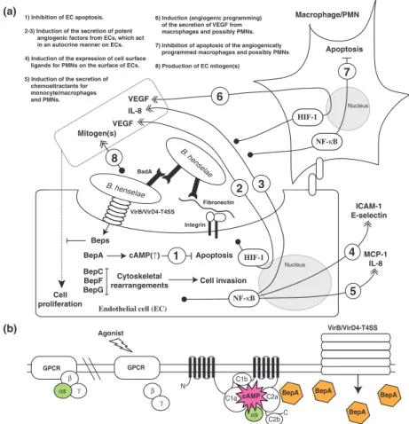
Hypothesis of the endothelial cells of the vascular wall The most widely appreciated primary niche hypothesis is the vascular wall, in more particular an intravacuolar compartment in an EC (Dehio, 2005). This is primarily based on the disease pathologies of B. quintana and B. ba-cilliformis infections in their natural reservoir host human, in addition to the wealth of information available for B. henselae–human EC interaction. Bartonella bacilli-formis is a deadly pathogen that causes a biphasic Car-rion’s disease in endemic areas of the Andes. The acute phase, called Oroya fever, is characterized by an intra-erythrocytic bacteremia that results in an often-fatal hemolytic anemia. The subsequent chronic phase, known as verruga peruana, manifests in vascular tumors that result from the proliferation of ECs where B. bacilliformis may form large cytoplasmic inclusions (Maguin˜a et al., 2009). A similar tropism for human erythrocytes is observed for B. quintana (Rolain et al., 2002; Rolain et al., 2003c). However, the bacteria do not appear hemo-lytic, and the development of vascular deformations/ tumors (BA) requires that the human host is immuno-compromised as an example suffering from AIDS (Ma-guin˜a et al., 2009). EC invasion by B. bacilliformis has been reproduced in vitro by several investigators (Garcia et al., 1990, 1992; Hill et al., 1992; Verma et al., 2000, 2001; Verma & Ihler, 2002; Cerimele et al., 2003). EC invasion by B. quintana appears more elusive in vitro, although the bacterium has been reported to adhere to and invade human ECs in vitro (Brouqui & Raoult, 1996;
Palmari et al., 1996; Mu¨ller et al., 2011) and potentially affect significant cellular processes such as apoptosis (Sch-mid et al., 2006) and proliferation (Palmari et al., 1996). Apart from B. quintana and B. bacilliformis, in vitro stud-ies of Bartonella spp. interaction with their reservoir host ECs have largely been neglected nor has such interaction ever been described in vivo. Only significant exception is one recent in vitro study where effects of B. henselae on human and feline macro- and microvascular ECs were compared (Berrich et al., 2011). The biological readouts with skin microvascular ECs clearly demonstrate that B. henselae increases the cell migration of human but not feline ECs in wound healing assays and that B. henselae strongly induces vascular endothelial growth factor (VEGF) secretion by human but not feline ECs (Berrich et al., 2011). These results may explain the reduced path-ogenic potential of B. henselae on cats as compared to humans. However, in the absence of direct cell adhesion and invasion data, the results appear inconclusive to answer the burning question if ECs truly constitute the primary niche of B. henselae in the cat. Further in vitro and in vivo studies in different mammals such as mouse or a rat with their respective Bartonella spp. colonizers are urgently needed. Conceptually, the EC primary niche hypothesis appears logical in the intravenous reservoir host models (Koesling et al., 2001; Schu¨lein et al., 2001). However, can it be applied to the more natural intradermal infection routes? Onset of the intraerythr-ocytic bacteremia after intradermal inoculation is pre-ceded by a significant lag (days–weeks) of abacteremia (Chomel et al., 1996; Abbott et al., 1997; Zhang et al., 2004; Marignac et al., 2010), which indicates that the bac-teria should enter the ECs from the apical surface, repli-cate, persist, and be synchronously seeded into the bloodstream.
Hypothesis of the circulatory hematopoietic stem cells ECs have a low proliferative potential, and therefore, vas-cular repair requires additional support. Vasvas-cular repair and neoangiogenesis are mediated, at least in part, by hematopoietic stem cells (HSCs) (Urbich & Dimmeler, 2004). It has been reported that B. henselae strain ATCC49882 adheres to and invades human HSCs, that is, CD133-positive cells that were enriched from peripheral blood mononuclear cells (Salvatore et al., 2008). CD133 is expressed on HSCs but is absent on mature ECs (Urb-ich & Dimmeler, 2004). This is a significant finding because circulating HSCs appear inherently resistant to invasion by a variety of microbial pathogens (Kolb-Ma¨urer et al., 2002). It was proposed that HSCs could carry B. henselae to peripheral tissues, in particular, to endothelium of microcirculation where vasoproliferative
disorders initiate (Salvatore et al., 2008). Although this might be the cellular basis of B. henselae-induced BA/peli-osis in the incidental host human, it has also been pro-posed that HSCs might constitute the primary niche of Bartonella spp. in their respective reservoir hosts (Ma¨ndle et al., 2005). This argument was based on an observation that intracellular B. henselae were detected in human ery-throid cells that were induced from HSCs, that is, CD34-positive cells that were enriched from peripheral blood (Ma¨ndle et al., 2005), by the addition of interleukin-3 (IL-3), granulocyte-macrophage-colony-stimulating factor (GM-CSF), and erythropoietin (epo) (Ma¨ndle et al., 2005). However, as B. henselae infection did not affect the differentiation of human HSCs into erythroid cells as judged by CD34 and glycophorin A cell surface markers, the results appear inconclusive given also the fact that B. henselae readily invade HSCs (Ma¨ndle et al., 2005). It is also well documented, as an example in the intravenous B. tribocorum-rat reservoir host infection model, that after injection the bacteria are rapidly cleared from circulating blood within hours and that no bacteria can be detected in the blood for about 4 days until the bacteremia peaks (Schu¨lein et al., 2001). Also, the lag of several days post-inoculation until the bacteria are sharply detected in the blood with high numbers in the intradermal reservoir host models (Chomel et al., 1996; Abbott et al., 1997; Zhang et al., 2004; Marignac et al., 2010) does not sup-port a circulatory nature of the primary niche.
Proposal of DCs and the draining lymph nodes
Upon exposure to microbial pathogens in peripheral tis-sues such as the inflamed skin, DCs migrate to lymph nodes and undergo maturation into potent immunostim-ulatory cells, especially to evoke a clonal expansion of antibody-producing B cells (Martı´n-Fontecha et al., 2009). We have already described the current knowledge of Bartonella spp.–DC interaction in ‘Means to affect effector functions of professional phagocytes’. In short, Bartonella spp. or at least B. grahamii in its natural reser-voir host mouse is not able to circumvent DC recogni-tion and activarecogni-tion, which appears of central importance for several pathogenic bacteria such as Brucella spp. to cause chronic infections (Billard et al., 2007). Actually, B. grahamii seems to overactivate cytokine responses of DCs, especially IFN-a/b secretion (Billard et al., 2007). Bartonella grahamii infection causes a more severe regio-nal lymphadenopathy in IFNAR1 (IFN-a/b receptor)-deficient mice compared with the wild-type mice (Kunz et al., 2008). This indicates that IFN-a/b overproduction is limiting the B-cell recruitment and/or B-cell prolifera-tion in the lymph nodes, the main cause of lymph node swelling/lymphadenopathy. The final outcome is the
inhibition of the antibody response and overall attenuation of host immune surveillance, which in the end allows the establishment of the chronic intraerythrocytic bacteremia. We would like to propose here that the draining lymph nodes are crucial anatomical sites first of all for the sup-pression of B-cell responses and thereby antibody pro-duction and secondly act as the anatomical site of the primary niche in reservoir hosts. The latter argument assumes that Bartonella spp. enter the draining lymph node within DCs that are migrating from the site of intradermal inoculation, although Bartonella spp. could also hypothetically gain access to the lumen of lymphatic vessels and eventually lymph nodes as a single bacterium and/or bacterial auto-aggregates, which are known to be formed by BadA of B. henselae (Kaiser et al., 2008) and Vomps of B. quintana (Zhang et al., 2004). Viability of the readily internalized Bartonella spp. in DCs (Vermi et al., 2006) has not been reported, but it could be sig-nificant based on the intracellular survival studies in macrophages, which inherently have more potent bacteri-cidal effects than DCs. Our proposal, like any other current hypothesis, cannot yet answer the peculiar peri-odicity of the bacterial appearance into the blood. Could it be envisioned that the bacterial replication to a certain density inside the lymph node–homed DCs would cause synchronous lysis of the infected DCs? One potential cytolytic bacterial factor is CAMP-like factor autotrans-porter Cfa, which was identified in B. henselae to induce hemolysis together with sphingomyelinase (Litwin & Johnson, 2005). The activity of this protein toward nucleated mammalian cells has not been reported, and it could also be functional in the arthropod host. Strong cytolytic activity of B. henselae has been identified toward tick Amblyomma americanum cell line (Billeter et al., 2009). There also appears to be a contact-dependent hemolytic activity in B. bacilliformis, which is protease sensitive, suggesting that it corresponds to a surface-exposed protein (Hendrix, 2000). It remains to be stud-ied whether this hemolysin is only present in B. bacilli-formis and how important this potential hemolytic activity actually is in the presence of the extremely potent hemophagocytic activity of monocyte/macrophages (Silva-Herzog & Detweiler, 2008). The lymph node pri-mary niche hypothesis could also explain the rapid clear-ance of bacteria from the circulating blood within hours postinoculation, with bacteremia peaking only after few days in the reservoir host intravenous infection models (Koesling et al., 2001; Schu¨lein et al., 2001). It is difficult to envision that bacteria could be migrating from the blood-filtering lymph nodes into a more peripheral tissue localization of the primary niche and then come back few days later. In part, the lymph node primary niche hypothesis is supported by the CSD in humans that is,
B. henselae infection in the incidental human host. Usu-ally, 2–3 weeks after a bite or a scratch of an infected cat, unusual lymphadenopathy of the lymph node(s) draining the area of the scratch or the bite develops, may suppurate and last for weeks (Klotz et al., 2011).
Bacterial factors involved in the colonization of the primary niche
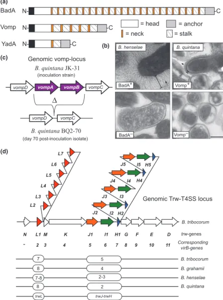
Several bacterial pathogens use type IV secretion systems (T4SSs) to translocate bacterial effector molecules (pro-teins or DNA) into the target host cells. These versatile transporters have evolved from bacterial conjugation sys-tems (Seubert et al., 2003; Christie et al., 2005). The prototypic T4SS is the VirB/VirD4 apparatus of Agrobac-terium tumefaciens, which mediates the transfer of the tumorigenic T-DNA complex into the infected plant cells. Mammalian pathogens have adapted T4SSs for the trans-fer of proteins directly into the host cell cytosol (for example, the CagA protein of Helicobacter pylori is trans-ported into gastric epithelial cells) or for the export of multisubunit protein toxins to the extracellular medium (for example, pertussis toxin secreted by Bordetella pertus-sis) (Christie et al., 2005), although they may still be capable of translocating DNA (Schro¨der et al., 2011). Bartonella spp. encode three distinct T4SSs, VirB/VirD4, Vbh, and Trw, which appear as key pathogenicity factors in mediating Bartonella spp.–host cell interactions (Schu-lein & Dehio, 2002; Seubert et al., 2003; Schmid et al., 2004; Vayssier-Taussat et al., 2010; Engel et al., 2011). The Trw–T4SS will be discussed in detail in ‘Adhesion to the erythrocytes’ and ‘Antigenic and phase variation of surface proteins’ in the context of erythrocyte adhesion and antigenic variation, respectively. The VirB/VirD4 T4SS has mainly been studied in B. henselae and B. tribocorum, but it appears to be well conserved also in other members of Bartonella spp., although it is absent in B. bacilliformis (Sweger et al., 2000; Alsmark et al., 2004; Saenz et al., 2007; Engel & Dehio, 2009; Engel et al., 2011). Bartonella spp. species of the clade 2 (Fig. 1), which apparently lack the VirB/VirD4-T4SS, contain a distinct T4SS that is homologous to VirB/VirD4-T4SS and is therefore designated as the virB-homolog-T4SS (Vbh-T4SS) (Saenz et al., 2007). The Vbh-T4SS is absent in B. bacilliformis and several species of the clade 4 (Saenz et al., 2007). Cellular functions of the Vbh-T4SS have not yet been studied in great detail (Engel et al., 2011).
The VirB/VirD4-T4SS of B. henselae is encoded by an operon composed of 10 genes (virB2–virB11) and a downstream-located virD4 gene (Schulein & Dehio, 2002; Schulein et al., 2005) (Fig. 5). Its closest bacterial relative is a genuine conjugation system, the AvhB/TraG system of the pATC58 of A. tumefaciens (Schulein & Dehio,
2002). A yeast two-hybrid interaction study of the com-ponents of the B. henselae VirB/VirD4-T4SS has largely confirmed the protein interactions that were previously identified in other T4SSs (Shamaei-Tousi et al., 2004; Christie et al., 2005) (Fig. 5). In analogy to the model developed for the topology and function of these related T4SSs (Christie et al., 2005), the B. henselae
VirB/VirD4-T4SS is considered to encompass a VirB2 pilin and VirB5 minor pilus components, which together form a pilus that apparently can mediate contact with the host cell; core components VirB3, VirB4, and VirB6–VirB11, which form a pore complex that spans both Gram-negative membranes; and the T4SS coupling protein VirD4, an inner membrane protein believed to function as an Fig. 5. Molecular characteristics of the VirB/VirD4-T4SS of Bartonella henselae. (a) A hypothetical model of the topology of B. henselae VirB/ VirD-T4SS based on studies on the archaetypical VirB/VirD4 T-DNA transfer system of plant pathogen Agrobacterium tumefaciens and yeast two-hybrid screen of protein–protein interactions between VirB proteins of B. henselae (Shamaei-Tousi et al., 2004; Christie et al., 2005). VirB6 of B. henselae has not yet been shown to interact with other VirB proteins. The coupling protein VirD4 is believed to recognize and target the effectors for translocation via the core T4SS machinery composed of VirB2-VirB11. CY, bacterial cytosol; IM, bacterial inner membrane; PP, bacterial periplasm; OM, bacterial outer membrane; EX, extracellular space. (b) Genetic organization of the genomic VirB/VirD4-T4SS locus. (c) Modular domain organization of Beps. The filamentous induced by cAMP (FIC) domain is characterized by a short amino acid motif (HPFXXGNG) of the catalytic center, which is highly conserved in Fic family proteins found in all domains of life and in viruses. Members of the Fic family of adenylyltransferases catalyze covalent addition of AMP moieties to target proteins, either at threonine or at tyrosine residues. The enzymatic activity was recently identified as the molecular basis how some pathogenic bacteria cause cytotoxicity through the modification of the switch I regions of Rho GTPases (Worby et al., 2009; Yarbrough et al., 2009; Palanivelu et al., 2011), the key regulators of cellular actin dynamics. The cellular functions of Bartonella spp. Fic proteins remain elusive. The BID domain and the adjacent positively charged C-tail are necessary for any given Bep to be translocated via the VirB/VirD4-T4SS (Schulein et al., 2005). Some of the Beps carry in their N-terminus short peptide motifs marked in black bars that resemble eukaryotic tyrosine phosphorylation motifs (e.g. EPLYA) (Selbach et al., 2009).
interface between the pore complex and the T4SS protein substrates (Fig. 5).
The proposed tip adhesin VirB5 (Fig. 5) is expressed in vivo as an immunodominant protein of B. henselae, as shown both after experimental infection of mice with B. henselae (Padmalayam et al., 2000) and by analyzing the sera of patients with CSD (Anderson et al., 1995). The homologous T4SS in B. tribocorum is indispensable for the establishment of the intraerythrocytic bacteremia in an intravenous natural rat reservoir host model as evidenced by comparative analysis of parental and VirD4- or VirB4-deficient mutant bacteria (Schulein & Dehio, 2002). Inter-estingly, a high fraction (> 80%) of VirB4-deficient mutant bacteria, which had been genetically complemented by the introduction of a virB4 copy in a replicative plasmid, were recovered from the blood without the complementation plasmid (Schulein & Dehio, 2002). The authors conclude that this finding indicates the functional importance of VirB/VirD4-T4SS for the initial abacteremic period of approximately 1 week during which the bacteria reside, replicate, and persist in the primary niche (Schu¨lein et al., 2001; Schulein & Dehio, 2002). Indeed, it has recently been reported that VirD4-deficient mutant of B. birtlesii is abac-teremic in an intradermal natural mouse reservoir host model, although the bacteria display equal erythrocyte adherence and invasion rates in vitro as compared to the parental strain (Vayssier-Taussat et al., 2010). At the moment, it remains unknown which of the VirB/VirD4-T4SS Bartonella spp. effector proteins, Beps (Fig. 5), are involved and how they are involved in the invasion and colonization of the primary niche and/or seeding of the bacteria from the primary niche into the bloodstream. However, BepD-deficient mutant has been identified in a signature-tagged mutagenesis (STM) screen as abacteremic (Saenz et al., 2007). This effector becomes tyrosine-phos-phorylated by the host cell–derived tyrosine kinases (Schu-lein et al., 2005) and therefore may interfere with the host cell signaling processes. Tyrosine phosphorylation is a con-served feature in several effectors translocated by bacterial pathogens, as an example in the T4SS-effector CagA of H. pylori (Odenbreit et al., 2000). Phosphorylation of CagA by Src family and Abl kinases recruits SH2-domain con-taining proteins such as tyrosine phosphatase Shp-2, tyro-sine kinase Csk, and adaptor protein Crk, all having a defined role in the H. pylori-induced cytoskeletal rear-rangements (Odenbreit et al., 2000; Backert & Selbach, 2005). Recently, a nonbiased quantitative proteomics approach was utilized to identify cellular target proteins that bind to the tyrosine-phosphorylated peptides of BepD, BepE, and BepF (Selbach et al., 2009). Several proteins such as Grb2, Grb7, Csk, Crk, Shp1, and Shp2 were identi-fied; however, the biology behind these interactions remains to be studied.
BadA mediates bacterial binding to endothelial and epithelial cells and extracellular matrix components in vitro (Riess et al., 2004, 2007; Kaiser et al., 2008; Mu¨ller et al., 2011). BadA-deficient mutants of B. triboco-rum and B. birtlesii have been identified as abacteremic in STM screens (Saenz et al., 2007; Vayssier-Taussat et al., 2010). Strikingly, the BadA-deficient mutants isolated in the B. birtlesii STM screen displayed equal erythrocyte adherence and invasion rates in vitro as compared to the parental strain (Vayssier-Taussat et al., 2010). This clearly indicates that in analogy to VirB/VirD4-T4SS, BadA is also required for the invasion and colonization of the mary niche and/or seeding of the bacteria from the pri-mary niche into the bloodstream. Perhaps this also applies to Vomps of B. quintana, because Vomp-deficient mutant is abacteremic in an intradermal macaque model (MacKi-chan et al., 2008). Several other bacterial factors with a pos-sible role in the primary niche colonization, such as inducible Bartonella autotransporter (iba) and several ABC transporters, have been identified in the STM screens (Sae-nz et al., 2007; Vayssier-Taussat et al., 2010). However, their detailed molecular functions remain to be studied.
Adhesion to the erythrocytes
The hallmark of chronic Bartonella spp. infection in their reservoir hosts, but not in the incidental hosts, is a long-lasting intraerythrocytic bacteremia. This also applies to the human-specific B. quintana (Rolain et al., 2002; Rolain et al., 2003c), a louse-transmitted bacterium that was initially detected during World War I as the causative agent of trench fever (5-day fever), and B. bacilliformis (Walker & Winkler, 1981; Benson et al., 1986), a sand fly-transmitted bacterium (Maguin˜a et al., 2009). Barto-nella bacilliformis is so far the only known species of Bartonella spp. that cause deleterious effects for the infected erythrocytes (Maguin˜a et al., 2009).
Bacterial adhesion is the first step in the erythrocyte invasion process. Bartonella bacilliformis is highly motile owing to the expression of multiple unipolar flagella (Benson et al., 1986; Scherer et al., 1993), and it has been reported that antibodies raised against the flagellin sub-unit partially inhibit erythrocyte binding and almost com-pletely abolish invasion (Scherer et al., 1993). However, the direct role of flagella in erythrocyte adhesin per se remains elusive by the lack of knowledge of the erythro-cyte ligand and genetic proof, that is, parallel analysis of wild-type and isogenic flagella-deficient mutant, although this mutant exists (Battisti & Minnick, 1999). The fla-gella-mediated motility could simply enhance bacteria– erythrocyte collisions, and other surface protein(s) might mediate the actual adhesion to the erythrocytes. This is in part supported by the fact that most Bartonella spp. are