Cerebral aneurysms in patients with autosomal dominant polycystic kidney disease—to screen, to clip, to coil?
4
0
0
Texte intégral
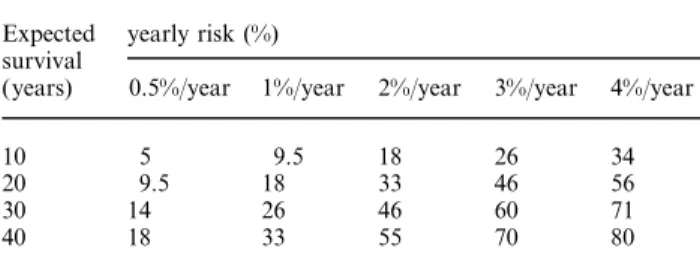
(2) 2320. L. Mariani et al.. years [18], size, location in the posterior circulation and a previous history of SAH from another aneurysm have been identified as predictors of rupture. The reported rate of rupture was surprisingly low: 0.05–0.5% per year for aneurysms smaller than 10 mm and approximately 1% per year for larger ones. The rupture rate for aneurysms larger than 25 mm was 6% per year during the first year. These results partially contradict several previous studies, which were included in a recent meta-analysis of the literature by Rinckel et al. [14]. They reported an overall risk of rupture of 1.9% per year (0.7% for aneurysms of 10 mm in diameter and less; 4% for larger ones) and a risk of 0.8% per year for asymptomatic aneurysms in patients without a previous history of SAH. In summary, the risk of aneurysm rupture depends mainly on its size, on a history of previous bleeding from another aneurysm, and on its location (the posterior circulation is at higher risk). Because most aneurysms detected in ADPKD patients are less than 10 mm in diameter the yearly risk of bleeding is low. However, the cumulated risk, remains significant. It obviously depends on the expected survival, which may be about 60 years in ADPKD patients [19]. Based on the annual risk of rupture and the expected survival, the approximate individual risk can be calculated (see Table 1). SAH from aneurysms is responsible for death in a relatively small proportion of patients with ADPKD [19]. However, the mean age at rupture in patients with ADPKD is between 35 and 40 years [19–21], that is 10–20 years earlier than in patients with sporadic SAH. This suggests that ADPKD per se is a risk factor for aneurysm rupture. The risk of developing new aneurysms in patients with a documented aneurysm may be as low as 2%. Although growth of aneurysms during long-term follow-up has been documented [22], it is probably a rare event.. The accuracy of Angio-MR examinations for screening The gold standard for the diagnosis and preoperative planning is the conventional four-vessel arteriography. Although the risk of death and permanent neurological injury of this procedure is approximately 0.5%, it might Table 1. Cumulative risk of aneurysm rupture depending on the yearly risk of rupture and expected survival at diagnosis according to the multiplicative law of probability Expected survival (years). yearly risk (%) 0.5%/year. 1%/year. 2%/year. 3%/year. 4%/year. 10 20 30 40. 5 9.5 14 18. 9.5 18 26 33. 18 33 46 55. 26 46 60 70. 34 56 71 80. Modified from Ref 30: risk of haemorrhage (in %)=1−(1−yearly risk) years of expected survival×100.. be higher in patients with ADPKD (see below). Angio-MR carries no risk and the contrast medium Gd-DTPA is not nephrotoxic. Its value in the detection of aneurysms is now well established [23,24]. Angiographically confirmed aneurysms of 6 mm or more in diameter have been detected with 100% sensitivity by two or more blinded readers with time-offlight-MRA [25]. The sensitivity decreased to 87.5, 68.2, 60 and 55.6% for aneurysms with a diameter of 5,4,3 and 2 mm respectively. There were no false positive results in these studies.. The risk of angiography Because cerebral panangiography is still an obligatory examination before treatment, its risk has also to be considered. There are few data on the specific risk of cerebral angiography in the subgroup of patients with ADPKD. The careful analysis of Chapman et al. [26 ], reported a 25% rate of transient complications, i.e. in eight of 32 patients: vasospasm with headache and nausea in two, severe headache in two, scotoma in two, scotoma and numbness of the hand in one, and asymptomatic dissection of one vertebral artery in one patient. All patients recovered completely after 48 h. None of the patients had significant elevation of the creatinine level after administration of contrast medium.. The risk of microsurgical clipping Craniotomy and microsurgical clipping is still the treatment of choice for aneurysms. A properly clipped aneurysm can be considered as cured. The great majority of authors do not report the efficiency of clipping, presumably because experienced neurosurgeons can rely on their intraoperative findings and perform control angiograms only in especially difficult cases. Microsurgical clipping of unruptured aneurysms is technically much easier and carries a lower risk of death or permanent morbidity than intervention after SAH. The results of surgery in unruptured or incidental aneurysms are well summarized in two recent metaanalyses of the literature [27,28]. The most important determinants for morbidity or mortality were the size and the location of the aneurysm. For non-giant (<25 mm) anterior circulation aneurysms mortality was 0.9% and morbidity 1.9% in contrast to 3 and 12.9% respectively for aneurysms in the posterior circulation. Giant aneurysms carried a much higher risk, i.e. 7.4% mortality and 26.9% morbidity for aneurysms in the anterior and even 9.6% mortality and 37.9% morbidity for aneurysms in the posterior circulation. In our opinion, however, the value of meta-analyses is limited. The risk of clipping an asymptomatic aneurysm has to be judged individually by an experienced neurosurgeon..
(3) Cerebral aneurysms with ADPKD. The role and risk of endovascular coiling As discussed above, there are patients in whom the operative risk is unreasonably high in relation to the natural risk. In such cases, especially for some large aneurysms and for aneurysms of the posterior circulation, the endovascular treatment is a good alternative. Using recent interventional neuroradiological techniques, a microcatheter can be navigated into the aneurysm. Its lumen is then occluded by deposition of electrolytically or mechanically detachable coils. However, the feasibility of this procedure depends on the local vascular anatomy, on the shape of the aneurysm, and on the experience of the endovascular neuroradiologist or neurosurgeon. Ideally, the aneurysm should have a small neck, i.e. the ratio maximal aneurysm diameter: neck diameter should be 2 or more. A definite cure has probably been achieved when the lumen of the aneurysm has been completely occluded. However, the long term follow-up is still unavailable. In a recent series of 115 patients with incidental aneurysms, a 63% rate of complete occlusion has been reported [29]. This underlines the importance of the proper selection patients with incidental aneurysms for coil therapy, because incomplete occlusion is not ideal. The overall morbidity and mortality was 5% in this study and was mostly due to embolism. However, a positive trend over time was observed suggesting a learning curve; the last 65 patients treated using intraoperative heparinization showed no complications [29]. Endovascular coiling is the preferred method for patients over 65 years of age.. Conclusion Because of (i) the relatively high prevalence of aneurysms in patients with ADPKD (approximately 10%), (ii) the significant annual risk of rupture (0.5–2%), and (iii) the potential catastrophic sequelae of SAH (>50% mortality and permanent disability), we feel that systematic screening with Angio-MR is advisable. Screening is specially indicated in relatives of patients with a known aneurysm. Because there is little chance to detect aneurysms before the age of 30 years and because then the risk of aneurysmal rupture is extremely small, screening is not recommended before the third decade of life. It remains unclear how often screening should be repeated, but a 5–10-year interval has been proposed and seems reasonable. If an aneurysm is detected by MR angiography, treatment options should be discussed with an experienced team of neurosurgeons and interventional neuroradiologists. Treatment is recommended when the individual risk of rupture is higher than the risk of treatment. The latter depends mainly on the age and general condition of the patient as well as on the size and location of the aneurysm. Microsurgical clipping is still the treatment of choice, when feasible, because it is curative.. 2321. Endovascular coiling is a good alternative in older patients. Acknowledgements. We would like to thank Dr B. O’Callaghan for the revision of the manuscript.. References 1. Schievink WI, Wijdicks EFM, Parisi JE, Piepgras DG, Whisnant JP. Sudden death from aneurysmal subarachnoid hemorrhage. Neurology 1995; 45: 871–874 2. Sacco RL, Wolf PA, Bharucha NE et al. Subarachnoid and intracerebral hemorrhage: natural history, prognosis, and precursive factors in the Framingham study. Neurology 1984; 34: 847–854 3. Longstreth WT Jr, Nelson LM, Koepsell TD, van Belle G. Clinical course of spontaneous subarachnoid hemorrhage: a population-based study in King County, Washington. Neurology 1993; 43: 712–718 4. Fogelholm R, Hernesniemi J, Vapalathi M. Impact of early surgery on outcome after aneurysmal subarachnoid hemorrhage: a population-based study. Stroke 1993; 24: 1649–1654 5. Inagawa T, Tokuda Y, Ohbayashi N, Takaya M, Moritake K. Study of aneurysmal subarachnoid hemorrhage in Izumo City, Japan. Stroke 1995; 26: 761–766 6. Schievink WI et al. Saccular intracranial aneurysms in autosomal dominant polycystic kidney disease. J Am Soc Nephrol 1992; 3: 88–95 7. Huston J III, Torres VE, Sullivan PP, Offord KP, Wiebers DO. Value of magnetic resonance angiography for the detection of intracranial aneurysms in autosomal dominant polycystic kidney disease. J Am Soc Nephrol 1993; 3: 1871–1877 8. Ruggieri PM et al. Occult intracranial aneurysms in polycystic kidney disease: screening with MR angiography. Radiology 1994; 191: 33–39 9. Ronkainen A et al. Familial intracranial aneurysms. Lancet 1997; 349: 1478–1479 10. Schievink WI. Intracranial aneurysms. Review article. N Engl J Med 1997; 336: 28–40 11. Linn FHH, Rinkel GJE, Algra A, van Gijn J. Incidence of subarachnoid hemorrhage: role of region, year and rate of computed tomography: a metanalysis. Stroke 1996; 27: 625–629 12. McCormick WF, Nofzinger JD. Saccular intracranial aneurysms: an autopsy study. J Neurosurg 1965; 22: 155–159 13. Inagawa T, Hirano A. Autopsy study of unruptured incidental intracranial aneurysms. Surg Neurol 1990; 34: 361–365 14. Rinkel GJ, Djibuti M, van Gijn J. Prevalence and risk of rupture of intracranial aneurysms: a systematic review. Stroke 1998; 29: 251–256 15. Wiebers DO, Whisnant JP, Sumdt TM Jr, O’Fallon WM. The significance of unruptured intracranial saccular aneurysms. J Neurosurg 1987; 66: 23–29 16. Schievink WI, Piepgras DG, Wirth FP. Rupture of previously documented small asymptomatic saccular intracranial aneurysms. Report of three cases. J Neurosurg 1992; 76: 1019–1024 17. Schievink WI, Prendergast V, Zabramski JM. Rupture of a previously documented small asymptomatic intracranial aneurysm in a patient with autosomal dominant polycystic kidney disease. Case report. J Neurosurg 1998; 89: 479–482 18. The International Study of Unruptured Intracranial Aneurysms Investigators (ISUIAI ). Unruptured intracranial aneurysms— risk of rupture and risks of surgical interventions. N Engl J Med 1998; 399: 1725–1733 19. Fick GM, Johnson AN, Hammond WS, Gabow PA. Causes of death in autosomal dominant polycystic kidney disease. J Am Soc Nephrol 1995; 5: 2048–2056 20. Chauveau D, Pirson Y, Verellen-Dumoulin C, Macnicol A, Gonzalo A, Gru¨nfeld JP. Intracranial aneurysms in autosomal dominant polycystic kidney disease. Kidney Int 1994; 45: 1140–1146 21. Lozano AM, Leblanc R. Cerebral aneurysms and polycystic kidney disease: a critical review. Can J Neurol Sci 1992; 19: 222–227.
(4) 2322 22. Juvela S, Porras M, Heiskanen O. Natural history of unruptured intracranial aneurysms: a long term follow-up study. J Neurosurg 1993; 79: 174–182 23. Gouliamos A, Gotsis E, Blahos L. et al. Magnetic resonance angiography compared to intra-arterial digital subtraction angiography in patients with subarachnoid hemorrhage. Neuroradiology 1992; 35: 46–49 24. Schuiere G, Huk WJ, Laub G. Magnetic resonance angiography of intracranial aneurysms: comparison with intra-arterial digital subtraction angiography. Neuroradiology 1992; 35: 50–54 25. Huston J III, Nichols DA, Luetmer PH et al. Blinded prospective evaluation of sensitivity of MR angiography to known intracranial aneurysms: importance of aneurysm size. Am J Neuroradiol 1994; 15: 1607–1614. L. Mariani et al. 26. Chapman AB et al. Intracranial aneurysm in adult polycystic kidney disease. N Engl J Med 1992; 327: 916–920 27. King JT, Berlin JA, Flamm ES. Morbidity and motality from elective surgery for asymptomatic, unruptured, intracranial aneurysms: a meta-analysis. J Neurosurg 1994; 81: 837–842 28. Raaymakers TWM, Rinkel GJE, Limburg M, Algra A. Mortality and morbidity of surgery for unruptured intracranial aneurysms. A meta-analysis. Stroke 1998; 29: 1531–1538 29. Murayama Y, Vinuela F, Duckwiler GR, Gobin YP, Guglielmi G. Embolization of incidental cerebral aneurysms by using the Guglielmi detachable coil system. J Neurosurg 1999; 90: 207–214 30. Kondziolka D, McLaughlin MR, Kestle JRW. Simple risk predictions for arteriovenous malformation hemorrhage. Neurosurgery 1995; 37: 851–855.
(5)
Figure
Documents relatifs
Neptunium sorption and redox speciation at the illite surface under highly saline conditions.. Nidhu Lal Banik, Rémi Marsac, Johannes Lützenkirchen, Christian Maquardt, Kathy
Grouping counselling approaches in this way allows the counsellor to examine the type of problem, relate it to an unmet need, and then attempt to match Client Problem.. Physical
Allez donc chercher la vérité dans cet imbroglio où, depuis toujours, tous avancent avec dans une main le Livre saint qui prône l’amour du prochain et dans
Toutes les salles sont équipées des technologies les plus récentes avec, pour les deux plus grandes, l’image en 3D et, surtout, dans la salle N°1, le nouveau son 3D Dolby Atmos
Cela rend également visible ce qui, pour des raisons de sécurité, se déroulera dans des halles fermées – de l’excavation des déchets par un système à ponts
(collective noun + singular or plural verb; audience can also be used as a countable noun with a normal plural: audiences are; also: class, club, committee, company,
After introducing some properties of the theory of ordinary differential equations, we provide a rigorous computational method for finding the periodic solution
The initial abstract model introduces the location grid (in- cluding the nest) with the distributed food and models the effect of ants activity – all the food is gradually
