M A J O R A R T I C L E
Genomic Epidemiology of Multidrug-Resistant
Mycobacterium tuberculosis During
Transcontinental Spread
Mireia Coscolla,1,2Pennan M. Barry,3John E. Oeltmann,6Heather Koshinsky,4Tambi Shaw,3Martin Cilnis,3Jamie Posey,6 Jordan Rose,5Terry Weber,3Viacheslav Y. Fofanov,4Sebastien Gagneux,1,2Midori Kato-Maeda,5and John Z. Metcalfe5
1
Medical Parasitology and Infection Biology, Swiss Tropical and Public Health Institute, and2University of Basel, Switzerland;3Division of Communicable Disease Control, Center for Infectious Diseases, California Department of Public Health, Richmond,4Eureka Genomics, Hercules, and5Division of Pulmonary and Critical Care Medicine, Francis J. Curry International Tuberculosis Center, San Francisco General Hospital, University of California; and
6
Centers for Disease Control and Prevention, Atlanta, Georgia
The transcontinental spread of multidrug-resistant (MDR) tuberculosis is poorly characterized in molecular epidemiologic studies. We used genomic sequencing to understand the establishment and dispersion of MDR Mycobacterium tuberculosis within a group of immigrants to the United States. We used a genomic ep-idemiology approach to study a genotypically matched (by spoligotype, IS6110 restriction fragment length poly-morphism, and mycobacterial interspersed repetitive units–variable number of tandem repeat signature) lineage 2/Beijing MDR strain implicated in an outbreak of tuberculosis among refugees in Thailand and con-secutive cases within California. All 46 MDR M. tuberculosis genomes from both Thailand and California were highly related, with a median difference of 10 single-nucleotide polymorphisms (SNPs). The Wat Tham Krabok (WTK) strain is a new sequence type distinguished from all known Beijing strains by 55 SNPs and a genomic deletion (Rv1267c) associated with increasedfitness. Sequence data revealed a highly prevalent MDR strain that included several closely related but distinct allelic variants within Thailand, rather than the occurrence of a sin-gle outbreak. In California, sequencing data supported multiple independent introductions of WTK with sub-sequent transmission and reactivation within the state, as well as a potential super spreader with a prolonged infectious period. Twenty-seven drug resistance–conferring mutations and 4 putative compensatory mutations were found within WTK strains. Genomic sequencing has substantial epidemiologic value in both low- and high-burden settings in understanding transmission chains of highly prevalent MDR strains.
Keywords. Mycobacterium tuberculosis; drug resistance; genomics; epidemiology; EmbR.
Mycobacterium tuberculosis is an ancient human path-ogen that continues to cause substantial morbidity and mortality, in part due to an expanding global epidemic of drug-resistant disease. In the United States, nearly 90% of multidrug-resistant (MDR) tuberculosis cases occur among foreign-born individuals [1], although
the relative proportion occurring through reactivation of latent MDR strains, direct importation of active dis-ease, and domestic transmission and reactivation, is not definitively known. Effective tuberculosis control strat-egies depend upon understanding these parameters among high-risk groups immigrating to the United States [2].
Analysis of data from next-generation whole-genome sequencing (WGS) allows detection of minute dif-ferences in genetic diversity and has contributed retrospectively to outbreak investigations [3–7] and population-based studies [8] in high-income settings. In the study of drug-resistant tuberculosis, WGS has improved understanding of causal mechanisms of drug resistance [9] and mutations compensatory for fit-ness costs associated with drug resistance [10]. Yet, Received 23 September 2014; accepted 6 January 2015; electronically published
18 January 2015.
Correspondence: John Z. Metcalfe, MD, PhD, MPH, University of California, San Francisco, Division of Pulmonary and Critical Care Medicine, San Francisco Ge-neral Hospital, 1001 Potrero Ave, Rm 5K1, San Francisco, CA 94110-0111 (john. [email protected]).
The Journal of Infectious Diseases®
2015;212:302–10
© The Author 2015. Published by Oxford University Press on behalf of the Infectious Diseases Society of America. All rights reserved. For Permissions, please e-mail: [email protected].
DOI: 10.1093/infdis/jiv025
transcontinental molecular epidemiology of drug-resistant tu-berculosis, including data from both high- and low-burden set-tings, is poorly represented in existing molecular epidemiologic studies of tuberculosis [11].
During 2004–2005, high MDR tuberculosis case rates among refugees living at Wat Tham Krabok (WTK) in Thailand coin-cided with thefinal major resettlement of Hmong peoples to the United States [12]. Transcontinental importation and evidence for domestic transmission of a lineage 2/Beijing MDR M. tuber-culosis strain led to major changes in Centers for Disease Control and Prevention (CDC) preimmigration tuberculosis screening protocols [13]. We generated and analyzed WGS data from M. tuberculosis genomes in both Thailand and the United States to clarify importation and establishment of the WTK strain within California.
METHODS Study Population
Since the late 1970s, WTK (a Buddhist temple in Saraburi Prov-ince, Thailand) has been home to Hmong refugeesfleeing po-litical persecution in Laos. Following recognition of high MDR tuberculosis case rates among Hmong refugees seeking resettle-ment and those recently resettled in the United States, a CDC-coordinated outbreak investigation at WTK between April 2004 and July 2005 identified 272 tuberculosis cases among 15 455 refugees, with 24 of 57 culture-positive individuals (42%) found to have MDR tuberculosis. Twenty isolates had identical IS6110 restriction fragment length polymorphisms (IS6110-RFLPs), spoligotyping results, and mycobacterial interspersed repetitive units–variable numbers of tandem repeat (MIRU-VNTR) signatures; of these, 15 (75%) had contact investigation data [12], were available among CDC-banked specimens, and were included in our analysis [12]. Documented exposure among several patients who had MDR tuberculosis simultane-ously, clustering of genotypes, concordant results of phenotypic drug susceptibility tests, and a high prevalence of tuberculin re-activity among household contacts were considered as evidence supporting an MDR tuberculosis outbreak within the camp.
California (2010 population, 37.2 million) state law requires reporting of all verified cases of tuberculosis (California Code of Regulations Title 17 §2500) to the California Department of Public Health (CDPH) Tuberculosis Registry. Routine genotyp-ing of clinical M. tuberculosis isolates has occurred since 2004, with cases prior to 2004 undergoing genotyping upon request (eg, in the course of an outbreak investigation and for special projects). MDR M. tuberculosis isolates in California with a genotype matching that yielded by the WTK investigation in Thailand (based on criteria described in the Conventional Genotyping subsection, below) were identified by searching genotyping results in the national and CDPH tuberculosis gen-otyping database. Of 225 MDR tuberculosis cases diagnosed
during 2004–2010, 22 (10%) occurred among persons of Hmong ethnicity. Five cases (23%) involved directly imported active MDR tuberculosis (symptomatic, culture-positive within 1 month of US arrival) and occurred concurrently with the Thailand outbreak. A systematic review of additional matching isolates that were identified during outbreak investigations and genotyped in California yielded 9 additional WTK MDR iso-lates collected during 1995–2003. Additional information from the epidemiologic investigation is available in the Supple-mentary Materials. All protocols were approved by the Califor-nia Health and Human Services Agency Committee for the Protection of Human Subjects and the University of California, San Francisco, Committee for the Protection of Human Subjects.
Conventional Genotyping
Extraction of genomic DNA from M. tuberculosis strains was per-formed during the log-phase growth of strains on culture medi-um. Spoligotyping, 24-locus MIRU-VNTR, and IS6110-RFLP genotyping were performed using standardized protocols. Isolates with an identical spoligotype (000000000003771), 24-locus MIRU-VNTR signature, and IS6110-RFLP (21-band pattern, ±1 band) were considered matching; for 12 of 31 California iso-lates (38%) and all Thai isoiso-lates, a 12-locus rather than 24-locus MIRU-VNTR genotype was available.
Phenotypic Drug Susceptibility Testing
California state law (California Code of Regulations Title 17 §2505) requires submission of all M. tuberculosis isolates to local public health laboratories and submission of all MDR M. tuberculosis isolates to the California Department of Public Health Microbial Diseases Laboratory (MDL). Tests for first-and second-line antituberculosis drug susceptibilities were performed at local laboratories or at the MDL, using the BAC-TEC 460 (Becton Dickinson Diagnostic Instruments, Sparks, Maryland), the MGIT 960 system (Becton Dickinson), or the agar proportion method.
Sequencing
Forty-six MDR strains (15 strains from Thailand, and 31 from California) were sequenced using HiSeq (Illumina;
Supplementary Table 1). Burrows-Wheeler Aligner v0.5.8c (BWA) and SMALT (https://www.sanger.ac.uk/resources/ software/smalt/) were used to map Illumina reads from these 46 genome sequences and 56 previously published lineage 2 genomes (Supplementary Table 1 and Supplemen-tary Table 2) against an inferred common ancestor of all M. tuberculosis complex lineages. An inferred common ancestor, rather than a previously sequenced strain (eg, H37Rv), was used as reference to avoid recovering mutations present only in the previously sequenced strain. The average number of reads that covers each position in the reference
genome ranged from 40× to 350× in different strains, with a median of 110×. Only nonredundant single-nucleotide poly-morphisms (SNPs) identified with BWA and SMALT map-ping were retained. For each strain, we called SNPs with Phred-scaled probability scores of >20, read depths lower than double the average read depth of the genome, and a min-imum of 5 reads. For filtering dense SNPs, a maximum of 2 SNPs were allowed within a window of size 10. Subclusters of strains were taken to represent putative transmission chains and were defined as genetically related M. tuberculosis isolates (≤4 SNP difference and proximal phylogenetic relationship according to our median joining network) from individuals with presumed or likely epidemiologic contact. Drug resis-tance–associated mutations identified in the Tuberculosis Drug Resistance Mutation Database [14] were retrieved from the SNP list (Table1andSupplementary Table 3). Drug resis-tance–conferring mutations (DRMs) in rpoB are noted in the
text by use of Escherichia coli notation; compensatory muta-tions were identified as nonsynonymous SNPs in rpoA or rpoC (Supplementary Table 3).
Phylogenetic Analysis
To examine the genetic diversity of the WTK strain, we sequenced and analyzed all 46 genomic sequences in conjunction with 56 widely diverse and geographically distributed lineage 2 strains se-lected from a global database (Supplementary Figure 1, Supple-mentary Table 1 and SuppleSupple-mentary Table 2). High-confidence DRMs were excluded from the diversity and phylogenetic analy-ses, since these are known to represent homoplastic events (ie, ste-reotyped evolution under the common selection pressure of antituberculosis medications).
Details about sequencing and phylogenetic analytic methods, including full references, are specified in the Supplementary Materials.
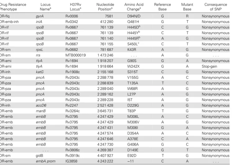
Table 1. Nonsynonymous Mutations According to Drug Resistance–Associated Locus Drug Resistance Phenotype Locus Namea H37Rv Locusa Nucleotide Positiona Amino Acid Changea Reference Base Mutant Base Consequence of SNP DR-flq gyrA Rv0006 7581 D94N/D G R Nonsynonymous
DR-emb-inh iniA Rv0342 412 280 Q481H G T Nonsynonymous
DR-rif rpoB Rv0667 761 139 H445Db C G Nonsynonymous
DR-rif rpoB Rv0667 761 139 H445Yb C T Nonsynonymous
DR-rif rpoB Rv0667 761 140 H445Rb A G Nonsynonymous
DR-rif rpoB Rv0667 761 155 S450Lc C T Nonsynonymous
DR-sm rpsL Rv0682 781 687 K43R A G Nonsynonymous
DR-sm rrs MTB000019 1 473 246 . . . A G . . .
DR-ami tlyA Rv1694 1 918 207 G90S G A Nonsynonymous
DR-ami tlyA Rv1694 1 918 664 W242X G A Stop-gain
DR-inh katG Rv1908c 2 155 168 S315T C G Nonsynonymous
DR-pza pncA Rv2043c 2 288 778 V155G A C Nonsynonymous
DR-pza pncA Rv2043c 2 288 839 T135A T C Nonsynonymous
DR-pza pncA Rv2043c 2 289 040 W68R A G Nonsynonymous
DR-pza pncA Rv2043c 2 289 162 L27P A G Nonsynonymous
DR-pza pncA Rv2043c 2 289 228 I5T A G Nonsynonymous
DR-inh accD6 Rv2247 2 521 428 D229G A G Nonsynonymous
DR-emb manB Rv3264c 3 645 731 T83P T G Nonsynonymous
DR-emb embB Rv3795 4 247 429 M306L A C Nonsynonymous
DR-emb embB Rv3795 4 247 429 M306V A G Nonsynonymous
DR-emb embB Rv3795 4 247 431 M306I G A Nonsynonymous
DR-emb embB Rv3795 4 247 574 D354A A C Nonsynonymous
DR-emb embB Rv3795 4 247 646 A378E C A Nonsynonymous
DR-emb embB Rv3795 4 247 730 G406A G C Nonsynonymous
DR-emb . . . Rv3806c 4 269 387 D149E G T Nonsynonymous
DR-sm gidB Rv3919c 4 407 927 E92D T G Nonsynonymous
DR-emb embA prom IG3858 4 243 222 −11 C A . . .
Abbreviation: SNP, single-nucleotide polymorphism.
a
Refer to the Mycobacterium tuberculosis H37Rv genome.
b
Corresponds to H526 in Escherichia coli.
c
Corresponds to S531 in Escherichia coli.
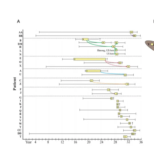
Figure 1. A, Epidemiologic linkages of multidrug-resistant tuberculosis cases in California and Thailand. California cases (yellow) are represented by the approximate date of US entry (left whisker), infectious period (box), and end of treatment (right whisker) or death (cross). Infectious periods were estimated as the interval from 3 months prior to symptom onset or abnormal chest radiograph through 2 weeks following initiation of appropriate antimicrobial therapy [15]. Epidemiologic links are indicated as colored arrows (solid lines indicate known epidemiologic links, and dashed lines indicate possible epidemiologic links). Twenty-nine patients for whom we had sequence data for the infecting strains are indicated in they-axis. B, Median joining network. The relationships of 46 Wat Tham Krabok (WTK) isolates were determined using 150 variable single-nucleotide polymorphisms (SNPs), excluding high-confidence drug resistance–associated mutations (Supplementary Table 3). Isolates and epidemiologic links are colored as described for panelA. Inferred nodes (unsampled) are represented by black circles. Shading indicates the relative SNP difference with respect to the central node. Note that cases D and M and cases R1andR2are paired longitudinal cases corresponding to 1 patient each.
T ranscontine ntal Spr ead of MDR M. tuber culosis
•
JID 2015:212 (15 July)•
RESULTS
Genomic Epidemiologic Investigations
In total, we performed WGS and analysis of 46 genotypically matched isolates from 15 patients in WTK and 29 patients (2 of whom had MDR tuberculosis twice) in California over a 22-year interval (Figure1andSupplementary Table 1). The over-all MDR tuberculosis incidence at WTK for 2004–2005 was 15 cases per 100 000. In California, the overall MDR tuberculosis in-cidence for 2004–2010 was 0.8 cases per 100 000 in the general population and 3.4 cases per 100 000 in the California Hmong population of 91 224 (in 2010). The median age of patients was in Thailand (35 years; interquartile range [IQR], 23–57 years) was similar to that in California (43 years; IQR, 20–66 years; P = .4). Approximately one-third of patients (n = 9) died during treatment in California, while mortality data were not available for Thailand. Three of 15 patients (20%) in Thailand and 6 of 29 (21%) in California were known to have previously received standardfirst-line tuberculosis treatment. Two individuals with a MDR WTK strain in California were born in the United States, one of whom was not of Hmong ethnicity. Among 7 individuals within household or community subclusters, the median time to reactivation following the end of the infectious period of an MDR tuberculosis putative source case was 6.2 years (IQR, 3.8–7.4 years). Among 13 individuals not within a well-supported trans-mission chain (genotypes of their isolates differed by >4 SNPs) and with tuberculosis not diagnosed upon arrival, the median time from US arrival to reactivation of MDR M. tuberculosis in-fection was 8.5 years (IQR, 3.8–12.0 years). During the study pe-riod in California, no tuberculosis due to a drug-susceptible WTK strain occurred (among any ethnic group), and all MDR tubercu-losis affecting persons of Hmong ethnicity was caused by a WTK strain.
Genome sequencing resolved the WTK cluster defined by conventional genotyping into several subclusters (≤4 SNP dif-ference, proximal phylogenetic relationships, and epidemiolo-gic linkages) in both California and Thailand (Figure 1). In Thailand, only 3 of 12 cases (25%) regarded as epidemiologi-cally linked in transmission chains were confirmed by genome sequencing. Moreover, we observed multiple branch points in the network, consistent with several closely related but distinct allelic variants (ie, a highly prevalent strain), rather than evidence of a single outbreak characterized by short genetic distances rep-resenting recent chains of transmission (eg, well-characterized in our study by the B-D-H-L-N group in California). The pres-ence of a highly prevalent strain was further supported by mul-tiple distinct combinations of DRMs, indicating drug resistance acquired independently on multiple occasions, rather than transmission of 1 drug-resistant strain from patient to patient (Figure2).
In California, genomic data supported a single case ( patient E) whose isolate occupied the central node in the WTK network
within California. Patient E arrived in the United States in the mid-1980s; received a diagnosis of cavitary, smear-positive MDR tuberculosis 2 years later; withdrew from treatment with-in a year; and had sputum smear-positive MDR tuberculosis diagnosed at death, 10 years later, indicating a potentially pro-longed infectious period. Seven cases (A, C, F, G, O, Q, and X) were contemporaneous with case E, and all had isolates that shared similar genotypes (≤4 SNP difference), suggesting that these cases may be in a chain of transmission. However, contact investigations could identify definitive epidemiologic links only among a subset of cases (O and X) that were extended family or household contacts (Figure1). Interestingly, the isolate from patient 17 (who had MDR tuberculosis diagnosed in Thailand and had not been in contact with patient E for >15 years) co-occupied the central node of the network with a nearly identical genotype. Epidemiological data integrated with the genomic network also demonstrated multiple independent importation events from Thailand with reactivation and transmission within the state. Patients I, S, P, Y, and R arrived in the United States following the death of patient E and likely represent indepen-dent importation events. Patients D and R1 had a second episode (M and R2, respectively) of MDR tuberculosis, the for-mer considered reinfection and the latter considered relapse following incomplete treatment. Public health contact investiga-tion activities at the time of the most recent Hmong resettle-ment (2004) also docuresettle-mented an MDR tuberculosis outbreak. This outbreak involved two neighboring households and pre-sumed transmission to a US-born person in a school setting. Genomic data demonstrated only minor differences in SNPs (≤4) between the isolate from patient B (the index case) and those from subsequent cases (H, L, M, and N) in this transmis-sion chain. Overall, genomic data supported all known links (100%; 10 of 10) and 78% of possible links (7 of 9). In addition, 7 other links (AA-BB, E-A, E-C, E-F, E-G, E-Q, and I-W-Y) were suggested by genomic data but were not supported epide-miologically. Of note, directionality according to WGS violated the temporal sequence of linked cases within 2 subclusters (eg, J-F and B-L); this could be explained by the presence of missing cases (eg, in subcluster J-F, 3 additional nongenotyped cases oc-curred in the same family), timing of transmission relative to specimen collection, or mixed infection with multiple strains. Phylogeny
WTK isolates from both Thailand and California were closely related (fixation index, 0.027) with a median of 10 SNPs (range, 0–20 SNPs) differentiating strains (Supplementary Fig-ure 1and Table2). In a sensitivity analysis, thefixation index did not significantly differ according to whether the 5 imported MDR tuberculosis cases were considered in the California or Thailand group. Moreover, the California strains showed a higher genetic diversification, compared with the Thai strains (π [±SD], 0.07 ± 0.005 vs 0.05 ± 0.005), suggesting multiple importation
events followed by the establishment and evolution of multiple WTK clones within California. This is further supported by the temporal appearance of the WTK strain in California and the finding that certain combinations of DRMs and compensatory mutations (eg, identical sets of embB resistance–conferring mu-tations) mapped exclusively to particular subclusters of the net-work (Figure2).
We found that the WTK strain was separated from all other lineage 2 strains sequenced to date and was defined by 55 speci-fic SNPs present in all WTK isolates but absent in other lineage 2 strains (Supplementary Figure 1and Table2). Fourteen of these SNPs were found in intergenic regions, and 41 were found in coding regions, 24 (59%) of which were nonsynonymous (Table2). Additionally, one intergenic SNP (between Rv0278c and Rv0279c) was homoplastic. All strains harbored genomic deletions previously described to be associated with lineage 2 strains (RD105, RD207, RD181, RD149, and RD152) [17], although only WTK strains harbored an additional deletion affecting the genetic locus Rv1267c.
Drug Resistance and Compensatory Mutations
High-confidence DRMs corroborated phenotypic drug-susceptibility test results, indicating resistance to isoniazid (katG S315T), rifampin (rpoB H526D/Y/R and S531L in 1 subset each), ethambutol (embB [A378E in all and M306L/V/I, D354A, and G406A in 1 subset each); Rv3806c D149E]), and streptomy-cin (rpsL K43R) were found (Table1andSupplementary Tables 1 and 3) [14]. An identical katG mutation in conjunction with dif-fering rpoB mutations indicates that a progenitor of the WTK strain was likely isoniazid resistant but not MDR. Additional pyr-azinamide (pncA) and capreomycin (tlyA) DRMs were present in subsets of isolates. Following misdiagnosis and known fluoro-quinolone exposure, patient X was found to have extensively drug-resistant tuberculosis with an isolate demonstrating hetero-resistance tofluoroquinolones (both wild type and mutation gyrA D94N were detected). Four possible compensatory muta-tions in rpoC (Supplementary Table 3) were found in strains with rpoB S531L:V775M (patients 6, W, and I), F831L ( patient X), W484G (patient 7), and P309S ( patients F and J; Figure2). In Figure 2. Median joining network with mapping of drug-resistance mutations. The relationships of 46 Wat Tham Krabok (WTK) isolates were determined using 150 variable single-nucleotide polymorphisms (SNPs), as described in Figure1B. Isolates are coded according to embB (fill color) and rpoB (border
color) mutation; other drug resistance and putative compensatory mutations are indicated in branches. Note that strains with matching drug resistance and/ or compensatory mutations are clustered together. Shading indicates the relative SNP difference with respect to the central node.
contrast, only a single rpoC mutation (S561P in patient 17, sub-clusters A-U and AA-BB) was associated with rpoB H526R.
In addition to 26 high-confidence DRMs (Table1and Supple-mentary Table 3), the WTK strain harbored 17 nonsynonymous or intergenic mutations recently proposed to be associated with multidrug resistance [18]. However, most of these mutations (12 of 17) have been previously identified as phylogenetic markers (6 SNPs are associated with lineage 2, 4 SNPs are associated with sublineage 2, and 2 SNPs are associated with lineages 2, 3, and 4) [16] and are therefore unlikely to have a causal association with drug resistance. Of the 5 remaining mutations, 1 each was distributed among 5 WTK strains, indicating a nonessential role in the propagation of the WTK strain.
DISCUSSION
We used next-generation sequencing data to delineate the lon-gitudinal clonal expansion of a lineage 2/Beijing MDR strain of M. tuberculosis among persons of Hmong ethnicity emigrating
from a Southeast Asian setting with a high burden of MDR tu-berculosis to the United States. We found that the domestic MDR tuberculosis rate among Hmong persons was >3 times that of the general population in California, a situation facilitat-ed by poverty and social isolation following resettlement in the United States [19,20]. Genomic data provided evidence against what was previously thought to be an MDR tuberculosis out-break in Thailand, indicated a central role for specific individ-uals in the establishment of the WTK strain in California, and confirmed multiple importation and subsequent reactivation events over a 22-year period.
Contact investigations aim to identify cases of active and latent M. tuberculosis infection among contacts in order to institute effective preventive therapy. This effort has been supplemented by M. tuberculosis genotyping based on mobile and repetitive ge-netic elements for >2 decades. However, conventional genotyping techniques examine <1% of the M. tuberculosis genome and are often insufficiently specific in outbreak situations, owing to a rate of change (the so-called molecular clock) that is slower than the rate of ongoing transmission and pathogenesis [21]. In contrast, WGS provides high-resolution molecular mapping of M. tubercu-losis that can identify short-term transmission events, even in the context of highly prevalent strains.
In our study, genomic data were decisive in resolving a puta-tive MDR tuberculosis outbreak in Thailand into multiple alle-lic variants of a highly prevalent strain. Despite extensive contact among cases and identical conventional genotypes [12], most cases were not related within recent transmission chains. Strains from lineage 2 (the East Asian lineage, which cludes the Beijing family of strains) are associated with an in-creased risk of drug resistance [22–24] and have been found to account for the majority of MDR tuberculosis cases in mono-phyletic fashion in other settings [8]. In California, a setting with a lower tuberculosis burden examined over a longer inter-val, conventional genotyping was sufficient to distinguish the WTK strain from other MDR strains and to discern relatedness to the 2004 investigation in Thailand. High-resolution molecu-lar techniques were necessary, however, to resolve short-term transmission chains, distinguish relapse from reinfection, and identify the central role of a potential super spreader in trans-mitting the WTK strain within California.
Interrogation of the complete M. tuberculosis genome is also advantageous in that it may identify genetic markers that ex-plain phenotypic consequences. We identified a deletion of Rv1267c (EmbR) within the WTK strain that may simultane-ously directly confer ethambutol resistance through mutations in the kinase-interacting domain of EmbR [25,26] and alter the ability of the host to mount an efficient immune response through functional changes in the ratio of lipoarabinomannan (LAM) to lipomannan (LM) [26,27]. The LAM/LM ratio has been associated with mycobacterial virulence, phagosome mat-uration, [28] apoptosis [29], and interferon signaling [30] in Table 2. Nonsynonymous Mutations Present in all Wat Tham
Krabok (WTK) Strains but Not Present in Other Lineage 2 Strains From the Global Collection
Reference Base Mutant Base Locusa Amino Acid Changea Genomic Positiona Locus Namea C G Rv0132c A212P 160 149 fgd2 C G Rv0592 P464R 691 891 mce2D A G Rv0614 I168V 709 857 . . . G C Rv0663 R290P 757 005 atsD C G Rv1166 T402R 1 297 356 lpqW T C Rv1524 F66L 1 718 921 . . . G T Rv1643 A123S 1 853 550 rplT G C Rv1784 A140P 2 021 051 . . . C T Rv1785c A58T 2 024 457 cyp143 C G Rv1813c A124P 2 055 743 . . . T C Rv1871c N71S 2 121 673 . . . T C Rv2520c N74S 2 837 395 . . . C G Rv2571c A212P 2 895 327 . . . A G Rv2601 I72V 2 928 601 speE G A Rv2700 R24Q 3 015 273 . . . A G Rv2702 K197E 3 017 446 ppgK C G Rv2763c D70H 3 073 402 dfrA G A Rv2834c T269I 3 140 509 ugpE C G Rv3201c V399L 3 575 842 . . . G A Rv3308 G233D 3 695 561 pmmB G A Rv3415c A16V 3 834 475 . . . G T Rv3447c S1233R 3 864 540 . . . G C Rv3596c L473V 4 039 288 clpC1 T C Rv3735 F87S 4 186 348 . . .
Data are from [16].
a
Refer to the Mycobacterium tuberculosis H37Rv genome.
macrophages and with interleukin 12 cytokine secretion by dendritic cells, all of which result in increasedfitness [31].
Previous work in several bacteria, including M. tuberculosis complex, has shown that mutations in rpoC can compensate for thefitness defects associated with mutations in rpoB that confer resistance to rifampin [10,32,33]. In M. tuberculosis, these rpoC mutations have been strongly associated with the rpoB S531L mutation, which is the most frequent mutation in rifampin-resistant clinical strains [10] and associated with a minorfitness defect in M. tuberculosis [34]. Hence, many differ-ent rpoC mutations seem to be able to compensate for the fit-ness defect associated with rpoB S531L. Our study supports this view, since 4 of 5 rpoC mutations that we identified were found in strains carrying rpoB S531L. Interestingly, an alternate rpoC mutation (S561P) was strongly associated with rpoB H526R. rpoB H526R has been shown to have a much greater fitness cost than rpoB S531L [34]. The fact that we found only a single rpoC mutation linked to rpoB H526R suggests that compensa-tion is somehow restricted in mutants carrying rpoB H526R, perhaps because the deleterious effect onfitness is stronger and therefore more difficult to compensate. More work is need-ed however to confirm this hypothesis.
Positive selection in strains exhibiting increasing levels of drug resistance map almost entirely to drug resistance–associated genes, and for drug-resistant M. tuberculosis (as in other mi-crobes) [35], the molecular antibiogram (ie, the collection of detected DRMs) has been used to corroborate other genotypic information in inferring chains of transmission [36]. In our study, embB resistance–conferring mutations were strikingly congruent within network subclusters. However, our study was not powered to examine the utility of DRMs as phylogenet-ic markers, and homoplasy would likely be a limiting factor for analyses undertaken on a broader scale [37].
Our study has potential limitations. First, estimates of clus-tering are typically based on a nonrandom sample of cases in a given community, and inference of transmission chains is subject to the same limitations as conventional genotyping with regard to sampling fraction and cluster size [21]. In Thai-land, and prior to 2004 in California, there was incomplete sam-pling of the population base. Thus, missed isolates identical to or highly similar to that infecting the putative super spreader ( patient E) might have provided an alternate explanation for some of the transmission of the WTK strain in California. Sec-ond, information on epidemiologic links was abstracted retro-spectively. Although detailed contact investigation data were often available, in particular for California cases, collecting addi-tional information prospectively was not feasible. Thus, some epidemiologic linkages and subsequent relationships between patients might be missed, including the identification of alternate source cases. Third, we avoided application of strict SNP cut points in inferring direct transmission. Accurate estimation of transmission trees relevant to public health practice will continue
to require a context of conventional epidemiologic information and a nuanced approach to SNP differences. This approach must account for within-person genetic variability (ie, pathogen variability during the same tuberculosis episode) and between-person genetic variability (ie, pathogen variability during se-quential transmission events) due to microevolution [38], mixed-strain infection [39], and heteroresistance [40]. Further, because SNP variability is heterogeneous in tempo across the full genome [41], accurate measurement of a molecular clock will require calibration of SNP changes to the gene regions they occupy.
In conclusion, WGS has epidemiologic added value in low-and high-burden settings low-and aids our understlow-anding of the transcontinental dispersion and transmission of MDR M. tuber-culosis. Used in real time, WGS may have alerted public health authorities to the presence of missing cases in chains of ongoing transmission or unknown sites of transmission in both Thailand and California. However, overall improvements to tuberculosis control or patient-important outcomes, along with questions of cost-benefit in low-burden settings, remain to be determined. Supplementary Data
Supplementary materialsare available at The Journal of Infectious Diseases online (http://jid.oxfordjournals.org). Supplementary materials consist of data provided by the author that are published to benefit the reader. The posted materials are not copyedited. The contents of all supplementary data are the sole responsibility of the authors. Questions or messages regard-ing errors should be addressed to the author.
Notes
Disclaimer. The authors alone are responsible for the views expressed in this publication, and they do not necessarily represent the decisions or policies of the Centers for Disease Control and Prevention.
Financial support. This work was supported by the National Institutes of Health (grants K23 AI094251 [to J. Z. M.] and R01 AI090928 [to S. G.]), the Robert Wood Johnson Foundation (Amos Medical Faculty Develop-ment Program award to J. Z. M.), the Swiss National Science Foundation (PP00P3_150750 to S. G.), and the European Research Council (309540-EVODRTB to S. G.).
Potential conflicts of interest. All authors: No reported conflicts. All authors have submitted the ICMJE Form for Disclosure of Potential Conflicts of Interest. Conflicts that the editors consider relevant to the con-tent of the manuscript have been disclosed.
References
1. Centers for Disease Control and Prevention. Reported tuberculosis in the United States, 2012.http://www.cdc.gov/tb/statistics/reports/2012/ pdf/report2012.pdf. Accessed 11 December 2013.
2. Schwartzman K, Oxlade O, Barr RG, et al. Domestic returns from in-vestment in the control of tuberculosis in other countries. N Engl J Med 2005; 353:1008–20.
3. Schurch AC, Kremer K, Daviena O, et al. High-resolution typing by in-tegration of genome sequencing data in a large tuberculosis cluster. J Clin Microbiol 2010; 48:3403–6.
4. Gardy JL, Johnston JC, Ho Sui SJ, et al. Whole-genome sequencing and social-network analysis of a tuberculosis outbreak. N Engl J Med 2011; 364:730–9.
5. Walker TM, Ip CL, Harrell RH, et al. Whole-genome sequencing to de-lineate Mycobacterium tuberculosis outbreaks: a retrospective observa-tional study. Lancet Infect Dis 2013; 13:137–46.
6. Roetzer A, Diel R, Kohl TA, et al. Whole genome sequencing versus tra-ditional genotyping for investigation of a Mycobacterium tuberculosis outbreak: a longitudinal molecular epidemiological study. PLoS Med 2013; 10:e1001387.
7. Kato-Maeda M, Ho C, Passarelli B, et al. Use of whole genome sequenc-ing to determine the microevolution of Mycobacterium tuberculosis during an outbreak. PLoS One 2013; 8:e58235.
8. Casali N, Nikolayevskyy V, Balabanova Y, et al. Evolution and transmis-sion of drug-resistant tuberculosis in a Russian population. Nat Genet 2014; 46:279–86.
9. Safi H, Lingaraju S, Amin A, et al. Evolution of high-level ethambutol-resistant tuberculosis through interacting mutations in decaprenylphos-phoryl-beta-D-arabinose biosynthetic and utilization pathway genes. Nat Genet 2013; 45:1190–7.
10. Comas I, Borrell S, Roetzer A, et al. Whole-genome sequencing of ri-fampicin-resistant Mycobacterium tuberculosis strains identifies com-pensatory mutations in RNA polymerase genes. Nat Genet 2012; 44:106–10.
11. Long R, Nobert E, Chomyc S, et al. Transcontinental spread of multi-drug-resistant Mycobacterium bovis. Am J Respir Crit Care Med 1999; 159:2014–7.
12. Oeltmann JE, Varma JK, Ortega L, et al. Multidrug-resistant tuberculo-sis outbreak among US-bound Hmong refugees, Thailand, 2005. Emerg Infect Dis 2008; 14:1715–21.
13. Lowenthal P, Westenhouse J, Moore M, Posey DL, Watt JP, Flood J. Re-duced importation of tuberculosis after the implementation of an en-hanced pre-immigration screening protocol. Int J Tuberc Lung Dis 2011; 15:761–6.
14. Sandgren A, Strong M, Muthukrishnan P, Weiner BK, Church GM, Murray MB. Tuberculosis drug resistance mutation database. PLoS Med 2009; 6:e2.
15. National Tuberculosis Controllers Association, Centers for Disease Control and Prevention. Guidelines for the investigation of contacts of persons with infectious tuberculosis. Recommendations from the National Tuberculosis Controllers Association and CDC. MMWR Recomm Rep 2005; 54:1–47.
16. Comas I, Coscolla M, Luo T, et al. Out-of-Africa migration and Neolith-ic coexpansion of Mycobacterium tuberculosis with modern humans. Nat Genet 2013; 45:1176–82.
17. Tsolaki AG, Hirsh AE, DeRiemer K, et al. Functional and evolutionary genomics of Mycobacterium tuberculosis: insights from genomic dele-tions in 100 strains. Proc Natl Acad Sci U S A 2004; 101:4865–70. 18. Zhang H, Li D, Zhao L, et al. Genome sequencing of 161
Mycobacteri-um tuberculosis isolates from China identifies genes and intergenic re-gions associated with drug resistance. Nat Genet 2013; 45:1255–60. 19. Leigh Brown P. A Hmong generationfinds its voice in writing. New
York Times 31 December 2011.http://www.nytimes.com/2012/01/01/ us/a-hmong-generation-finds-its-voice-in-writing.html?adxnnl=1& pagewanted=all&adxnnlx=1388281090-lpvZQbK691t1oUtDgHMZwQ. Accessed 28 December 2013.
20. Fadiman A. The spirit catches you and you fall down: a Hmong child, her American doctors, and the collision of two cultures. New York: Far-rar, Straus and Giroux, 1997.
21. Glynn JR, Vynnycky E, Fine PE. Influence of sampling on estimates of clustering and recent transmission of Mycobacterium tuberculosis de-rived from DNAfingerprinting techniques. Am J Epidemiol 1999; 149:366–71.
22. European Concerted Action on New Generation Genetic Markers and Techniques for the Epidemiology and Control of Tuberculosis. Beijing/W genotype Mycobacterium tuberculosis and drug resistance. Emerg Infect Dis 2006; 12:736–43.
23. Borrell S, Gagneux S. Infectiousness, reproductivefitness and evolution of drug-resistant Mycobacterium tuberculosis. Int J Tuberc Lung Dis 2009; 13:1456–66.
24. Ford CB, Shah RR, Maeda MK, et al. Mycobacterium tuberculosis mu-tation rate estimates from different lineages predict substantial differ-ences in the emergence of drug-resistant tuberculosis. Nat Genet 2013; 45:784–90.
25. Ramaswamy SV, Amin AG, Goksel S, et al. Molecular genetic analysis of nucleotide polymorphisms associated with ethambutol resistance in human isolates of Mycobacterium tuberculosis. Antimicrob Agents Che-mother 2000; 44:326–36.
26. Sharma K, Gupta M, Pathak M, et al. Transcriptional control of the my-cobacterial embCAB operon by PknH through a regulatory protein, EmbR, in vivo. J Bacteriol 2006; 188:2936–44.
27. Zhang N, Torrelles JB, McNeil MR, et al. The Emb proteins of myco-bacteria direct arabinosylation of lipoarabinomannan and arabinogalac-tan via an N-terminal recognition region and a C-terminal synthetic region. Mol Microbiol 2003; 50:69–76.
28. Fratti RA, Chua J, Vergne I, Deretic V. Mycobacterium tuberculosis gly-cosylated phosphatidylinositol causes phagosome maturation arrest. Proc Natl Acad Sci U S A 2003; 100:5437–42.
29. Rojas M, Garcia LF, Nigou J, Puzo G, Olivier M. Mannosylated lipoar-abinomannan antagonizes Mycobacterium tuberculosis-induced macro-phage apoptosis by altering Ca+2-dependent cell signaling. J Infect Dis 2000; 182:240–51.
30. Hmama Z, Gabathuler R, Jefferies WA, de Jong G, Reiner NE. Atten-uation of HLA-DR expression by mononuclear phagocytes infected with Mycobacterium tuberculosis is related to intracellular seques-tration of immature class II heterodimers. J Immunol 1998; 161: 4882–93.
31. Keane J, Remold HG, Kornfeld H. Virulent Mycobacterium tuberculosis strains evade apoptosis of infected alveolar macrophages. J Immunol 2000; 164:2016–20.
32. Brandis G, Hughes D. Genetic characterization of compensatory evolu-tion in strains carrying rpoB Ser531Leu, the rifampicin resistance muta-tion most frequently found in clinical isolates. J Antimicrob Chemother 2013; 68:2493–7.
33. Brandis G, Wrande M, Liljas L, Hughes D. Fitness-compensatory mu-tations in rifampicin-resistant RNA polymerase. Mol Microbiol 2012; 85:142–51.
34. Gagneux S, Long CD, Small PM, Van T, Schoolnik GK, Bohannan BJ. The competitive cost of antibiotic resistance in Mycobacterium tubercu-losis. Science 2006; 312:1944–6.
35. Harris SR, Cartwright EJ, Torok ME, et al. Whole-genome sequencing for analysis of an outbreak of meticillin-resistant Staphylococcus aureus: a descriptive study. Lancet Infect Dis 2013; 13:130–6.
36. Clark TG, Mallard K, Coll F, et al. Elucidating emergence and transmis-sion of multidrug-resistant tuberculosis in treatment experienced pa-tients by whole genome sequencing. PLoS One 2013; 8:e83012. 37. Ioerger TR, Feng Y, Chen X, et al. The non-clonality of drug resistance
in Beijing-genotype isolates of Mycobacterium tuberculosis from the Western Cape of South Africa. BMC Genomics 2010; 11:670. 38. Perez-Lago L, Comas I, Navarro Y, et al. Whole genome sequencing
analysis of intrapatient microevolution in Mycobacterium tuberculosis: potential impact on the inference of tuberculosis transmission. J Infect Dis 2014; 209:98–108.
39. Merker M, Kohl TA, Roetzer A, et al. Whole genome sequencing reveals complex evolution patterns of multidrug-resistant Mycobacterium tu-berculosis Beijing strains in patients. PLoS One 2013; 8:e82551. 40. Rinder H. Hetero-resistance: an under-recognised confounder in
diag-nosis and therapy? J Med Microbiol 2001; 50:1018–20.
41. Comas I, Chakravartti J, Small PM, et al. Human T cell epitopes of My-cobacterium tuberculosis are evolutionarily hyperconserved. Nat Genet 2010; 42:498–503.