HAL Id: inserm-00877866
https://www.hal.inserm.fr/inserm-00877866
Submitted on 29 Oct 2013
HAL is a multi-disciplinary open access
archive for the deposit and dissemination of
sci-entific research documents, whether they are
pub-lished or not. The documents may come from
teaching and research institutions in France or
L’archive ouverte pluridisciplinaire HAL, est
destinée au dépôt et à la diffusion de documents
scientifiques de niveau recherche, publiés ou non,
émanant des établissements d’enseignement et de
recherche français ou étrangers, des laboratoires
Intravitreal Aflibercept Injection for Macular Edema
Resulting from Central Retinal Vein Occlusion:
One-Year Results of the Phase 3 GALILEO Study.
Jean-Francois Korobelnik, Frank Holz, Johann Roider, Yuichiro Ogura,
Christian Simader, Ursula Schmidt-Erfurth, Katrin Lorenz, Miki Honda,
Robert Vitti, Alyson Berliner, et al.
To cite this version:
Jean-Francois Korobelnik, Frank Holz, Johann Roider, Yuichiro Ogura, Christian Simader, et al..
In-travitreal Aflibercept Injection for Macular Edema Resulting from Central Retinal Vein Occlusion:
One-Year Results of the Phase 3 GALILEO Study..
Ophthalmology: Journal of The
Ameri-can Academy of Ophthalmology, Elsevier, 2014, 121 (1), pp.202-8. �10.1016/j.ophtha.2013.08.012�.
�inserm-00877866�
Intravitreal Aflibercept Injection for Macular Edema Due to Central Retinal Vein
Occlusion: 1-Year Results of the Phase 3 GALILEO Study (135 of 135 characters allowed)
Jean-François Korobelnik, MD1-3; Frank G. Holz,MD4; Johann Roider, MD5; Yuichiro Ogura, MD6; Christian Simader, MD7; Ursula Schmidt-Erfurth, MD7; Katrin Lorenz,MD8; Miki Honda, MD9; Robert Vitti, MD10; Alyson J. Berliner, MD, PhD10; Florian Hiemeyer11; Brigitte Stemper,
MD11,12; Oliver Zeitz, MD11,13; Rupert Sandbrink, MD11,14 for the GALILEO Study Group
1Service d’ophtalmologie, Hopital Pellegrin—CHU de Bordeaux, Bordeaux, France; 2Université
Bordeaux Segalen, Bordeaux, France; 3INSERM, ISPED, Centre INSERM U897-Epidemiologie-Biostatistique, Bordeaux, France; 4Department of Ophthalmology, University of Bonn, Bonn, Germany; 5Department of Ophthalmology, University of Kiel, Kiel, Germany; 6Department of
Ophthalmology and Visual Science, Nagoya City University Graduate School of Medical Science, Nagoya, Japan; 7Department of Ophthalmology, Medical University of Vienna, Vienna, Austria; 8University Medical Center, Johannes Gutenberg-Universität Mainz, Mainz, Germany,
9Department of Ophthalmology, Juntendo University Urayasu Hospital, Chiba, Japan, 10
Regeneron Pharmaceuticals, Inc., Tarrytown, New York, USA; 11Bayer HealthCare AG, Berlin, Germany; 12Department of Neurology, University of Erlangen-Nürnberg, Germany, 13Klinik und Poliklinik für Augenheilkunde, Universitätsklinikum Hamburg-Eppendorf, Hamburg, Germany;
14Department of Neurology, Heinrich-Heine-Universität, Düsseldorf, Germany
Meeting presentation: This study was presented at the American Academy of Ophthalmology
Annual Meeting, November, 2012. This was an annual paper presentation.
Financial Support: The GALILEO study was funded by Regeneron Pharmaceuticals, Inc.,
Tarrytown, NY and Bayer HealthCare, Berlin, Germany. The sponsors participated in the design and conduct of the study, analysis of the data, and preparation of the manuscript.
Conflict of Interest: Dr. Korobelnik is a consultant to Alcon, Allergan, Bayer HealthCare, Carl
Zeiss Meditec, Novartis, and Thea. Dr. Holz is a consultant to Acucela, Alcon, Allergan, Bayer HealthCare, Genentech, Heidelberg Engineering, Novartis, and Pfizer and has received research funding from Alcon, Allergan, Bayer HealthCare, Carl Zeiss Meditec, GlaxoSmithKline, Heidelberg Engineering, Novartis, and Optos. He has also received travel support from Bayer HealthCare and lecture fee from Alcon, Bayer HealthCare, Heidelberg Engineering, Novartis, and Pfizer. Dr. Roider is a consultant to Bayer HealthCare, and has received travel support and fee for data monitoring/reviewing and statistical analysis from Bayer HealthCare. Dr. Ogura is a consultant to Alcon, Bayer HealthCare, Santen, and Wakamoto, and has received travel support from Bayer HealthCare. Dr. Simader’s institution has received payments from the Medical University of Vienna for data monitoring/reviewing, statistical analysis, and travel. Dr. Schmidt-Erfurth is a consultant to and has received travel support and fee for data monitoring/reviewing
and statistical analysis from Bayer HealthCare. She is also a consultant to and an advisory board member for Alcon, Allergan, Bohringer, and Novartis. She has received lecture fees from Alcon, Allergan, Bohringer, and Novartis. Dr. Lorenz is an advisory board member in
GeneSignal SAS and Sensimed AG, and a consultant to Bayer HealthCare. She has received research funding from FP7 European Union. She has also received lecture fee from Bayer HealthCare and MSD Pharmaceuticals, and travel support from Bayer HealthCare, Ivantis, Inc. Dr. Honda has no conflict of interest to disclose. Drs. Vitti and Berliner are employees of Regeneron Pharmaceuticals, Inc. Florian Hiemeyer and Drs. Stemper, Zeitz, and Sandbrink are employees of Bayer HealthCare.
Running head: Intravitreal aflibercept for macular edema due to CRVO (55 of 60 characters
allowed).
The investigators from the GALILEO study are listed in Appendix 1, available at http://aaojournal.org.
Corresponding author:
Jean-François Korobelnik, MD
Service d'Ophtalmologie, Hôpital Pellegrin Place Amélie Raba Léon, 33000 Bordeaux, France
Tel: +33 (0)5 56 79 57 41 Fax: +33 (0)5 56 79 47 58
email: jean-francois.korobelnik@chu-bordeaux.fr Manuscript length: 17 pages of 16-20 pages allowed.
Précis (35 of 35 words allowed)
The visual and anatomical improvements achieved after 6 monthly intravitreal doses of aflibercept were largely maintained over the second six months with as-needed dosing in patients with macular edema secondary to central retinal vein occlusion.
ABSTRACT (307 of 350 words)
Purpose: To evaluate the efficacy and safety of intravitreal aflibercept injections for treatment of
macular edema secondary to central retinal vein occlusion (CRVO).
Design: A randomized, multicenter, double-masked, phase 3 study.
Participants: A total of 177 treatment-naive patients with macular edema secondary to CRVO
were randomized in a 3:2 ratio.
Methods: Patients received either 2 mg intravitreal aflibercept or sham injections every 4 weeks
for 20 weeks. From week 24 to 48, the aflibercept group received aflibercept as needed (PRN), and the sham group continued receiving sham injections.
Main Outcome Measures: The primary efficacy endpoint was the proportion of patients who
gained ≥ 15 letters in best-corrected visual acuity (BCVA) at week 24. This study reports week-52 results including the proportion of patients who gained ≥ 15 letters in BCVA and the mean change from baseline BCVA and central retinal thickness (CRT). Efficacy endpoints at week 52 were all exploratory.
Results: At week 52, the mean percentage of patients gaining ≥15 letters was 60.2% in the
aflibercept group and 32.4% in the sham group (P = .0004). Aflibercept patients, compared with sham patients, had a significantly higher mean improvement in best-corrected visual acuity (+16.9 vs +3.8 letters, respectively) and reduction in central retinal thickness (-423.5 vs -219.3 µm, respectively) at week 52 (P < .0001 for both). Aflibercept patients received a mean (standard deviation) of 2.5 (1.7) injections during PRN dosing. The most common ocular adverse events in the aflibercept group were related to the injection procedure or the underlying disease and included macular edema (33.7%), increased intraocular pressure (17.3%), and eye pain (14.4%).
Conclusions: Treatment with intravitreal aflibercept provided significant functional and
monthly doses at week 24 were largely maintained until week 52 with as-needed dosing. Intravitreal aflibercept was generally well tolerated.
INTRODUCTION
The most common cause of vision loss in patients with central retinal vein occlusion (CRVO) is macular edema, which resolves spontaneously in only 30% of non-ischemic cases, and may not resolve in ischemic cases.1,2 Several lines of evidence indicate that vascular
endothelial growth factor (VEGF) may play a key role in the pathophysiology of macular edema secondary to CRVO. VEGF is released in response to retinal hypoxia, which occurs in CRVO due to impaired capillary blood flow.3 VEGF stimulates angiogenesis, and may result in neovascularization of the retina and/or the anterior segment, as well as vascular leakage resulting in macular edema.3 In CRVO patients, the vitreous level of VEGF correlates with the
severity of macular edema.4 Furthermore, intravitreal injections of the anti-VEGF agents,
ranibizumab or aflibercept, significantly improve visual and anatomical outcomes in patients with macular edema secondary to CRVO.5-9
Intravitreal aflibercept (historically known in the scientific literature as VEGF Trap-Eye [VTE]; Regeneron Pharmaceuticals, Inc.; Tarrytown, NY, USA, and Bayer Healthcare Pharmaceuticals, Berlin, Germany) is a fusion protein of key domains from human VEGF receptors 1 and 2 with human IgG Fc that binds to multiple VEGF-A isoforms with a higher affinity than ranibizumab and bevacizumab.10 Studies of intravitreal aflibercept injections in patients with neovascular age-related macular degeneration (AMD) demonstrated that aflibercept given monthly for three initial administrations and then once every two months improves visual and anatomical outcomes as effectively and safely as monthly ranibizumab over a one-year period.11 The efficacy and safety of intravitreal aflibercept for the treatment of
macular edema secondary to CRVO was investigated in two parallel trials performed in Europe and Asia/Pacific (GALILEO) and in the US (COPERNICUS).5,7,9 The primary efficacy endpoint of the GALILEO study was at week 24 and was published previously.9 Here we report the 52-week results of the GALILEO study.
METHODS Study Design
The GALILEO study is an 18-month, randomized, double-masked, phase 3 study comparing the efficacy and safety of intravitreal aflibercept with sham for the treatment of macular edema secondary to CRVO. The study protocol was approved by the institutional review board or ethics committee at each site. All patients signed a written consent form before initiation of the study-specific procedures. The study was registered with ClinicalTrials.gov (identifier no. NCT01012973) and was conducted across 63 sites in Europe and Asia/Pacific in compliance with ethical guidelines from the Declaration of Helsinki and International Conference on Harmonization. Data for this 52-week report were collected between October 2009 and July 2011.
The design and eligibility criteria for the GALILEO study have previously been described.9 Only one eye from each patient was included in the study. Patients were randomized in a 3:2 ratio to receive 2 mg intravitreal aflibercept (IVT-AFL 2Q4) or sham injections in the study eye once every 4 weeks for 20 weeks, for a total of 6 doses (Figure 1). From week 24 to week 52, patients in the aflibercept group were evaluated monthly and received aflibercept as needed (PRN) (IVT-AFL 2Q4 + PRN) if they had a > 50 µm increase in central retinal thickness (CRT) compared with the lowest previous measurement, new or persistent cystic changes within the neurosensory retina or subretinal fluid, persistent diffuse edema ≥ 250 µm in the central subfield, loss of ≥ 5 letters from the best prior measurement in conjunction with any increase in CRT, or an increase of ≥ 5 letters in best-corrected visual acuity (BCVA) from the most recent visit, suggesting potentially further improvements upon a subsequent injection. If none of the retreatment criteria were met, patients received a sham injection to maintain masking. Patients in the sham group continued to receive sham injections at all visits through week 52. All patients were eligible to receive panretinal laser
anterior segment, optic disc, or elsewhere in fundus. Given that there was no approved treatment for CRVO when the GALILEO study was designed, no other rescue treatment was prespecified. The GALILEO study design included a full year treatment with sham based on the request from health authorities. However, considering this long duration of sham treatment, the visual acuity and other ocular findings were carefully monitored by a team of masked medical reviewers. If, at any time, this review team had the impression that a patient might not benefit from further study participation or might be more adequately treated outside the study, the investigator was queried and asked to provide a re-assessment of the patient. Investigators then used their medical judgment to ultimately determine whether it would still be beneficial for the patient to continue the study.
Outcome Measures
The primary efficacy endpoint of the GALILEO study was the proportion of patients achieving a gain of ≥ 15 letters in BCVA from baseline to week 24, which was published previously.9 Here we report the 52-week results of the GALILEO study. Efficacy endpoints at
week 52 were all exploratory and included the proportion of patients who gained ≥ 15 letters in BCVA, mean change from baseline BCVA and CRT, proportion of patients progressing to neovascularization of the anterior segment, optic disc, or elsewhere in the fundus, and change from baseline in the mean National Eye Institute 25-item Visual Function Questionnaire (NEI VFQ-25) total and subscale (distance activities, near activities, and vision dependency) scores.
The efficacy and safety endpoints were assessed as described previously.9 BCVA and
CRT were assessed at baseline and every 4 weeks afterwards to week 52. Fundus
photography and fluorescein angiography (FA) were performed at screening (days -21 to -1) and weeks 12, 24, 36, and 52. Retinal perfusion status was determined by FA. Perfused and nonperfused retinas were defined as those with, respectively, < 10 disc area [DA] and ≥ 10 DA of capillary nonperfusion on FA. Vision-related quality of life was assessed at baseline and
weeks 24 and 52 using the NEI VFQ-25, which was administered by masked site personnel prior to intravitreal injections.
Statistical Analyses
The efficacy endpoints were analyzed in the full analysis set (FAS), which included all randomized patients who received any study treatment and had a baseline and at least one post-baseline BCVA assessment. In a prespecified analysis of proportions of patients who gained ≥ 15 letters at week 24 (the primary efficacy endpoint), patients who discontinued before week 24 were considered as non-responders. In a prespecified analysis of proportions of patients who gained ≥ 15 letters at week 52, the missing values were imputed by the last-observation-carried-forward (LOCF) method. Between-group differences in the proportion of patients who gained ≥ 15 letters were evaluated with a 2-sided Cochran-Mantel-Haenszel test.
Continuous variables were analyzed with an analysis of covariance, except for BCVA, which was assessed using an analysis of variance. LOCF approach was used to impute missing values. For sensitivity, additional analyses were performed using observed values at week 52. The proportion of patients developing neovascularization by week 52 was analyzed using a Cochran-Mantel-Haenszel test. Safety from baseline to week 24 was analyzed in the safety analysis set, which included all randomized patients who received any study treatment. Safety from week 24 to 52 was analyzed in week 24 completers within safety analysis set.
RESULTS
Of 240 patients screened, 106 patients were randomized to the IVT-AFL 2Q4 + PRN group, and 71 patients to the sham group. A total of 104 (98.1%) patients in the IVT-AFL 2Q4 + PRN group and 68 (95.8%) patients in the sham group were treated in the study and were included in the safety analysis set. One patient did not have any post-baseline BCVA value, and was, therefore, excluded from FAS. Thus, FAS included 103 patients in the IVT-AFL 2Q4 + PRN group and 68 patients in the sham group. Overall, 15 (14.2%) patients in the IVT-AFL 2Q4 + PRN group and 19 (26.8%) patients in the sham group discontinued the study before week 52. Major reasons for discontinuation in the IVT-AFL 2Q4 + PRN group were protocol violation (5 patients [4.7%]), withdrawal of consent (4 patients [3.8%]), and adverse events (4 patients [3.8%]). Major reasons for discontinuation in the sham group were lack of efficacy (6 patients [8.5%]), withdrawal of consent (6 patients [8.5%]), and adverse events (4 patients [5.6%]). No patient in the IVT-AFL 2Q4 + PRN group discontinued the study treatment due to a lack of efficacy.
Demographics and baseline disease characteristics of patients were similar in both treatment groups.9 Approximately half of patients had CRVO for less than 2 months (53.4% in the IVT-AFL 2Q4 + PRN group and 51.5% in the sham group, FAS). Most patients had a perfused retina (86.4% in the IVT-AFL 2Q4 + PRN group and 79.4% in the sham group) and a baseline BCVA of ≥ 35 letters (> 20/200; 83.5% in the IVT-AFL 2Q4 + PRN group and 82.4% in the sham group).9
Visual outcomes
At week 24, the proportion of patients who gained ≥ 15 letters in BCVA was 60.2% and 22.1% in the IVT-AFL 2Q4 and sham groups, respectively (patients who discontinued before week 24 were considered as non-responders; P < .0001).9 At week 52, the proportion of patients who gained ≥ 15 letters in BCVA was 60.2% in the IVT-AFL 2Q4 + PRN group versus
32.4% in the sham group (LOCF; Figure 2A). More patients in the sham group had ≥ 15 letters of improvement in BCVA at week 52 compared with week 24 (32.4% vs 22.1%, respectively). At week 52, patients treated with IVT-AFL 2Q4 + PRN maintained the improvements in BCVA achieved at week 24.
The proportion of patients who gained ≥ 10 and ≥ 30 letters or those who lost > 0, > 10, and > 15 letters at week 52 are shown in Table 1. Overall, higher proportions of sham patients lost > 0, > 10, and > 15 letters compared with patients treated with IVT-AFL 2Q4 + PRN at week 52 (Table 1).
The mean change from baseline BCVA in the IVT-AFL 2Q4 + PRN and sham groups was 18.0 versus 3.3 letters at week 24 and 16.9 versus 3.8 letters at week 52 (P < .0001 for both; Figure 2B). When stratified by the baseline retinal perfusion status, patients treated with IVT-AFL 2Q4 + PRN had a similar mean ( standard deviation [SD]) change from baseline BCVA in the perfused and nonperfused subgroups (+16.8 14.7 letters vs +17.4 16.1 letters, respectively) (Figure 2C). In contrast, eyes with a perfused retina in the sham group gained a mean (SD) of 6.8 (17.5) letters while those with a nonperfused retina lost a mean of 8.0 (15.8) letters at 52 weeks (Figure 2C). Regardless of the treatment group, patients with a baseline BCVA of ≤ 20/200 had a greater BCVA gain than those with a baseline BCVA of > 20/200 (9.4 vs 2.5 letters for sham and 21.1 vs 16.0 letters for IVT-AFL 2Q4 + PRN, respectively). Patients who had the disease for < 2 months in the sham and IVT-AFL 2Q4 + PRN groups gained a mean of 2.1 letters and 19.5 letters from baseline, respectively, while those with the disease for ≥ 2 months gained a mean of 5.5 letters and 13.7 letters from baseline, respectively.
Anatomical Outcomes
At week 24, the mean CRT reduction from baseline was 448.6 µm and 169.3 µm in the IVT-AFL 2Q4 and sham groups, respectively (P < .0001). With the start of PRN dosing at week
24, CRT slightly increased in the IVT-AFL 2Q4 + PRN group but then remained stable through week 52 (Figure 3). At week 52, the mean CRT reduction from baseline was significantly greater in the IVT-AFL 2Q4 + PRN group than in the sham group (423.5 µm vs. 219.3 µm, respectively; P < .0001). Regardless of the retinal perfusion status, patients treated with IVT-AFL 2Q4 + PRN had a greater CRT reduction ( SD) than those treated with sham
(respectively, 412.4 238.1 μm vs 201.2 226.4 μm for the perfused subgroup and 494.6 318.4 μm vs 294.3 258.6 μm for the nonperfused subgroup).During the 52-week study, 6 (5.8%) patients in the IVT-AFL 2Q4 + PRN group and 6 (8.8%) patients in the sham group developed neovascularization. In each group, 3 patients had a nonperfused retina at baseline, and five had disease duration of < 2 months at baseline. In the IVT-AFL 2Q4 + PRN group, 4 patients developed anterior segment neovascularization, 1 patient developed
neovascularization elsewhere in the fundus, and 1 patient developed neovascularization both in anterior segment and elsewhere in the fundus. In the sham group, 4 patients developed neovascularization of elsewhere in the fundus, 1 patient developed anterior segment neovascularization, and 1 patient developed neovascularization of optic disc. Panretinal photocoagulation was performed for 3 (4.4%) of the sham patients and 2 (1.9%) of the IVT-AFL 2Q4 + PRN patients.
Patient-Reported Outcomes
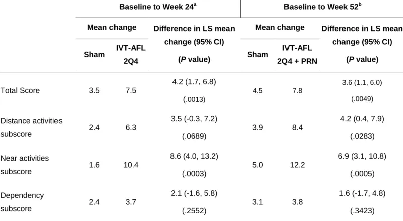
A clinically relevant improvement in the mean NEI VFQ-25 total score (≥ 4-point increase) was observed in both IVT-AFL 2Q4 + PRN (7.8 points) and sham (4.5 points) groups at week 52 (Table 2). The mean change from baseline to week 52 in near activities subscore was the highest among subscales, with IVT-AFL 2Q4 + PRN patients reporting a mean change of 12.2 points versus sham patients reporting a mean change of 5.0 points. No difference was noted between the two groups in the dependency subscale.
Study Drug Injections
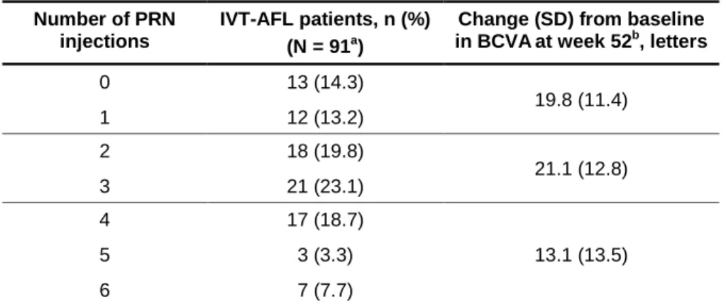
During the 52 weeks of treatment, the mean (± SD) number of injections was 11.8 ± 2.8 in the IVT-AFL 2Q4 + PRN group and 10.5 ± 4.2 in the sham group. Most IVT-AFL 2Q4 + PRN patients (64 of 91 patients completing week 52; [70.3%]) received ≤ 3 IVT-AFL during weeks 24 to 52, with a mean ± SD of 2.5 ± 1.7 injections during the PRN phase of study (Table 3). Patients who received ≤ 3 PRN injections had relatively higher BCVA gains than those who received 4-6 injections (Table 3). The median time (95% CI) to the first PRN intravitreal aflibercept injection was 83 days (62 to 88 days).
Safety
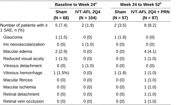
The percentage of patients experiencing at least one ocular treatment-emergent adverse event (TEAE) in the sham and intravitreal aflibercept groups was 64.7% and 54.8% from baseline to week 24, and 50.9% and 69.1% from week 24 to week 52, respectively. The most common ocular TEAEs reported for the study eye in the intravitreal aflibercept group as compared with the sham group were eye pain (11.5% vs 4.4%, respectively), increased intraocular pressure (IOP) (8.7% vs 5.9%, respectively), and conjunctival hemorrhage (8.7% vs 4.4%, respectively) from baseline to week 24, and worsening of macular edema (35.1% vs 10.5%, respectively), increased IOP (13.4% vs 3.5%, respectively), and reduced visual acuity (11.3% vs 1.8%, respectively) from week 24 to week 52. All adverse events of IOP elevation were mild, except for one severe event, which occurred in a sham patient before week 24. Ocular treatment-emergent serious adverse events (SAEs) are shown in Table 4. Most ocular SAEs were related to the disease state or injection procedure, and there were no clinically relevant differences between the treatment groups in terms of frequency or pattern of SAEs.
The incidence of non-ocular TEAEs was similar in the sham and intravitreal aflibercept groups from both baseline to week 24 (54.4% and 45.2%, respectively), and week 24 to week
52 (50.9% vs 51.5%, respectively). Nasopharyngitis was the most commonly reported non-ocular TEAE in both sham and intravitreal aflibercept groups from both baseline to week 24 (8.8% vs 7.7%, respectively) and week 24 to week 52 (19.3% vs 9.3%). Non-ocular SAEs occurred in a small group of patients with a similar frequency in both sham and intravitreal aflibercept groups from baseline to week 24 (7.4% and 5.8%, respectively) and week 24 to week 52 (8.8% and 6.2%, respectively). None of the non-ocular SAEs were reported for more than one patient from baseline to week 24. During weeks 24 to 52, non-ocular SAEs reported for more than one patient were pneumonia (one patient in each treatment group), and syncope (two patients in the sham group and one patient in the aflibercept group). No adverse event was adjudicated as an Anti-Platelet Trialists’ Collaboration (APTC)-defined arterial
thromboembolic event during the course of study. There were no deaths during the 52 weeks of this study.
DISCUSSION
The findings of the current study demonstrate that the improvements in BCVA and CRT achieved with monthly intravitreal aflibercept injections in the first 24 weeks of treatment were largely maintained during the PRN (as-needed) phase of study, with monthly monitoring and a mean of 2.5 injections from weeks 24 to 52. Of note, there was also a marked improvement in BCVA with aflibercept in a subgroup of patients with nonperfused retinas at baseline, in contrast to a particularly poor response in the sham group. The visual improvements with aflibercept enhanced vision-related quality of life, particularly in near visual activities. In this study, aflibercept was generally well tolerated, and most common adverse events were those typically associated with intravitreal injections or the underlying disease. The increase in macular edema seen in aflibercept patients during the PRN dosing phase suggests that some patients would have benefited from more regular dosing, rather than being treated in response to the recurrence of disease.
The sister study of GALILEO, the COPERNICUS study, demonstrated comparable improvements in BCVA and CRT with intravitreal aflibercept injections.5,7 However, the sham
groups in the two studies were not comparable during weeks 24 to 52 because, in the
COPERNICUS study, sham patients received aflibercept PRN starting from week 24 whereas, in the GALILEO study, sham patients continued to receive sham treatments through week 52. In the COPERNICUS study, patients receiving sham + IVT-AFL PRN did not achieve visual and anatomical improvements as robustly as those receiving aflibercept from the inclusion at week 52, suggesting that patients with macular edema secondary to CRVO may benefit from initiating treatment early with aflibercept.7
Treatment of CRVO with monthly intravitreal injections for 6 months, followed by monthly monitoring and PRN injections for an additional 6 months has been studied for ranibizumab in the CRUISE trial.8 Visual and anatomical outcomes reported from the CRUISE study are
the fixed monthly dosing phase largely maintained under PRN dosing with monthly monitoring.8,13 However, it is suggestive that the steeper decline in visual acuity between months 6 and 7 with 0.5 mg ranibizumab in the CRUISE study, compared with the smaller decline seen with aflibercept during the same time period in the GALILEO study, is reflective of a longer duration of effect with aflibercept.
The GALILEO results at week 52 corroborate the robust effect on visual and anatomical measures seen at week 24 in patients with macular edema secondary to CRVO, after 24 weeks of fixed monthly dosing with aflibercept. Originally, the PRN dosing regimen was introduced to investigate the feasibility of extending the treatment interval after the initial monthly aflibercept dosing phase. It has been demonstrated that the PRN dosing regimen largely maintained the improvements seen at week 24 with monthly monitoring. During the PRN dosing phase, an average of 2.5 injections was given in the IVT-AFL 2Q4 + PRN group, which approximates the 3 injections that would have been administered using a bimonthly dosing regimen, as has been established for aflibercept in wet AMD patients.11 From a practical perspective, the advantage
of PRN dosing is therefore questionable: while PRN dosing may lead to fewer injections than a fixed monthly regimen, it comes with the requirement of monthly visits. Therefore, a good alternative option would be flexibly adjusting the treatment interval using a “treat and extend” algorithm. This may potentially help to better preserve visual and anatomical gains over PRN dosing as well as reduce the challenges and cost of monthly monitoring.
ACKNOWLEDGEMENTS
Editorial assistance in the preparation of this manuscript was provided by Hadi Moini, PhD, Regeneron Pharmaceuticals, Inc.
REFERENCES
1. McIntosh RL, Rogers SL, Lim L, et al. Natural history of central retinal vein occlusion: an evidence-based systematic review. Ophthalmology 2010;117:1113-23 e15.
2. Wong TY, Scott IU. Clinical practice. Retinal-vein occlusion. N Engl J Med 2010;363:2135-44.
3. Pournaras CJ, Rungger-Brandle E, Riva CE, et al. Regulation of retinal blood flow in health and disease. Prog Retin Eye Res 2008;27:284-330.
4. Noma H, Funatsu H, Mimura T, et al. Role of soluble vascular endothelial growth factor receptor-2 in macular oedema with central retinal vein occlusion. Br J Ophthalmol 2011;95:788-92.
5. Boyer D, Heier J, Brown DM, et al. Vascular Endothelial Growth Factor Trap-Eye for Macular Edema Secondary to Central Retinal Vein Occlusion: Six-Month Results of the Phase 3 COPERNICUS Study. Ophthalmology 2012;119:1024-32.
6. Brown DM, Campochiaro PA, Singh RP, et al. Ranibizumab for macular edema following central retinal vein occlusion: six-month primary end point results of a phase III study. Ophthalmology 2010;117:1124-33 e1.
7. Brown DM, Heier JS, Clark WL, et al. Intravitreal Aflibercept Injection for Macular Edema Secondary to Central Retinal Vein Occlusion: 1-Year Results From the Phase 3 COPERNICUS Study. Am J Ophthalmol 2013;155:429-37 e7.
Mis en forme : Français (France)
8. Campochiaro PA, Brown DM, Awh CC, et al. Sustained benefits from ranibizumab for macular edema following central retinal vein occlusion: twelve-month outcomes of a phase III study. Ophthalmology 2011;118:2041-9.
9. Holz FG, Roider J, Ogura Y, et al. VEGF Trap-Eye for macular oedema secondary to central retinal vein occlusion: 6-month results of the phase III GALILEO study. Br J Ophthalmol 2013;97:278-84.
10. Papadopoulos N, Martin J, Ruan Q, et al. Binding and neutralization of vascular endothelial growth factor (VEGF) and related ligands by VEGF Trap, ranibizumab and bevacizumab. Angiogenesis 2012;15:171-85.
11. Heier JS, Brown DM, Chong V, et al. Intravitreal Aflibercept (VEGF Trap-Eye) in Wet Age-related Macular Degeneration. Ophthalmology 2012;119:2537-48.
12. Lucentis (ranibizumab injection) prescribing information.
http://www.gene.com/download/pdf/lucentis_prescribing.pdf. Accessed July, 2013. .
13. Heier JS, Campochiaro PA, Yau L, et al. Ranibizumab for macular edema due to retinal vein occlusions: long-term follow-up in the HORIZON trial. Ophthalmology 2012;119:802-9.
FIGURE LEGENDS
Figure 1. The GALILEO Study Design.
2Q4 = every 4 weeks; BCVA = best-corrected visual acuity; CRT = central retinal thickness; CRVO = central retinal vein occlusion; IVT-AFL = intravitreal aflibercept; PRN = pro re nata (as needed); PRP = panretinal photocoagulation.
Figure 2. Visual Outcomes During the 52 Weeks of Study. Percentage of patients who gained
≥ 15 letters at week 52 (A), mean change from baseline BCVA (B), and mean change from baseline BCVA by the status of retinal perfusion at baseline (C) are shown. Treatment
frequency with intravitreal aflibercept was 2Q4 and PRN, respectively, before and after week 24.
a
P = .0004 vs sham; b P < .0001 vs sham; c P < .001 vs sham.
2Q4 = every 4 weeks; BCVA = best-corrected visual acuity; ETDRS = Early Treatment Diabetic Retinopathy Study; IVT-AFL = intravitreal aflibercept; PRN = pro re nata (as needed).
Figure 3. Mean Change from Baseline CRT During the 52 Week of Study. Treatment
frequency with intravitreal aflibercept was 2Q4 and PRN, respectively, before and after week 24.
a
P < .0001 vs sham.
2Q4 = every 4 weeks; CRT = central retinal thickness; IVT-AFL = intravitreal aflibercept; PRN = pro re nata (as needed).
Table 1. Patients with Vision Gain and Loss at Week 52. Week 52 Sham (N = 68) IVT-AFL 2Q4 + PRN (N = 103) Vision gain, n (%) ≥ 30 letters 5 (7.4) 15 (14.6) ≥ 15 letters 22 (32.4) 62 (60.2) ≥ 10 letters 26 (38.2) 74 (71.8) Vision loss, n (%) > 0 letters 30 (44.1) 11 (10.7) > 10 letters 16 (23.5) 1 (1.0) > 15 letters 10 (14.7) 1 (1.0) IVT-AFL 2Q4 + PRN = intravitreal aflibercept injection monthly from baseline to week 24 and PRN from week 24 to week 52.
Table 2. Change From Baseline to Weeks 24 and 52 in the NEI VFQ-25 Score.
Baseline to Week 24
aBaseline to Week 52
bMean change
Difference in LS mean
change (95% CI)
(P value)
Mean change
Difference in LS mean
change (95% CI)
(P value)
Sham
IVT-AFL
2Q4
Sham
IVT-AFL
2Q4 + PRN
Total Score
3.5
7.5
4.2 (1.7, 6.8)
(
.0013) 4.5 7.8 3.6 (1.1, 6.0) (.0049)Distance activities
subscore
2.4
6.3
3.5 (-0.3, 7.2)
(.0689)
3.9
8.4
4.2 (0.4, 7.9)
(.0283)
Near activities
subscore
1.6
10.4
8.6 (4.0, 13.2)
(.0003)
5.0
12.2
6.9 (3.1, 10.8)
(.0005)
Dependency
subscore
2.4
3.7
2.1 (-1.6, 5.8)
(.2552)
3.1
3.8
1.6 (-1.7, 4.8)
(.3423)
an = 65 for sham and n = 96 for IVT-AFL 2Q4.
b
n = 67 for sham and n = 97 for IVT-AFL 2Q4 + PRN (except for the total score, which was n = 98).
CI = confidence interval; IVT-AFL 2Q4 = intravitreal aflibercept injection monthly from baseline to week 24; IVT-AFL 2Q4 + PRN
= intravitreal aflibercept injection monthly from baseline to week 24 and PRN from week 24 to week 52; LS = least square.
Table 3. Distribution of PRN Injections During Weeks 24-52 and BCVA Gains at
Week 52 in Patients Treated with IVT-AFL 2Q4 + PRN.
Number of PRN injections
IVT-AFL patients, n (%) (N = 91a)
Change (SD) from baseline in BCVAat week 52b, letters
0 13 (14.3) 19.8 (11.4) 1 12 (13.2) 2 18 (19.8) 21.1 (12.8) 3 21 (23.1) 4 17 (18.7) 13.1 (13.5) 5 3 (3.3) 6 7 (7.7)
BCVA = best-corrected visual acuity; IVT-AFL = intravitreal aflibercept; IVT-AFL 2Q4 + PRN = intravitreal aflibercept injection every 4 weeks from baseline to week 24 and PRN from week 24 to week 52; PRN = pro re nata (as-needed); SD = standard deviation.
a Patients completing week 52. b
Because of small number of patients in each injection category, BCVA gains at week 52 were shown for patients who received 0-1, 2-3, and 4-6 injections. The mean SD BCVA at baseline was 58.2 15.5, 49.4 15.9, and 55.4 15.0 letters for patients who received 0-1, 2-3, and 4-6 injections, respectively.
Table 4. Ocular SAEs in study eye Occurring From Baseline to Week 24 and Week 24 to Week
52.
Baseline to Week 24a Week 24 to Week 52b Sham (N = 68) IVT-AFL 2Q4 (N = 104) Sham (N = 57) IVT-AFL 2Q4 + PRN (N = 97)
Number of patients with ≥ 1 SAE, n (%)
5 (7.4) 2 (1.9) 2 (3.5) 8 (8.2) Glaucoma 1 (1.5) 0 (0) 1 (1.8) 0 (0) Iris neovascularization 0 (0) 1 (1.0) 0 (0) 0 (0) Macular edema 2 (2.9) 0 (0) 0 (0) 4 (4.1) Reduced visual acuity 1 (1.5) 0 (0) 0 (0) 1 (1.0) Vitreous detachment 0 (0) 1 (1.0) 0 (0) 0 (0) Vitreous hemorrhage 1 (1.5%) 0 (0) 1 (1.8) 1 (1.0) Macular fibrosis 0 (0) 0 (0) 0 (0) 1 (1.0) Macular ischemia 0 (0) 0 (0) 0 (0) 1 (1.0) Retinal detachment 0 (0) 0 (0) 0 (0) 1 (1.0) Retinal vein occlusion 0 (0) 0 (0) 0 (0) 1 (1.0)
a Safety analysis set. b
Week 24 completers within safety analysis set.
IVT-AFL 2Q4 = intravitreal aflibercept injection administered monthly from baseline to week 24; IVT-AFL 2Q4 + PRN = intravitreal aflibercept injection administered monthly from baseline to week 24 and PRN from week 24 to week 52; SAE = treatment-emergent serious adverse event.
Figure 2. (A)