HAL Id: inserm-00177752
https://www.hal.inserm.fr/inserm-00177752
Submitted on 9 Jan 2008HAL is a multi-disciplinary open access archive for the deposit and dissemination of sci-entific research documents, whether they are pub-lished or not. The documents may come from teaching and research institutions in France or abroad, or from public or private research centers.
L’archive ouverte pluridisciplinaire HAL, est destinée au dépôt et à la diffusion de documents scientifiques de niveau recherche, publiés ou non, émanant des établissements d’enseignement et de recherche français ou étrangers, des laboratoires publics ou privés.
Hepatitis B virus genotypes: a retrospective survey in
Southwestern France, 1999-2004.
Pascale Trimoulet, Mathieu Boutonnet, Maria Winnock, Muriel Faure,
Marc-Arthur Loko, Victor de Lédinghen, Pierre-Henri Bernard, Laurent
Castéra, Juliette Foucher, Michel Dupon, et al.
To cite this version:
Pascale Trimoulet, Mathieu Boutonnet, Maria Winnock, Muriel Faure, Marc-Arthur Loko, et al.. Hepatitis B virus genotypes: a retrospective survey in Southwestern France, 1999-2004.: HBV geno-types in Southwestern France. Gastroentérologie Clinique et Biologique / Research and Clinics in Hepatology and Gastroenterology, Elsevier Masson, 2007, 31 (12), pp.1088-94. �inserm-00177752�
HEPATITIS B VIRUS GENOTYPES: A RETROSPECTIVE SURVEY IN SOUTHWESTERN FRANCE, 1999-2004
ETUDE RETROSPECTIVE DES GENOTYPES DU VIRUS DE L’HEPATITE B EN AQUITAINE, 1999-2004
Pascale Trimoulet *1, Mathieu Boutonnet 2, Maria Winnock 3, 4, Muriel Faure 1, Marc-Arthur
Loko 4, Victor de Lédinghen 5, Pierre-Henri Bernard 6, Laurent Castéra 5, 6, Juliette Foucher 5,
6
, Michel Dupon 2, Jean-Marie Ragnaud 2, Marie-Edith Lafon 1, Patrice Couzigou 5, François
Dabis 4, Hervé Fleury 1, Didier Neau 2.
1
Laboratoire de Virologie, Hôpital Pellegrin, 33076 Bordeaux; 2Fédération de Maladies
Infectieuses, Hôpital Pellegrin, 33076 Bordeaux ; 3Service d’Information Médicale, Hôpital
Pellegrin; 4INSERM U593, ISPED, Université Victor Ségalen Bordeaux 2, 33076 Bordeaux ;
5Service d’Hépatogastroentérologie, Hôpital du Haut Lévêque, 33604 Pessac; 6
Service d’Hépatogastroentérologie, Hôpital Saint-André, 33075 Bordeaux, France
Corresponding author: Dr Pascale Trimoulet, Laboratoire de Virologie, Hôpital Pellegrin, place Amélie Raba-Léon, 33076 Bordeaux cedex, France. Telephone number: +33-556-795510, fax number: +33- 556-795673, E-mail address: pascale.trimoulet@chu-aquitaine.fr
SUMMARY
Objective. To determine the prevalence of HBV genotypes in Southwestern France and the
association between HBV genotypes and patients characteristics. Methods. 194 HBsAg-positive patients (median age: 45 yrs, range: 7-77, male: 78%) followed in Bordeaux Hospital in 1999-2004 were included. HBV genotype, pre-core (PC) and core promoter (CP) mutations were determined by sequencing. Results. Genotype distribution was A 51%, B 6.7%, C 5.7%, D 26.3%, E 7.7%, F 0.5%, G 2.1%. Among the 146 patients documented, 71.2% were Caucasians, 15.8% Africans, 13.0% Asians. Fifty-seven patients (36%) were HIV-infected. Eighty-two (42.3%) patients were HBeAg-positive. Genotype A was almost exclusively carried by Caucasians (96%), Africans were most frequent among genotype E (82%), and Asians were most prevalent among genotypes B and C (82% and 80%, respectively). Genotype A was associated with a higher prevalence of HBeAg than genotype D (53% versus 35.3%, p=0.03). PC variant was detected in 35% and CP variant in 43% of patients. PC variant was uncommon in genotype A patients (7.3%). Conclusion. Distribution of HBV genotypes differs according to ethnic origin, genotypes A and D being the most frequently found. Genotype A was more frequently associated with HBeAg-positivity and genotype D with HBeAg-negativity.
KEY WORDS: Hepatitis B virus infection; HBV genotypes; HBeAg positivity; Pre-core variants; Core promoter variant
RESUME
Objectifs. Déterminer la prévalence des génotypes du virus de l’hépatite B (VHB) en
Aquitaine et l’association entre génotypes VHB et caractéristiques des patients. Méthodes. 194 patients AgHBs-positif (âge médian : 45 ans, étendue : 7-77, hommes : 78%), vus entre 1999 et 2004 au CHU de Bordeaux ont été inclus. Le génotypage VHB, les mutations pré-C (PC) et promoteur de core (CP) ont été déterminés par séquençage Résultats. La distribution des génotypes était: A 51%, B 6,7%, C 5,7%, D 26,3%, E 7,7%, F 0,5%, G 2,1%. Parmi les 146 patients documentés, 71,2% étaient caucasiens, 15,8% africains, 13,0% asiatiques. 57 patients (36%) étaient VIH-positif et 82 (42,3%) AgHBe-positif. Le génotype A était prévalent chez les caucasiens (96%), le génotype E parmi les africains (82%), et les génotypes B et C chez les asiatiques (82% et 80%, respectivement). Le génotype A était associé à une plus forte prévalence de l’AgHBe que le génotype D (53% vs 35,3%, p=0.03). Les mutants PC et CP étaient présents chez 35% et 43% des patients, respectivement. Conclusion. La distribution des génotypes VHB diffère en fonction de l’origine ethnique des patients. Les génotypes A et D sont les plus fréquents en Aquitaine.
MOTS CLES: infection par le virus de l'hépatite B; génotypes VHB; AgHBe positif; mutants pré-core; mutants promoteur de core
INTRODUCTION
Chronic hepatitis B virus (HBV) infection remains a major public health problem worldwide with approximately 300 million chronic carriers and the development of severe complications such as liver cirrhosis and hepatocellular carcinoma [1]. The course of HBV infection depends on several factors modifying the immune response, including age at infection and host genetic factors [2], and it probably also depends on the genetic variability of the virus, which influences the expression of viral antigens. Indeed, HBV replicates via a reverse-transcription step and is estimated to have a high mutation rate [3]. On the basis of a comparison of complete genomic sequences, seven major viral genotypes, designated A to G, have been identified and correspond to viral isolates sharing more than 80% homology in their nucleotide sequences [4-7]. HBV genotypes have distinct geographical distributions. Genotype A is found in North America [4] and Northern Europe, as well as in parts of Africa [8,9], while genotypes B and C are the most common in Southeast Asia [4]. Genotype D is found universally [8]. Genotype E is predominantly found in Africa [8], and F clusters in Central and South America [10]. Genotype G has been reported in the United States and France [7].
Accumulating evidence suggests that HBV genotypes have an impact on the pattern of mutations in the pre-core (PC) and core promoter (CP) regions, the natural course of chronic HBV infection [11-15], or the severity of underlying liver disease [16,17]. Genotypes B and C have been studied most extensively due to their co-circulation in Asia, eliminating differences in ethnic or racial background of the patients. These studies have shown that genotype B, compared to genotype C, is associated with a higher rate of seroconversion from HBeAg to anti-HBe antibody, less active liver disease, and a lower rate of progression to cirrhosis. Correspondingly, genotype C is more prevalent among HBeAg-positive patients than
genotype B. Similarly, compared to genotype D, genotype A is more prevalent in HBeAg-positive than in anti-HBe-HBeAg-positive antibody patients. Several studies indicate that HBV genotypes may be associated with differences in treatment response [18-20]. Genotype C is associated with a lower response rate to alpha interferon therapy compared to genotype B. In Caucasian patients, a better response to peginterferon alpha-2b and lamivudine was found when patients were infected with genotype A vs genotype D. Investigating the clinical significance of HBV genotypes is becoming increasingly relevant. The identification of HBV genotypes could be useful to monitor HBV infection and related diseases and may also help in implementing appropriate therapeutic regimens.
To date, there are little data on the prevalence and clinical significance of HBV genotypes in France. One previous study suggested that HBV genotypes A and D were predominant in this country and that genotype A was associated with HBeAg positivity, while genotype D was associated with HBeAg negativity [21]. Another study by Ganne-Carrié et al. concluded that HBV genotypes A, B, C, D, and E circulated in the Seine Saint Denis District, close to Paris, reflecting the multiple geographical origins of patients [22].
The aim of this study was to determine the prevalence and the distribution of HBV genotypes among a consecutive sample of patients with HBV infection with or without HIV coinfection in Bordeaux hospital (France) that caters to patients from various parts of Southwestern France. Additional objectives were to determine whether there was an association of HBV genotypes with patients demographics, clinical status, and PC and CP variants.
MATERIALS AND METHODS
Patients. Patients with HBV infection and referred to our virological laboratory (Pellegrin
May 2004 were retrospectively and systematically included in the survey. The inclusion criteria were a serologically proven chronic HBV infection (HBsAg positivity > 6 months), HBV DNA positivity with concomitant HBeAg, and anti-HBe tests. Patients with HIV positivity or HBV treatment were also included.
Clinical findings. Information relating to the patients’ demographics, clinical and virological
status and hepatic disorders (clinical, biological and histological) were recorded anonymously and retrospectively from the patients’ medical files. Whenever a treated patient had a liver biopsy, it was performed before treatment. In HIV-infected patients, epidemiological, clinical, biological and therapeutic data were retrieved from the ANRS CO3 Aquitaine Cohort database of the Groupe d’Epidémiologie Clinique du Sida en Aquitaine (GECSA) [23]. Epidemiological characteristics included sex, age, and ethnicity. Coinfection with the immunodeficiency virus (HIV), the hepatitis C virus (HCV), and the delta virus (HDV) were also recorded. The presumed source of HBV infection was determined by inquiring about the patients’ sexual behaviour (intercourse with an HBV contaminated sexual partner), history of intravenous drug use at least once, blood transfusion, vertical transmission (HBsAg-positive mother), iatrogenic exposure (endoscopic or coelioscopy examination, history of surgery, acupuncture, hemodialysis).
Detection of virological markers in serum. The study population was tested routinely for
HBsAg, HBeAg, and anti-HBe antibody (Dade Behring Laboratories, Marburg, Germany), HIV antibodies (Dade Behring, Marburg, Germany), HCV antibodies (Ortho Clinical Diagnostics, Raritan, NJ), and anti-delta antibodies (DiaSorin, Saluggia, Italy).
Determination of HBV genotypes and detection of PC stop codon (G1896A) and CP
variants (A1762T, G1764A). HBV DNA was extracted from 200 µl of serum using QIAamp
DNA blood mini-kit (Qiagen, Hilden, Germany). HBV genotyping was performed by sequencing a part of the polymerase region. Two rounds of polymerase chain reaction
amplification were used for polymerase sequence studies [24]. The first round used primers
CHBV-3 (5’-CCTGCTGGTGGCTCCAGTTC-3’) and HBV17
(5’-CGTCCCGCGNAGGATCCAGTT-3’); nested polymerase chain reaction using primers
VT301 (5’-CYTGGCCWAAATTCGCAGTCCC-3’) and VT1022
(5’-GCAAANCCCAAAAGACAAAT-3’) yielded a 721-bp DNA fragment encoding part (nt 1214-1934, subdomains A-E) of the HBV DNA polymerase gene. Amplification of the precore sequence was performed by nested polymerase chain reaction as previously described [25]. The following primers were used for the amplification of the precore region (nt 1,814 to 1,900) and the core promoter (nt 1,742 to 1,849) of the HBV genome. The external primers were CATAAGAGGACTCTTGGACT-3’ (sense, nt 1,653 to 1,672), and 5’-GGCGAGGGAGTTCTTCTTCTAGGGG-3’ (antisense, nt 2,394 to 2,369), and primers for the second PCR were 5’-AATGTCAACGACCGACCTTG-3’ (sense, nt 1,679 to 1,698) and 5’-AGCTGAGGCGGTGTCGAGGAGATC-3’ (antisense, nt 1,985 to 2,009).
The amplified PCR products were purified by the QIA quick PCR purification kit (Qiagen) and then used for direct sequencing using internal antisense primer. Sequencing of the purified products was conducted on a CEQ automatic sequencer (Beckman Coulter, Fullerton, CA) with the CEQ DTCS Quick Start kit (Beckman Coulter). Analysis of each sequence was conducted using Seq Analysis and Seq Investigator software (Beckman Coulter).
For genotype characterization, all sequences obtained were compared to at least 24 Genbank sequences representative of all known HBV genotypes.
Statistical analysis.
Quantitative variables are described by their median and range and categorical variables by percentage. Comparisons between patients were performed using Kruskall Wallis non parametric test for quantitative variables and Fisher’s exact test for categorical variables. Multiple logistic regression with forward stepwise analyses were used to determine the
independent factors associated with HBV genotypes A and those associated with HBV genotype D. The variables related to genotype A and genotype D with P < 0.10 in univariate analysis were entered in the logistic model. Results were considered statistically significant at P < 0.05. Data were analyzed using SAS version 8.2 software package (SAS Institute Inc., Cary, NC, USA).
RESULTS
At total, 194 patients (152 males and 42 females) were identified and enrolled. Their median age was 45 years (range 7.2-76.9 years). The characteristics of the patients are listed in Table 1. Of the 146 patients with a documented ethnic background, 104 (71.2%) were Caucasians. HIV coinfection was present in 57/159 (36%) patients screened. Eighty-two patients (42.3%) were HBeAg-positive and 52 (26.8%) were currently receiving anti-HBV treatment (interferon: 9 patients, pegylated interferon: 3, lamivudine: 31, adefovir: 2, lamivudine and tenofovir: 7).
Seven HBV genotypes were found in the studied population. Genotypes A and D were the most common. Genotypes G and F were present in only 2.6% of the study population (Table
2). No association was found between median age and HBV genotypes (p = 0.28). There was a
strong association between ethnicity and HBV genotypes (P<0.001 for the overall comparison). Genotype A was almost exclusively carried by Caucasian patients (96%), africans were most frequent among genotype E (82%), and asians were most prevalent among
genotypes B and C (82% and 80%, respectively). The presumed mode of infection was known in 80 patients (41%). Of these, 58% were presumed to have acquired HBV infection
strong association between the presumed mode of infection and HBV genotypes (P=0.01 overall). Among genotype A, patients infected via sexual route were predominant, whereas among genotype D, patients infected via drug use were most prevalent (Table 2).
There was a strong association between HIV status and HBV genotypes (P<0.001). HIV
coinfection was common in patients with genotypes A and D (45.3% and 34.2%, respectively) and absent in patients with genotypes B and C (Table
2).
The proportion of patients with increased ALT over the upper limit of normal was not significantly different between HBV genotypes (p = 0.4). No association was found between liver activity and HBV genotypes (p = 0.47). On the other hand, there was an association between liver fibrosis and HBV genotypes (p = 0.01). The prevalence of severe liver fibrosis (F3 or F4 METAVIR score) was higher in HBV genotypes A and E-infected patients (Table
2).
HBV genotypes, HBeAg status and PC/CP variants.
Genotypes A and C were associated with a higher prevalence of HBeAg (53% and 55%, respectively) (Table 2). A strong correlation was found between HBV genotypes and PC as well as CP variants (Table 2 and Figure 1). Among the 188 patients documented, the overall prevalence of PC variant was 35%. PC variant (G1896A) was more common in patients with genotypes E (86.7%), B (61.5%), C (60%), and D (54.9%), and rare in patients with genotype A (7.3%) (P<0.001). Among the 186 patients documented, the overall prevalence of CP variants (A1762T and/or G1764A) was 43%. CP variant was most common among patients with genotype C (77.8%), followed by genotype A (46.9%), E (46.7%), and D (35.3%), and less common in patients with genotype B (15.4%) (P=0.05). PC (54% vs 8%, p<0.001) as well as CP variants (53% vs 29%, P=0.001) were more often found in HBeAg-negative than HBeAg-positive patients.
Factors relating to HBV genotypes A or D.
Factors that may be associated with the most prevalent HBV genotypes present in our patient population (genotypes A and D), including sex, age (<40 yrs vs >40 yrs), ethnicity, presumed mode of infection, PC and CP variant, HIV status, liver activity (A0-A1 vs A2-A3) and fibrosis (F0-F1 vs F2-F4) were analyzed by multiple logistic regression analyses. According to univariate analysis, 6 factors were significantly associated with HBV genotype A (P<0.1): age > 40 years, sexual transmission of HBV, HIV positive status, HBeAg positivity, absence of PC variant, METAVIR fibrosis score F2-F4. In multivariate analysis, age > 40 years, HIV positive status, absence of PC variant were independent factors associated with genotype A. Univariate analysis selected 3 factors associated with genotype D (P<0.1): intravenous drug use, non asian ethnicity, presence of PC variant. These variables were introduced in a multivariable logistic model to test the independent association with genotype D. In multivariate analysis, contamination via intravenous drug use, non-asian ethnicity and presence of PC variant remained independently associated with genotype D (Table 3).
DISCUSSION
In view of the distinct geographic distribution of HBV genotypes, it is important to survey their epidemiology in a given country or region, since HBV genotypes may have some influence on the course of liver disease. The seven major HBV genotypes (A-G) were represented in a sample recruited in one major French region outside of Paris. This distribution may reflect local differences caused by various ethnicities of inhabitants and their individual lifestyles that may also differ all over France. In our opinion, our study is the first to systematically investigate patients with chronic hepatitis B (CHB) providing HBV DNA
positivity. In addition, it is one of the first attempts to investigate genotypes in mono-infected or HIV co-infected patients. Therefore, the findings of the present survey are likely to be representative of the CHB patient population in Southwestern France.
Our results showed a predominance of genotypes A and D in agreement with other investigators [15,21]. As evidenced by previous studies concerning the geographic distribution of HBV, genotypes A and D were predominantly found in Caucasians, genotypes B and C in Asians and genotype E in Africans. Of particular note was the detection of genotype G in four of the 194 (2.1%) patients in Southwestern France. Because genotype G was discovered in 2000 [7], epidemiological data are limited. HBV genotype G has been
reported in France, Georgia, Germany, and Mexico, but not in Japan [7,26-28]. We found a strong correlation between HBV genotypes and the presumed mode of infection,
but the latter may merely be a surrogate marker of ethnicity or place of birth. However, multivariate analysis suggested rather the opposite since contamination via intravenous drug use and non-asian ethnicity were independently associated with genotype D. Genotype A was prevalent among patients infected via sexual route, genotype D was related to all modes of infection, and genotypes B and C were exclusively present among patients with vertical transmission or other routes of infection. Another important finding of our study was the strong association between HIV status and HBV genotypes. HIV coinfection was common in patients with genotypes A and D and absent in patients with genotypes B and C.
The majority of studies on the effect of genotypes on disease progression have been undertaken in South-east Asia where HBV is hyperendemic and genotypes B and C prevail. These studies showed that genotype C was associated with higher virus loads [29] and more aggressive liver disease [17,30]. On the other hand there is a dearth of studies comparing genotypes A and D. In India, genotype D is reported to be associated with more severe liver disease than that associated with genotype A. In contrast, in our study, genotype A was
associated with METAVIR fibrosis score F2-F4 in univariate analysis. This result was not confirmed in multivariate analysis. Maybe it is linked to the high prevalence of HIV seropositivity in this group (many studies having shown HIV coinfection as a factor in HBV disease progression), or to the high proportion of patients over 40 yrs. However we cannot exclude a pejorative specific evolution linked to genotype A.
In keeping with the results from other investigators, genotype A and C were associated with a higher prevalence of HBeAg compared with genotypes B and D [12,31-33]. Several studies reported a correlation between HBV genotype and HBeAg clearance. These studies found that the prevalence of HBeAg was higher in patients with genotype C compared to those with genotype B suggesting that HBeAg clearance occurred at higher rates among patients with genotype B [13,16]. Unfortunately, the cross-sectional design of our survey could not address the issue of the association of HBV genotypes with HBeAg seroconversion that has been recently suggested [14,16]. In multivariate analysis, HBeAg positivity was not an independent factor associated with genotype A. The PC/CP status could probably explain the difference between genotypes A and D concerning HBeAg positivity or negativity more than HBeAg clearance.
Our results clearly show in agreement with others [34] that HBeAg-negative chronic hepatitis is now the predominant form of chronic hepatitis B. In our population the overall proportion of HBeAg-negative chronic hepatitis B was 58%, lower than in a recent study [34]. This difference could be explained by a higher proportion of genotype A in our population, since genotype A is associated with a higher prevalence of HBeAg. Unfortunately, HBV genotyping was not performed by Zarski et al.
We found that PC and CP variants were detected in a substantial proportion (35% and 43%, respectively) of our patients. In patients with chronic HBV infection, HBeAg negative disease is becoming the predominant form of infection and has therapeutic implications [35,36]. This
form of chronic HBV infection is highly prevalent in the Mediterranean (50-80%) and Eastern Asian (45-55%) regions. Physicians managing patients with chronic HBV infection must be
aware of this condition because the natural course and treatment response are different from that of HBeAg-positive chronic hepatitis [37].
In accordance with previous reports [12,13,16,38], we found that PC variant (G1896A) was most common in patients with genotype D and rare in patients with genotype A. In our survey, CP variants (A1762T, G1764A) were evenly distributed among HBV genotypes A, C, D and E and relatively rare among HBV genotype B. The association between HBV genotypes and PC (G1896A) variant is related to base pairing in the stem-loop structure of the pregenome encapsidation sequence [11,12,38,39] but its basis has not been deciphered. Our study showed that PC variants were predominantly detected in HBeAg-negative patients, whereas CP variants were found in both HBeAg-negative and HBeAg-positive patients, as described by others [40,41]. A more marked increase in the prevalence of PC versus CP variants in HBeAg-negative patients is attributed to the fact that PC (G1896A) variants abolish whereas CP variants (A1762T, G1764A) only down-regulate HBeAg production. This study has identified three independent factors associated with genotype A in HBV infected patients: age > 40 yrs, HIV positive status, and absence of PC variant. The proportion of patients with age > 40 yrs was significantly higher, a finding consistent with the recent results of a survey performed in France by Ganne-Carrié et al [22] in which the mean age of patients infected by HBV genotypes B, C, and E, was significantly lower than HBV/A and /D. In keeping with the results from three recent studies [42-44] in which 57%, 92%, and 70% of HIV-HBV coinfected patients were infected by HBV genotype A, this genotype was strongly associated with HIV infection in our study.
In summary, seven HBV genotypes were present, and PC and CP variants could be detected in approximately one-third of patients with chronic HBV infection in Southwestern France.
HBV genotypes were related to various ethnicities of inhabitants and influenced the prevalence of serum HBeAg as well as PC and CP variants. Further studies are needed to determine if additional testing for HBV genotype as well as PC and CP variants may help in documenting and predicting clinical prognosis and thus guiding treatment decisions.
REFERENCES
1- Lee WM. Hepatitis B virus infection. N Engl J Med 1997; 337: 1733-1745.
2- Wright TL, Lau JYN. Clinical aspects of hepatitis B virus infection. Lancet 1993; 342: 1340-1344.
3- Orito E, Mizokami M, Ina Y, Moriyama EN, Kameshima N, Yamamoto M, et al. Host-independent evolution and a genetic classification of the hepadnavirus family on based nucleotide sequences. Proc Natl Acad Sci USA1989; 86: 7059-7062.
4- Okamoto H, Tsuda F, Sakugawa H, Sastrosoewignjo RI, Imai M, Miyakawa Y et al. Typing hepatitis B virus by homology in nucleotide sequence: comparison of surface antigen subtypes. J Gen Virol 1988; 69: 2575-2583.
5- Norder H, Hammas B, Lofdahl S, Courouce AM, Magnius LO. Comparison of the amino acid sequences of nine different serotypes of hepatitis B surface antigen and genomic classification of the corresponding hepatitis B virus strains. J Gen Virol 1992; 73: 1201-1208. 6- Norder H, Couroucé AM, Magnius L. Complete genomes, phylogenetic relatedness, and structural proteins of six strains of the hepatitis B virus, four of which represent two new genotypes. Virology 1994; 198: 489-503.
7- Stuyver L, De Gendt S, Van Geyt C, Zoulim F, Fried M, Schinazi RF, et al. A new genotype of hepatitis B virus: complete genome and phylogenetic relatedness. J Gen Virol 2000; 81: 67-74.
8- Norder H, Hammas B, Lee SD, Bile K, Couroucé AM, Mushahwar IK, et al. Genetic relatedness of hepatitis B viral strains of diverse geographical origin and natural variations in the primary structure of the surface antigen. J Gen Virol 1993; 74: 1341-1348.
9- Bowyer SM, van Staden L, Kew MC, Sim JG. A unique segment of the hepatitis B virus group A genotype identified in isolates from South Africa. J Gen Virol 1997; 78: 1719-1729.
10- Nakano T, Lu L, Hu X, Mizokami M, Orito E, Shapiro C, et al. Characterization of hepatitis B virus genotypes among Yucpa Indians in Venezuela. J Gen Virol 2001; 82: 359-365.
11- Lok AS, Akarca U, Greene S. Mutations in the pre-core region of hepatitis B virus serve to enhance the stability of the secondary structure of the pre-genome encapsidation signal. Proc Natl Acad Sci USA 1994; 91: 4077-4081.
12- Rodriguez-Frias F, Buti M, Jardi R, Cotrina M, Viladomiu L, Esteban R, et al. Hepatitis B virus infection: precore mutants and its relation to viral genotypes and core mutations. Hepatology 1995; 22: 1641-1647.
13- Orito E, Mizokami M, Sakugawa H, Michitaka K, Ishikawa K, Ichida T, et al. A case-control study for clinical and molecular biological differences between hepatitis B viruses of genotypes B and C. Japan HBV Genotype Research Group. Hepatology 2001; 33: 218-223. 14- Chu CJ, Hussain M, Lok AS. Hepatitis B virus genotype B is associated with earlier HBeAg seroconversion compared with hepatitis B virus genotype C. Gastroenterology 2002; 122: 1756-1762.
15- Sanchez-Tapias JM, Costa J, Mas A, Bruguera M, Rodes J. Influence of hepatitis B virus genotype on the long-term outcome of chronic hepatitis B in western patients. Gastroenterology 2002; 123: 1848-1856.
16- Lindh M, Hannoun C, Dhillon AP, Norkrans G, Horal P. Core promoter mutations and genotypes in relation to viral replication and liver damage in East Asian hepatitis B virus carriers. J Infect Dis 1999; 179: 775-782.
17- Kao JH, Chen PJ, Lai MY, Chen DS. Hepatitis B genotype correlate with clinical outcomes in patients with chronic hepatitis B. Gastroenterology 2000: 118: 554-559.
18- Kao JH, Wu NH, Chen PJ, Lai MY, Chen DS. Hepatitis B genotypes and the response to interferon therapy. J Hepatol 2000; 33: 998-1002.
19- Wai CT, Chu CJ, Hussain M, Lok AS. HBV genotype B is associated with better response to interferon therapy in HBeAg (+) chronic hepatitis than genotype C. Hepatology 2002; 36: 1425-1430.
20- Janssen HL, van Zonneveld M, Senturk H, Zeuzem S, Akarca US, Cakaloglu Y, et al. Pegylated interferon alfa-2b alone or in combination with lamivudine for HBeAg-positive chronic hepatitis B: a randomised trial. Lancet 2005; 365:123-129.
21- Halfon P, Bourliere M, Pol S, Benhamou Y, Ouzan D, Rotily M, et al. Multicenter study of hepatitis B virus (HBV) genotypes in France: correlation with liver fibrosis and hepatitis B e antigen status. J Viral Hepat 2006; 13: 329-335.
22. Ganne-Carrié N, Williams V, Kaddouri H, Trinchet JC, Dziri-Mendil S, Alloui C, et al. Significance of hepatitis B virus genotypes A to E in a cohort of patients with chronic hepatitis B in the Seine Saint Denis district of Paris (France). J Med Virol 2006;78:335-340. 23- Binquet C, Chêne G, Jacqmin-Gadda H, Journot V, Saves M, Lacoste D, et al. Modeling changes in CD4-positive T-lymphocytes counts after the start of antiretroviral therapy and the relation with risk of opportunistic infections: the Aquitaine Cohort, 1996-1997. Am J Epidemiol 2001; 153: 386-393.
24- Benhamou Y, Fleury H, Trimoulet P, Pellegrin I, Urbinelli R, Katlama C, et al. Anti-hepatitis B virus efficacy of tenofovir disoproxil fumarate in HIV-infected patients. Hepatology 2006; 43: 548-555.
25- Cho SW, Hahm KB, Kim JH. Reversion from precore/core promoter mutants to wild-type hepatitis B virus during the course of lamivudine therapy. Hepatology 2000; 32: 1163-1169. 26- Kato H, Orito E, Sugauchi F, Ueda R, Gish RG, Usuda S, et al. Determination of hepatitis B virus genotype G by polymerase chain reaction with hemi-nested primers. J Virol Methods 2001; 98: 153-159.
27- Sanchez LV, Maldonado M, Bastidas-Ramirez BE, Norder H, Panduro A. Genotypes and S-gene variability of Mexican hepatitis B virus strains. J Med Virol 2002; 68: 24-32.
28- Vieth S, Manegold C, Drosten C, Nippraschk T, Gunther S. Sequence and phylogenetic analysis of hepatitis B virus genotype G isolated in Germany. Virus Genes 2002; 24: 153-156. 29- Sugauchi E, Chutaputti A, Orito E, Kato H, Suzuki S, Ueda R, et al. Hepatitis B viral genotypes and clinical relevance. Hepatitis B virus genotypes and clinical manifestation among hepatitis B carriers in Thailand. J Gastroenterol Hepatol 2002;17:671-676.
30- Kao JH, Chen PJ, Lai MY, Chen DS. Genotypes and clinical phenotypes of hepatitis B virus in patients with chronic hepatitis B virus infection. J Clin Microbiol 2002;40:1207-1209. 31- Li JS, Tong SP, Wen YM, Vitvitski L, Zhang Q, Trepo C. Hepatitis B virus genotype A rarely circulates as an HBe-minus mutant: possible contribution of a single nucleotide in the precore region. J Virol 1993; 67: 5402-5410.
32- Mangia A, Chung YH, Hoofnagle JH, Birkenmeyer L, Mushahwar I, Di Bisceglie AM. Pathogenesis of chronic liver disease in patients with chronic hepatitis B virus infection without serum HBeAg. Dig Dis Sci 1996; 41: 2447-2452.
33- Chu CJ, Keeffe EB, Han SH, Perrillo RP, Min AD, Soldevila-Pico C, et al. Hepatitis B virus genotypes in the United States: results of a nationwide study. Gastroenterology 2003; 125: 444-451.
34- Zarski JP, Marcellin P, Leroy V, Trépo C, Samuel D, Ganne-Carrié N, et al. Characteristics of patients with chronic hepatitis B in France: predominant frequency of HBe antigen negative cases. J Hepatol 2006; 45:355-360.
35- Chu CJ, Keeffe EB, Han SH, Perrillo RP, Min AD, Soldevila-Pico C, et al. Prevalence of HBV precore/core promoter variants in the United States. Hepatology 2003; 38: 619-628. 36- Yuen MF, Sablon E, Wong DK, Yuan HJ, Wong BC, Chan AO, et al. Role of hepatitis B virus genotypes in chronic hepatitis B exacerbation. Clin Inf Dis 2003; 37: 593-597.
37- Hadziyannis SJ, Vassilopoulos D. Hepatitis e antigen-negative chronic hepatitis B. Hepatology 2001; 34: 617-624.
38- Grandjacques C, Pradat P, Stuyver L, Chevallier M, Chevallier P, Pichoud C, et al. Rapid detection of genotypes and mutations in the pre-core promotor and the pre-core region of hepatitis B virus genome : correlation with viral persistence and disease severity. J Hepatol 2000; 33: 430-439.
39- Junker-Niepmann M, Bartenschlager R, Schaller H. A short cis-acting sequence is required for hepatitis B virus pregenome encapsidation and sufficient for packaging of foreign RNA. EMBO J 1990; 9: 3389-3396.
40- Kidd-Ljunggren K, Oberg M, Kidd AH. Hepatitis B virus X gene 1751 to 1764 mutations: implications for HBeAg status and disease. J Gen Virol 1997; 78: 1469-1478.
41- Chan HL, Leung NW, Hussain M, Wong ML, Lok AS. Hepatitis B e antigen-negative chronic hepatitis B in Hong-Kong. Hepatology 2000; 31: 763-768.
42- Koibuchi T, Hitani A, Nakamura T, Nojiri N, Nakajima KJyuji T, et al. Predominance of genotype A HBV in an HBV-HIV-1 dually positive population compared with an HIV-1 negative counterpart in Japan. J Med Virol 2001;64:435-440.
43- Perez-Olmeda M, Nunez M, Garcia-Samaniego J, Rios P, Gonzalez-Lahoz J, Soriano VS. Distribution of hepatitis B virus genotypes in HIV-infected patients with chronic hepatitis B: therapeutic implications. AIDS Res Hum Retroviruses 2003;19:657-659.
44- Lacombe K, Massari V, Girard PM, Serfaty L, Gozlan J, Pialoux G, et al. Major role of hepatitis B genotypes in liver fibrosis during coinfection with HIV. AIDS 2006;20:419-427.
Table 1. General characteristics of the study population (N = 194) Tableau 1. Caractéristiques générales de la population étudiée (N = 194)
N (%) of patients Male gender 152 (78) Ethnicity (n=146) Caucasian African Asian 104 (71.2) 23 (15.8) 19 (13.0) Coinfection (n=159) HIV HCV HDV 57 (36) 15 (9) 3 (2) Presumed source of HBV infection
Intravenous drug use Transfusion Sexual Vertical Iatrogenic Unknown 16 (8) 3 (2) 46 (24) 3 (2) 12 (6) 114 (59) HBV phenotype HBeAg positive HBe antibody positive
82 (42.3) 105 (54.1)
ALT elevated (n=162) 71 (43.8)
Liver fibrosis (METAVIR scoring system) (n=83) F0 or F1 F2 or F3 F4 16 (19) 46 (56) 21 (25)
HIV, human immunodeficiency virus; HCV, hepatitis C virus; HDV, hepatitis delta virus; HBeAg, hepatitis B e antigen; HBe antibody, antibody to HBeAg; ALT, alanine aminotransferase
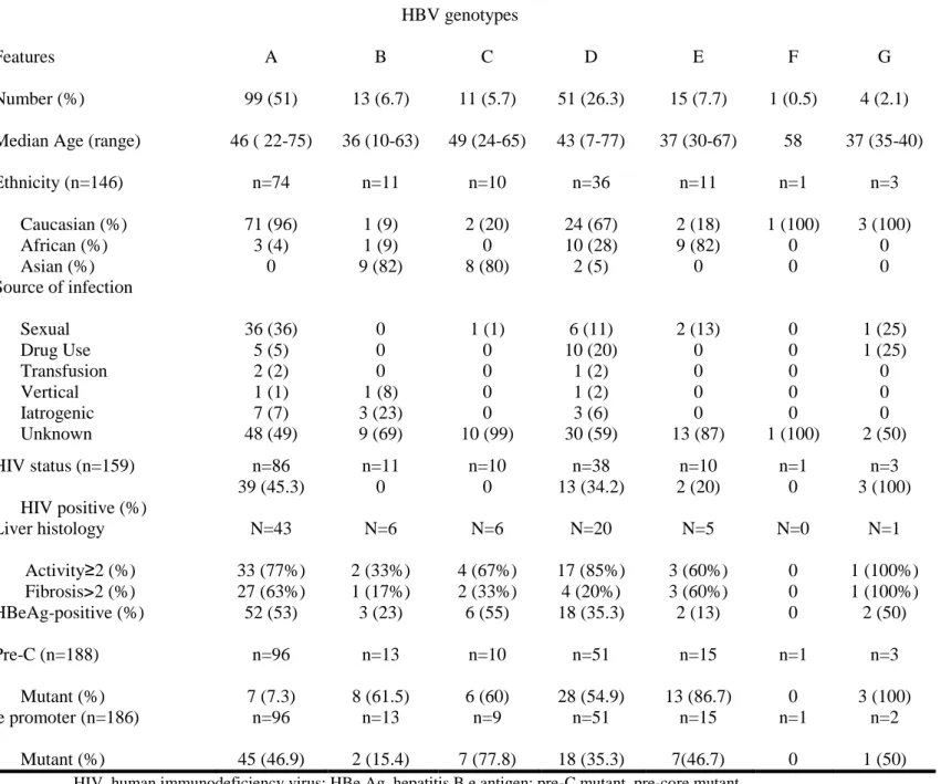
Table 2. Demographic, clinical, and virological characteristics of the 194 patients with reference to HBV genotypes
Tableau 2. Caractéristiques démographiques, cliniques et virologiques des 194 patients en fonction du génotype VHB.
HBV genotypes
Features A B C D E F G
Number (%) 99 (51) 13 (6.7) 11 (5.7) 51 (26.3) 15 (7.7) 1 (0.5) 4 (2.1) Median Age (range) 46 ( 22-75) 36 (10-63) 49 (24-65) 43 (7-77) 37 (30-67) 58 37 (35-40) Ethnicity (n=146) Caucasian (%) African (%) Asian (%) n=74 71 (96) 3 (4) 0 n=11 1 (9) 1 (9) 9 (82) n=10 2 (20) 0 8 (80) n=36 24 (67) 10 (28) 2 (5) n=11 2 (18) 9 (82) 0 n=1 1 (100) 0 0 n=3 3 (100) 0 0 Source of infection Sexual Drug Use Transfusion Vertical Iatrogenic Unknown 36 (36) 5 (5) 2 (2) 1 (1) 7 (7) 48 (49) 0 0 0 1 (8) 3 (23) 9 (69) 1 (1) 0 0 0 0 10 (99) 6 (11) 10 (20) 1 (2) 1 (2) 3 (6) 30 (59) 2 (13) 0 0 0 0 13 (87) 0 0 0 0 0 1 (100) 1 (25) 1 (25) 0 0 0 2 (50) HIV status (n=159) HIV positive (%) n=86 39 (45.3) n=11 0 n=10 0 n=38 13 (34.2) n=10 2 (20) n=1 0 n=3 3 (100) Liver histology Activity≥2 (%) Fibrosis>2 (%) N=43 33 (77%) 27 (63%) N=6 2 (33%) 1 (17%) N=6 4 (67%) 2 (33%) N=20 17 (85%) 4 (20%) N=5 3 (60%) 3 (60%) N=0 0 0 N=1 1 (100%) 1 (100%) HBeAg-positive (%) 52 (53) 3 (23) 6 (55) 18 (35.3) 2 (13) 0 2 (50) Pre-C (n=188) Mutant (%) n=96 7 (7.3) n=13 8 (61.5) n=10 6 (60) n=51 28 (54.9) n=15 13 (86.7) n=1 0 n=3 3 (100) Core promoter (n=186) Mutant (%) n=96 45 (46.9) n=13 2 (15.4) n=9 7 (77.8) n=51 18 (35.3) n=15 7(46.7) n=1 0 n=2 1 (50)
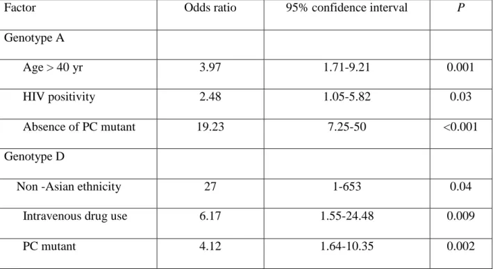
Table 3. Independent factors associated with genotypes A or D in multivariate analysis Tableau 3. Facteurs indépendants associés aux génotypes A et D en analyse multivariée.
Factor Odds ratio 95% confidence interval P
Genotype A
Age > 40 yr 3.97 1.71-9.21 0.001
HIV positivity 2.48 1.05-5.82 0.03
Absence of PC mutant 19.23 7.25-50 <0.001
Genotype D
Non -Asian ethnicity 27 1-653 0.04
Intravenous drug use 6.17 1.55-24.48 0.009
PC mutant 4.12 1.64-10.35 0.002
Figure 1. Prevalence of pre-core (PC) and core promoter (CP) variants in relation to HBV genotypes and HBeAg status.
Figure 1. Prévalence des mutants pré-core (PC) et promoteur de core (CP) en fonction du génotype VHB et du statut AgHBe.