Controlled Release Films and Functional Surfaces
Targeting Infection, Inflammation, and Bleeding
by
Anita Shukla
B.S., Chemical Engineering and Biomedical Engineering, Carnegie Mellon University, Pittsburgh, PA (2006)
M.S., Chemical Engineering Practice,
Massachusetts Institute of Technology, Cambridge, MA (2008)
SUBMITTED TO THE DEPARTMENT OF CHEMICAL ENGINEERING IN PARTIAL FULFILLMENT OF THE REQUIREMENTS FOR THE DEGREE OF
DOCTOR OF PHILOSOPHY IN CHEMICAL ENGINEERING TjTs-:C WI
AT THE c L-
MASSACHUSETTS INSTITUTE OF TECHNOLOGY JUNE 2011
D 2011 Massachusetts Institute of Technology. All rights reserved.
ARCHIV E S
Signature of Author...
Anita Shukla Department of Chemical Engineering May 11, 2011 C ertified by ... . ... ...
Paula T. Hammond Bayer Professor of Chemical Engineering and Executive Officer Thesis Supervisor C ertified by... . . . ... ,...
Robert S. Langer David H. Koch Institute Professor Thesis Supervisor A ccepted by ...
William M. Deen Professor of Chemical Engineering Chairman, Committee for Graduate Students
Controlled Release Films and Functional Surfaces
Targeting Infection, Inflammation, and Bleeding
by
Anita Shukla
Submitted to the Department of Chemical Engineering
on May 11, 2011, in partial fulfillment of the requirements for the degree of Doctor of Philosophy in Chemical Engineering
Abstract
Uncontrolled bleeding and infection are leading causes of patient morbidity and mortality following traumatic injury. Traditional pressure based methods of hemorrhage management are not suitable for incompressible or complex wounds. There is increasing interest in non-pressure based hemostatic dressings; however, many of these existing dressings are not amenable for use in complex sites and are often accompanied by adverse side effects. Additionally, patients are typically administered broad-spectrum antibiotics to prevent and eliminate existing infection. The systemic overuse of antibiotics has led to a worldwide increase in drug-resistant bacteria. As an alternative to these conventional treatments, local therapeutic delivery has the potential to effectively treat cellular dysfunction while avoiding drug toxicity.
This thesis focuses on developing degradable layer-by-layer (LbL) assembled multilayer films as local delivery coatings to address infection, inflammation, and bleeding. These films were engineered to deliver potent antibiotics such as vancomycin and exploratory drugs such as antimicrobial peptides, which prevent the development of drug resistant bacteria. Active films with large drug loadings and a range of drug release profiles were developed by taking advantage of film architectures, assembly techniques (spray versus dip LbL), and film component interactions. Due to the prevalence of infection and inflammation, degradable coatings for the concurrent release of antibiotics and anti-inflammatory therapeutics were also designed. These films have the potential to address a wide range of infection and inflammation requirements, from short term infection and inflammation eradication for trauma relief to infection prevention and long term inflammation mitigation from biomedical implants. All films were successfully applied to medically relevant substrates, including bandages and sutures, and were shown to be active in vitro against Staphylococcus aureus and cyclooxygenase. To address current complications with bleeding control, multilayer films were developed based on hydrogen bonding interactions found to occur between a polyphenol, tannic acid, and an essential clotting factor, thrombin. These thin films were used to coat a common clinically applied absorbent and porous gelatin sponge without reducing its liquid absorption capabilities. Coated sponges were shown to be highly effective in promoting hemostasis in a porcine spleen injury model. The therapeutic films developed in this thesis have the potential to be applied to any clinical substrate. Additionally, drug loading and release can be tuned based on the desired application. Thesis Supervisor: Paula T. Hammond,
Bayer Professor of Chemical Engineering and Executive Officer Thesis Supervisor: Robert S. Langer,
For my loving parents,
Arun and Vinita Shukla
Acknowledgments
There are so many individuals I would like to thank that have made this thesis possible. First, I would like to thank my advisor, Professor Paula Hammond. Paula has been a wonderful mentor who has taught me so much during my years at MIT. She is a truly inspiring person who has always been tremendously supportive of me. I would also like to thank my co-advisor, Professor Robert Langer, who has been a great source of guidance during my thesis research. Additionally, I am grateful to my thesis committee members, Professor Greg Stephanopoulos and Dr. James Bradner, who have always been enthusiastic about my research, providing a unique perspective and invaluable suggestions.
I am indebted to all of the Hammond Lab members who have been wonderful colleagues and friends during my time at MIT. In particular, the Layer-by-Layer Drug Delivery Subgroup has been a great source of intellectual input throughout my Ph.D. In particular, I would like to acknowledge Dr. Mara Macdonald who personally mentored me as an early graduate student, helping me get started on my research. Mara continues to be wonderful mentor and a great friend. I would also like to thank Dr. Kevin Krogman for teaching me about spray layer-by-layer assembly. I am very grateful to my wonderful collaborators, Dr. Rende Fuller and Dr. Amanda Engler. The work described in Chapter 4 could not have been completed without Renee; she is a wonderful person and good friend. Amanda initiated a collaboration in which together we characterized her antimicrobial polypeptides (work described in Chapter 7). I have learned so much from Amanda and thank her for her collaboration and friendship. I am also greatly indebted to Ferrosan and in particular, Flemming Jensen, who has been a great collaborator and helped coordinate testing of the devices described in Chapter 6 of this thesis. I have also been very blessed to have worked with several talented undergraduates during my time at MIT. In particular, I would like to thank Sravanthi Puranam and Jean Fang, who continue to amaze me; they are two of the most driven and bright students I have encountered. I would also like to acknowledge all of the staff in Chemical Engineering and the Institute for Soldier Nanotechnologies for their help throughout my graduate career. I would especially like to thank Christine Preston whose kind smile, sense of calm, and organizational skills have always made my life so much easier.
Much of the reason I am at MIT is due to the support of several faculty members who I encountered in my years as an undergraduate student at Carnegie Mellon University. I would like to thank Professor Lynn Walker, who was my research advisor during my time at CMU. Thank you, Professor Walker, for telling me to get my act together if I ever wanted to get into a graduate school like MIT during my first semester at CMU. I would also like to thank Professor Krystyn Van Vliet who was my research mentor during a summer research experience for undergraduates program at MIT. Professor Van Vliet is a wonderful mentor and has continued to be very supportive of throughout my graduate career. Additionally, I would like to thank Professor Hadley Sikes, who has provided me with great mentorship and career advice during graduate school.
Last but not least, I would like to acknowledge my family and friends. In particular, I would like to thank my husband, Vikas Srivastava. Vikas balances me perfectly and has really been
very instrumental in me completing this Ph.D. while remaining sane. My parents, Arun and Vinita Shukla, are the most wonderful parents anyone could ask for. For as long as I can remember, they have taught me that there is no substitute for hard work and that I should always be proud of what I do. They are my inspiration and motivation. My brothers, Anish and Kush, always know how to make me laugh. My sister-in-law, Jenn, has always made me feel proud of whatever I accomplish. In particular, I want to thank Anish and Jenn for giving me the two most wonderful nieces, Arianna and Alexis. They really put things in perspective for me and make me forget about tough days in lab. I would also like to thank my in-laws and the rest of my family in India who have all been very supportive of me during my Ph.D.
Financial support was provided by the U.S. Army Research Office under contract W91 1NF-07-D-0004 through the MIT Institute of Soldier Nanotechnologies. Research described in Chapter 5
and Chapter 6 was partially funded by Ferrosan (Soeborg, Denmark). A. Shukla gratefully acknowledges support through a National Science Foundation Graduate Research Fellowship. This work utilized the Center for Materials Science and Engineering MRSEC Shared Experimental Facilities supported by the National Science Foundation under award number DMR-02-13282 and facilities at the MIT Institute for Soldier Nanotechnologies.
Contents
A cknow ledgm ents ... 5
Contents 8 Figures 11 Tables 17 Chapter 1 Introduction...18
1.1 Controlled Local Drug Delivery ... 19
1.2 Layer-by-Layer Assembly ... 21
1.2.1 Layer-by-Layer Assembly for Drug Delivery ... 22
1.3 Thesis Overview ... 23
Chapter 2 Controlling the Release of Peptide Antimicrobial Agents from Surfaces .... 26
2.1 Introduction ... 26
2.2 Materials and Methods... 29
2.2.1 M aterials ... 29
2.2.2 Polyelectrolyte and Peptide Solution Preparation... 30
2.2.3 Layer-by-Layer Assembly ... 30
2.2.4 Film Growth and Degradation Characterization... 31
2.2.5 Therapeutic Release Studies ... 31
2.2.6 Therapeutic Quantification ... 31
2.2.7 Bacterial Growth Inhibition Assays... 32
2.2.8 Bacterial Attachment Assay... 33
2.2.9 Biocompatibility ... 33
2.2.10 Statistical Analysis... 34
2.3 Results and Discussion ... 35
2.3.1 Film Architecture and Morphology ... 35
2.3.2 Ponericin GI Loading, Release, and Film Degradation ... 39
2.3.3 Bacterial Growth Inhibition... 43
2.3.4 Bacterial Attachment ... 45
2.3.5 Film Biocompatibility... 46
2.4 C onclusions... 47
Chapter 3 Tunable Vancomycin Releasing Surfaces for Biomedical Applications... 48
3.1 Introduction ... . 48
3.2 Materials and Methods... 50
3.2 .1 M aterials ... . . 50
3.2.2 Polyelectrolyte-Drug Interaction Studies... 51
3.2.3 F ilm A ssem bly ... 5 1 3.2.4 B andage C oating ... 52
3.2.5 Film Growth and Morphology Characterization... 53
3.2.6 D rug R elease... 53
3.2.7 Bacterial Growth Inhibition... 53
3.2.8 B iocom patibility ... 54
3.2.9 Long Term Storage Stability... 55
3.2.10 Statistical Analysis... ... 55
3.3 R esults and D iscussion ... ... ... 55
3.3.1 Choice of Film Architecture ... 55
3.3.2 Film Growth and Morphology ... 58
3.3.3 Drug Incorporation and Release ... 62
3.3.4 Combining Dip and Spray LbL Assembly for Practical Application... 67
3.3.5 F ilm E fficacy ... 69
3.3.6 Vancomycin Storage Stability in LbL Films for On-Demand Care ... 71
3 .4 C on clu sion s... . 74
Chapter 4 Design of Multi-Drug Release Coatings Targeting Infection and Inflammation... 76
4 .1 Introdu ction ... . 76
4.2 Materials and Methods... 78
4.2 .1 M aterials ... . . 78
4.2.2 F ilm A ssem bly ... 79
4.2.3 D rug R elease... 80
4.2.4 Studying Film Component Interactions ... 80
4.2.5 Measuring Drug Activity ... 81
4.2.6 Statistical Analysis ... 81
4.3 Results and Discussion ... ... ... 82
4.3.1 Film Component Interactions ... ... 82
4.3.2 Film Assembly and Drug Release Characteristics... 86
4.3.3 Therapeutic Potential of Optimal Film Architectures... 89
4.3.4 Composite Film Summary ... 91
4 .4 C on clu sion s... . 92
Chapter 5 Release of Vancomycin from Multilayer Coated Absorbent G elatin Sponges ... 94
5 .1 Introdu ction ... . 94
5.2 Materials and Methods... 95
5.2.1 M aterials ... . . 95
5.2.2 Film Assembly on Surgifoam@... 96
5.2.3 Characterization of Film and Surgifoam@ Properties ... 96
5.2.4 Vancomycin Release from Surgifoam® ... 97
5.2.5 Bacterial Growth Inhibition ... 98
5.2.6 Statistical A nalysis... 98
5.3 Results and Discussion ... 99
5.3.1 Surgifoam @ Coating Characterization ... 99
5.3.2 Vancom ycin Release and Therapeutic Potential... 102
5.3.3 Drug Activity ... 107
5.4 Conclusions... 108
Chapter 6 Layer-by-Layer Assem bled Hem ostatic Coating... 110
6.1 Introduction... 110
6.2 M aterials and M ethods... 111
6.2.1 M aterials ... 111
6.2.2 Film Preparation... 112
6.2.3 Characterization of Film Properties ... 113
6.2.4 Film Activity ... 114
6.2.5 Statistical Analysis... 115
6.3 Results and Discussion ... 115
6.3.1 Determ ining Film Architecture... 115
6.3.2 Spray Film Assembly and Characterization ... 117
6.3.3 Film Activity ... 121
6.4 Conclusions... 123
Chapter 7 Effects of Side Group Functionality and Molecular Weight on the Activity of Synthetic Antim icrobial Polypeptides ... 125
7.1 Introduction... 125
7.2 M aterials and M ethods... 128
7.2.1 Polypeptide Synthesis and Physical Characterization ... 128
7.2.2 Polypeptide Antim icrobial Characterization... 131
7.3 Results and Discussion ... 133
7.3.1 Antim icrobial Polypeptide Synthesis... 133
7.3.2 Bacterial Growth Inhibition... 136
7.3.3 Bacteria Attachm ent Inhibition... 142
7.3.4 Polypeptide Biocompatibility ... 144
7.4 Conclusions... 145
Chapter 8 Conclusions and Future Directions... 147
8.1 Thesis Sum m ary... 147
8.2 Future Directions ... 149
8.3 Concluding Rem arks... 151
Bibliography ... 153
Appendix A Fitting Data That Transitions Between Two Linear Regimes... 167
A .1 Instantaneous Transition... 167
A.2 Sm ooth Transition... 167
Appendix B Effect of Wash Ionic Strength on Vancomycin Films... 169
Appendix C Antim icrobial Polypeptide Surface Coatings ... 171
C. 1 Calculation of Thickness... 171
C.2 Substrate Coating Experim ents... 172
Figures
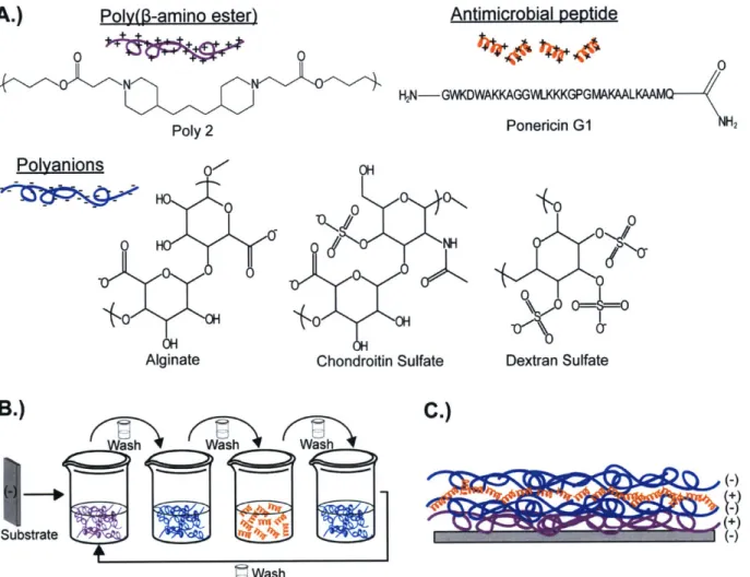
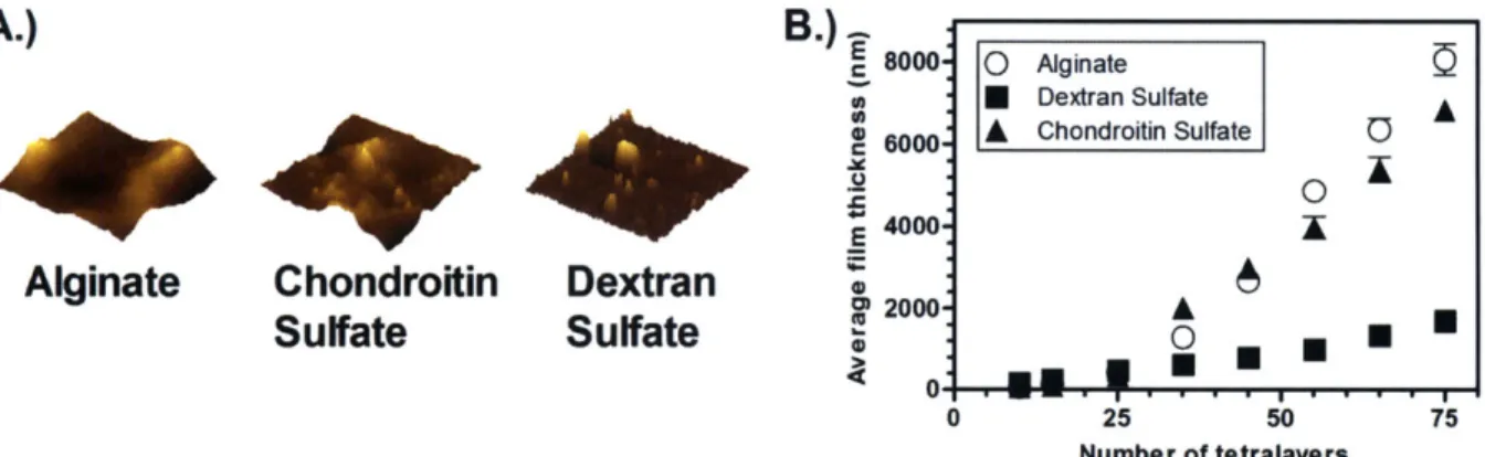
Figure 2-1: Film components, layer-by-layer film assembly, and film architecture. A.) Structure of poly 2, alginate, chondroitin sulfate, dextran sulfate, and ponericin GI. B.) LbL assembly process. C.) Single tetralayer of the 75 tetralayer films assembled in this study. ... 3 6 Figure 2-2: Film morphology and growth profiles. A.) Atomic force microscopy images (10 pm
by 10 ptm; z-scale = 500 nm. B.) Growth profiles of films based on polyanion used... 37 Figure 2-3: Ponericin GI release versus time for 75 tetralayer films. A.) Total ponericin GI
released over time based on polyanion used in film. B.) Normalized ponericin GI release profiles based on polyanion used in film. C.) Normalized ponericin GI release over first 3 0 h o u rs... 4 0 Figure 2-4: Normalized film thickness versus time of films starting at 75 tetralayers... 42 Figure 2-5: Normalized bacteria inhibition versus ponericin GI concentration. A.) Inhibition of
S. aureus growth by a standard of non-film-released ponericin GI. B.) Inhibition of S. aureus growth by (poly 2/alginate/ponericin G1/alginate)75 film release solution... 44
Figure 2-6: S. aureus growth inhibition assay for all film constructs and control. A.) Uncoated silicon control. B.) (Poly 2/alginate/ponericin G1/alginate)75 film. C.) (Poly
2/chondroitin sulfate/ponericin G1/chondroitin sulfate)75 film. D.) (Poly 2/dextran
sulfate/ponericin GI/dextran sulfate)75 film. Scale bar = 200 pm... 44
Figure 2-7: S. aureus attachment assay for all film constructs and control. A.) Uncoated silicon control. B.) (Poly 2/alginate/ponericin Gl/alginate)75 film. C.) (Poly 2/chondroitin
sulfate/ponericin GI/ chondroitin sulfate)75 film. D.) (Poly 2/dextran sulfate/ponericin
GI/dextran sulfate)75 film . Scale bar = 200 pm ... 46
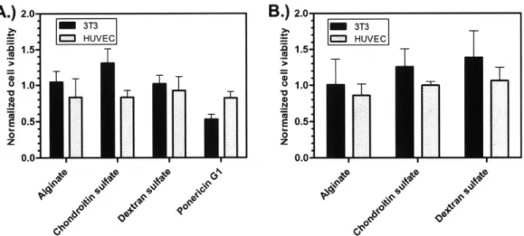
Figure 2-8: Normalized cell viability for all film constructs. A.) Viability of cells exposed to media containing dissolved polyanions or ponericin GI standards. B.) Viability of cells exposed to release media based on polyanion used in film architecture. ... ... 47
Figure 3-1: Film assembly and components. A.) LbL assembly schematic (dipped and sprayed assembly); tetralayer film architecture. B.) Film component structures (cations highlighted in green and purple, anions in red). ... 56 Figure 3-2: Drug-polyelectrolyte interaction studied via HPLC. The dashed red traces in A-C
represent pure 5 pg/mL vancomycin solution at pH 7.4 in 0.01 M PBS. The dashed red traces in D-F represent pure 5 pg/mL vancomycin solution at pH 7.4 in 1 M NaCl. The solid traces represent mixtures containing 5 pg/mL vancomycin and 5 pg/mL polyelectrolyte. A.) Vancomycin-dextran sulfate mixture, pH 7.4, 0.01 M PBS. B.) Vancomycin-chondroitin sulfate mixture, pH 7.4, 0.01 M PBS. C.) Vancomycin-alginate mixture, pH 7.4, 0.01 M PBS. D.) Vancomycin-dextran sulfate mixture, pH 7.4, 1 M NaCl. E.) chondroitin sulfate mixture, pH 7.4, 1 M NaCl. F.) Vancomycin-alginate m ixture, pH 7.4, 1 M N aCl... 57
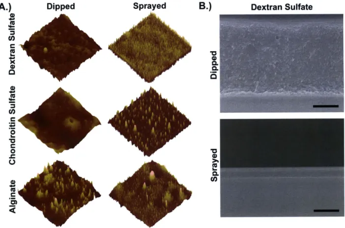
Figure 3-3: Film growth profiles. A.) (Poly 2/dextran sulfate/vancomycin/dextran sulfate), sprayed and dipped. B.) (Poly 2/chondroitin sulfate/vancomycin/chondroitin sulfate), sprayed and dipped. C.) (Poly 2/alginate/vancomycin/alginate), sprayed and dipped.... 59 Figure 3-4: Final film morphology. A.) Atomic force microscope images (10 gm x 10 pm) for sprayed and dipped films of architecture (poly 2/polyanion/vancomycin/polyanion)o. The maximum z-scale for each of the polyanions is as follows: dextran sulfate (dipped,
zmax = 250 nm; sprayed, Zmax = 4 nm), chondroitin sulfate (dipped, zmax = 150 nm;
sprayed, zmax = 7 nm), and alginate (dipped, Zmax = 200 nm; sprayed, Zmax = 15 nm). B.)
Scanning electron microscope cross-section images for (poly 2/dextran sulfate/vancomycin/dextran sulfate)6o dipped and sprayed films (scale bar = 1 [tm)... 62
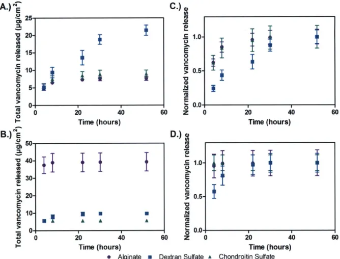
Figure 3-5: Vancomycin release from (poly 2/polyanion/vancomycin/polyanion)60 films. A.) Total vancomycin release over time from dipped films. B.) Total vancomycin release over time from sprayed films. C.) Normalized vancomycin release over time from dipped films. D.) Normalized vancomycin release over time from sprayed films... 63 Figure 3-6: Interdiffusion in vancomycin dipped and sprayed films. A.) HPLC spectra of film release eluent from a representative dipped (poly 2/dextran sulfate/vancomycin/dextran sulfate)60 film. B.) Vancomycin release from sprayed film of architecture (poly 2/dextran
sulfate/vancom ycin/dextran sulfate)120 ... ... . . . 65 Figure 3-7: Vancomycin release from composite dip and spray film of architecture: (poly
2/dextran sulfate/vancomycin/dextran sulfate)o,i, - (poly 2/alginate/vancom ycin/alginate)o,spray...68 Figure 3-8: Spray coating of a commercial bandage with film architecture (poly
2/alginate/vancomycin/ alginate)60. A,) Aerial view scanning electron microscope
images of an uncoated and coated bandage (scale bar = 100 pm). B.) Total vancomycin release from spray coated bandage. ... 69 Figure 3-9: Staphylococcus aureus growth inhibition. A.) S. aureus growth inhibition by (poly 2/alginate/vancomycin/alginate)60 coated bandage. Zone of inhibition surrounding coated
no inhibitory zone surrounding uncoated bandage. B.) S. aureus growth inhibition by film released eluent from a dipped (poly 2/dextran sulfate/vancomycin/dextran sulfate)60
film. For release from 0 - 10 hours, dilution 1 contains 7 pig/mL of vancomycin and subsequent dilutions correspond to 50% reduction in concentration of the previous dilution. For the remaining release, dilution 1 contains 16 ptg/mL vancomycin and subsequent dilutions correspond to 50% reduction in concentration of the previous d ilu tio n ... 7 0 Figure 3-10: Normalized cell viability for vancomycin films. A.) Viability of cells in response to film release media for all film architectures. B.) Viability of cells in response to non-film incorporated drug and polyanions... 71 Figure 3-11: Vancomycin release profiles upon storage. Profiles shown here correspond to storage for 0 months, 3 months at 25 'C, 6 months at 25 'C, and 6 months at 4 C... 72 Figure 3-12: Normalized S. aureus density upon exposure to vancomycin film release solution. Films stored for A.) 0 months, B.) 3 months at 25 'C, and C.) 6 months at 25 C. ... 73 Figure 3-13: Vancomycin film behavior after 1 month storage at 37 'C. A.) Vancomycin release
profiles of films stored for 0 and 1 month at 37 'C. B.) Normalized S. aureus density upon exposure to vancomycin film release solution for films stored for 1 month at 37 'C. ... 7 4 Figure 4-1: Layer-by-layer film architectures. A.) Antibiotic-only and NSAID-only LbL film architectures. B.) Composite antibiotic and NSAID LbL film architectures. Note: orange = polyanion, blue = poly(p-amino ester), green = vancomycin, red = diclofenac encapsulated within poly(carboxymethyl-p-cyclodextrin). ... 78 Figure 4-2: Solution based film component interactions. A.) Vancomycin-polyCD interaction. B.) Vancomycin-diclofenac interaction. All interactions were studied at four conditions: 0.1 M sodium acetate buffer and 1 M NaCl, pH 5 and 6... 83 Figure 4-3: Study of diffusion and exchange behavior in single-therapeutic films... 85 Figure 4-4: Composite film drug release profiles. A.) Drug release from dipped LbL film: (poly 2/dextran sulfate/vancomycin/dextran sulfate)60 + (poly 2/polyCD-diclofenac)20. B.)
Drug release from sprayed LbL film: (poly 2/chondroitin sulfate/vancomycin/chondroitin sulfate)60 + (poly 2/polyCD-diclofenac) 20. ... ... ... 87
Figure 4-5: Multi-drug release device coatings. Scanning electron microscopy images of coated medical devices (scale bar = 20 ptm for IOL and bandage; 100 pim for suture). The uncoated IOL image shows both the lens and haptic regions. The visible crack on the coated IOL is a scratch on the film showing the existence of a smooth film on the lens. 89 Figure 4-6: Composite film-released drug efficacy. A.) COX activity of diclofenac released from LbL bandage coating at Day 1, 2, 4, and 6 of release. Controls of pure polyCD, pure vancomycin, and pure diclofenac were also included. B.) Vancomycin activity against agar coated S. aureus of (i) LbL coated bandage, (ii) uncoated bandage, and (iii)
vancomycin control (30 ptg) (scale bar = 9 mm). C.) Normalized S. aureus inhibition by vancomycin released from dipped LbL film architecture: (poly 2/dextran sulfate/vancomycin/dextran sulfate)60 + (poly 2/polyCD-diclofenac) 20... ... . . . 90 Figure 4-7: Total drug release from composite film architectures. A.) (Poly 2/chondroitin sulfate/vancomycin/chondroitin sulfate)60 + (poly 2/polyCD-diclofenac)20 dipped. B.)
(Poly 2/polyCD-diclofenac) 20 + (poly 2/chondroitin sulfate/vancomycin/chondroitin
sulfate)6o dipped. C.) (Poly 2/alginate/vancomycin/alginate)6o + (poly
2/polyCD-diclofenac)2o dipped. D.) (Poly 2/polyCD-diclofenac)20 + (poly 2/alginate/vancomycin/alginate)6o dipped. E.) (Poly 2/polyCD-diclofenac)20 + (poly 2/dextran sulfate/vancomycin/dextran sulfate)6o dipped. F.) (Poly
2/polyCD-diclofenac)20 + (poly 2/chondroitin sulfate/vancomycin/chondroitin sulfate)60 sprayed.. 92
Figure 5-1: Spray layer-by-layer assembly for porous substrates. Each airbrush aerosolizes and sprays film components or the rinse solution at the substrate; a vacuum is applied to pull solutions through the substrate. For the vancomycin LbL films, 1 = poly 2, 2 and 4 =
dextran sulfate, and 3 = vancom ycin. ... 100 Figure 5-2: SEM micrographs of uncoated and (poly 2/dextran sulfate/vancomycin/dextran sulfate), spray LbL coated Surgifoam@. Scale bar = 500 ptm and 50 tm for top and bottom row micrographs, respectively, for both plan-view and cross-section images (except 60 tetralayer cross-section top row, where scale bar = 200 tm), respectively. .101 Figure 5-3: Absorbency ratio of phosphate buffered saline by film coated compared to uncoated S u rg ifo am @ ... 10 2 Figure 5-4: Vancomycin release profiles from Surgifoam@ coated with (poly 2/dextran sulfate/vancomycin/ dextran sulfate), where n = 60 and 120. A.) Drug release expressed in ptg of vancomycin per mg of Surgifoam®. B.) Drug release expressed in ptg of vancomycin per Surgifoam® projected in-plane area (cm2). ... 103
Figure 5-5: Normalized vancomycin release profiles. A.) Complete release from Surgifoam® and flat substrates coated with (poly 2/dextran sulfate/vancomycin/dextran sulfate)60 spray LbL films and vancomycin-soaked Surgifoam@ (no film). B.) Data shown in (A.) up to 52 hours of release. C.) Complete release from Surgifoam® and flat substrates coated with (poly 2/dextran sulfate/vancomycin/dextran sulfate)120 spray LbL films and vancomycin-soaked Surgifoam® (no film). D.) Data shown in (C.) up to 77 hours of relea se . ... 10 4 Figure 5-6: Vancomycin release from (poly 2/dextran sulfate/vancomycin/dextran sulfate)120 coated Surgifoam®. A.) Release from three individual samples is shown; the average of these three samples leads to the results shown in Figure 5-5C and 5-5D. B.) The total vancomycin released for the last three time points of significant release showing that each individual sample releases a significant quantity of vancomycin through 150 hours. ... 106 Figure 5-7: Staphylococcus aureus growth inhibition. A.) Normalized S. aureus density upon
exposure to dilutions of film release solutions from LbL coated Surgifoam® and a standard of film released vancomycin (dilution 1 = 2.3, 2.3, and 1.9 [tg/mL for
non-film released vancomycin, n = 60, and n = 120, respectively; each subsequent dilution is half the concentration of the previous dilution). B.) Agar coated with S. aureus exposed to 60 tetralayer LbL film coated pieces of Surgifoam@ (i and ii), an uncoated piece of Surgifoam@ (iii), and a 30 pg vancomycin control disc (iv). Sample (i) is the top two-thirds of the coated sponge, while sample (ii) is the bottom one-third... 108 Figure 6-1: Film growth monitored by quartz crystal microbalance. The growth of two different architectures on a monolayer of BPEI are shown: (thrombin/tannic acid)n and (mannitol/tannic acid),. The start of BPEI, thrombin or mannitol, and tannic acid flow is indicated by labels above the graph, along with each PBS wash step indicated by an arro w ... 1 17 Figure 6-2: Thickness of sprayed (thrombin/tannic acid), films for n = 10, 25, and 50. Both average film thickness and the change in thickness per bilayer are shown for each growth region (0 to 10 bilayers, 10 to 25 bilayers, and 25 to 50 bilayers)... 118 Figure 6-3: Sprayed (thrombin/tannic acid), film morphology measured by atomic force
microscopy for n = 10, 25, and 50. Each image is 10 tm x 10 tm, and Zma= 360 nm,
380 nm, and 440 nm for n = 10, 25, and 50, respectively. ... 119 Figure 6-4: Sprayed (thrombin/tannic acid), film degradation for n = 10, 25, and 50. A.) Film
thickness over time. B.) Normalized film thickness over time... 120 Figure 6-5: Sprayed (thrombin/tannic acid)n coated Surgifoam@ morphology for n = 0, 10, 25, and 50 (scale bar = 200 ptm ). ... 12 1 Figure 6-6: Activity of film coated Surgifoam@ at n = 10, 25, and 50. A.) Activity expressed as
international units (IU) per square centimeter of coated Surgifoam@ and IU per milligram of Surgifoam@. B.) Flat surface film thickness and activity of coated Surgifoam@ (U/cm2). Activity data was provided by Ferrosan (Soeborg, Denmark). ... 122 Figure 6-7: In vivo activity of (thrombin/tannic acid), coated Surgifoam@ for n = 10, 25, and 50, and BPEI control in a porcine spleen bleeding model. A.) Representative image of the porcine spleen bleeding model. B.) Time to hemostasis following sample application. Figure data provided by Ferrosan (Soeborg, Denmark). ... ... 123 Figure 7-1: Alkyne-azide cycloaddition click functionalization of PPLG and various amine side
groups. The abbreviation
Q indicates that the amine is quaternary and Cn indicates a
carbon chain length w ith n repeat units. ... 127 Figure 7-2: A.) 1H-NMR spectrum of PPLG (DP = 140) in d7 DMF. B.) 'H-NMR spectrum of PPLG (DP = 140) functionalized with QC1 in D20. C.) 'H-NMR spectrum of PPLG (DP= 140) functionalized with QC6 in CD30D ... 134 Figure 7-3: Molecular weight distribution of PPLG obtained using a DMF GPC and calculated using PMMA standards. The degree of polymerization was determined by 'H-NMR. 135
Figure 7-4: Bacteria growth inhibition for primary amine functionalized polymers based on normalized turbidity measurements. A.) S. aureus normalized bacteria density at varying polymer concentrations. B.) E. coli normalized bacteria density at varying polymer concentrations (*high turbidity was observed for DP = 140 polypeptides at concentrations of 4500 - 1125 pg/mL due to polypeptide precipitate forming; in these cases, however, complete bacteria growth inhibition was observed based on clear solution surrounding the polypeptide precipitate). ... 137 Figure 7-5: Bacteria growth inhibition by QC8 (DP = 75) coating for both S. aureus and E. coli.
... 14 0 Figure 7-6: Circular Dichroism of QC8 functionalized PPLG in methanol (DP = 75, concentration = 1.67 m g/m L) ... 141 Figure 7-7: Representative FTIR of QC8 functionalized PPLG... 142 Figure 7-8: S. aureus attachment inhibition by quartemary amine functionalized polypeptides with varying hydrophobicity (QC4 - QC12; control = uncoated substrate)... 143 Figure 7-9: E. coli attachment inhibition by quartemary amine functionalized polypeptides with varying hydrophobicity (QC4 - QC12; control = uncoated substrate)... 143 Figure 7-10: Normalized hemolysis for QC8 polypeptide. ... 145 Figure A -1: Tw o phase linear transition. ... 168 Figure B-1: Growth profiles for: (poly 2/polyanion/vancomycin/polyanion)n. Rinse steps following each deposition were conducted in deionized water (no ionic strength of pH adju stm ent)... 169 Figure B-2: Vancomycin release from dipped and sprayed 60 tetralayer films with water rinse step s... 17 0 Figure C -1: Polypeptide schem atic... 171 Figure C-2: Average polypeptide film thickness... 172 Figure C-3: QC10 solvent cast substrate morphology (10 im x 10 pm). A.) Before media
treatment (maximum z-scale = 22.1 nm). B.) After media treatment (maximum z-scale = 1.7 n m )... 17 3
Tables
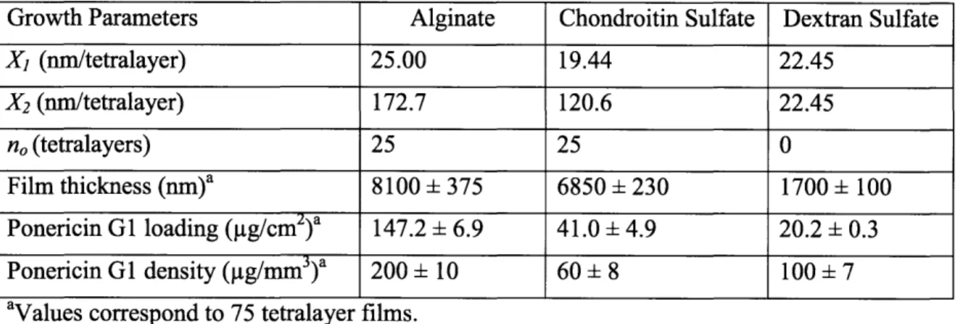
Table 2-1: Growth and loading parameters based on polyanion used in AmP films. ... 38
Table 3-1: Film morphology and drug loading properties at sixty tetralayers. ... 61
Table 4-1: Diffusion and exchange behavior in single-therapeutic films... 86
Table 4-2: Total drug loading and release timescale of single-therapeutic and composite films. 91 Table 5-1: V ancom ycin release kinetics... 105
Table 6-1: Sprayed (thrombin/tannic acid), film characteristics... 120
Table 7-1: Sum m ary of polypeptides tested. ... 128
Table 7-2: Staphylococcus aureus growth inhibition properties. ... 137
Table 7-3: Escherichia coli growth inhibition properties... 138
Table 7-4: Bacteria response to QCn (n > 4) polypeptides... 140
Chapter 1
Introduction
Bleeding and infection are two major causes of patient morbidity and mortality following traumatic injury [1-3]. Traditional options for hemorrhage control include the application of pressure-based devices such as tourniquets and compressive bandages. However, these devices are often unsuitable for application to complex wounds or incompressible sites, and recently there has been much interest in the development of non-pressure based hemostatic dressings [1, 4]. Fibrin based glues and dressings have been used to augment the natural clotting cascade. However, fibrin products are not practical due to their short shelf-life, exorbitant prices, and risk of disease transfer. Zeolite-based powders have also been applied; they function by absorbing water and concentrating the clotting factors in a wound. Again, these powders are not ideal due to their lack of applicability in windy environments and the inherent exothermic reaction that accompanies their use, which is known to cause severe burns to surrounding tissue [1, 5]. Freeze-dried chitosan bandages have shown the greatest promise of currently available hemostatic dressings and have been adopted for use by the Department of Defense [2, 4]. The mode of action of these bandages is not entirely understood; however, their ability to promote hemostasis is thought to be due to their tissue adhesive properties, ability to cause vasoconstriction, and attraction of negatively charged red blood cells and platelets [1]. The primary drawback of these chitosan bandages is difficulty in application to complex non-planar wound sites [1].
To treat potential infection following injury, patients are typically administered broad-spectrum antibiotics orally or intravenously. This systemic exposure allows drug-resistant bacteria to flourish and greatly increases difficulty in treating infection [6]. In fact, in wounded soldiers, there is a close correlation with the increasing use of broad-spectrum antibiotics and a rise in antibiotic-resistant bacteria, such as methicillin-resistant Staphylococcus aureus (MRSA) [3]. Additionally, colonization of bacteria on implant surfaces or dead-tissue can give rise to biofilms, which further complicate treatment [7]. There has been much research in the area of localized antibiotic delivery methods to avoid systemic administration, particularly in the area of orthopedic devices, wound dressings, periodontal devices, and vascular grafts, to name a few [8].
Scaffolds and dressings embedded with the antimicrobials allowing for passive drug release [8-10], drug-loaded hydrogels [11], degradable coatings [12-14], and antimicrobial polymeric surfaces [15-19] have been developed. In many cases, these devices are unable to bear large drug loads without compromising device integrity. Often, there is inadequate control over drug release timescales and concentrations. This is especially concerning as prolonged exposure to antibiotics, particularly at concentrations that are below the minimum inhibitory concentration of the therapeutic against common bacteria, is directly implicated in causing drug resistance [6, 8].
It is clear that there is vast area for improvement in both the control of bleeding and the treatment of infection. Namely, for bleeding control, there is a need for an effective non-pressure based technology that is applicable to a variety of wound configurations. For infection treatment, local delivery methods that demonstrate superior control over drug release timescale and drug loading are needed.
1.1 Controlled Local Drug Delivery
Use of local drug delivery devices has the potential to alleviate many of the non-ideal outcomes of systemic drug delivery. Most often drugs are administered either topically, orally, intravenously, intramuscularly, subcutaneously, or sublingually. Local delivery offers significant advantages over these methods. These include requiring lower drug doses, a greater control over drug toxicity and bioavailability, potential for extended release, potential for control over drug release profile directly at the site, and requiring lower number of drug administrations during treatment [20, 21]. There are numerous local drug delivery devices currently approved by the United States Food and Drug Administration (FDA) that are routinely utilized. Some of the prominent areas of use include drug-eluting stents, catheters, orthopedic devices, and wound dressings [8, 20]. In most of these devices, polymers are utilized to both load and control drug release behavior. Commercial drug-eluting stents contain micron-scale coatings of polyisobutylene or polymethacrylate copolymers loaded with drug. In the case of the widely-studied sirolimus-releasing stent, CYPHER7 (manufactured by Cordis), drug loadings of approximately 140 pg/cm2 are attained with a burst-release upfront followed by extended drug release up to 6 weeks. For antimicrobial catheters, two methods of functionalization have been employed, including dip coating of catheters in drug solution prior to use where drugs will adsorb onto and absorb into the catheter material, as well as drug impregnation prior to injection
molding or extrusion to produce the device [20]. To combat osteomyelitis, antibiotics are loaded into bone cement, polymethylmethacrylate (PMMA), or PMMA beads. PMMA bone cement is a non-degradable polymer in which drugs can be combined during the curing process, in which a solid and liquid component are combined immediately prior to use. The solid component contains the PMMA powder, an initiator, and the drug, while the liquid component contains the methyl methacrylate monomers. Upon mixing, the glassy polymer cement is formed within minutes accompanied by an exothermic reaction [8]. Drug release behavior from PMMA is governed by drug loading, porosity, surface area, and roughness [20]. Wound dressings are also another application of great interest for localized drug delivery and there are several currently approved devices in use. Acticoat@ (manufactured by Smith and Nephew) is a gauze dressing with silver-coated polyethylene meshes on both the outer layers; wetting the gauze several times a day allows the release of antimicrobial silver to the wound site. To eliminate need for changing dressings, biodegradable dressings utilizing lactide-caprolactone copolymers have found utility in the clinic [20]. Aside from the few mentioned here, numerous other formulations and applications of local drug delivery devices are currently in clinical use.
Despite their widespread use, there are many areas for improvement of currently used drug delivery devices. Many of these devices release drug at concentrations that are sub-therapeutic which can exacerbate the condition and increase difficulty in treatment. This is especially prevalent with PMMA use, where often less than 10% of the loaded drug is eventually released from these non-degradable matrices [8]. Additionally, it has been shown that many of the organisms in contact or close proximity to the drug-releasing PMMA, develop resistance to the drug, most likely due to the sub-minimum inhibitory concentration drug exposure [20]. Also, there is often a lack of adequate control over drug delivery timescales, where prolonged drug exposure is problematic and may give rise to drug-resistance in the case of antibiotics. These limitations can often not be overcome with current design methods, due to the fact that the mechanical and functional properties of the drug loaded devices must be maintained. Most of the current methods do not allow for complex delivery profiles, such as sequential delivery of multiple therapeutics. In current devices, release is often a combination of drug diffusion and degradation or dissolution of polymer matrix [21], rather than a stimuli-responsive release. Additionally, as apparent with PMMA use, many of the current local delivery techniques are inherently harsh in regards to temperature or solvent used during device preparation [8]. This is
especially problematic when trying to incorporate sensitive components into these devices, such as proteins, which will denature at these conditions. Recognizing the benefits of controlled local drug delivery while trying to overcome the many drawbacks of current techniques, this thesis explores the development of therapeutic coatings using the layer-by-layer assembly technique to target infection, inflammation, and bleeding.
1.2 Layer-by-Layer Assembly
Layer-by-layer (LbL) assembly is used for the fabrication of polymer multilayer films [22]. In this technique, films are assembled by the sequential adsorption of materials with complementary functionality, including electrostatic interactions [22], hydrogen-bonding [23], and biological interactions [24]. Following each deposition step, a wash step is used to remove any non-specifically bound material. Functionality reversal at each deposition step allows the film to continue growing with each subsequent deposition [22]. Although dip LbL assembly, in which the substrate is submerged in deposition solutions allowing substantial time for diffusion and adsorption of species to the growing film surface, is the most common method of LbL assembly, both spray [14, 25-28] and spin LbL assembly [29] have also been explored. Spray LbL assembly, in particular, has shown great promise in achieving thin films in a time-effective manner by eliminating diffusion limitations of the LbL process. LbL assembly is an aqueous technique, and unlike many of its bulk coating counterparts, it is able to support the direct encapsulation of a variety of sensitive molecules and macromolecular species, including proteins [30-34], peptides [13, 35, 36], nucleic acids [24, 37-40], etc. This assembly technique has found utility in a variety of areas, including biological engineering, electrochromics, and fuel cells, amongst numerous others [41-47]. Additionally, due to ease of application, LbL films have been applied to a wide variety of substrates, including stents [37], electrospun materials [27], microparticles [38], mammalian cells [48, 49], biological tissues [50], etc. Properties of the materials and solvents used to assemble these films are critical in determining film characteristics, including molecular weight and degree of ionization, related directly to solution ionic strength and pH [13, 51-53].
1.2.1 Layer-by-Layer Assembly for Drug Delivery
Due to the fact that a wide range of species can be encapsulated and delivered from LbL assembled systems, these films are being examined extensively for various therapeutic applications [44]. Of particular interest to this thesis are films developed to deliver antimicrobials, anti-inflammatory drugs, and proteins. In specific, previous research has shown the incorporation and release of a variety of therapeutics from LbL films, including antibiotics such as gentamicin [12, 54, 55] and triclosan [56], chemotherapeutics such as doxorubicin [57], non-steroidal anti-inflammatory agents such as diclofenac [58], and proteins such as FGF-2 and BMP-2 [30, 31, 33, 34]. In much of this work, it has been shown that changing the number of layers allows for tunable drug loading.
Many groups have explored the encapsulation of drugs in these films by absorption after film assembly where the drug is not one of the direct building blocks of the film. For example, in one study bilayer films were composed of poly(L-lysine) (PLL) and hyaluronic acid, following which the film was soaked in paclitaxel allowing for absorption of the drug. These films had an anti-proliferative effect on a cancerous cell line [59]. In another recent study, a unique method of using LbL films to obtain antimicrobial drug incorporation and release was examined. In this study, bilayer films were assembled with poly(methacrylic acid) (PMAA) and poly(N-vinylpyrrolidone) (PVPON) which were subsequently crosslinked and the PVPON removed, leaving behind a PMAA hydrogel. Antimicrobial drugs, proteins, and peptides were incorporated post hydrogel formation via absorption. The drug release here was based on hydrogel swelling in response to changes in pH and ionic strength [55]. Others have explored the direct incorporation of the drug as a component of the LbL film. In one study, the antimicrobial peptide (AmP), defensin, was adsorbed as a layer into a bilayer film of poly(L-glutamic acid) (PGA) and PLL. The cationic defensin was deposited following an anionic PGA deposition, and PLL was needed in order to obtain complete charge reversal. Release of drug from these films was not quantified or examined; however, it was noted that bacteria needed to be in close contact with the film for antimicrobial activity to be visualized [35].
Rather than relying simply on passive release of drugs, stimuli-responsive drug release may be desirable. For example, biodegradable polypeptides including PLL and PGA have been used extensively in LbL films for this purpose. In one particular study, both TGF-p and BMP-2 were incorporated as layers into a PGA and PLL containing film and used to create polymeric
capsules. In vivo bone formation upon administration of these capsules was examined in mice. Film activity in this study was attributed to the enzymatic degradation of the PGA and PLL, liberating the active proteins [31]. In a similar study, films containing PGA, PLL, DNA, and cyclodextrins, were found to demonstrate in vitro activity only in the presence of cells and not due to passive release of drug [60]. Although these biodegradable polymers have been utilized extensively, their properties are often not as easily manipulated as entirely synthetic polymers, which has implications on drug loading and release properties of LbL films. Many researchers have therefore examined the use of synthetic degradable polymers in these films, such as hydrolytically degradable poly(p-amino esters) [61, 62]. The properties of these polymers can help tune the degradation rate of LbL films in the specific environment in which the films are applied. For example, the level of hydrophobicity of poly(p-amino esters) has been shown to greatly influence the mechanism of film degradation, including surface and bulk erosion, and the rate at which these processes occur [63].
It may also be desirable to sequentially deliver multiple therapeutics from a single LbL assembled film. Sequential drug delivery is complicated by the phenomena of interdiffusion which occurs when one or more components within a film architecture are sufficiently mobile within the underlying film during assembly, allowing rearrangement in the film structure [52]. Methods of blocking interdiffusion of one segment of a multilayer film from another have been examined in the form of thermally crosslinked barrier layers for the sequential delivery of macromolecular model therapeutics [64]. Additionally, current methods in the Hammond Lab at the Massachusetts Institute of Technology such as the incorporation of catechol modified polymers into LbL films have also shown great initial promise in controlling film component interdiffusion.
1.3 Thesis Overview
This thesis focuses on using LbL assembly to design device coatings that target infection, inflammation, and bleeding. The remainder of the thesis is divided into 7 chapters discussing the design of these coatings. In Chapter 2, a hydrolytically degradable polymer multilayer film is developed for the delivery of an AmP, ponericin GI. Several film architectures are designed in which the AmP is directly incorporated via its net cationic charge. Depending on film architecture, a variety of drug loadings are obtained and AmP is released over a timescale of 10
days with varying release profiles. The film-released ponericin G1 is found to maintain complete activity against S. aureus. Additionally, film-coated surfaces prevent bacterial attachment which may be useful in preventing biofilm formation [13].
Although AmP films are highly promising, production of natural AmPs is not cost-effective. As a viable alternative, Chapter 3 describes the use of dip and spray LbL assembly to develop hydrolytically degradable films for the delivery of a small weakly charged antibiotic, vancomycin. These films are found to have unprecedented drug loadings (up to 20 weight percent) and a variety of release profiles including bolus and linear drug release. Drug loading and release profiles are strong functions of both film architecture and LbL assembly technique (spray versus dip LbL). This work also provides insight into the importance of interdiffusion in these films for promoting non-electrostatic secondary interactions that are necessary for enhancing film stability [14]. Additionally, Chapter 3 describes studies on the long-term storage stability of these vancomycin containing films. These films are found to be highly stable at both room and elevated temperatures and entirely maintain vancomycin activity against S. aureus, providing a valuable alternative to typical intravenous formulations of the drug which are known to degrade at these conditions.
Chapter 4 describes the design of coatings aimed at simultaneously targeting infection and inflammation. These composite films are assembled using the antibiotic films described in Chapter 3 and modularly combining with LbL films previously developed for the delivery of diclofenac, a non-steroidal anti-inflammatory drug [58]. Studies uncovering novel interactions between vancomycin and diclofenac as well as other film components allow a priori prediction of the behavior of these composite films. Dip and spray LbL assembly are used to further control drug loading and release. The versatility of these films in coating medical devices, including bandages, intraocular lenses, and sutures is also demonstrated in this work, and complete retention of film-released therapeutic efficacy is demonstrated.
In Chapter 5, the vancomycin films developed in Chapter 3 are applied to a clinically relevant substrate, an absorbent and porous gelatin sponge, Surgifoam@. These sponges are typically used in combination with clotting factors, to promote hemostasis, and would clearly benefit from releasing antibiotics as well. A vacuum spray assembly technique is used to assemble vancomycin films on Surgifoam@ in order to take advantage of the large surface area available for coating and minimize liquid exposure. The effect of the sponge on the drug loading
and release kinetics of these vancomycin films along with the effect of the film on the substrate are thoroughly examined. Overall, an increase in drug loading and release time is noted along with increased liquid absorption by film-coated sponges. This increased absorption is attributed to the hydrophilicity and absorptive quality of the vancomycin film. Additionally, the coated sponges show activity against S. aureus compared to no activity with uncoated sponges, as expected.
Chapter 6 describes the development of multilayer films aimed at promoting hemostasis. These nanoscale films are designed based on novel hydrogen bonding interactions found to occur between a polyphenol, tannic acid, and an essential clotting factor, thrombin. These films are developed primarily using spray LbL assembly. The growth and morphology of these protein films is thoroughly described. The films are also applied to Surgifoam@. In collaboration with Ferrosan these films are tested for both in vitro and in vivo activity. The Surgifoam@ coated films are found to be highly active in promoting hemostasis in a porcine spleen injury model.
As previously mentioned, use of AmPs as antimicrobial therapies is often impractical. In Chapter 7, the development and testing of a cost-effective synthetic antimicrobial polypeptide is described. A library of these polypeptides is synthesized by the ring opening polymerization of y-propargyl-L-glutamate N-carboxyanhydride and the alkyne-azide cycloaddition click reaction. The polypeptides range in length from 30 to 140 repeat units and have varied side group functionality, including primary, secondary, tertiary, and quaternary amines with hydrocarbon side chains ranging from 1 to 12 carbons long. Overall, these polypeptides are shown to have broad-spectrum antimicrobial activity against both Gram-positive and Gram-negative bacteria and also prevent bacterial attachment on functionalized surfaces. Additionally, these polypeptides display very low hemolytic activity, far lower than most natural AmPs, an initial test of their biocompatibility [19].
Finally, Chapter 8 concludes with a discussion of the primary conclusions from Chapters 2 through 7. Additionally, Chapter 8 provides suggestions for future work that may advance the current findings of this thesis.
Chapter 2
Controlling the Release of Peptide Antimicrobial
Agents from Surfaces
2.1 Introduction
A sharp rise in the emergence of antibiotic resistant bacteria over the last several decades is of major concern. Methicillin-resistant Staphylococcus aureus (MRSA), which was initially found to be limited to hospitals, has become widespread in community settings during the last two decades despite efforts to contain its spread. Over time, staph resistance to several other classes of antibiotics is also being observed [65]. Additionally, soldiers returning from overseas are often seen to have contracted multi-drug resistant infections [3]. This vast spread in antibiotic resistance is due in part to the sophisticated mechanisms by which these bacteria gain resistance, including the ability of bacteria to exchange genetic material from one strain to another [65]. This exchange of genetic information is much more likely to occur upon repeated exposure to large concentrations of particular antibiotics. Current therapies, such as prophylaxis with broad spectrum antibiotics, only augment this critical problem [66]. For these reasons, a localized rather than systemic administration of antimicrobial agents is highly desirable.
Scientists have recently begun to understand the mechanisms behind the development of biofilms, which are a common source of infection. These sessile bacteria colonies develop on medical device surfaces and dead tissue. They damage surrounding tissues and lead to implant failure, while giving rise to planktonic cells and spreading infection [7]. Biofilms are rarely treatable with conventional antibiotics, and methods of resistance differ from those which planktonic cells have adopted [7, 67]. The most effective method for preventing biofilms, is to stop the initial bacterial attachment on the surface of the implant material. Bacteria attachment can be prevented by using appropriately functionalized implant coatings.
Localized antimicrobial delivery leads to time-effective handling of infection, while potentially eliminating issues associated with systemic toxicity and excessive exposure of healthy human bacterial flora to agents that may cause resistant bacteria to develop [68].
Localized antimicrobial delivery has been explored in the context of drug loaded orthopedic device coatings, cements, and wound dressings with conventional antibiotics in attempts to prevent biofilm formation and treat infections caused by planktonic bacteria [8]. Although controlled local release resolves some of the issues of systemic treatment, an additional challenge is the ability to gain therapeutic efficacy of antibacterial treatments while avoiding contributions to the rise in antimicrobial resistance. The systematic and controlled release of antimicrobial peptides (AmPs) provides a means to achieve this goal. These broad spectrum peptides are found as part of the innate immune system in eukaryotes, and are used as a first line of defense against invading pathogens. AmPs are active against Gram-positive, Gram-negative, and multi-drug resistant bacteria, and are also known to be antifungal and antiviral [69, 70]. The exact modes of action of these peptides remain to be elucidated; several proposed mechanisms [69-71] suggest that AmPs are very unlikely to cause the development of resistance. Additionally, it has been found that AmPs, such as lactoferrin, are able to block biofilm development [72]. AmPs might have this effect on biofilms due to their diverse modes of action and cellular targets [73]. Recognizing AmPs as a promising agent intended to combat and prevent infection and formation of biofilms, we have explored the controlled and extended delivery of AmPs from layer-by-layer (LbL) assembled polyelectrolyte multilayer films, which can coat a variety of materials for local delivery.
Layer-by-layer assembly of multilayer thin films is a versatile tool used in the past to formulate thin films for numerous applications such as drug delivery [12, 30, 40, 62, 64, 74, 75], biosensors [76], selective membranes, fuel cells [77], nanomechanical films, and electrochromic devices [41]. These films can be formed by exploiting electrostatic, hydrogen bonding, and covalent interactions between film components [22, 23, 78, 79]. Compared to other proposed methods of local drug delivery, such as the use of hydrogels, LbL films offer the ability to gain temporal control over drug release from a variety of substrates and the potential for pairing AmP release for sequential delivery of multiple therapeutic agents [64]. By incorporating hydrolytically degradable polymers into these films, surface erosion based temporal control of drug release can be attained. The family of cationic poly(p-amino esters) [61] has proven to be very useful for this particular application [43, 80]. These polymers have been shown to be effective in the delivery of several model drugs [62], proteins [30], antibiotics [12], and DNA [40]. Additionally, compared to other coating techniques, LbL assembly has the advantage of
being a gentle, aqueous assembly process, which has the ability to conformally coat virtually any substrate, with nanometer level control over the composition of the layers. An advantage of AmPs that enables facile assembly with negatively charged polymer systems is their largely cationic composition.
A few recent studies have recognized the need for local delivery of new antimicrobials, and have focused on AmP incorporation in LbL films. Non-degradable and non-eluting films were the focus of initial studies in which LbL films containing antibacterial and antifungal cationic AmPs were assembled [35, 81]. These films were found to have strong action against various Gram-positive and Gram-negative bacteria and fungi placed in contact with the resulting film surfaces. The results attributed importance to the initial bacterial adhesion to the film and the resulting proximity of the peptide to promote bacteria killing; no drug was found to elute from the films. A more recent study looked at construction of a film containing a hydrophobic AmP, and qualitatively determined that this AmP was partially leached from the non-degradable film system and remained active in solution over a timescale of 24 hours. These films also exhibited strong activity against Gram-positive bacteria; this action was attributed to both the contact of bacteria with the film surface as well as activity of released peptide in the surrounding media [36]. The level of control in peptide loading and release timescales that are required in applications that could benefit from such antimicrobial films has thus far not been demonstrated; the introduction of thin film coatings that can release AmPs in a controlled manner are therefore of interest for biomedical coating and wound healing applications.
With the initial successes of AmP incorporation in LbL films, there remain challenges in achieving sustained AmP release from surfaces with a highly tunable amount of drug loading. It is also important to achieve fully decomposable films that erode completely in the body for applications such as coatings on degradable bandages, sutures, and medical implants. For these purposes, we examined a previously unexplored aspect of local delivery of AmPs: delivery from degradable polymer films. As mentioned earlier, use of degradable LbL films allows for temporal control over drug release as well as a potential for sequential release with a multitude of agents, including those needed for hemostasis and wound healing, as well as tissue regeneration and repair. A form of the natural AmP, ponericin G 1, was the focus of this study. Found in the venom of predatory ants, the ponericin family has shown strong activity against a wide range of bacteria, including S. aureus, and has low hemolytic activity [82]. Here, we demonstrate for the
first time, the incorporation and release of an AmP from a hydrolytically degradable polymer thin film formulated using LbL assembly. It is well known that factors such as degree of ionization, molecular weight, and secondary interactions can affect film assembly [51, 52,
83-85]. Considering this and in attempts to tune antimicrobial delivery properties via nanolayer film
composition, we have examined several different architectures for ponericin G1 film assembly and explored the effects of varying the alternating polyanion on film growth properties.
2.2 Materials and Methods
2.2.1 Materials
Poly(p-amino ester) 2 (poly 2) was synthesized as previously described [61, 86] (molecular weight, Mn = 6.7 kDa). Chondroitin sulfate sodium salt was purchased from TCI International (Tokyo, Japan; Mn estimated using water GPC to be approximately 85 kDa). Alginate (Mn = 120 - 190 kDa) was purchased from Sigma-Aldrich (St. Louis, MO). Dextran sulfate sodium salt (Mn = 500 kDa) was purchased from Polysciences (Warrington, PA). Silicon substrates (test grade, n type) were obtained from Silicon Quest International (Santa Clara, CA). Ponericin G1 was synthesized by the Massachusetts Institute of Technology Biopolymers Lab
(Cambridge, MA) with amino acid sequence:
GWKDWAKKAGGWLKKKGPGMAKAALKAAMQ [82] and an amidated C-terminus. All agents were utilized as provided without further purification. Deionized water (18.2 ME,