SHORT COMMUNICATION
A REMARKABLE CASE OF A SHARK-BITTEN ELASMOSAURID PLESIOSAUR
KENSHU SHIMADA,∗,1,2TAKANOBU TSUIHIJI,3TAMAKI SATO,4and YOSHIKAZU HASEGAWA5;
1Environmental Science Program and Department of Biological Sciences, DePaul University, 2325 North Clifton Avenue, Chicago, Illinois 60614, U.S.A., [email protected];2Sternberg Museum of Natural History, Fort Hays State University, 3000 Sternberg Drive, Hays, Kansas 67601, U.S.A.;3National Museum of Nature and Science, 3-23-1 Hyakuhin-cho, Shinjuku-ku, Tokyo 169-0073, Japan, [email protected];4Department of Astronomy and Earth Sciences, Tokyo Gakugei University, 4-1-1 Nukui-Kita-Machi, Koganei City, Tokyo 184-8501, Japan, [email protected];5Gunma Museum of Natural History, 1674-1 Kamikuroiwa, Tomimoka, Gunma 370-2345, Japan, [email protected]
Futabasaurus suz ukii Sato, Hasegawa, and Manabe, 2006, is an
elasmosaurid plesiosaur from the Upper Cretaceous in central Japan. The holotype and the only known specimen of this taxon is a partial skeleton (Fig. 1), which co-occurred with “several tens of shark teeth” (Sato et al., 2006:468). The shark teeth were pre-viously identified as those of ‘Odontaspis sp.’ (e.g., Obata et al., 1970), but we re-identified them as an extinct lamniform shark,
Cretalamna (= Cretolamna) appendiculata (Agassiz, 1835). This
fossil represents the first direct evidence indicating the diet and feeding behavior of C. appendiculata. As described below, it is also one of the most remarkable cases of shark feeding in the fos-sil record. The purpose of this paper is to properly describe the shark teeth and to discuss their significance.
SPECIMEN DESCRIPTION
The teeth of Cretalamna appendiculata described here (NSM PV 21870) are housed in the National Museum of Nature and Science (formerly known as National Science Museum), Tokyo, Japan. The assemblage consists of 87(+?) teeth that co-occurred with the holotype of Futabasaurus suz ukii (NSM PV15025), 82 of which are loose (Fig. 2) and five of which are fragmentary teeth embedded in four separate bones of this plesiosaur (Fig. 3). As shown in Figures 2 and 3, we assigned numbers 1 through 87 to these teeth for the purpose of description.
The teeth exhibit variation in size, ranging from 8 mm (Tooth 39) to 27 mm (Tooth 43) in total tooth height where complete. Some are almost bilaterally symmetrical, whereas others are highly asymmetrical, reflecting the differences in their tooth posi-tions (see Shimada, 2007). All 87 teeth are well mineralized, sug-gesting that they were either functional or near functional. At least 35 teeth can be identified as anterior teeth based on their narrow, erect or nearly erect main cusp (Fig. 2: Teeth 2, 4, 5, 7, 8, 11, 15, 24, 28, 30, 37, 41, 44, 46, 53–55, 57, 58, 60, 64–68, 70, 74, 75, 78, 81, and 83–87). The rest of the 52 teeth are identified as post-anterior teeth because of their broad, distally inclined main cusp in which the majority of them are represented by medium-sized to large, mesially located lateral teeth (e.g., Teeth 3, 10, 14, 20–22, 31, 33, 34, 36, 38, 42, 43, 49, 50, 62, and 69), although some of these may include upper intermediate teeth because of their strongly curved main cusp (e.g., Teeth 9, 16, 35, and 48). Distally located lateral teeth characterized by a small, highly inclined main cusp are present (e.g., Teeth 29, 39, 51, and 73), but are poorly repre-sented in the tooth set.
There are five broken anterior teeth (Teeth 83–87 represent-ing tips of their main cusp; Fig. 3) embedded in four bones of
Futabasaurus suz ukii: one in a humerus and four in three
verte-brae (Verteverte-brae “#6,” “#25,” and “#34,” which are original field numbers used by Sato et al., 2006; see Fig. 1). Tooth 85 (Fig. 3B)
*Corresponding author.
is embedded near the anterodorsal corner of the right humerus immediately distal to its tuberosity (Fig. 3A). The labial face of Tooth 85 faces the posterior side of the right humerus, suggest-ing that the shark bit the forelimb from its anterior side. Two teeth, Teeth 83 and 84, are embedded in the posterodorsal cor-ner of the neural spine of “Vertebra #34,” a posterior cervical vertebra (Fig. 3C). These two teeth are presently well exposed on the broken surface of a bone fragment that came off from the neural spine during fossil preparation (Fig. 3D). Tooth 86 (Fig. 3F) is embedded in the right lateral side of the posterior edge of the neural spine about one-third from the dorsal tip of “Verte-bra #25,” a pectoral verte“Verte-bra (Fig. 3E). Tooth 87 (Fig. 3G, H) is embedded in the right lateral side of the neural spine of “Ver-tebra #6” near its posterodorsal corner. The ver“Ver-tebra represents the second to the last dorsal (i.e., near the sacral vertebrae: see Sato et al., 2006:text-fig. 6), and the posterodorsal tip of its neu-ral spine apparently broke off along the plane of the embedded shark tooth during fossil preparation. The labial face of Teeth 83, 84, 86, and 87 (Fig. 3C–H) faces the anteroventral side of the ver-tebrae, suggesting that the bite took place from the posterodorsal side of each of these vertebrae. There are no apparent bite marks, including those from teeth of adjacent and opposing tooth rows, recognized on any of the bones; however, this lack may be due in part to the poor preservation of the bone surfaces.
The two teeth embedded in “Vertebra #34,” Teeth 83 and 84, come into contact (Fig. 3D) as they pierce the bone. The tip of Tooth 83 points towards the right side of the vertebra, whereas that of Tooth 84 points towards its left side, suggest-ing that they likely represent a pair of oppossuggest-ing ‘functional teeth’ from one shark that broke off upon biting. Because
Cre-talamna appendiculata had a mouth with upper jaws over
bit-ing the lower jaws (Shimada, 2007), and because Tooth 83 is situated labial to Tooth 84, Teeth 83 and 84 were prob-ably an upper tooth and a lower tooth, respectively. If so, the dorsal side of the shark was facing the left side of the plesiosaur, which does not contradict the taphonomy of the ple-siosaur skeleton that was buried with the left side of the vertebral column facing stratigraphically upward (Fig. 1; see also below). On the other hand, whether Tooth 85 in the humerus and Teeth 86 and 87 in the other two vertebrae (Fig. 3B, F, H) represent up-per or lower teeth cannot be ascertained because these teeth are too fragmentary. However, if the taphonomy of the plesiosaur skeleton is used as evidence and assuming that the shark or sharks did not engage in biting the bones up-side-down, these three teeth likely represent the lower teeth because they are embedded into the underside of each respective bone (i.e., the dorsal surface of the humerus and the right side of the two vertebrae that were all facing stratigraphically downward; Fig. 1).
The 82 loose teeth include many incomplete teeth. Most of the damage likely occurred during the excavation or preparation be-cause they exhibit fresh, sharp breaks (Teeth 6, 8, 9, 14, 16, 19, 26, 592
FIGURE 1. Holotype of Futabasaurus suz ukii Sato, Hasegawa, and Manabe (NSM PV15025) that was associated with 87 teeth of Cretalamna
appendiculata (Agassiz) (NSM PV 21870; see Figs. 2, 3). Single asterisk
(∗) indicates a bone oriented dorsal side up (most remaining skeletal el-ements are oriented either left lateral side up or ventral side up); double asterisk (∗∗) indicates displaced cervical vertebra; black elements indicate gastroliths; gray element indicates ventral skeletal apparatus displaced dorsal to vertebral column; wavy line indicates riverbank. Four arrows (A–D) point to positions of five Cretalamna teeth embedded in four sep-arate bones (see Fig. 3; direction of arrow implies direction of bite; see Fig. 4). Scale equals 1 m.
28, 32, 37, 52, 60, 61, 70, 77, and 82). There are also a number of teeth in which the crown tip apparently became damaged before fossilization (e.g., Teeth 4, 7, 21, 48–50, 53, 57, 62, 78, and 79). The exact locations of the 82 loose teeth were not recorded, but they were noted to be concentrated near the four limbs during the preparation (Hasegawa and Obata, 1972, 1976).
DISCUSSION
Plesiosaur carcasses are assumed to have provided a large sup-ply of food for many organisms, including sharks, in the Late Cre-taceous seas (e.g., Carpenter, 2006). Some examples supporting this contention include: (1) the association of a plesiosaur mate-rial with one or more teeth of sharks, such as Notidanodon
pecti-natus (Agassiz, 1843) (e.g., Cione and Medina, 1987), Archae-olamna kopingensis judithensis Siverson, 1992 (Siverson, 1992),
and Squalicorax cf. S. pristodontus (Agassiz, 1843) (Cicimurri and Everhart, 2001); (2) plesiosaur bones with putative bite marks of
Squalicorax sp. (Schwimmer et al., 1997) and Cretoxyrhina man-telli (Everhart, 2005); and (3) the association of probable
gas-troliths of a plesiosaur interpreted to be a possible undigested gastric residue of C. mantelli (Shimada, 1997). However, previous reports do not include any case of a plesiosaur fed by Cretalamna
appendiculata.
Cretalamna appendiculata was a medium-sized shark and had
a ‘lamnoid tooth pattern’ with cutting function, a subterminal mouth, lamnoid vertebral pattern, and likely a fusiform body pos-sibly with a lunate caudal fin (Shimada, 2007). Its dental mor-phology, suited for both cutting and grasping, and its long tempo-ral range and wide geographic distribution are interpreted to be the reflection of its success as an ecological generalist (Shimada, 2007). Martin and Rothschild (1989) reported a skeletal specimen of Cretalamna with putative stomach contents and a series of
Cre-talamna-bitten caudal vertebrae of a mosasaur. However, those
two Cretalamna specimens were later re-identified as remains of
Cretoxyrhina mantelli (Agassiz, 1843) (Shimada, 1997). Similarly,
Martin and Fox (2007) reported a tooth of Cretalamna associated with a mosasaurs skeleton and considered it to be a tooth lost during scavenging, but because the tooth is represented only by a crown and measures only 2 mm in height, it may not belong to
Cretalamna. Concomitantly, there is no convincing fossil record
that suggests the diet of Cretalamna, although it has been inferred to be piscivorous because jaws and teeth of Cretalamna resem-ble those of extant Lamna spp. that are piscivorous (Shimada, 2007). Thus, the Cretalamna-bitten plesiosaur described here is important by shedding new insights into the feeding biology of this shark.
The specimen described here occurred in the upper part of the Tamayama Formation, which consists of a sandy mudstone rich in mica and carbonized wood fragments (Obata and Hasegawa, 1970). Besides wood pieces, fossil amber and remains of vari-ous bivalve taxa, such as Inoceramus amakusensis (Nagao and Matsumoto), Apiotrigonia minor (Yabe and Nagano), and
Gly-cymeris(?) sp., were associated with the plesiosaur, suggesting
that the site was a shallow, nearshore marine environment (e.g., Obata et al., 1970). Ando et al. (1995) concluded that the ple-siosaur horizon is a part of a transgressive sequence deposited at the ‘lower shoreface’ to ‘inner shelf.’ The mud-dominant lithol-ogy indicates a ‘nearshore mud belt’ beyond the ‘foreshore-shoreface sand belt’ where mud particles settled out of suspen-sion below the fair-weather wave-base in water ranging 5–20 m deep (see Tucker, 1991). Ando et al. (1995) noted that the upper part of the formation was the ‘highstand systems tract,’ showing evidence for storm-generated waves as the sea advanced toward the land with extensive fluvial systems that generated high detri-tal input into the sea, resulting in a high rate of sedimentation.
The bones of the plesiosaur skeleton were articulated except for the skull, a cervical vertebra, the pelvic girdle, and a few phalanges of the left forelimb (Fig. 1). The posterior portion of the skull, most of the phalanges of the right hind limb, and vir-tually all the caudal vertebrae are lost apparently due to ero-sion by the Ohisa River. One disarticulated cervical vertebra was located near the right forelimb, but most of the cervical verte-brae are missing. The skull is displaced to the posterior end of the skeleton adjacent to the right hind limb. The dislocated skull and the cervical vertebra indicate that the head and neck became detached from the rest of the body, and were transported posteri-orly before burial. Most of the neck might have been also eroded away. Whereas the lack of most of the left ribs are largely due to damage during the excavation and preparation, gastralia and the pectoral girdle other than the clavicular arch were not discovered at the site, and their fate is unknown. Nevertheless, the fact that most of the bones are articulated suggests that the plesiosaur was buried rapidly.
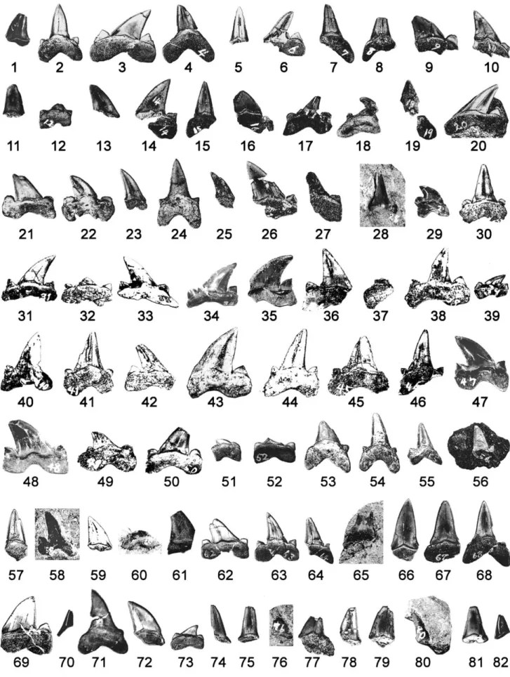
FIGURE 2. Eighty-two loose teeth of Cretalamna appendiculata (Agassiz) (NSM PV 21870, which also include five teeth depicted in Fig. 3) collected in association with holotype of Futabasaurus suz ukii Sato, Hasegawa, and Manabe. Numbers (‘Tooth 1’ through ‘Tooth 82’) indicate arbitrarily assigned tooth numbers used in text. Except Teeth 28, 29, 35, 39, 47, 48, 56, 58, 60, 65, and 76 that are in labial view, all teeth show lingual view. Scale equals 2 cm.
FIGURE 3. Tooth tips of Cretalamna appendiculata (Agassiz) (part of NSM PV 21870; cf. Fig. 2) embedded in bones of holotype of
Futabasaurus suz ukii Sato, Hasegawa, and Manabe. Numbers (‘Tooth 83’
through ‘Tooth 87’) indicate arbitrarily assigned tooth numbers used in text. A, right humerus of F. suz ukii in dorsal view (proximal to top) show-ing position (arrow) of Tooth 85 (corresponds to arrow ‘A’ in Fig. 1); B, close-up view of Tooth 85; C, posterior cervical vertebra of F. suz ukii in right lateral view (dorsal to top) showing the position (asterisk) of de-tached bone fragment that contains Teeth 83 and 84 (pointed by arrow ‘B’ in Fig. 1); D, close-up view of bone fragment in ventral aspect with Teeth 83 (right) and 84 (left); E, anteriorly located dorsal vertebra of F.
suz ukii in right lateral view (dorsal to top) showing position (arrow) of
Tooth 86 (pointed by arrow ‘C’ in Fig. 1); F, close-up view of Tooth 86;
G, neural spine of posterior dorsal vertebra of F. suz ukii in posterior view
(dorsal to top) showing position (arrow) of Tooth 87 (pointed by arrow ‘D’ in Fig. 1); H, close-up view of Tooth 87. Vertical scale equals 5 cm; horizontal scale equals 1 cm.
The plesiosaur individual was at least 6.4 m in length and pos-sibly as long as 9.2 m (Sato et al., 2006), and the body was posi-tioned belly-side up with a slight roll to its right side when buried (Fig. 1). The skeleton shows a simple, near-vertical collapse of the thoracic region where about 40 gastroliths of the plesiosaur are distributed. In contrast, the taphonomy of the pelvic girdle is rather puzzling. The right hind limb is slightly dislocated poste-riorly from the acetabulum of the pelvic girdle and has its dorsal surface facing upward. The most intriguing aspect is that the ver-tebral column is placed on the upward-facing ventral surface of the pelvic girdle articulated with the left hind limb (Obata et al., 1970; Sato et al., 2006).
The displacement of skeletal parts of the plesiosaur was pre-viously considered to be the result of wave action (Obata et al., 1970). However, this idea is tenuous because the mud-dominant lithology suggests water with low kinetic energy. The shark teeth were concentrated around the plesiosaur skeleton (see below) rather than distributed over a wider area.
Hasegawa and Obata (1972, 1976) remarked the association of shark teeth and noted that the plesiosaur was likely attacked by multiple sharks. We do not necessarily agree with the ‘shark
attack’ scenario. However, as described below, we concur that this specimen represents a case of Futabasaurus suz ukii fed by multiple individuals of Cretalamna appendiculata.
Cretalamna appendiculata had eight rows of anterior teeth in
each individual, and they had multiple replacement teeth lingual to generally one truly functional, labial-most tooth in each tooth row (Shimada, 2007). It is highly unlikely that all eight of the functional anterior teeth in a single individual could have been lost at a single feeding event. Even if such an unlikely case is as-sumed, the preservation of at least 35 anterior teeth with the ple-siosaur implies a minimum of five individuals that participated in the feeding.
At least three additional lines of evidence suggest that the tooth set consists of teeth from multiple individuals. First, some pairs of teeth likely represent teeth from the same tooth position in different individuals based on their close morphological resem-blance but with size differences (e.g., Teeth 30 and 41). Second, whereas the range of size variation (e.g., height) among anterior teeth is narrow in an individual of Cretalamna appendiculata (Shi-mada, 2007), the tooth set includes anterior teeth that are of dif-ferent sizes (e.g., Teeth 41, 54, and 55). Third, whereas the largest post-anterior teeth in C. appendiculata are the first and second upper lateral teeth and they are almost as tall as the tallest ante-rior teeth in its dentition (Shimada, 2007; see below), the tallest lateral tooth (Tooth 43) in the tooth set is much taller than all co-occurring anterior teeth (e.g., including Tooth 41), indicating that the lateral tooth came from yet a different individual.
The minimum number of individual sharks represented by the preserved teeth, including those embedded in the plesiosaur bones, would be in the range of six to seven individuals if only the specific examples given above are counted. However, the tooth set consists of at least 87 teeth that include at least 35 anterior teeth, with a wide range of size differences among teeth of the same, or similar, tooth types as well as with conceivable mor-phological variations that can be interpreted as individual differ-ences (e.g., sharp versus blunt lateral cusplets). Thus, it is not un-reasonable to consider that there were more than six or seven sharks.
Shimada (2007) estimated the total length (TL) of an indi-vidual of Cretalamna appendiculata from the Niobrara Chalk of Kansas to be 2.3–3.0 m. The individual possessed anterior teeth that had a narrow range of crown height (CH), 15.0–16.0 mm, with the average of about 15.5 mm (Shimada, 2007:table 1). The tallest lateral teeth in the individual were the first and second up-per lateral teeth, measuring about 13.5 mm. Because each species of modern, non-embryonic lamniform sharks shows a reason-able, linear, positive correlation between the tooth size and body size (e.g., see Shimada, 2005, and references therein), these tooth measurements can be used to extrapolate the TL of the smallest and largest individuals of C. appendiculata that putatively partici-pated in the feeding in this case. One of the shortest anterior teeth represented in the tooth set is Tooth 55 that measures about 10 mm in CH (Fig. 2). This measurement is only about 64.5% of the approximate average CH of anterior teeth reported by Shimada (2007; i.e., 15.5 mm), suggesting that Tooth 55 came from an indi-vidual that measured 1.5–1.9 m TL. The tallest tooth in the tooth set is Tooth 43, which is identified as either the first or the second upper lateral tooth and has the CH of about 19 mm (Fig. 2). This tooth is about 1.41 times larger than the tallest lateral teeth re-ported by Shimada (2007; i.e., 13.5 mm), suggesting that it came from an individual measuring 3.2–4.2 m TL. Thus sizes of indi-viduals that likely participated in the feeding on the plesiosaur probably range from 1.5 to 4.2 m TL. However, the estimated sizes of most other sharks associated with F. suz ukii likely ranged 2.3–3.0 m TL.
Earlier reports (e.g., Hasegawa and Obata, 1972, 1976) indi-cated that the plesiosaur could have died as a result of fighting with the sharks. However, the specimen does not offer the exact
cause of its death. If the plesiosaur had indeed been attacked by one or more individuals of C. appendiculata, it must have been a fatal attack because bones of the plesiosaur immediately around the embedded teeth do not show any indication of bone healing (e.g., Shimada, 1997).
Sato et al. (2006:468) attributed the putative shark feeding as extensive “predation and/or scavenging” but added that scaveng-ing must be at least responsible in part for the damage in the plesiosaur specimen. Furthermore, they stated that some “loss of ventral elements may indicate that the damage (by predation?) to the ventral portion of the carcass prevented the collection of gas in the carcass and subsequent drifting,” and “the damage by scavenger(s) may have occurred at an early stage in postmortem drift of the carcass, allowing the relatively complete skeleton to settle on the sea floor” (Sato et al., 2006:468). Modern large ma-rine vertebrates die of natural causes such as predation, parasites, illness, old age, and hazards at birth (Sch ¨afer, 1972). Because the plesiosaur was an ‘old adult’ (Sato et al., 2006), its cause of death can be any of these possibilities if not predation.
Regardless of the cause of death, modern large marine verte-brates (e.g., whales and seals) generally sink to the bottom of the ocean after death and may begin to float several days later as gas develops and fills the body cavity as part of the decay process (Sch ¨afer, 1972). What exactly happened between the death of the plesiosaur and its final descent to the seafloor is difficult to deci-pher from the fossil record. Nevertheless, the belly-side-up posi-tion of much of the plesiosaur body may reflect the expansion of gas in the body cavity ventral to the vertebral column that cre-ated the tendency for the carcass to float with the belly up. How-ever, two lines of evidence suggest that the skeleton likely did not leave the sea floor to become a so-called ‘bloat-and-float’ carcass (e.g., Schwimmer, 1997). First, a drifting carcass generally loses its skeletal parts one by one from its distal elements for weeks, as the integument and connective tissues tear where skeletal parts are spread over a wide area on the sea floor (Sch ¨afer, 1972). The plesiosaur skeleton is broken into some large segments, but they are not widely distributed, and each segment is largely well ar-ticulated. Second, associated shark teeth were clustered in high concentration near the plesiosaur skeleton. If it were a drifting carcass, they would have been distributed widely and sparsely on the ocean floor.
Several factors, such as the size of the body, temperature of the water, and salinity of the sea, determine how quickly the flesh of a vertebrate carcass decays (Sch ¨afer, 1972). Studies by Sch ¨afer (1972) have shown that a body of an extant seal or fish generally decomposes within 3 months after its death to become a disarticulated skeleton. The body size of these animals is much smaller than the plesiosaur individual, but the fact that the skele-ton is largely articulated and lacks evidence for the bloat-and-float scenario suggests that this plesiosaur was likely exposed on the ocean floor not for an extended period of time after its death and before the burial, possibly no more than a few months.
A possible explanation for the involvement of multiple sharks is ‘feeding frenzy’ (e.g., Springer, 1967), feeding action by a group of highly motivated organisms in aggregation. Motta (2004:168) dismissed the existence of this phenomenon by stating that “feed-ing frenzies of sharks appear to be noth“feed-ing more than highly motivated feeding events involving generally many individuals.” However, abandonment of this term is premature because the phenomenon does bear some behavioral significance in light of the fact that certain sharks such as the white shark, Carcharodon
carcharias (Linnaeus), prefer solitary feeding (e.g., Pratt et al.,
1982). Whereas a feeding frenzy is generally a short-term event, one major challenge of identifying such a short-lived episode in the fossil record is the fact that every geological record involves ‘time-averaging’ at some scale (see Kowalewski, 1996). The case with Futabasaurus suz ukii is no exception to this situation. The feeding could have occurred multiple times on separate
occa-FIGURE 4. Schematic drawing showing inferred posture of holotype of
Futabasaurus suz ukii Sato, Hasegawa, and Manabe (NSM PV15025; see
Fig. 1) in map view soon after it came to rest on ocean floor after its death. Four sharks in silhouette (1–4) are Cretalamna appendiculata (Agassiz) at four places (with their directions) where shark bite took place unequiv-ocally based on the locations of preserved embedded teeth. Wavy lines indicate minimal number of positions where body parts presumably be-came detached, and arrows (A–C) show their inferred directions of dis-placement relative to body that may, or may not, have caused by sharks (see text for detail). Scale equals 1 m.
sions, and whether one or more episodes of feeding frenzy took place is inconclusive. Nevertheless, the occurrence of over 80 conspecific shark teeth, including five piercing in the plesiosaur bones, makes the present record one of the most remarkable cases of shark feeding in the fossil record.
CONCLUSION
Our study represents the first report of a Cretalamna-bitten plesiosaur. Figure 4 shows a schematic illustration of the approx-imate posture of the plesiosaur carcass and four places where shark bite took place unequivocally (Figs. 1, 3). The plesiosaur is depicted to be about 7 m TL based on Sato et al.’s (2006) es-timate. Sharks are scaled to an average estimated TL of about 2.5 m. The taphonomy indicates that the remains were likely buried within a few months. Although Figure 4 simply implies that there were at least four places where sharks bit on the bones of the plesiosaur, our study suggests that at least six or seven sharks ranging between 1.5 and 4.2 m TL scavenged the ple-siosaur. The fact that the teeth consist of different sizes indicates that small (young) and large (old) individuals of C. appendiculata inhabited the same water.
ACKNOWLEDGMENTS
This report could not have been made without the contribu-tion of T. Suzuki who discovered the specimen in 1968. I. Obata (NSM) led the initial investigation with one of us (Y. Hasegawa). For various logistical assistances for the present study, we thank
many staff members of NSM, including K. Fujita, T. Kase, and M. Manabe. T. Tsuihiji was supported by the Japan Society for the Promotion of Science Research Fellowship. T. Sato was financially supported by KAKENHI (grant number 18.6288) and Tokyo Gakugei University. Additional financial support was provided by the Environmental Science Program of DePaul University. Comments made by M. J. Everhart, three anonymous reviewers, and editors significantly improved the quality of this paper.
LITERATURE CITED
Agassiz, L. 1833–1843 [1835, 1843]. Recherches sur les poissons fossiles [5 volumes]. Imprimerie de Patitpierre, Neuch ˆatel, 1420 pp. Ando, H., M. Seishi, M. Oshima, and T. Matsumaru. 1995.
[Fluvial-shallow marine depositional system of the Futaba Group (Upper Cretaceous)—depositional facies and sequences]. Journal of Geog-raphy 104:284–303. [Japanese]
Carpenter, K. 2006. Comparative vertebrate taphonomy of the Pembina and Sharon Springs Members (Middle Campanian) of the Pierre Shale, Western Interior. Paludicola 5:125–149.
Cicimurri, D. J., and M. J. Everhart. 2001. An elasmosaur with stom-ach contents and gastroliths from the Pierre Shale (Late Creta-ceous) of Kansas. Transactions of the Kansas Academy of Science 104:129–143.
Cione, A. L., and F. A. Medina. 1987. A record of Notidanodon pectinatus (Chondrichthyes, Hexanchiformes) in the Upper Cretaceous of the Antarctic Peninsula. Mesozoic Research 1:79–88.
Everhart, M. J. 2005. Bite marks on an elasmosaur (Sauropterygia; Ple-siosauria) paddle from the Niobrara Chalk (Upper Cretaceous) as probable evidence of feeding by the lamniform shark, Cretoxyrhina
mantelli. PalArch 2:14–24.
Hasegawa, Y., and I. Obata. 1972. [Notes on the excavation of a new ple-siosaur]. Natural Science and Museum, Tokyo 39:1–15. [Japanese] Hasegawa, Y., and I. Obata, I. 1976. [Restoration of a plesiosaur]. Land
and Education 6:2–7. [Japanese]
Kowalewski, M. 1996. Time-averaging, overcompleteness and the geolog-ical record. Journal of Geology 104:317–326.
Martin, J. E., and J. E. Fox. 2007. Stomach contents of Globidens, a shell-crushing mosasaur (Squamata), from the Late Cretaceous Pierre Shale Group, Big Bend area of the Missouri River, central South Dakota; pp. 167–176 in J. E. Martin and D. C. Parris (eds.), The Geology and Paleontology of the Late Cretaceous Marine Deposits of the Dakotas. Geological Society of America, Special Paper 427.
Martin, L. D., and B. M. Rothschild. 1989. Paleopathology and diving mosasaurs. American Scientist 77:460–467.
Motta, P.J. 2004. Prey capture behavior and feeding mechanics of elas-mobranchs; pp. 165–202 in J. Carrier, J. Musick, and M. Heithaus (eds.), Biology of Sharks and Their Relatives. CRC Press, Boca Ra-ton, Florida.
Obata, I., and Y. Hasegawa. 1970. [Plesiosaur—marine “dinosaur”]. Ma-rine Sciences Monthly 2:56–62. [Japanese].
Obata, I., Y. Hasegawa, and T. Suzuki. 1970. [Discovery of an elasmosaur from the Upper Cretaceous Futaba Group]. Journal of the Geolog-ical Society of Japan 76:161–164. [Japanese]
Pratt, H. L., Jr., J. G. Casey, and R. B. Conklin. 1982. Observation on large white sharks, Carcharodon carcharias, off Long Island, New York. United States Fishery Bulletin 80:153–156.
Sato, T., Y. Hasegawa, and M. Manabe. 2006. A new elasmosaurid ple-siosaur from the Upper Cretaceous of Fukushima, Japan. Palaeon-tology 49:467–484.
Sch ¨afer, W. 1972. Ecology and Palaeoecology of Marine Environments. University of Chicago Press, Chicago, Illinois, 568 pp.
Schwimmer, D. R. 1997. Late Cretaceous dinosaurs in eastern USA: a taphonomic and biogeographic model of occurrences; pp. 203–211 in D. L. Wolberg, E. Stump, and G. D. Rosenberg (eds.), Dinofest International Proceedings. Academy of Natural Science, Philadel-phia, Pennsylvania.
Schwimmer, D. R., J. D. Stewart, and G. D. Williams. 1997. Scavenging by sharks of the genus Squalicorax in the Late Cretaceous of North America. Palaios 12:71–83.
Shimada, K. 1997. Paleoecological relationships of the Late Cretaceous lamniform shark, Cretoxyrhina mantelli (Agassiz). Journal of Pale-ontology 71:926–933.
Shimada, K. 2005 (date of imprint 2004). The relationship between the tooth size and total body length in the sandtiger shark, Carcharias
taurus (Lamniformes: Odontaspididae). Journal of Fossil Research
37:76–81.
Shimada, K. 2007. Skeletal and dental anatomy of lamniform shark, Cretalamna appendiculata from Upper Cretaceous Nio-brara Chalk of Kansas. Journal of Vertebrate Paleontology 27:584– 602.
Siverson, M. 1992. Biology, dental morphology and taxonomy of lamni-form sharks from the Campanian of the Kristianstad Basin, Sweden. Palaeontology 35:519–554.
Springer, S. 1967. Social organization of shark populations; pp. 149–174 in P. W. Gilbert, R. F. Mathewson, and D. P. Rall (eds.), Sharks, Skates, and Rays. Johns Hopkins University Press, Baltimore, Mary-land.
Tucker, M. E. 1991. Sedimentary Petrology: An Introduction to the Ori-gin of Sedimentary Rocks, second edition. Blackwell Scientific Pub-lications, London, 260 pp.