Caffeine induces endothelial tissue factor expression via
phosphatidylinositol 3-kinase inhibition
Cathérine Gebhard1,2,3*;Erik W. Holy1,2,3*; Giovanni G. Camici1,2; Alexander Akhmedov1,2; Simon F. Stämpfli1,2,3;Barbara E. Stähli1,2,3; Bettina von Rickenbach1,2; Alexander Breitenstein1,2,3; Helen Greutert1,2; Zhihong Yang4; Thomas F. Lüscher1,2,3; Felix C. Tanner1,2,3
1Cardiovascular Research, Physiology Institute, University of Zurich, Zurich, Switzerland; 2Center for Integrative Human Physiology, University of Zurich, Zurich, Switzerland; 3Cardiology, Cardiovascular Center, University Hospital Zurich, Zurich, Switzerland; 4Vascular Biology, Department of Medicine, University of Fribourg, Fribourg, Switzerland
Summary
Tissue factor (TF) is the key activator of coagulation and is involved in acute coronary syndromes. Caffeine is often reported to increase car-diovascular risk; however, its effect on carcar-diovascular morbidity and mortality is controversial. Hence, this study was designed to investigate the impact of caffeine on endothelial TF expression in vitro. Caffeine concentration-dependently enhanced TF protein expression and sur-face activity in human endothelial cells stimulated by tumour necrosis factor (TNF)-α or thrombin. Caffeine inhibited phosphatidylinositol 3-kinase (PI3K) activity and this effect was comparable to that of the known PI3K inhibitor LY294002. Consistently, treatment of endothelial cells with LY294002 enhanced TNF-α induced TF expression to a similar extent as caffeine, and adenoviral expression of the active PI3K mutant (p110) reversed the effect of both caffeine and LY294002 on TF ex-pression. Caffeine and LY294002 increased DNA binding capacity of the
Correspondence to:
Felix C. Tanner, MD
Cardiology, Cardiovascular Center, University Hospital Zürich Rämistrasse 100, 8091 Zürich, Switzerland
Tel.: +41 44 255 39 79 , Fax: +41 44 635 68 27 E-mail: [email protected]
transcription factor nuclear factor κB, whereas the activation pattern of mitogen-activated protein kinases (MAPK) remained unaltered. Luci-ferase reporter assay revealed a caffeine dependent activation of the TF promoter, and RT-PCR revealed a dose dependent increase in TF mRNA levels when stimulated with caffeine in the presence of TNF-α. In con-clusion, caffeine enhances TNF-α-induced endothelial TF protein ex-pression as well as surface activity by inhibition of PI3K signalling. Since the caffeine concentrations applied in the present study are within the plasma range measured in humans, our findings indicate that caffeine enhances the prothrombotic potential of endothelial cells and under-score the importance of PI3K in mediating these effects.
Keywords
Tissue factor / factor VII, signal transduction, coronary syndrome, en-dothelial cells, nutrition
Financial support:
This study was supported by the Swiss National Science Foundation (grant no. 3200B0–113328/1 to FCT, grant no. 3100–068118.02/1 to TFL and grant no. 310030_130500 to GGC), Bonizzi-Theler Foundation, Velux Foundation, Wolfermann Nägeli Foundation, and the Swiss Heart Foundation.
Received: September 11, 2011
Accepted after major revision: January 16, 2012 Prepublished online: March 22, 2012
doi:10.1160/TH11-09-0624
Thromb Haemost 2012; 107: 884–894
* These authors contributed equally to this work.
Introduction
Arterial thrombosis is the critical event in acute vascular syndromes such as unstable angina, myocardial infarction, peripheral ischemia, and stroke (1–3). Thrombus formation is determined by activation of coagulation factors, in particular tissue factor (TF), a membrane-bound glycoprotein playing a central role in initiation and propa-gation of thrombosis. Its expressionis upregulated by inflammatory mediators such as tumour necrosis factor (TNF)-α and can be de-tected in different cell typesof the atherosclerotic plaque (4, 5).
Caffeine (1,3,7-trimethylxanthine) is the most widely used drug in the world; indeed, 80–90% of adults report regular con-sumption of caffeine-containing beverages such as coffee, tea, chocolate, and cola drinks. Caffeine is often regarded as a cardio-vascular risk factor; however, there is conflicting epidemiological evidence on the relation of caffeine consumption to the occurrence
of acute cardiovascular events (6–13). The conflicting evidence may in part be due to the various pleiotropic effects caffeine has on cells at physiologically achievable concentrations. A number of pu-tative mechanisms for caffeine-related cardiovascular effects, such as its ability to inhibit phosphodiesterases (14), and to directly an-tagonise adenosine receptors (15), have been proposed, although the molecular basis of these effects has not been established.
Additionally, caffeine is a direct inhibitor of class I and II phos-phatidylinositol 3 kinases (PI3K), with the most potent effects seen against the p110δ isoform, and this inhibitory effect may explain some of its physiological and pharmacological properties (14). PI3Ks are important negative regulators of TF expression in human endothelial cells (16, 17); and this pathway suppresses in-flammation and coagulation in endotoxaemic mice (18). Targeting PI3K activity in various inflammatory conditions has become an active area of investigation (19, 20).
Based on the fact that a) the PI3k pathway negatively regulates TF expression and thrombus formation, and b) caffeine is a direct inhibitor of the PI3K pathway, we hypothesised that, under these conditions, caffeine may promote endothelial TF expression.
Materials and methods
Cell cultureHuman aortic endothelial cells (HAEC) and vascular smooth muscle cells (both from Clonetics, Allschwil, Switzerland)were cultured as described (21). THP-1 cells (LGC Promochem, Mol-sheim, France) were cultured in RPMI containing 10% fetal calf serum. Adhering cells were grown toconfluence in 3 cm dishes and rendered quiescent for24 hours (h) before stimulation with 5 ng/ ml TNF-α or 1 U/ml thrombin (both from Sigma, Buchs, Switzer-land). Cells were pretreated with caffeine (Sigma) for 15 minutes (min) before stimulation, as preliminary experiments demon-strated that its effect was similar from 15 to 60 min of pretreatment (data not shown). LY294002 (Cell Signaling, Allschwil, Switzer-land), 1,3-Dipropyl-8-cyclopentylxanthine (DPCPX), 3,7-Di-methyl-1-(2-propynyl) xanthine (DMPX) (all from Sigma) were added to the dishes 60 min before stimulation. Cytotoxicity was as-sessed with a colourimetric assay to detect lactate dehydrogenase release (Roche, Basel, Switzerland).
Western blot
Protein expression was determined by Western blot analysis as de-scribed (22).Equal loading was confirmed by Ponceau S staining. Antibodies against human TF and tissue factor pathway inhibitor (TFPI) (both from American Diagnostica, Stamford, CT, USA) were used at 1:2,000dilution. Antibodies against IκBα (Santa Cruz Biotechnology, Santa Cruz, CA, USA), phosphorylated p38 MAP kinase (p38), p44/42 MAP kinase (extracellular signal-regulated kinase [ERK]), and c-Jun NH2-terminal kinase (JNK; all from Cell Signaling) were used at 1:1,000, 1:1,000, 1:5,000, and 1:1,000 dilu-tion, respectively. Antibodies against total p38, ERK, and JNK (all from Cell Signaling) were used at 1:2,000, 1:10,000, and 1:1,000 di-lution, respectively. All blots were normalised to Glyceraldehyde-3-phosphate dehydrogenase (GAPDH; Chemicon, Temecula, CA, USA) or α-Tubulin (aT) expression (1:20,000 dilution, Sigma).
TF activity
TF activity was analysed at the surface of HAEC using a colorimet-ric assay (Amecolorimet-ricanDiagnostica) according to the manufacturer’s recommendations with some modifications as described (23). TF/ FVIIa complex converted human FX to FXa,which was measured
by its ability to metabolise a chromogenicsubstrate. A standard curve with lipidated human TF was performed to assure that measurements were taken in the linear range of detection.
Real-time PCR
All real-time PCR experiments were performed in triplicate using the SYBR Green JumpStart kit (Sigma) in an MX3000P PCR cycler (Stratagene, Amsterdam, Netherlands). Each reaction (25 μl) con-tained 2 μl cDNA, 1 pmol of each primer, 0.25 μl of internal reference dye, and 12.5 μl of JumpStart Taq ReadyMix (containing buffer, dNTPs, stabilisers, SYBR Green, Taq polymerase, and JumpStart Taq antibody). The following primers were used: human tissue factor (F3): sense primer: 5’-TCCCCAGAGTTCACACCTTACC–3’ (bases 508–529 of F3 cDNA; NCBI no. NM 001993), antisense primer: 5’–TGACCACAAATACCACAGCTCC–3’ (bases 892–913 of F3 cDNA; NCBI no. NM 001993); human L28: sense primer: 5’–GCATCTGCAATGGATGGT-3’, antisense primer: 5’–TGTTCTTGCGGATCATGTGT-3’. The amplification program consisted of one cycle at 95°C for 10 min followed by 40 cycles with a denaturing phase at 95°C for 30 seconds, an annealing phase at 60°C for 1 min, and an elongation phase at 72°C for 1 min. PCR products were analysed on an ethidium bromide stained 1% agarose gel, and a melting curve analysis was performed after amplification to verify the accuracy of the amplicon. In each real-time PCR run for F3 and L28, a calibration curve was included that was generated from serial dilutions of the respective purified amplicon.
In vitro PI3K assay
PI3K activity in endothelial cells was estimated using a commer-cially available PI3K ELISA kit (catalog number K-1000; Echelon, Salt Lake City, UT, USA) according to the manufacturer’s instruc-tions. Briefly, endothelial cells were lysed, supernatant was mixed with anti-PI3K p85 antibody(Millipore, Billerica, MA, USA) and incubated for 1 h. Then, protein A agarose beads were added to equal amounts of total protein and the samples rocked (4°C) for 1 h at 4°C. The immunoprecipitated PI3K was incubated with phosphatidylinositol-4,5-bisphosphate (PIP2) substrate and reac-tion buffer for 1 h. The amount of PIP3 formed from PIP2 by PI3-K activity was detected using a competitive ELISA.
NFκB DNA binding capacity
NFκB DNA binding activity in endothelial cells was estimated using a commercially available colorimetric NFκB assay (TransAM NFκB, cataolg number 40096; Active Motif, Carlsbad, CA, USA) according to the manufacturer’s instructions. Briefly, for isolation of nuclear proteins a Nuclear Extract Kit (Active Motif) was
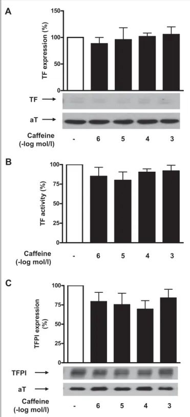
ap-Figure 2: Caffeine does not affect basal TFPI and TF expression in human aortic endothelial cells. Caffeine does not alter TF protein ex-pression under basal conditions (A). Values are indicated as percent of un-stimulated control. p=NS vs. control. Caffeine does not alter TF activity under basal conditions (B). Values are indicated as percent of unstimulated control. p=NS vs. control. Caffeine did not alter endothelial TFPI expression under basal condition (C). Values are indicated as percent of unstimulated control. p=NS vs. control. Values are representative of at least four different experi-ments; all blots are normalised to α-Tubulin (aT) expression.
plied. Nuclear extracts were then added to a 96-well plate on which oligonucleotide containing the NFκB consensus site (5´GGG -ACTTTCC-3´) had been immobilised. Then, a primary antibody against an epitope on p65 that is accessible when NF?B is bound to its target DNA was added for 1 h. This was followed by addition of a secondary antibody conjugated to horseradish peroxidase. Co-lorimetric readout was achieved by spectrophotometry.
Adenoviral expression of PI3K active mutant rCD2-p110α
Generation of recombinant adenovirus expressing an active PI3K mutant (pEF-BOSrCD2-p110α) was carried out as described (24). For transduction, the vector was added to HAEC at 100 pfu/cell for 1 h and then removed. HAEC were kept in growth medium for 24 h and then serum-starved for 24 h prior to TNF-α stimulation in the presence or absence of caffeine or LY 294002 for 5 h.
TF promoter activity
The TF promoter (- 227 bp to +121 bp) was cloned upstream of the luciferase cDNA and the SV40 PolyA signal into the multiple cloning site of the helper vector VQAd5K-NpA (ViraQuest Inc., North Liberty, IA, USA). In a first step, HindIII and BamHI restric-tion sites of VQAd5K-NpA were used to insert a 2.7 kb HindIII/ BamHI restriction fragment of pGL2-Basic vector (Promega, Madison, WI, USA) containing the luciferase cDNA and the SV40 PolyA signal. In a second step, a 0.3 kb KpnI restriction fragment from a human TF promoter plasmid including the TF minimal promoter kindly provided by Dr. Nigel Mackman (University of North Carolina, Chapell Hill; NC, USA) was ligated into the KpnI site of the resulting construct. The whole insert was sequenced to confirm its orientation and the absence of any nucleotide substitu-tions. This construct named VQAd5/hTF/Luc was used for pro-duction of an adenoviral vector (Ad5/hTF/Luc). For transpro-duction, the vector was added to HAEC at 60 pfu/cell for 1 h and then re-moved. HAEC were kept in growth medium for 24 h and then serum-starved for 24 h prior to TNF-α stimulation for 5 h. Firefly luciferase activity was determined in cell lysates using a
lumino-Figure 1: Caffeine enhances TF expression in human aortic endothe-lial cells. Caffeine enhances TNF-α (A) and thrombin (B) -induced TF protein expression in a concentration-dependent manner. Values are indicated as percent of TNF-α or thrombin alone. *p<0.05 vs. TNF-α or thrombin alone. Caffeine enhances TNF-α (C) and thrombin (D) -induced TF surface activity in a concentration-dependent manner. Values are indicated as percent of TNF-α or thrombin alone. *p<0.05 vs. TNF-α or thrombin alone. Caffeine does not affect TNF-α (E) and thrombin (F) -induced TFPI protein expression. Values are indicated as percent of TNF-α or thrombin alone. p=NS vs. TNF-α or thrombin alone. Values are representative of at least four different experi-ments; all blots are normalised to α-Tubulin (aT) expression.
post-hoc Tukey’s test as appropriate. A probabilityvalue of <0.05 was considered significant.
Results
Caffeine enhances endothelial TF protein expression and activity
HAECs were stimulated with TNF-α (5 ng/ml) or thrombin (1 U/ml) for 5 h in the presence or absence of caffeine (10–6–10–3 M). Caffeine at 10–4M was calculated to correspond to 2–3 cups of coffee in a human weighing 70 kg(15, 25, 26). Caffeine enhanced TNF-α-induced TF expression in a concentration-dependent manner (n=9; p<0.05; 씰Fig. 1A), with a maximal effect occurring at 10–3 M and resulting in a 2.3-fold induction as compared to TNF-α alone. Similarly, caffeine enhanced thrombin induced TF expression by 2.2-fold as compared to thrombin alone (n=7; p<0.05; 씰Fig. 1B). The effect of caffeine on TF expression was par-alleled by an increased TF surface activity which reached 1.3 times the level induced by TNF-α alone (n=6; p<0.05; 씰Fig. 1C) and 1.7 times that by thrombin alone, respectively (n=5; p<0.05; 씰Fig. 1D). Caffeine did not affect TF expression and TF activity under basal conditions (n=4; p=NS; 씰Fig. 2A and B).
Similar to HAECs, TNF-α (5 ng/ml) and thrombin (1 U/ml) in-duced TF expression in THP-1 cells (n=11; p<0.05; data not shown) and vascular smooth muscle cells (VSMC) (n=4; p<0.05, data not shown). Caffeine, however, did not affect TF expression in either one of these cell types (n>4; p=NS; data not shown).
To exclude a cytotoxic effect, HAECs, VSMCs or THP-1 cells were incubated with caffeine for 5 h, and cell death was assessed by LDH release. No significant increase in LDH release of any cell type was observed for any concentration of caffeine used (n=4; p=NS; data not shown).
Caffeine does not affect TFPI expression
Treatment of TNF-α-stimulated HAECs with increasing concen-trations of caffeine (10–6 to 10–3 M) did not affect TFPI expression (n=5; p=NS; 씰Fig. 1E). Similarly, caffeine did not alter endothelial TFPI expression after thrombin stimulation (n=4; p=NS; 씰Fig. 1F) or in unstimulated cells (n=8; p=NS; 씰Fig. 2C).
Caffeine enhances TF mRNA and protein expression time-dependently
Real time-PCR revealed that treatment with caffeine (10–3M) en-hanced TNF-α induced TF mRNA expression in a time-dependent manner reaching a maximal induction of 1.6-fold after 1 h (n=5; p<0.05; 씰Fig. 3A). Accordingly, stimulation with caffeine (10–3 meter (Berthold Technologies, Bad Wildbad, Germany). Protein
concentration of the cell lysates was determined for normalisation of luciferase activity.
Statistics
Data are presented as mean ± SEM. Statistical analysiswas per-formed by two-tailed unpaired Student’s t-test or ANOVA with
Figure 3: Caffeine increases TF mRNA and protein expression in a time-dependent manner. Caffeine enhances TNF-α induced TF mRNA ex-pression after 1 h of stimulation (A). Values are determined by real-time PCR, indicated as percent of TNF-α alone, and normalised to L28 expression. *p<0.05 vs. TNF-α alone. Caffeine enhances TNF-α-induced TF protein ex-pression after 3 h of stimulation (B). Values are indicated as percent of TNF-α alone. *p<0.05 vs. TNF-α alone.
M) led to a time-dependent induction of TF protein expression up to 1.7-fold the level induced by TNF-α alone; a maximal effect was observed after 5 h (n=5; p<0.05; 씰Fig. 3B). Moreover, treatment with caffeine after TNF-α enhanced TNF-α-induced TF protein expression in a time-dependent manner: the shorter the delay in
caffeine administration after TNF-α stimulation, the more pro-nounced was its effect on TF expression; with 2 h delay TF induc-tion was 1.8 fold the level induced by TNF-α alone after, with 4 h delay TF expression remained unaltered (n=5; p<0.05; data not shown).
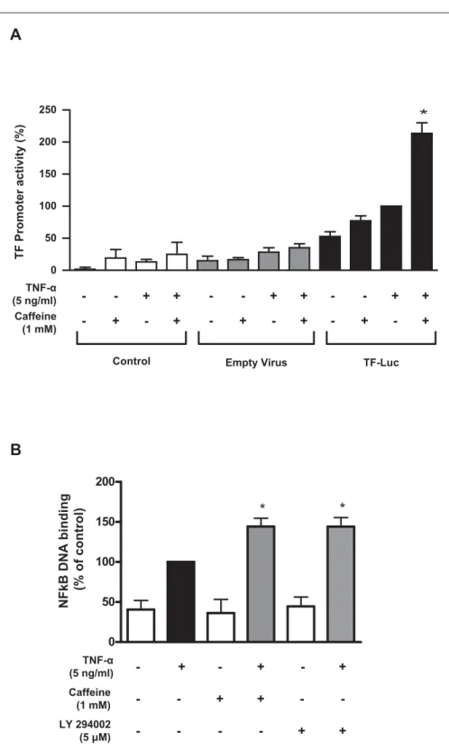
Figure 4: Caffeine enhances TF promoter activity and TNF-α-induced DNA binding of NFκB. Caffeine enhances TNF-α-induced TF promoter activation (A). Values are normalised to total protein concentration, are represen-tative of at least four different experiments, and are indicated as percent of TNF-α alone *p<0.05 vs. TNF-α alone. Caffeine and LY294002 further enhances TNF-α-induced DNA binding of NFκB by 1.5-fold (B). Under basal condition NFκB DNA binding remains un-altered in the presence of caffeine or LY294002 (B). Values are indicated as percent of TNF-α alone. *p<0.05 vs. TNF-α alone.
Caffeine enhances TNF-α induced NFκB DNA binding and TF promoter activity
To assess whether the effect on TF mRNA was mediated by enhanced transcription, the impact of caffeine on TF promoter activity was analysed after transfection of HAEC with a plasmid expressing firefly luciferase under control of the human TF promoter (- 221 bp to +121 bp). Caffeine enhanced TF promoter activity by 2.1-fold as compared to TNF-α alone (n=4; p<0.05; 씰Fig. 4A). Caffeine did not affect basal promoter activity (n=4; p=NS; 씰Fig. 4A). Next, the effect of caffeine and LY294002 on NFκB activation in the presence and absence of TNF-α was examined. The capacity of NFκB to bind to its DNA consensus site (5´-GGGACTTTCC-3´) was determined in nuclear extracts by using a colourimetric assay. Caffeine further enhanced TNF-α-induced DNA binding of NFκB by 1.5-fold (n=4; p<0.05; 씰Fig. 4B). This effect compared well to that of LY294002, which enhanced NFκB DNA binding by 1.5-fold in the presence of TNF-α (n=4; p<0.05; 씰Fig. 4B). Under basal conditions, NFκB DNA binding remained unaltered in the presence of caffeine or LY294002 (n=4; p=NS; 씰Fig. 4B). Nuclear translocation of NFκB, as determined by degradation of its inhibitor IκBα remained unaf-fected by both caffeine and LY294002 (n=3; p=NS; data not shown).
Caffeine does not alter MAPK activation
To assess whether caffeine altersMAP kinase activation, HAECs were examined at different time points aftercytokine stimulation. The MAP kinases p38, ERK, and JNK were transiently activated by TNF-α (5 ng/ml; 씰Fig. 5). Maximal activation of ERK, p38 and JNK was observed after 15 min. Quantitative analysis of four inde-pendent experiments revealed that this phosphorylation pattern remained unaffected by caffeine as compared to TNF-α alone (씰Fig. 5, original records). Total expression of p38, ERK, and JNK remained unaltered at all time points examined (씰Fig. 5).
Caffeine enhances TF expression by inhibiting PI3K
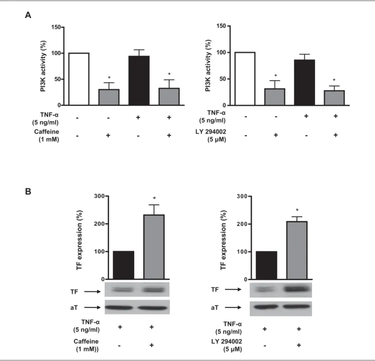
PI3K activation was determined by assaying PI3K lipid kinase activ-ity in p85 immunoprecipitates. Caffeine (10–3 M) inhibited endothe-lial PI3K activity under both, basal and TNF-α stimulated con-ditions, by 72% and 68%, respectively (n=4; p<0.05; 씰Fig. 6A). The effect of caffeine was less potent at lower concentrations (10–4 M; n=4; p<0.05 vs. control; data not shown). This effect of caffeine on
Figure 5: Caffeine does not affect MAP ki-nase activation. Stimulation with TNF-α leads to transient phosphorylation (Pho) of the MAP kinases ERK (A), p38 (B), and JNK (C). Caffeine does not alter the pattern of MAP kinase acti-vation. Total (Tot) levels of ERK, p38, and JNK remain unchanged. Values are representative of at least four different experiments.
PI3K was similar to that of the well known PI3K inhibitor LY294002 which inhibited PI3K activity by 69% under basal conditions and by 74% in TNF-α stimulated cells (n=4; p<0.05; 씰Fig. 6A right panel). When HAECs were treated with LY294002 before stimulation with TNF-α, TF expression was enhanced by 2.1-fold (n=7; p<0.05;
씰Fig. 6B right panel) as compared to TNF-α alone. These effects
compared well to that of caffeine, which enhanced TF expression by 2.3-fold (n=9; p<0.05; 씰Fig. 6B left panel). Adenoviral over-expression of a constitutively active PI3K (rCD2-p110α) reversed the effect of caffeine (n=6; p<0.05 vs. control virus; 씰Fig. 7A) and LY294002 (n=6; p<0.05 vs. control virus; 씰Fig. 7B) on TF ex-pression in TNF-α stimulated HAECs.
Figure 6: Caffeine inhibits PI3K activity. Caffeine (10–3 M) inhibits en-dothelial PI3K activity under basal conditions and in the presence of TNF-α (A, left panel). LY294002 (5x10⁻⁶ M) inhibits endothelial PI3K activity under basal conditions and in the presence of TNF-α (A, right panel). Values are de-termined by lipid kinase activity in p85 immunoprecipitates, are represen-tative of at least four different experiments, and are indicated as percent of
unstimulated controls. *p<0.05 vs. unstimulated control. Caffeine enhances TNF-α-induced TF protein expression by 2.3-fold (B, left panel). This effect compares well to that of the PI3K inhibitor LY294002 which enhances TF ex-pression by 2.1-fold (B, right panel). Values are indicated as percent of TNF-α alone and are representative of at least seven different experiments; all blots are normalised to α-Tubulin (aT) expression. *p<0.05 vs. TNF-α alone.
The effect of caffeine on TF expression is not mediated via adenosine receptors
Caffeine is a non-selective adenosine receptor blocker. Hence, the effect of the adenosine A1 receptor antagonist
1,3-dipropyl-8-cyclopentylxanthine (DPCPX) and the A2a receptor antagonist 3,7-dimethyl-1-(2-propynyl) xanthine (DMPX) on TF expression was assessed. Pre-treatment with increasing concentrations of DPCPX or DMPX did not alter TNF-α-induced TF protein ex-pression (n=4; p>0.2 for DPCPX or DMPX vs. % of the respective value for TNF-α alone, data not shown).
Discussion
This study demonstrates that caffeine increases endothelial TF ex-pression and activity induced by inflammatory and procoagulant mediators and accelerates thrombus formation in vivo. This effect occurs at a transcriptional level and is mediated via PI3K in-hibition leading to an increase of p65/NFκB DNA binding activity and induction of the TF promoter.
The estimated overall caffeine intake is 200–250 mg/person per day in the USA and Canada, and 300–400 mg/person per day in Northern Europe and Great Britain (26, 27). The caffeine concen-tration applied for in vivo experiments (10 mg/kg) and the dose used for in vitro experiments (10–4 M) correspond to intake of 250 mg caffeine or 2–3 cups of coffee by a human weighing 70 kg (15, 18–20, 25, 26); hence the caffeine concentrations applied in the presented experiments are physiologically relevant. This interpre-tation is in line with the observation that caffeine exerted its effect independent of whether it was added before or after TNF-α.
TNF-α and thrombin are both well-known inducers of en-dothelial TF expression. Caffeine enhanced TNF-α- and throm-bmediated TF expression with similar potency and may thus in-crease TF expression under both inflammatory and prothrom-botic conditions. Biologically active TF is located at the cell surface, and the effect of caffeine on TF expression was indeed paralleled by an increased TF surface activity. The latter, however, was less pro-nounced than that observed for protein expression, which may be related to the presence of encrypted TF or the distribution of TF in different cellular compartments. TF activity is inhibited by TFPI, and the balance between these factors is essential for vascular homeostasis (28). Caffeine did not affect endothelial TFPI ex-pression under basal and stimulated conditions and in the pres-ence of TNF-α or thrombin. Hence, our data indicate that the ef-fect of caffeine on TF expression is not attenuated by a concomi-tant increase in TFPI expression and is specific for TF.
PI3Ks are key players in various inflammatory, autoimmune, and allergic processes. The inhibitory role of PI3K on TF ex-pression is well documented, as inhibition of these kinases en-hances TF protein expression in response to different mediators (17). PI3Ks are divided into three distinct classes based on their primary structure. The class I enzymes are further divided into α, β, γ, and δ isoforms based on their distinct p110 catalytic subunits and modes of regulation. Selective targeting of PI3K isoform p110β and p110γ has been proposed as an antithrombotic strategy and isoform-selective p110 inhibitors are currently developed by pharmaceutical companies (19, 20). Caffeine is known to inhibit the in vitro activity of PI3K with the most potent effect seen against
Figure 7: The effect of caffeine on TF expression is reversed by a con-stitutively active PI3K mutant. Adenoviral expression of the concon-stitutively active PI3K mutant rCD2-p110 reverses the effect of caffeine on TF ex-pression in TNF-α-stimulated HAECs (A). Adenoviral expression of the consti-tutively active PI3K mutant rCD2-p110 reverses the effect of LY294002 on TF expression in TNF-α-stimulated HAECs (B). Values are indicated as percent of TNF-α alone and are representative of at least four different experiments; all blots are normalised to GAPDH expression. *p<0.05 vs. TNF-α alone.
p110δ (14). The present study confirms that caffeine inhibited PI3K activity in endothelial cells and demonstrates that inhibition of PI3K by caffeine enhanced TF promoter activity leading to an increase in TF mRNA and protein expression. This effect was re-versed by over-expression of a constitutively active PI3K p110α mutant in endothelial cells and compared well to that of the classic unspecific PI3K inhibitor LY294002. Hence, these observations suggest that caffeine may exert prothrombotic actions by in-hibition of PI3K, particularly that of p110α isoform, at concen-trations that are within the pharmacological and physiological range.
Extracellular adenosine levels vary within tissues and with dif-ferent degrees of cellular stress; its plasma levels increase up to the micromolar range as a consequence of vascular inflammation and tissue ischaemia (29–32). Caffeine antagonises adenosine recep-tors in the cardiovascular system (33–35), and the effect of caffeine on TF protein is consistent with the observation that adenosine re-leased from human umbilical vein endothelial cells inhibits TF ex-pression in an autocrine or paracrine manner, and that adenosine receptor antagonists upregulate TF expression (29, 36). In our study, blocking either group of receptors did not alter endothelial TF expression, suggesting that the effect of caffeine does not de-pend on adenosine receptor inhibition under our experimental conditions. Thus, it is likely that the xanthin directly inhibits the PI3 kinase pathway.
Previous studies indicate that pharmacologic inhibition of PI3K enhances LPS-induced IκBα degradation and nuclear trans-location of NFκB. However, PI3K has been shown to act both posi-tively and negaposi-tively on NFκB-dependent gene expression, and exactly how the PI3K pathway regulates nuclear translocation of NFκB remains unclear (37, 38). We observed here that inhibition of PI3K did not affect degradation of IκBα in endothelial cells. However, we found a caffeine dependent increase in NFκB DNA binding capacity, and this effect was again congruent to that of LY294002.
Hence, these observations suggest that inhibition of PI3K by caffeine enhances nuclear activity of NFκB leading to an increase in TF promoter activity. Interestingly, the effect of caffeine on NFκB activation occurred only under inflammatory or prothrom-botic conditions which supports the interpretation that caffeine requires additional stimuli to display its full effect on TF. Since caf-feine consumption has been linked to an increased risk of cardio-vascular events in some, but not all prospective studies (6, 8, 9), with some results supporting no association between coffee intake and cardiovascular disease (39) or even beneficial effects of caf-feine on cardiovascular morbidity and mortality (7), this mechan-ism of action may explain, at least to some extent, the ongoing con-troversy.
MAP kinase activation mediates endothelial TF expression by promoting transcription in response to many stimuli (4, 22) and earlier studies have shown that MAPK activation is required for PI3K-dependent TF activation. Accordingly, p38, ERK, and JNK were transiently activated by TNF-α. Caffeine, however, did not
af-fect the pattern of MAPK activation, indicating that the efaf-fect of caffeine on endothelial TF expression occurs independently of MAPK activation.
Caffeine did not enhance TNF-α-induced TF expression in THP-1 cells or VSMC, indicating that the effect of caffeine on TF is cell specific, and supporting the interpretation that caffeine spe-cifically exerts its action on endothelial TF expression via the PI3K pathway; indeed, the latter does not exert any negative feedback on TF expression in VSMC or THP-1 cells (4).
Overall, our findings provide further evidence for a prothrom-botic potential of caffeine, by its ability to directly inhibit phosp-hoinosite metabolism and thereby potentiate endothelial TF ex-pression at sites of inflammation. Moreover, our study underlines the importance of the PI3K system in modulating cardiovascular disease. Since the effect of caffeine was reversed by over-expression of a constitutively active PI3K p110α mutant, however, the present data question the overall beneficial therapeutic effects of isoform selective p110 inhibitors, which have recently been proposed as a novel antithrombotic strategy. Further studies are needed to understand the mechanism by which PI3Ks are exerting their ef-fects and to clarify the role of the selective PI3K inhibitors in spe-cific inflammatory conditions.
Conflicts of interest
None declared.
What is known about this topic?
● Tissue factor (TF) is the key activator of coagulation and plays a central role in initiation and propagation of thrombosis. Its ex-pression is upregulated by inflammatory mediators such as tumour necrosis factor-α and can be detected in different cell types of the atherosclerotic plaque.
● Caffeine is often reported to increase cardiovascular risk; however, its effect on cardiovascular morbidity and mortality is controver-sial. Caffeine is a direct inhibitor of the phosphatidylinositol 3- kinase (PI3K) pathway
● PI3K negatively regulates TF expression and thrombus formation. Targeting PI3K activity in various inflammatory conditions has be-come an active area of investigation.
What does this paper add?
● Caffeine induces TF expression, thereby enhancing the prothrom-botic potential of endothelial cells.
● Inhibition of PI3K signalling mediates caffeine induced TF ex-pression leading to activation of nuclear factor κB and upregu-lation of TF.
● Caffeine concentrations applied in the present study are within the plasma range measured in humans. Caffeine intake may promote arterial thrombosis and its consumption should be controlled in patients at risk of cardiovascular complications.
References
1. Libby P. Inflammation in atherosclerosis. Nature 2002; 420: 868-874. 2. Corti R, Hutter R, Badimon JJ, et al. Evolving concepts in the triad of
athero-sclerosis, inflammation and thrombosis. J Thromb Thrombolysis 2004; 17: 35-44. 3. Owens AP, 3rd, Mackman N. Tissue factor and thrombosis: The clot starts here.
Thromb Haemost 2010; 104: 432–439.
4. Steffel J, Luscher TF, Tanner FC. Tissue factor in cardiovascular diseases: molecu-lar mechanisms and clinical implications. Circulation 2006; 113: 722-731. 5. Roque M, Reis ED, Fuster V, et al. Inhibition of tissue factor reduces thrombus
formation and intimal hyperplasia after porcine coronary angioplasty. J Am Coll Cardiol 2000; 36: 2303-2310.
6. Happonen P, Voutilainen S, Salonen JT. Coffee drinking is dose-dependently re-lated to the risk of acute coronary events in middle-aged men. J Nutr 2004; 134: 2381-2386.
7. Woodward M, Tunstall-Pedoe H. Coffee and tea consumption in the Scottish Heart Health Study follow up: conflicting relations with coronary risk factors, coronary disease, and all cause mortality. J Epidemiol Community Health 1999; 53: 481-487.
8. LaCroix AZ, Mead LA, Liang KY, et al. Coffee consumption and the incidence of coronary heart disease. N Engl J Med 1986; 315: 977-982.
9. Baylin A, Hernandez-Diaz S, Kabagambe EK, et al. Transient exposure to coffee as a trigger of a first nonfatal myocardial infarction. Epidemiology 2006; 17: 506-511.
10. Jee SH, He J, Appel LJ, et al. Coffee consumption and serum lipids: a meta-analy-sis of randomized controlled clinical trials. Am J Epidemiol 2001; 153: 353-362. 11. Jacques PF, Bostom AG, Wilson PW, et al. Determinants of plasma total homocys-teine concentration in the Framingham Offspring cohort. Am J Clin Nutr 2001; 73: 613-621.
12. Ammon HP, Bieck PR, Mandalaz D, et al. Adaptation of blood pressure to con-tinuous heavy coffee drinking in young volunteers. A double-blind crossover study. Br J Clin Pharmacol 1983; 15: 701-706.
13. Jick H, Miettinen OS, Neff RK, et al. Coffee and myocardial infarction. N Engl J Med 1973; 289: 63-67.
14. Foukas LC, Daniele N, Ktori C, et al. Direct effects of caffeine and theophylline on p110 delta and other phosphoinositide 3-kinases. Differential effects on lipid ki-nase and protein kiki-nase activities. J Biol Chem 2002; 277: 37124-37130. 15. Huang ZL, Qu WM, Eguchi N, et al. Adenosine A2A, but not A1, receptors
me-diate the arousal effect of caffeine. Nat Neurosci 2005; 8: 858-859.
16. Viswambharan H, Ming XF, Zhu S, et al. Reconstituted high-density lipoprotein inhibits thrombinduced endothelial tissue factor expression through in-hibition of RhoA and stimulation of phosphatidylinositol 3-kinase but not Akt/ endothelial nitric oxide synthase. Circ Res 2004; 94: 918-925.
17. Steffel J, Latini RA, Akhmedov A, et al. Rapamycin, but not FK-506, increases en-dothelial tissue factor expression: implications for drug-eluting stent design. Cir-culation 2005; 112: 2002-2011.
18. Schabbauer G, Tencati M, Pedersen B, et al. PI3K-Akt pathway suppresses coagu-lation and inflammation in endotoxemic mice. ArteriosclerThrombVascBiol 2004; 24: 1963-1969.
19. Fougerat A, Gayral S, Gourdy P, et al. Genetic and pharmacological targeting of phosphoinositide 3-kinase-gamma reduces atherosclerosis and favors plaque sta-bility by modulating inflammatory processes. Circulation 2008; 117: 1310-1317.
20. Jackson SP, Schoenwaelder SM, Goncalves I, et al. PI 3-kinase p110beta: a new tar-get for antithrombotic therapy. Nat Med 2005; 11: 507-514.
21. Stahli BE, Camici GG, Steffel J, et al. Paclitaxel enhances thrombin-induced en-dothelial tissue factor expression via c-Jun terminal NH2 kinase activation. Circ Res 2006; 99: 149-155.
22. Steffel J, Akhmedov A, Greutert H, et al. Histamine induces tissue factor ex-pression: implications for acute coronary syndromes. Circulation 2005; 112: 341-349.
23. Camici GG, Steffel J, Akhmedov A, et al. Dimethyl sulfoxide inhibits tissue factor expression, thrombus formation, and vascular smooth muscle cell activation: a potential treatment strategy for drug-eluting stents. Circulation 2006; 114: 1512–1521.
24. Ming XF, Barandier C, Viswambharan H, et al. Thrombin stimulates human en-dothelial arginase enzymatic activity via RhoA/ROCK pathway: implications for atherosclerotic endothelial dysfunction. Circulation 2004; 110: 3708-3714. 25. Sudano I, Binggeli C, Spieker L, et al. Cardiovascular effects of coffee: is it a risk
factor? Prog Cardiovasc Nurs 2005; 20: 65-69.
26. Fredholm BB, Battig K, Holmen J, et al. Actions of caffeine in the brain with special reference to factors that contribute to its widespread use. Pharmacol Rev 1999; 51: 83-133.
27. Mandel HG. Update on caffeine consumption, disposition and action. Food ChemToxicol 2002; 40: 1231-1234.
28. Pedersen B, Holscher T, Sato Y, et al. A balance between tissue factor and tissue fac-tor pathway inhibifac-tor is required for embryonic development and hemostasis in adult mice. Blood 2005; 105: 2777-2782.
29. Pearson JD, Gordon JL. Vascular endothelial and smooth muscle cells in culture selectively release adenine nucleotides. Nature 1979; 281: 384-386.
30. Engler RL. Adenosine. The signal of life? Circulation 1991; 84: 951-954. 31. Ely SW, Berne RM. Protective effects of adenosine in myocardial ischemia.
Circu-lation 1992; 85: 893-904.
32. Downey JM, Liu GS, Thornton JD. Adenosine and the anti-infarct effects of pre-conditioning. CardiovascRes 1993; 27: 3-8.
33. Fredholm BB. Astra Award Lecture. Adenosine, adenosine receptors and the ac-tions of caffeine. Pharmacol Toxicol 1995; 76: 93-101.
34. White PJ, Nguyen TT. Chronic caffeine treatment causes changes in cardiac ade-nosine receptor function in rats. Pharmacology 2002; 65: 129-135.
35. Aqel RA, Zoghbi GJ, Trimm JR, et al. Effect of caffeine administered intravenously on intracoronary-administered adenosine-induced coronary hemodynamics in patients with coronary artery disease. Am J Cardiol 2004; 93: 343-346. 36. Deguchi H, Takeya H, Urano H, et al. Adenosine regulates tissue factor expression
on endothelial cells. ThrombRes 1998; 91: 57-64.
37. Kim YH, Choi KH, Park JW, et al. LY294002 inhibits LPS-induced NO production through a inhibition of NF-kappaB activation: independent mechanism of phos-phatidylinositol 3-kinase. Immunol Lett 2005; 99: 45-50.
38. Ojaniemi M, Glumoff V, Harju K, et al. Phosphatidylinositol 3-kinase is involved in Toll-like receptor 4-mediated cytokine expression in mouse macrophages. Eur J Immunol 2003; 33: 597-605.
39. Wilson PW, Garrison RJ, Kannel WB, et al. Is coffee consumption a contributor to cardiovascular disease? Insights from the Framingham Study. Arch Intern Med 1989; 149: 1169-1172.