HAL Id: hal-02481326
https://hal.archives-ouvertes.fr/hal-02481326
Submitted on 10 Nov 2020
HAL is a multi-disciplinary open access
archive for the deposit and dissemination of
sci-entific research documents, whether they are
pub-lished or not. The documents may come from
teaching and research institutions in France or
abroad, or from public or private research centers.
L’archive ouverte pluridisciplinaire HAL, est
destinée au dépôt et à la diffusion de documents
scientifiques de niveau recherche, publiés ou non,
émanant des établissements d’enseignement et de
recherche français ou étrangers, des laboratoires
publics ou privés.
Distributed under a Creative Commons Attribution| 4.0 International License
Annie Lebreton, Erwan Corre, Jean-Luc Jany, Loraine Brillet-Guéguen,
Carlos Pèrez-Arques, Victoriano Garre, Misharl Monsoor, Robert Debuchy,
Christophe Le Meur, Emmanuel Coton, et al.
To cite this version:
Annie Lebreton, Erwan Corre, Jean-Luc Jany, Loraine Brillet-Guéguen, Carlos Pèrez-Arques, et al..
Comparative genomics applied to Mucor species with different lifestyles. BMC Genomics, BioMed
Central, 2020, 21 (1), pp.135. �10.1186/s12864-019-6256-2�. �hal-02481326�
R E S E A R C H A R T I C L E
Open Access
Comparative genomics applied to Mucor
species with different lifestyles
Annie Lebreton
1, Erwan Corre
2, Jean-Luc Jany
1, Loraine Brillet-Guéguen
2,3, Carlos Pèrez-Arques
4, Victoriano Garre
4,
Misharl Monsoor
2, Robert Debuchy
5, Christophe Le Meur
1, Emmanuel Coton
1, Georges Barbier
1and
Laurence Meslet-Cladière
1*Abstract
Background: Despite a growing number of investigations on early diverging fungi, the corresponding lineages have not been as extensively characterized as Ascomycota or Basidiomycota ones. The Mucor genus, pertaining to one of these lineages is not an exception. To this date, a restricted number of Mucor annotated genomes is publicly available and mainly correspond to the reference species, Mucor circinelloides, and to medically relevant species. However, the Mucor genus is composed of a large number of ubiquitous species as well as few species that have been reported to specifically occur in certain habitats. The present study aimed to expand the range of Mucor genomes available and identify potential genomic imprints of adaptation to different environments and lifestyles in the Mucor genus.
Results: In this study, we report four newly sequenced genomes of Mucor isolates collected from non-clinical environments pertaining to species with contrasted lifestyles, namely Mucor fuscus and Mucor lanceolatus, two species used in cheese production (during ripening), Mucor racemosus, a recurrent cheese spoiler sometimes described as an opportunistic animal and human pathogen, and Mucor endophyticus, a plant endophyte. Comparison of these new genomes with those previously available for six Mucor and two Rhizopus (formerly identified as M. racemosus) isolates allowed global structural and functional description such as their TE content, core and species-specific genes and specialized genes. We proposed gene candidates involved in iron metabolism; some of these genes being known to be involved in pathogenicity; and described patterns such as a reduced number of CAZymes in the species used for cheese ripening as well as in the endophytic isolate that might be related to adaptation to different environments and lifestyles within the Mucor genus.
Conclusions: This study extended the descriptive data set for Mucor genomes, pointed out the complexity of obtaining a robust phylogeny even with multiple genes families and allowed identifying contrasting potentially lifestyle-associated gene repertoires. The obtained data will allow investigating further the link between genetic and its biological data, especially in terms of adaptation to a given habitat.
Keywords: Early divergent fungi, Genome, Adaptation, CAZymes, Peptidases, Iron uptake Background
The Mucor genus belongs to the most prominent order of the Mucorales, a phylogenetically ancient group of fungi pertaining to the “early diverging fungi” [1]. From the first microscopic observation of a Mucor specimen in 1665 up until now, several hundreds of potential
Mucorspecies have been reported [2]. Mucor species are common and predominantly saprotrophs [3]. These ubi-quitous microorganisms may colonize multiple and con-trasted environments from dungs or dead plant materials to plant and animal tissues. Members of the Mucor genus have an ambivalent impact on human ac-tivities. Regarding their negative impact, some Mucor species, in particular the thermotolerant species Mucor indicus, Mucor ramosissimus and seven members of the Mucor circinelloidescomplex [4] have been shown to be
© The Author(s). 2020 Open Access This article is distributed under the terms of the Creative Commons Attribution 4.0 International License (http://creativecommons.org/licenses/by/4.0/), which permits unrestricted use, distribution, and reproduction in any medium, provided you give appropriate credit to the original author(s) and the source, provide a link to the Creative Commons license, and indicate if changes were made. The Creative Commons Public Domain Dedication waiver (http://creativecommons.org/publicdomain/zero/1.0/) applies to the data made available in this article, unless otherwise stated. * Correspondence:laurence.meslet@univ-brest.fr
1Univ Brest, Laboratoire Universitaire de Biodiversité et Ecologie Microbienne,
F-29280 Plouzané, France
human and animal pathogens responsible for mucormy-cosis [2]. Mucormycosis has been recently described as the third most common angioinvasive fungal infection and can lead to death [5]. Another negative impact con-cerns the ability of different species of the genus to spoil raw and transformed foods and feeds [6]. On the con-trary, some Mucor species have an important biotechno-logical potential thanks to their high growth rates in a large range of temperatures [7], existence of a yeast state in certain Mucor spp. [8], and high proteolytic and lipo-lytic enzymatic activities [9], making them good candi-dates for biotechnology. Interestingly, some species are also used in food manufacturing of Asian fermented food production (such as ragi, tempeh, furu or mureha) or for French cheese ripening (such as Tomme or Saint-Nectaire) [2].
The increasing number of infections associated with Mucorspecies, as well as the biotechnological potential of the genus, have led to a large effort to better know these fungi. In this context, Vongsangnak et al. proposed a meta-bolic network of the oleaginous strain Mucor circicinelloides WJ11 [10, 11], Corrochano et al. shed new light on Mucor sensory perception [12], and multiple genes potentially in-volved in virulence were investigated and discovered in Mucor spp. [13–21]. Following the annotation of the first Mucorgenome sequence (Mucor circinelloides CBS 277.49), researches on Mucor benefited and will continue to benefit from different sequencing projects including the Zygolife initiative (http://zygolife.org/home/) which aims to provide a better phylogenetical classification to the formerly called Zygomycetes which include the Mucor genus (see [1]).
This phylogenetical classification appears to be challen-ging as stated by inconsistencies among previous works; e.g. Mucor indicus CDC-B7402 placement was modified between the phylogeny of Álvarez et al. and that of Whal-ter et al., Mucor endophyticus CBS 385–95 placement was modified between Whalter et al. and Lebreton et al. re-spective studies [22–24]. Moreover, the uncertain taxo-nomic assignment of some Mucor isolates used in published studies may lead to confusion. For example, fol-lowing genomic studies, the 97–1192 isolate was reas-signed from Mucor racemosus to Rhizopus oryzae and isolate CDC-B9738 (initially Rhizopus microsporus) was consecutively assigned to Mucor racemosus by Chibucos et al. and more recently reassigned to R. microsporus by Gryganskyi et al. [13, 25]. As stated by Gryganskyi et al., the closely related genus Rhizopus cannot be deciphered with a single or even a handful of gene families [25]. How-ever, the range of Mucor genome sequences exploited is limited and those available with annotations even scarcer. Furthermore, these genome sequencing projects are mainly limited to Mucor species with a biotechnological or patho-genic potential. Indeed, at the beginning of this study, only six Mucor annotated genomes were freely available, five of
them corresponding to isolates pertaining to the M. circi-nelloidescomplex.
The present study aimed to use comparative genomics to identify potential genomic imprints of adaptation to different environments and lifestyles in the Mucor genus. To do so, four genomes, corresponding to Mucor fuscus 109160 and Mucor lanceolatus UBOCC-A-109153 (used in cheese production, during ripening), Mucor racemosus UBOCC-A-109155 (a cheese spoiler sometimes described as an opportunistic animal and hu-man pathogen) [26] and Mucor endophyticus CBS 385–
95 (a plant endophyte [27]), were sequenced, annotated and compared to those of six publicly available Mucor and two Rhizopus (formerly identified as Mucor) isolates.
Results
Genome description
Genome sequences and assembly
The genomes of M. fuscus UBOCC-A-109160, M. lanceola-tus UBOCC-A-109153, M. racemosus UBOCC-A-109155 and M. endophyticus CBS 385–95 were sequenced, assem-bled and annotated in the context of this study. Their gen-ome features were compared to eight previously sequenced and annotated genomes from Mucor isolates (n = 6) and Rhizopusisolates formerly identified as Mucor spp. (n = 2) [12, 13, 28] (Table 1). Due to the recent reassignment of the Mucor circinelloides complex members to distinct species [4], isolates identified as M. circinelloides were renamed according to their current phylogenetic place-ment: Mucor circinelloides CBS 277.49 was renamed into Mucor lusitanicus CBS 277.49, Mucor circinelloides CDC-B5328 into Mucor velutinosus CDC-CDC-B5328 and Mucor circi-nelloidesNBRC 6742 (synonym of Mucor ambiguus NBRC 6742) into Mucor griseocyanus NBRC 6742. For simplicity, with the exception of M. circinelloides and R. microsporus representatives (two isolates were analysed both for M. cir-cinelloidesand R. microsporus species), the different isolates investigated will, hereafter, be named by their species name. The number of scaffolds in M. lusitanicus assembly was the lowest with 21 scaffolds. M. endophyticus was the second less fragmented assembly with 159 scaffolds. The 10 other assemblies were composed of 470 to 3888 scaffolds (Table1).
Despite these differences in genome fragmentation, at least 95% of the 290 single copy fungal orthologous genes searched by BUSCO were found complete in all genomes. It is worth noting that respectively 52 and 87% of the searched genes were found duplicated in Rhizopus micro-sporusCDC-B9645 and Rhizopus microsporus CDC-B9738 genomes whereas, in the other genomes, duplicated genes only represented 17 to 26% of the searched genes. The two Rhizopus microsporus isolates also exhibited the highest genome size with 65 Mb and 75 Mb for CDC-B9645 and CDC-B9738 isolates, respectively, while the average size for
Table 1 Genome assembly features and structural annotation of the studied Mucor and Rhizopus isolates. The four isolates sequenced in the context of this work are in red. IG: intergenic regions, TE: transposable elements
the other geomes is approximately 39 Mb. The endophytic species M. endophyticus, had the smallest genome size (35 Mb) (Table1).
Genome annotation
The number of predicted genes was in accordance with genome sizes. In Mucor species, the number of predicted genes fluctuated from 9997 to 12,571, i.e. a gene density ranging from 247 to 341 genes/Mb, while 15,153 and 21, 229 genes were predicted for R. microsporus CDC-B9645 and CDC-B9738, i.e. a gene density of 234 and 283 genes/Mb, respectively (Table1).
Gene characteristics were well conserved among the Mucor and Rhizopus genomes: the average gene length was 1568 bp, 83% of genes had predicted introns, genes had an average frequency of 4.1 exons and average in-tron size was approximately 60 bp.
Noteworthy, within all genomes, the intergenic dis-tance was variable: 25% of intergenic regions were shorter than ~ 300 bp whereas the largest intergenic re-gions exceeded 20 kb. When repeated elements were taken into account for the analysis, regions up to 15 kb with neither gene nor repeated elements were still de-tected (Fig.1).
Genome comparisons Phylogenomic reconstruction
The 181,601 proteins predicted from the 10 Mucor, three Rhizopus and the Phycomyces blakesleeanus ge-nomes were grouped in 20,588 orthogroups. Among
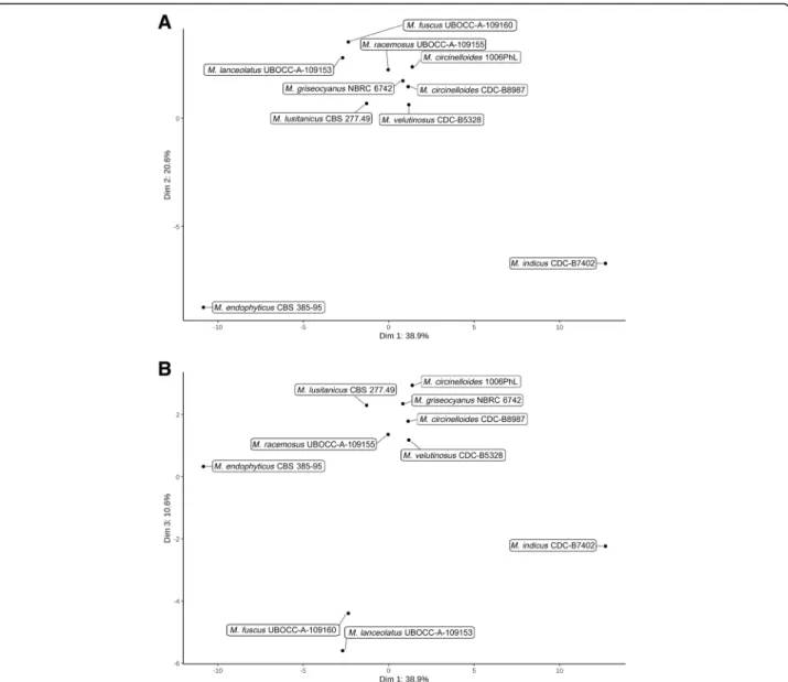
them, 4240 orthogroups were composed of predicted proteins belonging to all genomes while 64 were com-posed of single copy orthologs. Among the 64-single copy orthogroups, 51 were selected to reconstruct the species tree using Maximum Likelihood and Bayesian methods, R. delemar and P. blakesleeanus being used as outgroups (Fig.2a). The present study confirmed the re-cently described phylogenetic positions of the former M. circinelloides formae: M. griseocyanus, M. lusitanicus and M. velutinosus as distinct clades from M. circinel-loides[4]. The placement of M. indicus was altered com-pared to the topology obtained by Whalter et al. but concurred with the topology published by Álvarez et al. [22, 24]. Similarly, the placement of R. microsporus CDC-B9645 and R. microsporus CDC-B9738 was con-cordant with the topology of Chibucos et al. which iden-tified these two isolates as M. racemosus, but clearly differentiated from the one by Gryganskyi et al. in which the two isolates were clustered with R. microsporus iso-lates (and renamed accordingly) [13,25]. This phylogen-etic position was not affected when four previously sequenced R. microsporus were added to the phyloge-nomic reconstruction (Additional file 1: Figure S1). These results clearly raise questions concerning their ac-tual position and the genetic bases associated with these incongruences. The other studied isolates had concord-ant phylogenetic placements with previously published studies [13,22,23,25].
From the biological point of view, M. fuscus and M. lanceolatus that are close in the tree, have mainly been
Fig. 1 Repartition of the intergenic length within the 12 studied Mucor and Rhizopus genomes. The four isolates sequenced in the context of this study are in red. The boxes represent intergenic length harbored by 25 to 75% (sorted by length) of the intergenic regions, the line within the box represent the median length, dots represent each value corresponding to the 10% longest intergenic units represented
encountered in cheeses, the basal singleton M. endophy-ticushas only been described as a wheat leaf endophyte, whereas the other Mucor species, grouped in the same clade, are usually described as potential human patho-genic species.
Transposable elements
The studied genomes contained contrasting transposable element (TE) coverages (Fig.2b). The plant endophyte, M. endophyticus, held the lowest TE coverage (5%) whereas the ubiquitous M. racemosus contained the highest TE coverage (37%). Even between close species, TE coverage and composition differed notably, e.g. between M. lanceo-latusand M. fuscus or between M. griseocyanus and the other species from the M. circinelloides complex (M. circi-nelloides, M. lusitanicus and M. velutinosus). M. lanceola-tus showed higher TE coverage than M. fuscus (23 and 14%, respectively) for both transposons and retrotranspo-sons but the predicted elements in M. lanceolatus were mainly non-autonomous (i.e. not including all the do-mains necessary for their transposition). On the contrary, almost all M. fuscus predicted TE were autonomous, the main represented category being terminal inverted repeat (TIR) but few retrotransposons, long interspersed nuclear elements (LINE) and long terminal repeat (LTR), were also detected (Additional file2: Figure S2).
Among members of the M. circinelloides complex, three species displayed a similar pattern in terms of TE compos-ition and, except for one of the M. circinelloides isolates
(1006PhL), in terms of coverage. The fourth species, M. griseocyanus,displayed striking differences from the other species. Indeed, the TE coverage of M. griseocyanus was reduced by at least twofold compared to the other mem-bers (Fig. 2b). Furthermore, the main TE category was identified as belonging to the helitron order, a marginal category in the three other members of the clade.
Predicted proteins repartition
The predicted protein repertoires encoded by the 14 Mucoromycota investigated were compared with one an-other. This led to the identification of a set of core pre-dicted proteins shared by all studied Mucor species and to the determination of the proteins also occurring in the studied R. microsporus isolates. Dispensable proteins (shared by at least two species) and species-specific pro-teins (only found in a single species) were also identified (Fig.2c). For each category, duplicated proteins were iden-tified. Despite the phylogenetic divergence between spe-cies, at least one third of the predicted proteins for each species were part of the core predicted proteome and the number of species-specific proteins was restricted.
As suggested by BUSCO results, most of R. micro-sporus predicted proteins were duplicated (Fig. 2c). This result explained the relatively low number of single copy core proteins gathered for phylogeny recon-struction despite a core Mucor proteome of approxi-mately 6000 proteins.
Fig. 2 Representation of the phylogenomic tree, repetitions coverage and gene number for the genomes pertaining to the studied Mucor and Rhizopus isolates (initially assigned to M. racemosus). a Phylogenomic tree of the studied isolates reconstructed with RAxML based on 51 single copy ortholog families using R. delemar RA-99880 and P. blakesleeanus NRRL1555 as outgroup and calibrated on time scale with r8S program. The branch length corresponds to the estimated time before present. Color dots represent bootstrap support. The four isolates sequenced in this study are in red. b Representation of the repetitions coverage in each genome. Unknown: potential transposable element that could not be identified. c Representation of the protein numbers in the studied isolates. Duplicated proteins count represent extra copy number of each gene family
Evolution of gene families across phylogeny
When using the whole genome dataset, CAFE-, Dupli-PhyML- and Notung-based analyses yielded non-concordant results (data not shown) with inconsistent placements of expansion/contraction events within the cluster encompassing R. microsporus CDC-B9738, R. microsporus CDC-B9645 and M. racemosus. This be-haviour was interpreted as a side effect of a putative whole genome duplication or hybridization compatible with the observations done on the two R. microsporus genomes. The phylogenetic placement of these two isolates within the genus Mucor being also ques-tioned, we decided to remove them from the CAFE analysis. CAFE identified 44 rapidly evolving gene families on the M. lanceolatus/M. fuscus branch (per-taining to the two species associated to cheese ripen-ing). Among these families, two were associated to secondary metabolism, namely an acyl-CoA synthetase and a cytochrome p450 encoding gene families, both with reduced number of genes in cheese ripening spe-cies (M. fuscus and M. lanceolatus) compared to other species. A cysteine hydrolase gene family was also reduced in the two cheese-associated species. An-other family (less conserved) with genes identified as encoding putative transcriptional activators of glyco-lytic enzyme was expanded in the latter species ge-nomes. Other families were either unknown or similar to TE sequences.
In the M. endophyticus endophyte, at the node sep-arating cheese-associated species from pathogenic spe-cies and at the node separating M. indicus from other species, 49, 4 and 9 gene families were considered as rapidly evolving, respectively. However, these gene families were either of unknown function or similar to TE sequences.
Genes coding for CAZymes, peptidases and small secreted proteins
As different CAZyme and peptidase repertoires are associ-ated to different lifestyles, comparison of the annotations of CAZyme and peptidase genes annotation of M. lusitanicus were publicly available in the MycoCosm database (https://
mycocosm.jgi.doe.gov/mycocosm/home). However, for the
sake of the comparison, those annotations were performed again in this study following the same pipeline as the other studied genomes. This allowed to show that, for M. lusita-nicus, the number of peptidase genes was found higher (351 against 304) and the number of CAZymes lower (229 against 339) in this study compared to annotation per-formed by the Join Genome Institute and displayed in the MycoCosm database. Taking this difference into account, the total numbers of CAZyme (155–306) (Table 2) and peptidase (332–404) encoding genes (Table3) were found concordant with what is observed in other sequenced spe-cies of the Mucoromycota phylum as indicated in the MycoCosm protein database. In particular, the total num-ber of CAZyme encoding genes in the Mucor genomes ana-lysed in this study was lower than what is observed in average in Ascomycota and Basidiomycota (Additional file
3: Figure S3). Moreover, among CAZyme genes, those en-coding glycosyl-transferases (GT) were found more numer-ous than those encoding glycoside hydrolases (GH) which is usual in the Mucoromycota but not in Dikarya (Add-itional file3: Figure S3).
Clear differences in terms of CAZyme encoding gene composition among the different genomes were de-tected. Among the investigated species, M. indicus pos-sessed the highest content in all the CAZyme gene classes. In contrast, the number of all classes of CAZyme genes (except those encoding redox enzymes classified in auxiliary activities -AA-) was drastically reduced in M.
Table 2 Number of genes encoding Carbohydrate Active enZymes (CAZymes). Major CAZymes classes are shown separately, Auxiliary redox enzymes (AA), Carbohydrate-Binding Modules (CBM), Carbohydrate Esterases (CE), Glycoside Hydrolases (GH), Glycoside Transferases (GT) and Polysaccharide Lyases (PL). Enzymes substrats are indicated: Cell Wall (CW), Fungal Cell Wall (FCW) and Plant Cell Wall (PCW). The four isolates sequenced in the context of this study are in red
Isolate AA CBM CE GH GT PL Total CAZymes CW FCW PCW
12 2 15 56 67 2 155 10 19 8 12 7 18 68 107 1 214 12 21 12 10 5 16 62 105 2 201 11 19 12 11 5 20 81 115 2 235 18 22 14 M. griseocyanus NBRC_6742 12 7 25 83 113 2 243 18 25 13 M. circinelloides 1006PhL 15 7 23 83 121 2 252 18 24 14 M. circinelloides CDC-B8987 15 7 21 83 110 2 239 17 24 14 M. lusitanicus CBS 277.49 11 5 23 75 112 2 229 16 22 12 M. velutinosus CDC-B5328 16 6 24 78 112 2 239 17 22 13 M. indicus CDC-B7402 25 11 34 96 136 3 306 20 28 15
endophyticus. The number of genes coding for catabolic CAZymes was also reduced in M. fuscus and M. lanceo-latus. Interestingly, these reductions were noticeable for genes coding for cell wall degradation enzymes and more specifically in plant cell wall (PCW) degradation enzymes in M. endophyticus (Table 2). Differences among groups of species sharing a same putative lifestyle in terms of number of CAZyme families encoding genes were illustrated by the principal component analysis (PCA) of CAZymes in particular along axes 1, 2 and 3 (Fig. 3). The first axis represented the opposition be-tween the thermophilic opportunistic pathogen M. indi-cus and the endophyte M. endophyticus. This axis was constructed using mainly 28 CAZyme families (correl-ation above 0.75) all contracted in M. endophyticus com-pared to M. indicus, the other species presenting in almost all cases an intermediary pattern.
The second axis opposed M. endophyticus and M. indicus to all the other species. Three CAZyme families were mainly involved (correlation above 0.75) in this axis construction: GH8, GH20 and GT77. It is noteworthy that the GH20 β-hexosaminidase family was expanded in M. endophyticus and M. indicus, whereas the GH8 and GT77 tend to be reduced in the two latter species. The third axis represented the opposition between two species used for cheese ripening (M. fuscus and M. lan-ceolatus) and all the other species except M. indicus, the latter one presenting an intermediary pattern. Only four CAZymes families were mainly involved (correlation above 0.75) in this axis construction: GH9, GH37 and GT4 were contracted and GH45 expanded in M. fuscus and M. lanceolatus compared to almost all the other studied species.
Noteworthy, this analysis highlighted the GH3, GH5 (first axis) and GH9 (third axis) CAZyme families which are among the major cellulases involved in plant cell
wall degradation. They were less represented in M. endo-phyticusand to a lesser extent in M. fuscus and M. lan-ceolatus(Additional file4: Table S1).
Noteworthy, in addition to its expanded CAZyme reper-toire, M. indicus also displayed the largest peptidase (Table
3) repertoire and, with M. circinelloides isolate 1006PhL, the highest frequency of genes coding for secreted proteins and small secreted proteins (SSPs) (Fig.4). In contrast, M. fuscusand M. lanceolatus exhibited the lowest frequencies of secreted protein and SSP encoding genes (Fig.4).
Genes involved in secondary metabolism
As secondary metabolism genes can be associated with habitat adaptation, genes encoding PKS, NRPS, TPS and DMATS were investigated. Among genes associated with terpene biosynthesis, some corresponding to squalene cyclases, squalene synthases, bifunctional lycopene cy-clases, squalene/phytoene synthases and geranylgeranyl pyrophosphate (ggpp) synthases were identified in each species. The number of genes in each category was well conserved among the different species (Table4).
In all studied species, a single complete nrps gene (i.e. having at least one condensation domain, one carrier do-main, one phosphopantetheine attachment site and 1 AMP-binding domain) was detected. Three other genes containing three of the four mandatory NRPS domains were found in M. indicus, both of them lacking the con-densation domain, and one in M. lusitanicus, lacking the AMP-binding domain. In all species, no gene coding for DMATS were identified.
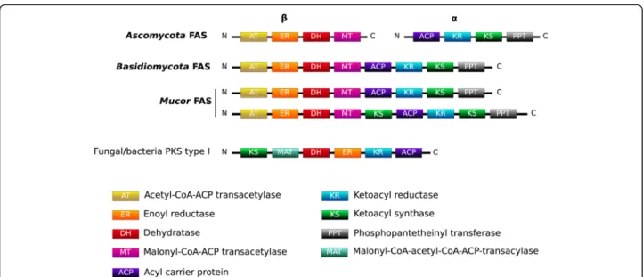
Interestingly, some gene sequences encoding putative PKS can be determined by automated annotation; how-ever, using a manual approach, these genes actually cor-respond to fatty acid synthases (FAS) type I encoding genes. They were detected in the investigated genomes: three in M. indicus, one in M. fuscus and M. lanceolatus
Table 3 Number of genes encoding proteolytic enzymes (peptidases) and their inhibitors (MEROPS database). The four isolates newly sequenced in the context of this study are in red
Peptidases
Isolate Aspartic Cysteine Metallo Serine Threonine Total peptidases Inhibitors
31 80 107 118 19 355 11 28 75 103 117 21 344 10 24 77 101 115 20 337 10 31 85 100 117 20 353 12 M. griseocyanus NBRC_6742 32 84 101 110 20 347 10 M. circinelloides 1006PhL 32 82 101 114 20 349 13 M. circinelloides CDC-B8987 30 78 94 109 21 332 11 M. lusitanicus CBS 277.49 34 86 103 108 20 351 10 M. velutinosus CDC-B5328 29 85 103 109 21 347 13 M. indicus CDC-B7402 44 88 114 132 26 404 11
and two in the other genomes. The different domains of these genes were similar in terms of composition and organization to FAS encountered in Basidiomycota, while FAS are encoded by two genes in Ascomycota (Fig.5). In M. indicus, M. racemosus and in M. velutino-sus, one of the FAS genes held a second KS domain. Recent transcriptomic analyses indicated that these genes were not expressed on PDA medium [23]. Note-worthy, none of the potential secondary metabolism associated genes determined in the different Mucor ge-nomes were organized in metabolic clusters.
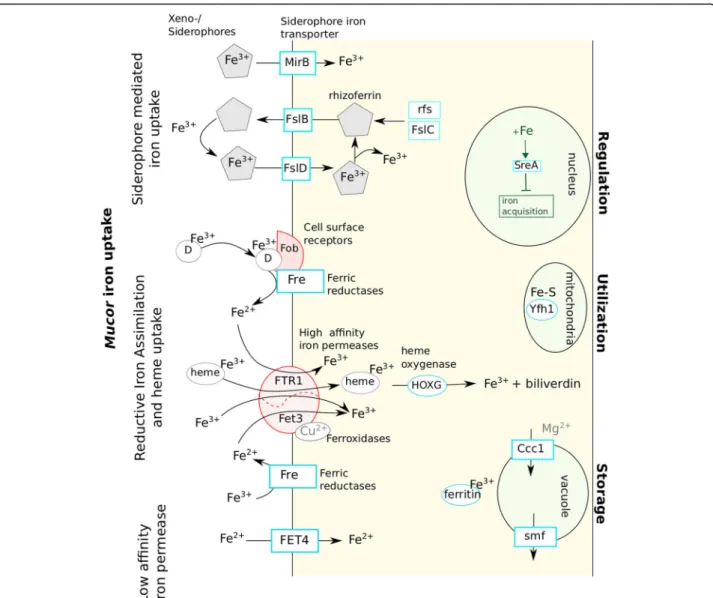
Iron uptake
As the involvement of iron uptake in mucormycosis has been reported, the associated genes were studied [30].
Homologs of genes encoding proteins involved in the different iron uptake mechanisms identified so far in fungi [31] were found in the analysed Mucor genomes (Table5; Fig.6).
Regarding the siderophore-mediated iron uptake, genes playing a role in the carboxylate and hydroxa-mate siderophore synthesis were searched for. At least one ortholog of the R. delemar rfs gene, necessary for the carboxylate siderophore rhizoferrin production [33] was found in each Mucor species. Other genes that might be involved in this rhizoferrin-mediated iron uptake mechanism were identified based on their homology to the bacterial genes of the Francisella tularensis rhizoferrin operon [34]. Homologs of the FslB and FslC F. tularensis genes were detected in each Mucor genome and numerous potential Mucor
Fig. 3 Representation of Mucor isolates repartition within the PCA analysis on their CAZYmes family proteins content. a Representation of the first two dimensions. b Representation of dimensions 1 and 3
genes belonging the major facilitator family matched to FslD. However, FslA, FslE, FslF and the operon regulator Fur could not be detected in Mucor ge-nomes. Genes involved in hydroxamate siderophore synthesis were not detected but predicted orthologous genes corresponding to the Aspergillus MirB sidero-phore permease (group 1) encoding gene and another gene coding for a MirB-like siderophore permease (group 2) were identified (Table5).
Regarding the reductive iron assimilation iron uptake, homologous sequences of the gene encoding the FTR1 high-affinity permease and fet3 ferroxidase genes were de-tected (fet3a was not dede-tected in M. fuscus, M. lanceolatus and M. racemosus genomes). Except for the FTR1 encoding gene, no gene involved in heme uptake was identified. The FET4 low affinity iron permease encoding gene was identi-fied in the different Mucor genomes. When focusing on the iron uptake regulation, homologs to the SreA iron uptake
Fig. 4 Representation of the number of secreted proteins within the genomes of the studied Mucor isolates. SSP: secreted proteins containing less than 300 aa. Secreted: secreted proteins with more than 300 aa. The four isolates sequenced in the context of this study are in red
Table 4 Number of genes involved in secondary metabolites found in the 10 studied Mucor isolates. The four species sequenced in this study are in red. For each gene category, maxima are highlighted in orange and minima in blue
*
repressor gene were detected but no gene involved in acti-vation of iron acquisition pathways such as HapX or Aft genes were identified. Two ferritin encoding genes of simi-lar lengths were present in each studied species; in each case, the two genes shared 70% similarity and 40% identity. Interestingly, the number of genes involved in iron uptake was always higher in the genome of the opportunistic pathogen M. indicus than in the other genomes (Table5). Indeed, in the M. indicus genome, (i) for the siderophore pathways, the rfs rhizoferrin biosynthesis gene was dupli-cated as well as a MirB-like siderophore permease encoding gene; (ii) for the reductive iron assimilation (RIA), the fet3b and fet3c ferroxidase genes were duplicated; (iii) for heme degradation, a supplementary heme oxygenase gene homo-log was detected and (iv) for direct Fe2+ uptake, three orthologs of the FET4 low affinity permease were identified, whereas two genes were found for the M. circinelloides complex and one for the other studied species. Finally, only one copy of the sreA-like gene, involved in down regulation of iron acquisition, was found in M. indicus whereas at least two copies were found for all other species.
On the contrary, the two cheese-associated species (M. fuscus and M. lanceolatus) exhibited a reduced number of genes involved in iron uptake within their genomes. MirB-like siderophore permease encoding gene was absent from the M. fuscus genome whereas one copy of fslB-like siderophore permease gene was lost. M. lanceolatus lost one copy of the cell surface re-ceptor fob gene and of the FTR1 high affinity permease, both species lacked the fet3a ferroxidase genes. The lat-ter genes were also absent from the cheese contaminant M. racemosusgenome.
Antifungal resistance
According to their habitat, fungi can be confronted to chemical biocide. In particular, at the hospital or in agricul-ture, fungicides are used and fungi can develop resistance. Based on the genome comparison we focused our attention on the antifungal resistance of the studied Mucor to triazoles.
The deduced aminoacyl sequences of the two lanos-terol 14α-demethylase paralogues CYP51 F1 and CYP51 F5 involved in ergosterol biosynthesis were globally well conserved over the Mucor genomes with the occurrence of the F128 residue in Helix I of CYP51 F5 which has been reported to be responsible for short-chain azole re-sistance in Mucorales [35]. However, a two-residue sub-stitution (AA to TS at residues 290–291) of Helix I of CYP51 F5 in M. lanceolatus was observed (Fig.7).
According to Caramalho et al., this residue could play a role in the intrinsic resistance against triazoles. These observations raised the question of the impact of these amino acid substitutions on M. lanceolatus azole suscep-tibility. Growth studies on azole supplemented media clearly showed that M. lanceolatus was the most suscep-tible species to both long- (posaconazole) and short-chain (voriconazole) azoles among the tested Mucor species (Table6) [35].
Discussion
Although benefiting from a growing interest due to their involvement in mucormycosis but also their biotechno-logical potential, a restricted number of Mucor genomes is available to this date, leading to scarce whole genome comparative studies [13, 15, 36]. These comparative
Fig. 5 Domain organization of FAS within the studied Mucor genomes in comparison to Ascomycota and Basidiomycota reported FAS organizations
studies mainly focused on human/clinical environments species. Yet, only a handful of Mucor species are known to cause human infections [2]. In this context, this study ex-panded the range of Mucor genomes available by includ-ing genomes from species collected from non-clinical environments. In particular, M. endophyticus was only re-ported as a wheat endophyte [27] and M. lanceolatus was, so far, only collected from cheeses [26].
The determined phylogeny, based on a large set of ortho-logous genes (51), that integrated the four genome se-quences obtained in this study is partially concordant with published phylogenies. In particular it confirmed the phylogenetic relationships amongst different members of the M. circinelloides complex [4] but was non-concordant with the latest published phylogeny concerning the phylo-genetic position of R. microsporus B9738 and
CDC-B9645 isolates, as they appeared in a sister group of the M. circinelloidescomplex and closely related to M. racemosus. This was observed both in the present study and that of Chibucos et al. (76 analysed orthologs) while they clustered into the R. microsporus clade with M. circinelloides species as an outgroup in Gryganskyi et al. study (192 orthologues analysed) [13,25]. Topologies may vary depending on the selected genes and on the reconstruction pipeline. The contrasted placements of the 2 R. microsporus isolates, which harbour larger-sized genomes (Fig. 2c), among the different studies may arise from the whole/partial-genome duplication events and/or hybridization observed in their genomes [25]. As a confirmation, when considering gene families involved in secondary metabolism and iron acquisition (which were investigated in this study by comparative genomics approaches), R. microsporus
Table 5 Number of genes involved in iron uptake found in the 10 studied Mucor. The four species sequenced in this study are in red. For each gene category, maxima are highlighted in orange and minima in blue. Proteins encoded by the different genes and their role in iron uptake mechanisms are presented in Fig.6
CDC-B9738 and CDC-B9645 genes were often found duplicated. Phylogenetic reconstructions performed using the duplicates yielded incongruent topologies with either CDC-B9738 or CDC-B9645 isolates clus-tering with R. delemar clade or alternatively with M. racemosus clade suggesting a possible hybrid genomic content for these two isolates.
Whatever the phylogenetic placement of the studied species or their proposed habitat/lifestyle, the current study revealed that the gene features among Mucor spe-cies (gene number, size, exon length etc.) were globally conserved. However, as indicated by the lack of macro-and micro-synteny (data not shown), species within this genus experienced extensive genomic rearrangements. These rearrangements can be partially explained by transposable elements (TE), which displayed high degree of diversity within the available genomes and have
already been reported to have a major role in fungal genomic diversity and genome evolution [37].
The average genome size of the Mucor species ana-lysed in this study (39 Mb) is congruent with what is observed at the scale of the Mucoromycotina subphy-lum (38 Mb) and also in Ascomycota (37 Mb). The gene number (9997 to 12,571) is also in concordance with the gene numbers observed in Mucoromycotina and As-comycota[38]. The core genome detected in this study includes approximately 6000 genes. The species-specific genes and the genes shared by species with similar lifestyles (data not shown) mostly encode pro-teins with unknown functions. They are thought to de-termine specific traits, such as adaptation to different environmental niches or preferential colonization of certain habitats, and determining their function would be of special interest.
Fig. 6 Proposed mechanisms of iron uptake by Mucor spp. as well as elements used for iron regulation, utilization and storage. Elements in red were described in literature as important for Mucor pathogenicity [13,16,32]. D: Deferoxamine
As osmotrophic organisms, fungi depend, for gaining nutrients, on lytic enzyme secretion to externally break down surrounding resources prior to absorption of the ca-tabolized products [39]. Depending on the lifestyle (sapro-trophic, pathogenic or mutualistic) and the habitat, the way of gaining nutrients and energy may vary. In particu-lar qualitative and quantitative differences in terms of CAZymes can be potent reporters of fungal lifestyle [38,
40–42]. In the present study, diverging CAZyme reper-toires among species or species groups sharing the same ecology were identified. The genome of M. endophyticus CBS385–95, isolated from wheat leaves and described as an endophyte, shares similar traits with genomes of other fungal endophytes. It harbours a low number of catabolic CAZymes (CBM, GH and PL) and esterase (CE) genes [43] including genes coding for plant cell wall degrading enzymes, such as the GH5, GH3 and GH9 fungal cellu-lases encoding genes. This observation suggests that M. endophyticus, like other fungal endophytes and ectomy-corrhizal fungi, is indeed adapted to intercellular growth and to the avoidance of the plant immune system [43,44]. The two species almost only isolated from cheeses and in-volved in cheese ripening (M. fuscus and M. lanceolatus) also displayed, to a lesser extent than M. endophyticus, a reduced repertoire of carbohydrate active enzyme encod-ing genes. The reduction was less significant for the GT encoding genes whose roles are not directly connected to
the external environment. It could be hypothesized that this reduced set of catabolic CAZymes is an imprint of specialization to a less ubiquitous lifestyle and an adapta-tion to the cheese environment. In contrast, the thermo-philic opportunistic pathogen M. indicus harbours the largest set of CAZyme genes. This trait is not shared by M. circinelloides, a species that may cause mucormycosis. This could be related rather to the high capacity of M. indicusto degrade plant materials [45] than to an oppor-tunistic pathogenic lifestyle. However, it is worth noting that M. indicus possesses a much larger protease reper-toire compared to the other Mucor species including the M. circinelloides. Given that proteases [46,47] were found clearly correlated to pathogenicity in Ascomycota oppor-tunistic pathogens one might expect a higher pathogen-icity potential of M. indicus over M. circinelloides.
Examination of secondary metabolic pathways can also shed light on fungal ecology and adaptation to a particu-lar lifestyle [48]. In this study, we investigated more specifically genes that are essential to most of the sec-ondary metabolite biosynthesis (PKS, NRPS, DMATS…) but also genes involved in iron uptake which have been shown to be fundamental for fungal virulence in oppor-tunistic pathogens [49, 50] and for cheese colonizing microorganisms given that cheese is a strongly iron-restricted medium [51]. The search for genes involved in secondary metabolite biosynthesis are often initiated by
Fig. 7 Partial amino acid sequence alignment of lanosterol 14α-demethylase F5 in different Mucor isolates. The consensus sequence is represented in blue and polar amino acids appear in green. ML: M. lanceolatus; MF: M. fuscus; ME: M. endophyticus; MR: M. racemosus; MLu: M. lusitanicus
Table 6 Growth diameter (cm) of representative Mucor isolates on RPMI medium supplemented or not with different concentrations of voriconazole (VCZ) or posaconazole (PCZ) VCZ concentration PCZ concentration Isolate 0 mg/L 10 mg/L 50 mg/L 100 mg/L 500 mg/L 1000 mg/L 0 mg/L 10 mg/L 50 mg/L 100 mg/L 500 mg/L 1000 mg/L M. endophyticus CBS 385-95 ≥ 5,4 ≥ 5,4 ≥ 5,4 ≥ 5,4 0 0 ≥ 5,4 ≥ 5,4 ≥ 5,4 ≥ 5,4 0 0 M. fuscus UBOCC-A-109160 ≥ 5,4 3,2 0,8 0,2 0,2 0,2 ≥ 5,4 3 3 3 1,5 0,2 M. lanceolatus UBOCC-A-109153 ≥ 5,4 4,5 0 0 0 0 ≥ 5,4 0 0 0 0 0 M. racemosus UBOCC-A-109155 ≥ 5,4 ≥ 5,4 ≥ 5,4 ≥ 5,4 3,2 0,5 ≥ 5,4 ≥ 5,4 ≥ 5,4 ≥ 5,4 ≥ 5,4 ≥ 5,4 M. lusitanicus CBS 277.49 ≥ 5,4 ≥ 5,4 ≥ 5,4 ≥ 5,4 0,3 0,3 ≥ 5,4 ≥ 5,4 ≥ 5,4 ≥ 5,4 ≥ 5,4 ≥ 5,4
the search for PKS, NRPS or DMATS genes clustered with additional genes involved in the considered biosyn-thesis pathway and known as BGC (biosynthetic gene cluster). In the framework of this study, no typical BGCs involved in secondary metabolism, including a PKS, FAS, NRPS, NRPS-like gene or terpene synthesis related genes, were detected within the studied Mucor genomes. Still, secondary metabolites pathways have been charac-terized in Mucorales [3] and different genes involved in secondary metabolism were identified in the present and previous studies. The apparent absence or at least scar-city of BGCs within the genus Mucor (and possibly at a broader scale within the Mucoromycotina) might appear enigmatic since the genus encompasses species with contrasting lifestyles similar to what exists in Ascomy-cota in which BGCs are abundant. This raises the ques-tion why the apparent selective advantage conferred by BGCs to higher fungi would not apply to Mucor species. The answer to this question might lie in some ecological specificities of the Mucorales which are considered as ruderal species avoiding stress and competition [52, 53] or to structural specificities such as their coenocytic structure [54], and may shed new light regarding the BGC evolution in eukaryotes. All Mucor genomes inves-tigated in this study included a NRPS gene which role has still to be determined as well as different genes en-coding for enzymes associated with terpene biosynthesis. As noticed by Voigt et al. who analysed different Mucor genomes, no PKS genes but genes with a typical struc-ture and domain order of Type I fatty acid synthases (FAS) were detected in the different Mucor genomes in-vestigated in the present study [3]. These Mucor FAS have their domains allocated within a single gene as in Basidiomycotaand not over two genes as in Ascomycota (Fig. 5) [55,56]. This result supports the hypothesis of a primordial contiguous FAS gene encoding the entire FAS [55]. Most of the Mucor genomes analysed here in-clude two putative FAS genes, but a single FAS gene was found in the cheese-related species M. lanceolatus and M. fuscus, and up to three genes were found in the op-portunistic pathogen M. indicus. Whatever the number of FAS genes included in the genome, only one FAS was found expressed when the isolate was cultivated on PDA medium [23]. It could be hypothesized that the add-itional FAS genes have lost their functionality or display a different role since FAS gene are expected to be expressed in such conditions to participate to the fatty acid synthesis for membrane synthesis and energy stor-age [56]. Whether or not this additional FAS might be involved in secondary metabolisms like specialized sec-ondary metabolism FAS [57] has to be determined.
Among the secondary metabolites, terpenes play a role in natural pigment synthesis such as the carotenoid bio-synthesis [3]. Carotenoid synthesis has been described in
M. circinelloidesand in particular in overproducing iso-lates [3, 58, 59]. Although the different species within the Mucoromycotina subphylum are expected to produce and accumulate large amount of carotenoids, differences might exist among species [60]. Terpenes play also an important role in flavour production which is an import-ant trait for cheese-ripening fungi that could have been under human-directed selection in species used for cheese making [61]. This study indicated that the main genes involved in terpene biosynthesis were conserved among the analysed Mucor species without any import-ant differences in terms of gene number with the excep-tion of GGPP genes for which the number is lower in the endophytic species as well as in M. griseocyanus and in the M. indicus opportunistic pathogen.
Iron is an essential nutrient involved in variety of cel-lular processes such as respiration, oxidative stress and amino acid synthesis [62]. Iron uptake has been de-scribed as a virulence factor for pathogenic fungal iso-lates [49, 50] and of primary importance in cheese microorganisms since cheese is a highly iron-depleted medium [51]. In fungi, four different iron uptake mecha-nisms have been described so far: (i) siderophore medi-ated Fe3+ uptake, (ii) reductive iron assimilation (RIA), (iii) heme uptake and (iv) direct Fe2+ uptake [49] (Fig.
6). Homologs of genes coding for proteins participating to the four mechanisms were found in the analysed Mucorgenomes. These results suggest that the different Mucor species investigated here rely on carboxylate sid-erophore rhizoferrin as it is the case for M. circinelloides CBS 277.49 and R. delemar [33]. This type of sidero-phore is not used by Ascomycota and Basidiomycota, and would thus be a specificity of early diverging fungi in the fungal kingdom. Noteworthy, rhizoferrin encoding gene sequences have only been described in bacterial ge-nomes so far [32, 63] and coding sequences pertaining to the rhizoferrin operon in F. tularensis were used here to search for homologs in Mucor spp. In F. tularensis, genes involved in rhizoferrin synthesis and transport are located in an operon regulated by the Fur gene [34]. Based on these results, new candidate genes involved in rhizoferrin synthesis, import and export are proposed in the present study (Fig. 6). Although no actual evidence of Mucor ability to produce hydroxamate siderophores was detected, some of these siderophores could be used by Mucor species as xeno-siderophores as suggested by the presence of mirB-like siderophore transporter genes in some of the Mucor genomes. It is worth noting that the bacterial siderophore deferoxamine is also used by Mucorspp. as xeno-siderophores [64].
Interestingly, the three isolates sampled from cheese presented a reduced number of genes related to iron ac-quisition compared to the other isolates. Indeed, the ge-nomes of these isolates lack a FTR1 gene copy as well as
the fet3a gene. It could be hypothesized that the latter genes would have a specific role in Mucor pathogenicity since R. delemar mutants with FTR1 reduced gene copies or with decreased FTR1 expression had reduced virulence in a deferoxamine-treated mouse model of mucormycosis [30]. The fet3a gene appeared as the less important among the fet3 genes regarding M. circinel-loides pathogenicity, but inactivation of two fet3 genes led to a reduced virulence [17] and the loss of fet3a led to an increased sensibility to the mutations on fet3b/ fet3cin terms of fungal pathogenicity. Furthermore, one copy of the ferroxamine binding (Fob) cell surface pro-tein gene is absent in the M. lanceolatus genome, the production of this protein being required for full viru-lence of R. delemar in a deferoxamine-treated mouse model of mucormycosis [14]. M. fuscus also lacks MirB-like and one copy of fslB-MirB-like siderophore permease genes, which might reduce its potential to acquire iron. These results provide possible evidence of safety of these food-related species although the number of studied iso-lates is too low to definitely assert it. On the contrary, M. indicus, a species that is considered as the most threatening opportunistic human and animal pathogen amongst the Mucor species [7] harbours an increased set of genes involved in iron uptake which might be an asset to its opportunistic pathogenic lifestyle.
Susceptibility to antifungal drugs, and in particular to azole antifungal agents which are widely used for mucor-mycosis treatments as well in agriculture [35, 65], is of interest as it may vary in Mucor species according to their specific habitats, e. g. cheese isolates are probably less exposed to antifungals and antifungal resistance is unlikely to offer a selective advantage to non-pathogens. Azole drugs inhibit lanosterol 14α-demethylase (LDM) thus blocking ergosterol biosynthesis and resulting in a build-up of toxic sterols [66]. In Mucorales, two lanos-terol 14α-demethylase paralogues CYP51 F1 and F5 co-exist with a substitution in CYP51 F5 at residue 128 responsible for innate resistance against short-tailed tria-zoles, and a V to A substitution at residue 291 of CYP51 F5 also potentially playing a role [35]. Interestingly, we found that M. lanceolatus, only isolated so far from cheese, did not harbour the V to A substitution at resi-due 291 of the CYP51 F5 predicted protein but bears in-stead a unique two aminoacyl substitution (AA to TS) at residues 290 and 291 (in consensus sequence). Azole tests performed in this study demonstrated that these substitutions were associated with a notable higher sus-ceptibility to both short- and long-tailed azoles.
Conclusions
In conclusion, this study expanded the range of Mucor genomes available by including genomes from species with contrasted lifestyles represented by isolates
collected from non-clinical environments (more specific-ally, cheese and plant). In addition to yield a better over-view of the Mucor pan-genome, the expanded range of genomes allowed identifying contrasting features that could contribute to habitat and niche adaptation al-though distinguishing divergences due respectively to evolutive adaptation or to non-ecologically based evolu-tionary forces may appear difficult given that the differ-ent taxa did not diverged recdiffer-ently. The obtained data will allow further investigating the link between genetic and biological data, especially in terms of niche or habi-tat adaphabi-tation.
Materials and methods Biological material
The genomes of 12 representative isolates were investi-gated in the present study (Table7). Four of them were sequenced in the framework of this study while the eight others were publicly available [12, 13, 28]. M. fuscus UBOCC-A-109160, M. lanceolatus UBOCC-A-109153, M. endophyticus CBS 385–95 (UBOCC-A-113049) and M. racemosus UBOCC-A-109155 used for genome se-quencing were cultivated in the dark at 25 °C on PDA solid medium (Difco Laboratories, Detroit, Michigan). Spore suspensions of each isolate were produced as pre-viously described by Morin-Sardin et al. [7]. Concentra-tions were adjusted to 107 to 108 spores·mL− 1 prior to storage at − 80 °C until use. For genomic DNA extrac-tion, each of the studied monospore isolates was grown on PDA solid medium at 25 °C for 7 days.
Species assignment within M. circinelloides complex
Species assignment of isolates pertaining to the M. circi-nelloides complex was performed using a Maximum Likelihood phylogenetic reconstruction based on a cyclo-propane-fatty-acylphospholipidsynthase gene (cfs) se-quence alignment including cfs sese-quences available in Wagner et al. [4].
Genome sequencing and assembly
Genomic DNA from M. fuscus, M. racemosus, M. lanceo-latusand M. endophyticus was extracted from fresh myce-lium, following the genomic DNA extraction method as outlined by the 1000 Fungal Genomes Project (http://1
000.fungalgenomes.org/home/protocols/high-quality-gen-omic-dna-extraction/#Fulton1995) which is based on a
protocol by Spanu et al. [72]. with an optional step using Qiagen genome-tips (Qiagen). Due to the low efficiency of the CTAB method for M. lanceolatus, genomic DNA of this isolate was also extracted following the protocol de-veloped by Cheeseman et al. with a purification by a cesium chloride gradient with DAPI [73].
Genomes were sequenced with Illumina technology (San Diego, CA) at different sequencing facilities
(Additional file 5: Table S2). For each of the four species, DNA were paired end sequenced (read length 2 × 100 bp, insert size 500 bp). An additional mate pair sequencing was performed for M. lanceo-latus and M. endophyticus (read length 2 × 100 bp, insert size 9–12 kb). Sequences were quality checked with FastQC [74]. Adaptors were removed, reads were quality trimmed (bases kept had a phred score above 25) and reads shorter than 20 bp were dropped with Cutadapt [75]. Mate pair reads of M. lanceolatus were mapped with STAR [76] on a preliminary version of the assembly (by pro-viding only M. lanceolatus paired end data to CLC Genom-ics Workbench -CLCbio, Seoul, Korea-). Mate pair reads separated by less than 500 bp and oriented in forward-reverse were dropped. This new set of reads was used in further M. lanceolatus assemblies. M. lanceolatus and M. endophyticus genomes were assembled using Velvet [77] (option “shortMatePaired”, k-mer of 67 for M. lanceolatus and k-mer of 85 for M. endophyticus), while M. racemosus and M. fuscus genomes were as-sembled with SOAPdenovo [78]. Genome assembly quality was checked with BUSCO v3 [79] using the fun-gal dataset and Rhizopus Augustus training.
Genome annotation of the four newly sequenced genomes
Genome assembly scaffolds were annotated using combina-tions of ab initio predictors, RNAseq data support and homology search. As for ab initio predictors, Genemark-ES [80], with self-training, and Augustus [81], with Rhizopus training available within the Augustus tool, were used. RNAseq transcripts were extracted and sequenced as previ-ously described in Lebreton et al. and reconstructed using two methods [23]: (i) by mapping RNAseq reads on gen-ome with STAR [82] and reconstructing transcripts with Cufflinks [83], and (ii) by de novo transcript reconstruction with Trinity [84] and mapping the obtained transcripts on the genomes with GMAP [85]. Predicted proteins of M. lusitanicus [12], R. delemar [9] and P. blakesleeanus [12] were searched on genomes with Exonerate [86]. Consensus gene models were generated from all predictions by EVi-denceModeler [87].
The obtained gene predictions were functionally anno-tated as follows: transmembrane domains were predicted with TMHMM [88], peptide signal with SignalP v4 [89] and Pfam domains with HMMER [90] using the PFAM-A database [91]. Sequence homologies were searched
Table 7 List of isolates used in this study and their reported habitat. The four isolates sequenced in the context of this study are in red
Species Isolate Isolation source Reported habitat of the
species Reported habitat references Genome reference or accession CBS 385–95 (UBOCC-A-113049) Triticum aestivum, leaves
Plant endophyte [27] This study
UBOCC-A-109160 Cheese Cheese [26] This study
UBOCC-A-109153 Cheese Cheese [26] This study
UBOCC-A-109155 Cheese Cheese, yogurt, walnuts,
sausages, grassland soil, decaying vegetables, human
[24,26,67] This study
M. griseocyanusa NBRC 6742 Unknown Vegetable, tanned sole
leather [68] BBKB00000000 M. circinelloides f. circinelloides 1006PhL Skin of a healthy human
Cheese, sufu starter, decaying vegetables, human, soda, air, soil, dung, sediment
[22,24,26,69] [29]
M. circinelloides CDC-B8987 Human BL line [13]
M. lusitanicusb CBS 277.49
(UBOCC-A-108085)
Unknown [12]
M. velutinosusc CDC-B5328 Human: nasal [13]
M. indicus CDC-B7402 Human: unknown Human, dung,
Dioscorea tuber, sorghum malt
[24,70,71] [13]
R. microsporus (formerly M. racemosusc)
CDC-B9645 Clean room floor Human, dust,
sorghum malt, stored cereals
[13,24] [13]
R. microsporus (formerly M.
racemosusc) CDC-B9738 Human abdomen GCA_000697275.1
CBS (Centraalbureau voor Schimmelcultures, Netherlands), NBRC (Biological Ressource Center, NITE), NRRL (Agricultural Research Service Culture Collection, USA), CDC (Centers for disease Control and Prevention) and UBOCC (University of Brest Culture Collection)
a
Originally M. ambiguus NBRC 6742.b
Originally M. circinelloides CBS 277.49. These isolates were reassigned according Wagner et al. [4].c
Initially referenced as Mucor racemosus [13] but later assigned to Rhizopus microsporus [25]
using tBLASTx and BLASTp [92] (with an e-value threshold inferior to 10− 5, against Swissprot-Uniprot and Uniref90 databases as well as M. lusitanicus CBS 277–49, R. delemar RA-99880 and P. blakesleeanus NRRL1555 filtered proteins obtained from the JGI plat-form [93]. EC numbers were predicted using PRIAM [94] and were transferred from homology researches. GO terms were transferred from homology search. Gene names were assigned with AHRD (Automated Assign-ment of Human Readable Descriptions) available on Github (https://github.com/groupschoof/AHRD).
Non-coding RNA were predicted with tRNAscan-SE [95], RNAmmer [96] and Infernal [97] using the Rfam database [98]. The obtained data were integrated in an instance of the genome viewer Apollo [99] allowing ex-perts to validate gene prediction quality and perform manual curation.
Complementary annotation of the full set of genomes
Transposable elements (TE) were annotated using the REPET pipeline [100] that includes a de novo prediction and TE classification [101]. Carbohydrate-active en-zymes were searched using dbCAN2 [102] based on se-quences available in the CAZy database [103] with HMMER (E-Value <1e− 15, coverage > 0.35), DIAMOND [104] (E-Value <1e− 102) and Hotpep [105] (Frequency > 6.0, Hits > 2.6). Only annotations predicted by at least two different tools were subsequently considered. Pepti-dases and their inhibitors available in the MEROPS data-base [106] were searched using BLASTp (E-Value 10− 5).
Phylogenetic reconstruction
Predicted proteomes of the 12 studied isolates, as well as those of R. delemar and P. blakesleeanus (both latter spe-cies being considered as outgroups), were compared based on sequence similarity to identify orthologous proteins using the Orthofinder v.2.2.0 software [107] (E-value 10− 5, inflation 1.5). The 64 obtained single copy orthologs were selected to reconstruct the phylogeny of the studied spe-cies. Multiple alignment was inferred using PRANK v.170427 [108], run with default settings. Spuriously aligned regions were excluded with TrimAl v1.4.r15 [109] with a 0.2 gap threshold. Based on the alignments, 13 orthogroups were manually discarded due to low percent-age of identical sites or high number of gaps among ortho-logs. Subsequent alignments were concatenated in a supermatrix of 23,398 sites. This matrix was used to recon-struct the species tree in one hand using Bayesian Monte Carlo Markov Chain (MCMC) samples with PhyloBayes v3.3 MCMC samplers [110] with a CAT+GTR model and 3 chains and in the other hand by Maximum Likelihood in-ference with RAxML PTHREADS v. 8.2.9 [111], with a par-titioned LG + G model, in which each data partition represented a single input gene family. A bootstrap analysis
with 100 replicates under the same model was performed in RAxML in order to assess tree branch support. To con-firm the obtained phylogenetic position of the 2 R. micro-sporusisolates, an expanded species tree including 11 other sequenced Mucoromycotina (25 isolates overall) was restructed as follows. The 29 single copy genes families con-taining at least 20 isolates were independently aligned with PRANK v.170427 and trimmed with TrimAl v1.4.r15. For each gene family a gene tree was reconstructed using RAxML with the auto-detection of the best amino-acid model and a bootstrap analysis of 100 samples. The species tree was then reconstructed using the 29 gene trees with Clann v4.2.2 using a bootstrap analysis of 100 samples [112]. The obtained RAxML tree of the main species was used to estimate the divergence time between species with the Langley-Fitch method with r8s v1.8 [113] by calibrating against the assessed origins of P. blakesleeanus and R. dele-marat 468 MY [114].
Evolution of genes families
Based on OrthoFinder results and the obtained ultra-metric tree, expansion and contraction of gene families were reconstructed with CAFE v4 [115]. Birth and death parameters were estimated independently using ortholo-gous groups containing less than 75 genes per isolate. The analysis was done on all isolates except R. micro-sporus CDC-B9645 and R. microsporus CDC-B9738. Rapidly evolving families were predicted by CAFE using the Viterbi algorithm.
Secondary metabolism related genes
Secondary metabolism associated genes (polyketide syn-thase -PKS-, non-ribosomal peptide synthetase -NRPS-, terpene synthase -TPS-, dimethylallyl tryptophan syn-thase -DMATS-) and other genes potentially involved in adaptation to the environment (e.g. genes related to iron acquisition and to antifungal resistance) were searched in each species. Gene cluster associated with secondary metabolites were searched with antiSMASH v.4 fungal version (FungiSMASH) [116] and SMURF [117].
Antifungal susceptibility
M. lusitanicus, M. endophyticus, M. fuscus, M. lanceola-tus and M. racemosus, as well as Byssochlamys fulva UBOCC-A-101005 (as a positive control), were grown in 5.4 cm Petri dishes on solid RPMI medium (Gibco BRL, Gaithersburg, MD) with 2% glucose and different con-centrations of azole antifungal compounds: 0 mg.l− 1, 10 mg.l− 1, 50 mg.l− 1, 100 mg.l−1, 500 mg.l− 1 and 1000 mg.l− 1 of voriconazole (VCZ) (OHRE Pharam, Tours, France) or posaconazole (PCZ) (MSD France, Courbe-voie, France), respectively. Each antifungal concentration was assayed in triplicates. Growth diameters were mea-sured after 7 days.
Availability of supporting data
Raw sequence data were deposited at the European Nucleotide Archive (ENA) (http://www.ebi.ac.uk/ena/data/
view/PRJEB30975) and genome assemblies, annotations and
genome browser are available on the ABiMS platform
(http://application.sb-roscoff.fr/project/mucor/) as well as on
the JGI Mycocosm platform (https://genome.jgi.doe.gov/
programs/fungi/index.jsf). Noteworthy, functional
annota-tions found on the Mycocosm platform differ from the ones exploited here since the structural annotation was function-ally re-annotated by the pipelines associated with the Mycocosm platform (https://genome.jgi.doe.gov/Mucrac1,
https://genome.jgi.doe.gov/Mucend1,https://genome.jgi.doe.
gov/Mucfus1, https://genome.jgi.doe.gov/Muclan1). All the
genome sequences compared to the genomes sequenced in the present study were found in NCBI Genome except for M. lusitanicusgenome available at the Mycocosm platform.
Supplementary information
Supplementary information accompanies this paper athttps://doi.org/10. 1186/s12864-019-6256-2.
Additional file 1: Figure S1. Phylogenomic tree for the genome of 25 Mucoromycotina. The tree was reconstructed using Clann based on 29 genes trees. Each tree corresponded to one of the single copy gene families included at least 20 of the 25 isolates investigated. Bootstrap supports are indicated under the branch.
Additional file 2: Figure S2. Representation of the TE coverage in each genome depending on the TE category (classification of Wicker et al.) [118]. The four species sequenced in this study are in red. DHX: Helitron transposon. DTX: TIR transposon. DXX-MITE: unknown non-autonomous transposon, MITE-like. noCat: potential transposable element that could not be identified. RIX: LINE retrotransposons. RLX: LTR retrotransposon. RXX unknown retrotransposon. RXX-LARD: unknown non-autonomous retrotransposon, LARD-like. RXX-TRIM: unknown non-autonomous retro-transposon, TRIM-like.
Additional file 3: Figure S3. Number of CAZymes in the different fungal phyla. Major CAZymes classes are shown separately, Auxiliary redox enzymes (AA), Carbohydrate-Binding Modules (CBM), Carbohydrate Esterases (CE), Glycoside Hydrolases (GH), Glycoside Transferases (GT) and Polysaccharide Lyases (PL).
Additional file 4: Table S1. Number of identified CAZyme encoding genes involved in the degradation of plant cell wall components (Cellulose active, Hemicellulose active and Pectin active). The presence of genes has been confirmed with manual annotation.
Additional file 5: Table S2. Extraction and sequencing information corresponding to the four newly sequenced Mucor isolates Abbreviations
CAZymes:Carbohydrates Active enZymes; CYP51: 14α-demethylase paralogues; DMATS: DiMethylAllyl Tryptophan Synthase; FAS: Fatty Acid Synthase; NRPS: NonRibosomal Peptide Synthetase; PCZ: Posaconazole; PKS: Polyketide Synthase; RIA: Reductive Iron Assimilation; SSP: Small secreted protein; TPS: Terpene Synthase; VCZ: Voriconazole
Acknowledgements
The authors are thankful to Vincent Bruno, for providing the annotation of the Mucor CDC-B8987, CDC-B7402, CDC-B9645, CDC-B5328 and CDC-B9738 isolates, to Jonathan Dorival, for advices on PKS, Antoine Branca and Emma-nuelle Morin for stimulating discussion, Antoine Hermet for his involvement at the early stage of the project and Stephen Mondo for the integration of Mucor genomes in the Mycocosm platform. The authors are also thankful to the two anonymous reviewers for their constructive comments. The authors
thank the UBOCC strain collection (https://www.univ-brest.fr/ubocc) for pro-viding Mucor isolates. This research was funded by the Région Bretagne (ARED program) and EQUASA, a technological platform of the Université de Bretagne Occidentale, in the framework of the MUCORSCOPE project. The M. fuscus, M. lanceolatus and M. racemosus genome sequences were obtained in the context of the French National Agency for Research (ANR)project "Food-Microbiomes".
Authors’ contributions
Research idea conception: ECn, GB, LMC and JLJ. DNA Extraction: AL and RD. Antifungal assay: CLM. Analytical approach design: ECe, JLJ and LMC. Apollo instance deployment for manual annotation: LG and MM. Manual annotation: AL, JLJ, CPA, VG and LMC. Data analysis: AL, ECe, JLJ and LMC. Manuscript writing: AL, JLJ and LMC. Manuscript discussion and revision: ECn, GB and ECe. All authors read and approved the final manuscript.
Funding
This research was funded by the the Région Bretagne (ARED program) and EQUASA, a technological platform of the Université de Bretagne Occidentale, in the framework of the MUCORSCOPE project.
Availability of data and materials
Raw sequence data were deposited at the European Nucleotide Archive (ENA) (http://www.ebi.ac.uk/ena/data/view/PRJEB30975), genome assemblies and annotations are available on the ABiMS platform ( http://application.sb-roscoff.fr/project/mucor/).
Ethics approval and consent to participate Not applicable.
Consent for publication Not applicable. Competing interests
The authors declare that they have no competing interests. Author details
1Univ Brest, Laboratoire Universitaire de Biodiversité et Ecologie Microbienne,
F-29280 Plouzané, France.2Station Biologique de Roscoff, Plateforme ABiMS,
CNRS: FR2424, Sorbonne Université (UPMC), Paris VI, Place Georges Teissier, 74 29682 Roscoff Cedex, BP, France.3CNRS, Integrative Biology of Marine
Models (LBI2M), Station Biologique de Roscoff (SBR), Sorbonne Université, 29680 Roscoff, France.4Department of Genetics and Microbiology, Faculty of
Biology, University of Murcia, 30100 Murcia, Spain.5Institute for Integrative
Biology of the Cell (I2BC), CEA, CNRS, University Paris-Sud, Université Paris-Saclay, CEDEX 91198 Gif-sur-Yvette, France.
Received: 13 May 2019 Accepted: 31 October 2019
References
1. Spatafora JW, Aime MC, Grigoriev IV, Martin F, Stajich JE, Blackwell M. The fungal tree of life: From molecular systematics to genomescale phylogenies. Microbiology Spectrum. 2017;5(5). https://doi.org/10.1128/microbiolspec.FUNK-0053-2016Retrieved fromhttps://escholarship.org/uc/item/4485m01m. 2. Morin-Sardin S, Nodet P, Coton E, Jany J-L. Mucor: a Janus-faced fungal
genus with human health impact and industrial applications. Fungal Biol Rev. 2017;31(1):12–32.
3. Voigt K, Wolf T, Ochsenreiter K, Nagy G, Kaerger K, Shelest E, Papp T. In: Hoffmeister D, editor. 15 Genetic and Metabolic Aspects of Primary and Secondary Metabolism of the Zygomycetes. Cham: Springer International Publishing; 2016. p. 361–85.
4. Wagner L, Stielow B, Hoog S, Schwartze V, Kurzai O, Walther G. A new species concept for the clinically relevant Mucor circinelloides complex. Persoonia - Molecular Phylogeny and Evolution of Fungi; 2019. 5. Petrikkos G, Skiada A, Lortholary O, Roilides E, Walsh TJ, Kontoyiannis DP.
Epidemiology and Clinical Manifestations of Mucormycosis. Clin Infect Dis. 2012;54(suppl_1):S23–34.