HAL Id: hal-02756069
https://hal.inrae.fr/hal-02756069
Submitted on 3 Jun 2020
HAL is a multi-disciplinary open access
archive for the deposit and dissemination of
sci-entific research documents, whether they are
pub-lished or not. The documents may come from
teaching and research institutions in France or
abroad, or from public or private research centers.
L’archive ouverte pluridisciplinaire HAL, est
destinée au dépôt et à la diffusion de documents
scientifiques de niveau recherche, publiés ou non,
émanant des établissements d’enseignement et de
recherche français ou étrangers, des laboratoires
publics ou privés.
How to visualisation and localisation of acid phosphatase
transcripts in common bean nodules (Phaseolus
vulgaris) by in situ RT-PCR
Laurie Amenc, Ingrid van Aarle, Gaëlle Viennois, Saber Kouas, Doan Luu,
Claude Plassard, Jean-Jacques Drevon
To cite this version:
Laurie Amenc, Ingrid van Aarle, Gaëlle Viennois, Saber Kouas, Doan Luu, et al.. How to visualisation
and localisation of acid phosphatase transcripts in common bean nodules (Phaseolus vulgaris) by in
situ RT-PCR. Rhizosphere 2, Aug 2007, Montpellier, France. 2007. �hal-02756069�
In situ RT-PCR to visualise the distribution of
transcripts in symbiotic-root tissues
Obje c t if :
In o rde r t o de t e c t ge n e e x pre s s io n in diffe re n t t is s u e s by i n s i t u Re v e rs e Tran s c ript as e
Po ly m e ras e Ch ain Re ac t io n (RT-PCR), e n z y m e -labe lle d flu o re s c e n t s u bs t rat e was u s e d t o i n s i t u wit h e
pi-flu o re s c e n c e m ic ro s c o py an d t o v is u alis e it at t h e le v e l o f t ran s c ript s . Tis s u e s u s e d we re ro o t -n o du le s o f
Phaseolus vulgaris and hypae of Hebeloma cylindrosporum.
Results
Conclusion
The use of the ELF substrate to reveal activity of the alkaline phosphatase-conjugated
antibody used in the technique enabled visualisation of gene expression in different organisms
that could be related to specific structures.
L.K. Amenc
1, I.M. van Aarle
1, G. Viennois
2, L.Bouhmama
1, D. Luu
2, C.Plassard
1, J.J Drevon
1 11SUPAGRO.M-INRA UMR 1222, Place Viala, 34060 Montpellier Cedex 1, France.21SUPAGRO.M-INRA UMR BPMP, Place Viala, 34060 MontpellierCedex 1, France.
References: Cellier F, Con éjér o G, Rica u d L, Lu u DT, Le p et it M, Gos t i F, Ca s s e F (2 0 0 4 ) Pla n t J 3 9 : 8 3 4 846. J e b a r a M., Aou a n i E .A., Pa yr e H., Dr evon J .J . (2 0 0 4 ) J Plan t Physiol 1 6 2 : 3 0 9 -315. Kolt a i H, Bir d DM (2 0 0 0 ) Pla n t Ph ys iol 1 2 3 : 1 2 0 3 1212. I.M. van Aa r le, G.Vie n n ois , L.K. Am e n c, M.V. Ta t r y, Doa n T. Lu u , C. Pla s s a r d .. (2 0 0 7 ). Mycorrhiza, Published online. Tatry M.V. (2003) PhD
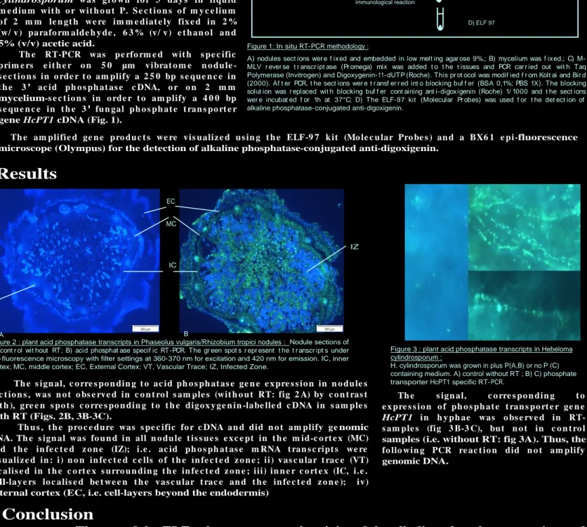
Figure 2 : plant acid phosphatase transcripts in Phaseolus vulgaris/Rhizobium tropici nodules : Nodule sections of A) cont r ol wit hout RT; B) acid phosphat ase specif ic RT-PCR. The gr een spot s r epr esent t he t r anscr ipt s under epi-fluorescence microscopy with filter settings at 360-370 nm for excitation and 420 nm for emission. IC, inner cortex; MC, middle cortex; EC, External Cortex; VT, Vascular Trace; IZ, Infected Zone.
IC VT MC EC IZ A B
Figure 3 : plant acid phosphatase transcripts in Hebeloma cylindrosporum :
H. cylindrosporum was grown in plus P(A,B) or no P (C) containing medium. A) control without RT ; B) C) phosphate transporter HcPT1 specific RT-PCR.
Th e s ign al, c o rre s po n din g t o ac id ph o s ph at as e ge n e e x pre s s io n in n o du le s s e c t io n s , was n o t o bs e rv e d i n c o n t ro l s am ple s (wit h o u t RT: fig 2 A) by c o n t ras t wit h ), gre e n s po t s c o rre s po n din g t o t h e digo x y ge n in -labe lle d c DNA in s am ple s with RT (Figs. 2B, 3B-3C).
Th u s , t h e pro c e du re was s pe c ific fo r c DNA an d did n o t am plify ge nomic DNA. Th e s ign al was fo u n d i n all n o du le t is s u e s e x c e pt in t h e m id-c o rt e x (MC) an d t h e in fe c t e d z o n e (IZ); i. e . ac id ph o s ph at as e m RNA t ran s c ript s we re v is u aliz e d in : i) n o n i n fe c t e d c e lls o f t h e in fe c t e d z o n e ; ii) v as c u lar t rac e (VT) lo c alis e d in t h e c o rt e x s u rro u n din g t h e in fe c t e d z o n e ; iii) in n e r c o rt e x (IC, i.e . cell-lay e rs lo c alis e d be t we e n t h e v as c u lar t rac e an d t h e in fe c t e d z o n e ); iv ) external cortex (EC, i.e. cell-layers beyond the endodermis)
Th e s ign al, c o rre s po n di n g t o e x pre s s io n o f ph o s ph at e t ran s po rt e r ge n e
HcPT1 i n h y ph ae was o bs e rv e d in
RT-s am ple RT-s (fig 3 B-3 C), bu t n o t in c o n t ro l samples (i.e. without RT: fig 3A). Thus, the fo llo wi n g PCR re ac t io n did n o t am plify genomic DNA.
A
B
C
Figure 1: In situ RT-PCR methodology :
A) nodules sect ions wer e f ixed and embedded in low melt ing agar ose 9%.; B) mycelium was f ixed.; C) M-MLV r ever se t r anscr ipt ase (Pr omega) mix was added t o t he t issues and PCR car r ied out wit h Taq Polymerase (Invitrogen) and Digoxygenin-11-dUTP (Roche). This pr ot ocol was modif ied f r om Kolt ai and Bir d (2000). Af t er PCR, t he sect ions wer e t r ansf er r ed int o blocking buf f er (BSA 0,1%; PBS 1X). The blocking solut ion was r eplaced wit h blocking buf f er cont aining ant i-digoxigenin (Roche) 1/ 1000 and t he sect ions wer e incubat ed f or 1h at 37°C; D) The ELF-97 kit (Molecular Pr obes) was used f or t he det ect ion of alkaline phosphatase-conjugated anti-digoxigenin.
Material & methods
A) Fixation, inclusion, sections 50 µM (vibratom) nodule
C) Reverse Transcription, PCR, immunological reaction
D) ELF 97 Agarose low melting
mycelium
B) Fixation of 2 mm length section
Ph a s e o l u s v u l g a r i s was in o c u lat e d wit h
Rh i z o b i u m t r o p i c i CIAT8 9 9 an d gro wn in h y dro ae ro po n ic c u lt u re s fo r 5 we e k s u n de r P deficiency. No du le s o f 3 m m diam e t e r we re fix e d an d s t o re d in 4 % (w/ v ) parafo rm alde h y de , 4 5 % (v / v ) e t h an o l an d 5 % (v / v ) ac e t ic ac id, be fo re e m be ddin g in lo w melting point agarose 9% (w/v).
Th e e c t o m y c o rrh iz al fu n gu s Hebeloma cylindrosporum was gro wn fo r 5 day s in liqu id
m e diu m wit h o r wit h o u t P. S e c t io n s o f m y c e liu m o f 2 m m le n gt h we re im m e diat e ly fix e d in 2 % (w/ v ) parafo rm alde h y de , 6 3 % (v / v ) e t h an o l an d 5% (v/v) acetic acid.
Th e RT-PCR was pe rfo rm e d wit h s pe c ific prim e rs e it h e r o n 5 0 µm v ibrat o m e n o du le -s e c t io n -s in o rde r t o am plify a 2 5 0 bp -s e qu e n c e in t h e 3 ac id ph o s ph at as e c DNA, o r o n 2 m m mycelium-s e c t io n s in o rde r t o am plify a 4 0 0 bp s e qu e n c e in t h e 3 fu n gal ph o s ph at e t ran s po rt e r gene HcPT1 cDNA (Fig. 1).
Th e am plifie d ge n e pro du c t s we re v is u aliz e d u s in g t h e ELF-9 7 k it (Mo le c u lar Pro be s ) an d a BX6 1 e pi-fluorescence microscope (Olympus) for the detection of alkaline phosphatase-conjugated anti-digoxigenin.