HAL Id: hal-01784461
https://hal.archives-ouvertes.fr/hal-01784461
Submitted on 3 May 2018
HAL is a multi-disciplinary open access
archive for the deposit and dissemination of
sci-entific research documents, whether they are
pub-lished or not. The documents may come from
teaching and research institutions in France or
abroad, or from public or private research centers.
L’archive ouverte pluridisciplinaire HAL, est
destinée au dépôt et à la diffusion de documents
scientifiques de niveau recherche, publiés ou non,
émanant des établissements d’enseignement et de
recherche français ou étrangers, des laboratoires
publics ou privés.
Ghislain Bidaut, Karsten Suhre, Jean-Michel Claverie, Michael Ochs
To cite this version:
Ghislain Bidaut, Karsten Suhre, Jean-Michel Claverie, Michael Ochs. Determination of strongly
overlapping signaling activity from microarray data. BMC Bioinformatics, BioMed Central, 2006, 7
(1), �10.1186/1471-2105-7-99�. �hal-01784461�
Open Access
Methodology article
Determination of strongly overlapping signaling activity from
microarray data
Ghislain Bidaut
1,2,3, Karsten Suhre
2, Jean-Michel Claverie
2and
Michael F Ochs*
1Address: 1Fox Chase Cancer Center, 333 Cottman Avenue, Philadelphia, PA, 19111, USA, 2Structural and Genomic Information Laboratory, UPR2589-CNRS, 13288 Marseille, France and 3Center for Bioinformatics, Department of Genetics, University of Pennsylvania School of Medicine, 1423 Blockley Hall, 423 Guardian Drive, Philadelphia, PA 19104-6021, USA
Email: Ghislain Bidaut - [email protected]; Karsten Suhre - [email protected]; Michel Claverie - [email protected]; Michael F Ochs* - [email protected]
* Corresponding author
Abstract
Background: As numerous diseases involve errors in signal transduction, modern therapeutics
often target proteins involved in cellular signaling. Interpretation of the activity of signaling pathways during disease development or therapeutic intervention would assist in drug development, design of therapy, and target identification. Microarrays provide a global measure of cellular response, however linking these responses to signaling pathways requires an analytic approach tuned to the underlying biology. An ongoing issue in pattern recognition in microarrays has been how to determine the number of patterns (or clusters) to use for data interpretation, and this is a critical issue as measures of statistical significance in gene ontology or pathways rely on proper separation of genes into groups.
Results: Here we introduce a method relying on gene annotation coupled to decompositional
analysis of global gene expression data that allows us to estimate specific activity on strongly coupled signaling pathways and, in some cases, activity of specific signaling proteins. We demonstrate the technique using the Rosetta yeast deletion mutant data set, decompositional analysis by Bayesian Decomposition, and annotation analysis using ClutrFree. We determined from measurements of gene persistence in patterns across multiple potential dimensionalities that 15 basis vectors provides the correct dimensionality for interpreting the data. Using gene ontology and data on gene regulation in the Saccharomyces Genome Database, we identified the transcriptional signatures of several cellular processes in yeast, including cell wall creation, ribosomal disruption, chemical blocking of protein synthesis, and, criticially, individual signatures of the strongly coupled mating and filamentation pathways.
Conclusion: This works demonstrates that microarray data can provide downstream indicators
of pathway activity either through use of gene ontology or transcription factor databases. This can be used to investigate the specificity and success of targeted therapeutics as well as to elucidate signaling activity in normal and disease processes.
Published: 28 February 2006
BMC Bioinformatics2006, 7:99 doi:10.1186/1471-2105-7-99
Received: 22 September 2005 Accepted: 28 February 2006 This article is available from: http://www.biomedcentral.com/1471-2105/7/99
© 2006Bidaut et al; licensee BioMed Central Ltd.
This is an Open Access article distributed under the terms of the Creative Commons Attribution License (http://creativecommons.org/licenses/by/2.0), which permits unrestricted use, distribution, and reproduction in any medium, provided the original work is properly cited.
Data analysis flowchart
Figure 1
Data analysis flowchart. The data was downloaded from Rosetta Inpharmatics and filtered to include only genes and
exper-iments that showed significant variation. Bayesian Decomposition analysis generated patterns and associated gene lists for all dimensionalities between 3 and 25. ClutrFree was used to interpret these results, including use of the MIPS database of ontol-ogies.
Background
Many diseases develop because of errors in signaling, and newer therapeutics specifically target proteins involved in cellular signaling [1,2]. However, these therapies are not always effective [3], and the reason for failure, whether inherent poor interaction or complex cellular response, is unknown. In order to understand the development of dis-ease and drug resistance in these cases, the recovery of the process that led to the specific cellular malfunction must be identified. Such errors generally involve the cellular sig-naling networks that control cell growth, differentiation, apoptosis, and motility [4,5]. Because of the extreme underlying biological complexity of these pathways, dis-eases that involve errors in signaling processes arise from a myriad of different cellular malfunctions, for example in cancers [6,7] and diabetes [8,9]. It is from this complex background that functional genomics attempts to glean insight to improve our understanding of diseases. One of the major uses of microarrays has been elucidation of gene expression in cancer, often focused on refining cancer identification using computational and statistical approaches [10-12]. In addition, the discovery of biomar-kers in the form of differential levels of production of mRNA has been a focus in a number of studies [13-15]. The fact that determination of the mRNA levels of a single gene is easier than using an entire array has driven the shift to the use of arrays to generate potential biomarkers, so that the expression levels of these individual genes can be screened for in a more economical way (see, for exam-ple, [16]). For diabetes, microarrays have been used to elucidate gene expression in both type I and type II dis-eases, and customized chips targeting genes of interest have been developed [17].
Many tools for statistical inference, pattern recognition, and data mining have been developed for microarray data analysis. Statistical tests include SAM [18], VERAandSAM [19], ANOVA techniques [20,21], Bayesian approaches [22,23], and rank tests [24]. Pattern recognition and data mining techniques comprise both unsupervised tech-niques, such as hierarchical clustering [25], singular value decomposition [26], multidimensional scaling [27], Bayesian mixture models [28,29], and other clustering methods [30-34], and supervised techniques, such as sup-port vector machines [35] and artificial neural networks [36], (for a review see [37]).
While these techniques are useful, they have certain limi-tations as regards more advanced uses in the elucidation of mechanisms operating in diseased tissues. New thera-peutics specifically target proteins involved in cellular sig-naling [1-3,38-40]. As noted above, these therapies are not always effective, and a method to understand the rea-son for their ineffectiveness is highly desirable. If the
fail-ure modes for the targeted therapeutics are understood, new therapeutics can be designed or combination thera-pies undertaken. In addition, to design new therathera-pies that work alone or in combination with other therapies, an understanding of signaling networks is required. Microar-ray measurements can provide insight into these issues. Unfortunately, the recovery of pathway information from transcriptional data requires complex analysis, since sign-aling protein activity is not generally linked to the mRNA expression levels of genes encoding the signaling proteins themselves [41], nor are protein levels tightly coupled to transcript levels even in yeast [42,43]. This makes it impossible to directly link an increase in mRNA expres-sion of the gene encoding a signaling protein, such as the therapeutic target, with activity of the protein and there-fore of the signaling pathway. Instead, an analysis must treat changes in mRNA levels as downstream indicators of activity.
An important issue to resolve in order to correctly inter-pret patterns in microarray data is the underlying dimen-sionality of the data, since statistical analysis of genes in groups relies on correct separation. The dimensionality provides an estimate of the number of patterns required to explain the variation in the data not related to noise, which is equivalent to the number of basis vectors required mathematically to describe the data or the number of principal components required to span the data.
We present here a new application of Bayesian Decompo-sition [44-48] and ClutrFree [49] that estimates dimen-sionality by measuring the consistency of assignment of genes to patterns. With this approach, transcriptional sig-natures are linked to signaling activities through gene ontology [50] using the MIPS database [51] and through analysis of transcription factor activity [52]. We demon-strate this technique on the Rosetta deletion mutant data-set [53], which is a compendium of genome-wide transcription measured for 6300 genes across 300 condi-tions (mostly deletion mutants, but some chemical treat-ments). Figure 1 details the workflow of our analysis. Previous studies of the compendium were performed using hierarchical clustering [53], non-negative matrix factorization [54], and Bayesian Decomposition [44]. The dimensionality of the data was estimated in various ways in these studies leading to estimates from 7 to 50 dimen-sions.
Results
Dimensionality of the data
We propose a value for the Rosetta dataset dimensionality based on the average persistence calculations at each tree level made with ClutrFree using multiple Bayesian
Decomposition simulations. The dimensionality has been inferred from the average persistence defined in the Meth-ods section. As the number of basis vectors (i.e., patterns) k is increased, the curve shows a dramatic drop for k > 15 (see Figure 2). This drop is due to the reorganization of the groups of mutants constituting the basis vectors for k > 15 patterns, leading to an overly low average persistence. The freedom to move between branches leads also to some loss of consistency in the annotations as one moves down a branch. This contrasts with the behavior of basis vectors obtained for less than 15 patterns where biological functions split logically as the number of patterns increases. We also observed a reduction in the number of genes related to each basis vector for 16 patterns in com-parison to 15. Also, the standard deviation across samples of the obtained vectors is significantly higher for 16 pat-terns (1.7 × 10⁻³) than for 15 patterns (7.9 × l0-4)
indicat-ing that the Markov chain samplindicat-ing is not as tightly constrained by the probability distribution. The behavior observed occurs because of the potential to overfit the data with 16 basis vectors allowing the algorithm to find multiple configurations to explain the variation in the data.
Identifying patterns and functions
Bayesian Decomposition retrieves the two linked matri-ces: the P matrix (pattern matrix) groups mutants that share cellular functions, which can be deduced from the genes linked to each pattern contained in the A matrix.
Each mutant (a column of the P matrix) can belong to multiple patterns, which models the fact that each mutant will have many cellular functions active. Each gene (a row of the A matrix) can be assigned to multiple patterns, reflecting the fact that evolution has led to genes being involved in multiple cellular processes. Interpretation of the results involves identifying cellular processes from the genes that are significantly expressed in a pattern (i.e., within a column of A).
We proceed by using the dimensionality estimate of 15 patterns and exploring for each pattern the genes associ-ated with that pattern. These genes are interpreted using the MIPS ontology for yeast [51] in order to predict the cellular processes associated with a pattern. In addition, for patterns that can be linked to signaling pathways, we discuss the use of data on genes regulated by specific tran-scription factors and validate the results by analysis of spe-cific key deletion mutants. For each pattern that shows enhancement of ontological terms we provide the terms, the enhancement (as defined in the methods section), and the p value for a hypergeometric test on the term. We summarize the results in terms of patterns previously identified in other studies using this data set, then we present the new results isolating signatures for activity of the mating and filametation pathways.
Patterns identified in previous studies
Examination of pattern 1 shows expression of the overall common minimal processes necessary for survival, with 386 annotated genes associated with this pattern at a 3σ level. Measure of enhancement, e, of cellular functions, reveals two highly represented functional groups: 1) groups related to protein synthesis and 2) groups related to DNA synthesis. Group 1 includes genes enhanced in Protein Targeting, Sorting and Translocation (Term 14.04, e = 1.84, p = 0.0022), Protein Synthesis (Term 12, e = 1.55, p = 0.024), and Ribosome Biogenesis (Term 12.01, e = 1.60, p = 0.059). Group 2 includes DNA Processing (Term 10.01, e = 1.32, p = 0.097), DNA Recombination and DNA Repair (Term 10.01.05, e = 1.31, p = 0.16), and DNA Synthesis (Term 10.01.03, e = 1.58, p = 0.16). The p-values for the ontology terms remain high, due to the large number of genes associated with this pattern.
This pattern, which essentially includes genes necessary for viability, contains all the mutants of the dataset, although the Ssn6∆ mutant shows a lower level for this pattern than other mutants. As the Ssn6∆ mutant exhibits substantially greater overall expression than any other mutant (including the Tup1∆ mutant with the second highest level), this may reflect the high association of the Ssn6∆ mutant seen in almost all patterns, which will have some gene overlap with this pattern.
The average persistence across all dimensions
Figure 2
The average persistence across all dimensions. The
average persistence across the dimensions is plotted for 3 to 25 dimensions. The significant drop between 15 and 16 dimensions suggests that 15 patterns provides the correct dimensionality for analysis.
0 5 10 15 20 25 0 2 4 6 8 10 12 14 16 18
16
15
Dimensionality (K) Average Persistence PPattern 5 contains 172 annotated genes. Highly enhanced ontologies include transport-related functions: Trans-ported Compounds (Term 20.01, e = 2.02, p < 10-4),
C-compound and Carbohydrate Transport (Term 20.01.03, e = 2.60, p < 3 × 10⁻⁴), and Cellular Transport, Transport
Facilitation and Cellular Routes (Term 20, e = 1.62, p < 6 × 10⁻⁴), in addition to other transport terms at p < 10-3.
The pattern contains the two deletion mutants, Ssn6∆ and Tup1∆, and represents the strong response seen in the original study [53]. Ssn6p and Tup1p form a system of transcriptional repression that appears to be highly con-served in eukaryotes [55]. In yeast, the complex acts as a global transcriptional repressor over a large number of genes (more that 150), coordinating several cellular sys-tems, including haploid specific genes, glucose repressible genes, and oxygen utilization genes [56]. Turning off this repression leads to a large overall increase in gene expres-sion (the overall expresexpres-sion in these two mutants is many fold higher than in other mutants).
Pattern 7 is related to the lack of cell wall functions (Cell Wall, Term 42.01, e = 0.0), as 28 cell wall genes (of 32 total) are absent from this pattern, while the other four genes have multiple annotations suggesting they have roles unrelated to Cell Wall function. Enhancement is present for Protein Modification (Term 14.07, e = 4.7, p < 10-4) and Fermentation (Term 02.16, e = 4.5, p < 0.01).
This pattern contains the mutants Gas1∆ and Fks1∆, which impair cell-wall synthesis, as well as the mutant YER083c∆, annotated as disrupting the cell wall in the original study [53]. The pattern contains other mutants disrupting ergosterol biosynthesis as well, including Erg2∆, She4∆, as well as YER044c∆. In addition, the pat-tern includes yeast treated with the drugs that are known to disrupt the cell wall, such as Tunicamycin and Glu-cosamine.
Pattern 11 is related to ribosomal function, with enhance-ments in terms for Ribosome Biogenesis (Term 12.01, e = 6.32, p < 4 × 10⁻⁴) and Protein Synthesis (Term 12, e =
3,89, p < 6 × 10⁻³). The pattern contains 8 mutants related
to ribosomal proteins, Rpll2a∆, Rpl27a∆, Rpl34a∆, Rpl6b∆, Rp18a∆, Rps24a∆, Rps24a∆ (haploid), and Rps27b∆, as well as some mutants with deleted ORFs of unknown function, YOR078w∆, YMR269w∆, and YHR034c∆, proposed to be involved in ribosomal func-tions [53].
Patterns related to cellular signalling pathways
The two patterns that represent new insights into this data are 13 and 15, which appear related to two strongly cou-pled developmental pathways in yeast. Previous studies [44,53] have identified the mating pathway transcrip-tional response, however this has included both the fila-mentation response and the mating response. It is difficult to separate these signatures, as the mating and filamenta-tion pathways share many common elements in a MAPK cascade as shown in Figure 3[57-59].
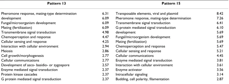
Gene ontology (GO) was used to determine the biological function described by each pattern, with a term added specifically for transposable elements, as these are known to play a role during filamentation [60,61]. The terms that showed enhancement are summarized in Table 1. The patterns show strong overlap, since many genes are shared between the mating and filamentation responses. How-ever, the filamentation ontology term is significantly higher only in pattern 15, which also shows a strong sig-nature of transposable element genes. Meanwhile, the GO terms for meiosis and morphogenesis (such as for bud-ding in S. cerevisiae) are significantly enhanced only in pattern 13. This allows association of pattern 13 with acti-vation of the mating pathway, and pattern 15 with activa-tion of the filamentaactiva-tion pathway.
Yeast MAPK signaling for mating and filamentation
Figure 3
Yeast MAPK signaling for mating and filamentation.
The strongly linked MAPK signaling pathways for mating and filamentation are shown schematically with black arrows indi-cating mating pathway signaling and gray arrows showing fila-mentation pathway signaling. The mating pathway is initiated by binding to Ste2p or Ste3p receptors, while the causative molecular trigger for filamentation is unclear. The pathways share many components.
Ste2/ Ste3 Ste20 α β γ G Protein Ste5 Ste11 Ste7 Fus3 Ste12 Tec1 Kss1 Cdc42 Bmh1/2 Ras2 mating signaling filamentation signaling
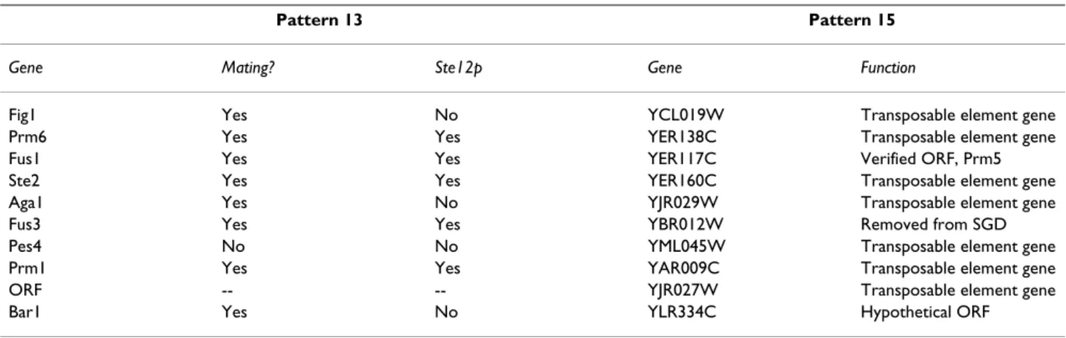
In addition, we analyzed the 10 genes whose expression is most strongly linked to each pattern. These are shown in Table 2, which summarizes which genes are known to be regulated by the transcriptional activators related to the mating (Ste12p) and filamentation (Ste12p-Tec1p com-plex) pathways. The results show that the top 10 genes related to pattern 13 have 9 genes of known function, with 8 related to the mating response, of which five are known to be regulated by Ste12p. For pattern 15, 7 of the top 10 genes are known to be transposable element genes, with three other genes having unknown functions. This again links pattern 13 to mating and pattern 15 to filamenta-tion.
In order to validate that the patterns were actually meas-uring activity of the mating and filamentation pathways, we explored deletion mutants related to these pathways [61,62]. The mating response in S. cerevisiae is mediated via a MAPK signaling cascade initiated by binding to the Ste2p or Ste3p membrane receptors (Figure 3). The signal is transduced through Ste11p, Ste7p, and Fus3p with Ste5p serving as a scaffolding protein. The signal activates the Ste12p transcription factor, leading to transcription of mating response genes. In addition, the signal is trans-duced to the MAPK cascade from the membrane receptor by a G protein complex or through the Ste20p protein. Pattern 13 shows near zero signal for the deletion mutants Ste11∆, Ste7∆ Fus3∆, Ste12∆, Ste5∆, and Ste2∆, while showing signal for deletion mutants of Ste20∆ and Tec1∆. This is exactly as expected, with the membrane receptor, all signaling proteins in the cascade, and the transcription factor necessary to generate the transcriptional response related to the mating signal (note that the Ste3∆ mutant is not in the data set). Ste20p is not necessary to raise the mating response, since the G-protein complex can trigger
activation of Ste11p directly. For pattern 15, the response is very similar. The signal is near zero for the deletion mutants Ste2∆, Ste11∆, Ste7∆, and Ste12∆. The Fus3∆ mutant shows a signal for pattern 15, as appropriate, while the Fus3∆, Kss1∆ double deletion mutant does not. In addition, the Tec1∆ mutant shows no signal for pattern 15, indicating that Tec1p is required for filamentation [61]. Finally, the signal is greatly reduced for the Ste20∆ deletion mutant in pattern 15 relative to pattern 13, which agrees with previous work suggesting that the filamenta-tion pathway is more dependent on Ste20p signaling than is the mating pathway [62].
Discussion
Microarrays and GeneChips™ have become the tools of choice for the investigation of genome-wide transcription in most biological systems. The resulting data comprises noisy estimates of transcription levels for roughly 6,000 genes in yeast to more than 20,000 genes in typical mam-malian studies. Numerous statistical and data mining methods have been applied to this data in order to iden-tify individual genes showing differential expression, to identify patterns related to physiological states, and to identify groups of genes comprising biological processes. These studies generally have not focused on the estima-tion of cellular signaling from the data, despite the preva-lence of cellular signaling in many diseases.
As noted in the introduction, the recovery of signaling pathway information from transcription data requires complex analysis, since protein levels do not correlate well with mRNA levels and signaling protein activity is not generally linked to the mRNA expression levels of genes encoding the signaling proteins themselves. As such, changes in mRNA levels are limited to being a
Table 1: The most enhanced gene ontology terms in patterns 13 and 15. Each term is presented together with a measure of how overrepresented it is compared to a random draw of the same number of genes. These were also confirmed to be significant by hypergeometric tests.
Pattern 13 Pattern 15
Pheromone response, mating-type determination 6.31 Transposable elements, viral and plasmid 8.42 development 6.09 Pheromone response, mating-type determination 7.26 Fungal/microorganism development 6.09 Transmembrane signal transduction 6.41 Mating (fertilization) 6.09 G-protein mediated signal transduction 6.10 Transmembrane signal transduction 4.98 development 5.69 Chemoperception and response 4.47 Fungal/microorganism development 5.69 Cellular sensing and response 4.25 Mating (fertilization) 5.69 Interaction with cellular environment 2.94 Chemoperception and response 5.47
Meiosis 2.86 Cellular sensing and response 5.21
Cell growth/morphogenesis 2.77 Cellular communications 4.45 Cellular communications 2.77 Enzyme mediated signal transduction 3.81 Development of asco- basidio- or zygospore 2.57 Interaction with cellular environment 3.61 Enzyme mediated signal transduction 2.37 Enzyme activator 3.56 Protein kinase cascades 2.37 Intracellular signaling 3.14 G protein mediated signal transduction 2.37 Budding, cell polarity, filamentation 2.87
downstream indicator of activity. If a complete model for the transcription of genes, including all known transcrip-tional regulators and biological processes regulating tran-scription, was available, the inference of activity would be straightforward. Unfortunately, the network models and even gene annotations are still far from being complete. In addition, the growing evidence supporting the important role of non-coding RNAs in regulation of gene expression (including antisense transcripts and micro-RNAs, see for example [63-65]) further undermines the potential of using mRNA species as markers for proteins and their activities [66].
In order to overcome this incompleteness, we have cre-ated the method described here. We couple identification of transcriptional signatures with our Bayesian Decompo-sition algorithm to a consistency analysis for gene assign-ment to patterns determined by comparison of different dimensionalities using ClutrFree. This allows the identifi-cation of the correct dimensionality to be applied to sub-sequent ontology and transcription factor analyses. ClutrFree is also used to determine the ontological terms enhanced within each pattern and to obtain a list of genes tied to this pattern, which can then be linked to specific transcription factors. In this way, the biological processes associated with conditions can be identified, and infer-ences can be made on the activity of specific transcription factors. This then allows inference on the activity of sign-aling pathways, which cannot be obtained with methods previously applied to microarray data. Overall, the method requires many separate steps, each modeling an aspect of the biological system, in order to make proper inferences on signaling from the data.
In the application to the Compendium data presented here, our analysis was able to extract the common features for a set of mutants that eliminated related pathways. As
in previous studies, the global transcriptional repressor complex Ssn6-Tupl has been isolated in a single group. In addition, patterns for cell-wall synthesis, ribosomal func-tion, and the global functions necessary for continued via-bility of yeast were isolated. In contrast to previous analyses of this data, two pathways related to the MAPK cascade were isolated, one related to mating and the other to filamentation. Once the correct dimensionality was determined, Bayesian Decomposition was able to identify transcriptional signatures unique for each pathway. The assignment was validated by an investigation of the dele-tion mutants known to adversely affect these pathways.
Conclusion
Microarray studies have been widespread in biological and medical research, often focusing on identification of genes significantly correlated with various disease states. However, many diseases arise from disruptions in cellular signaling, and in these cases gene expression only pro-vides a downstream indicator of signaling activity. This greatly complicates the analysis. The new approach intro-duced here recovered signatures allowing us to make vali-dated inferences on strongly overlapping signaling pathways.
The results demonstrate that for Saccharomyces cerevisiae, the mating and filamentation pathways can be distin-guished from transcriptional signatures determined from analysis of microarray data, despite the intrinsic high noise, confounding transcriptional activity, and tightly coupled nature of the pathways. The next step will be to apply these methods to more complex signaling networks in worms, flies, and mammals.
Table 2: The genes most strongly associated with patterns 13 and 15 in order of strength of association. For pattern 13, it is noted whether the genes are known to be regulated in the mating process, and whether the gene is known to be directly regulated by Stel2p. For pattern 15, the gene function is shown. All data is from the Saccharomyces Genome Database [73, 74].
Pattern 13 Pattern 15
Gene Mating? Ste12p Gene Function
Fig1 Yes No YCL019W Transposable element gene
Prm6 Yes Yes YER138C Transposable element gene
Fus1 Yes Yes YER117C Verified ORF, Prm5
Ste2 Yes Yes YER160C Transposable element gene
Aga1 Yes No YJR029W Transposable element gene
Fus3 Yes Yes YBR012W Removed from SGD
Pes4 No No YML045W Transposable element gene
Prm1 Yes Yes YAR009C Transposable element gene
ORF -- -- YJR027W Transposable element gene
Methods
The Rosetta deletion mutant data set
The Rosetta Compendium comprises 300 conditions, including 276 deletion mutants, 11 tetracycline regulated genes, and 13 drug treatments, in S. cerevisiae growing in rich medium [53]. The data were generated from a two color cDNA microarray hybridization assay [67], and transcriptional profiles were measured both with techni-cal replication and biologitechni-cal replication (151 mutants). In parallel with the 300 experiments, 63 controls of wild-type S. cerevisiae were grown in identical conditions and compared against each other, permitting creation of a gene-specific error model. The data was downloaded from Rosetta Inpharmatics.
Data preprocessing
The data was filtered to retain only conditions character-ized by at least a variation of 3 fold in a minimum of 2 genes. Then all genes that did not vary by 3-fold in at least 2 conditions were also removed, leading to a data matrix comprising 764 genes and 228 conditions.
The data used by Bayesian Decomposition included both the mean log ratio for each data point and the uncertainty in this measurement determined by the Rosetta error model. Since Bayesian Decomposition as applied here requires positivity [45], the log ratios were converted to ratios and the uncertainties propagated to uncertainties on the ratios. Although analysis of residuals suggested that seven dimensions fit the data [44], the analysis pre-sented here suggests that this is due to overestimation of uncertainty in the data. Bayesian Decomposition is not highly sensitive to minor misestimations of noise how-ever, so that this should not be a problem for this analysis. Analysis with Bayesian Decomposition
Bayesian Decomposition (BD) has been applied to multi-ple types of data: in vivo spectroscopic data [68], medical imaging [69], microarray data from single cell organisms [44,45], mammalian model organisms [47], humans [70], and on phylogenomic sequence data [71]. A detailed description of the algorithm [46] and a review of applica-tions [48] have been published.
Briefly, BD models the microarray data, comprising a matrix of estimates of the ratio of expression between the experimental condition and a control, as the result of the multiplication of two matrices describing behaviors across conditions (the P or pattern matrix) and the distri-bution of genes within those behaviors (the A or ampli-tude matrix). Naturally, the data, D, includes noise, so that the full relationship is defined by
where Dij is the estimated ratio for gene i in mutant j, Aik is the strength of gene i in pattern k, Pkj is the strength of mutant j in pattern k, and εij is the noise for gene i in
mutant j estimated by the Rosetta error model. BD esti-mates A and P by a Markov chain Monte Carlo (MCMC) simulation. For the fixed noise estimate, ε, A and P are inferred from the marginal probability distribution p (D | A,P) p (A,P) [2]
where p(A,P) is the prior probability and p (D | A,P) is given by the likelihood such that
The prior is used here to require positivity and to mini-mize structure in the estimates of A and P [46].
The analysis with BD is similar mathematically to an anal-ysis with singular value decomposition (SVD) or with principal component analysis (PCA), since all methods estimate two matrices that together reconstruct the data. In both PCA and SVD, orthogonality conditions force each row of P to be linearly independent, deriving P from either the data matrix (SVD) or the covariance matrix (PCA) using deterministic algorithms. Since patterns of expression related to biological processes will not gener-ally be independent, BD uses the MCMC approach to avoid orthogonality. The resulting rows of P are usually easier to relate to biological processes than those from SVD or PCA.
BD was run at each posited dimensionality, K (as in equa-tion 1), between 3 and 25, generating a mean estimate and an uncertainty (i.e., standard deviation from samples of the posterior probability distribution) for each element of A and P. The dimensionality is equivalent to the number of patterns, as the patterns can be viewed as non-orthogonal basis vectors for D.
Persistence and dimensionality
The results of Bayesian Decomposition across the differ-ent estimated dimensionalities, K, were compared with ClutrFree in order to visualize stable basis vectors and a persistence measurement on them [49]. Each independ-ent BD analysis provides a tree level, with each pattern represented by a node (see Figure 4). The analysis with the fewest patterns is placed at the top of the tree, and addi-tional levels of analyses are added creating a tree from
Dij A Pik kj ij k K ≅ + =
∑
ε 1 1 [ ] log ( | , )p D A P . ij ij ik kj k K j i D A P = − − =∑
∑
∑
1 2 2 1 2 ε [[ ]3fewer to larger numbers of patterns. Connections in the tree are created in a greedy way. Level N+1 (e.g., 4) is con-nected to level N (e.g., 3) by finding the node in level N+1 with the highest correlation to a node in level N. The cor-relation is given by the Pearson corcor-relation for the strength of the assignment of mutants to a pattern (i.e., between Pkj for the nodes). These nodes are connected and removed. From the remaining nodes, the maximum cor-relation between a node at level N+1 and one at N is found again, and this process is repeated until only a sin-gle node remains at level N+1. This node is then con-nected to the node at level N that yields the highest correlation.
Following our previous work [49], we use a measure of persistence to quantify the robustness of a pattern across the variation of the number of patterns. An example cal-culation is shown in Figure 5. The assignment of a mutant to a pattern (i.e., a node) is binarized based on the mean
and uncertainty of the assignment of the mutant to the pattern determined by the MCMC sampling, using a requirement that the mutant be assigned to the pattern at greater than 3σ above zero. In Figure 5, it is assumed there are four mutants in a pattern, thus there are four binarized values. Then at each node, each mutant is compared for consistency in presence of the mutant in linked nodes within the tree. For example, for the highlighted node in Figure 5, the first mutant is present in the node above and the node below, so it is present in all 3 connected nodes. For the second mutant it is present in 2, and the third and fourth mutants are absent. The average persistence for the node is therefore (3+2+0+0)/4 = 1.25 as noted in Figure 5. For branches, the mutant status is only required to agree in a single branch to be counted. The average persistence at a dimension is then the average of the persistence for all nodes at that dimension (i.e., a row in Figure 4).
Relationship of patterns across dimensionalities
Figure 4
Relationship of patterns across dimensionalities. The results for all patterns identified in all runs of Bayesian
Decompo-sition are summarized here. The top row shows three patterns from an analysis with 3 dimensions, while the bottom row shows 25 dimensions. The highlighted node is pattern 13 in 15 dimensions, which is the pattern identified as the mating response. Nodes are connected as described in the text using Pearson correlation measures. The numbers within the nodes are indices and have no intrinsic meaning. Each number provides the row index for P and column index for A for the analysis at that level. Results of Algorithms 1 2 3 1 2 4 3 1 2 4 5 3 1 2 3 4 5 6 1 2 3 4 5 7 6 1 5 6 4 8 7 2 3 1 2 3 4 5 6 7 8 9 1 2 3 4 5 6 7 9 10 8 1 2 3 4 10 7 8 6 11 9 5 1 2 3 9 6 8 10 7 5 4 11 12 1 2 3 4 9 5 6 7 8 10 11 12 13 1 2 11 10 8 13 5 7 6 9 4 3 14 12 1 2 3 4 5 6 7 8 9 15 14 12 10 11 13 1 2 3 5 4 6 13 10 14 7 9 15 11 12 8 16 1 6 4 2 3 5 7 8 9 11 17 16 10 12 13 14 15 1 2 3 4 7 5 10 11 18 6 17 16 9 13 12 8 15 14 1 2 3 15 6 7 8 19 16 13 17 12 10 4 5 11 14 9 18 1 2 4 3 5 9 16 13 10 18 20 8 6 12 19 15 14 11 7 17 1 2 3 4 11 12 8 5 10 6 13 9 7 14 20 17 15 19 18 16 21 1 2 4 3 5 6 7 9 8 10 12 21 16 19 11 13 20 14 22 17 15 18 1 2 6 4 7 3 5 8 9 18 13 11 15 12 17 19 16 22 23 10 20 14 21 1 2 5 12 4 10 3 6 11 13 8 9 15 7 14 16 19 18 20 21 22 24 17 23 1 2 3 4 6 5 7 8 21 16 17 20 24 13 23 10 14 22 9 18 15 25 19 12 11
We assessed the dimensionality of the data using meas-urements of persistence. The persistence was measured for analyses from 3 – 25 patterns and the dimension chosen where a significant drop occurred in an otherwise slow monotonic decline, which was expected due to the branching nature of the tree. Figure 2 shows the signifi-cant drop between 15 and 16 dimensions, so 15 patterns were chosen for further analysis.
Ontology and function
To assign ontological terms to the genes contained in basis vectors, we annotated our data using the gene ontol-ogies from the Comprehensive Yeast Genome Database (CYGD) hosted at the Munich Information center for Pro-tein Sequences (MIPS) [51,72]. The analysis here utilizes the Functional Catalog (FunCat) format that describes each gene using a hierarchical ontological model. Similar to persistence, we defined enhancement as a measure of the over-representation, or under representa-tion, of a gene function in a subset of the data [47]. It is the ratio of the frequency of occurrence of genes anno-tated by a particular ontological term in the pattern to the frequency of occurrence of the same term in the whole dataset,
with gp being the number of genes annotated with the term t in pattern p, np the total number of genes in the
pat-tern p, G the number of genes annotated by the term t in the data, and N being the total number of genes in the dataset. In addition, we apply a hypergeometric test to estimate a p-value for each term. Function was then deter-mined by inspection of enhanced ontological terms. Transcription factor analysis
The genes were also analyzed for the patterns determined to be related to signaling pathways by exploration of the ten genes most strongly associated with the pattern. Each gene was analyzed using the Saccharomyces Genome Database [73] to determine whether it was known to be associated with mating or filamentation processes and to determine if it was directly regulated by the Ste12p tran-scription factor.
Authors' contributions
GB performed analysis with BD and ClutrFree, including identification of biological processes from gene ontology measurements. KS and JMC provided advice and guidance on development of ClutrFree for these analyses. MFO oversaw the project and did gene ontology and transcrip-tion factor analysis on patterns 13 and 15.
Acknowledgements
We thank the National Institutes of Health, National Library of Medicine (LM008309 to mfo) and National Cancer Institute (CCCG CA06927 sup-porting mfo), and the Pennsylvania Department of Health (grant to mfo). JMC and KS acknowledge the support from the Réseau National des Génopoles (RNG). e t p g n G N p p ( , ) / / , [ ] = 4
A sample calculation of the average persistence for a single node
Figure 5
A sample calculation of the average persistence for a single node. The average persistence is calculated by comparing
the persistence at each node in the tree given in Figure 4. Each assignment of each mutant (4 are shown here) to a pattern is binarized as described in the text, then the average persistence for a node is calculated by checking on the number of times the mutant assigned to the pattern occurs in the connected nodes. The mutant can occur in any branch below the node of interest to be considered as present. If it occurs in multiple child nodes at a single level, that is still treated as a single occurrence for that level. The average for a dimension is then the average of the persistence of all nodes at that level.
1,1,0,0
1,0,1,1
1,1,0,0
1,0,1,0
1,1,0,0
1,0,0,1
1,0,1,0
1,1,0,1
0,1,0,0
P = 1.5
P = 0.75
P = 1.25
References
1. Mauro MJ, Druker BJ: STI571: targeting BCR-ABL as therapy for CML. Oncologist 2001, 6:233-8.
2. Repka T, Chiorean EG, Gay J, Herwig KE, Kohl VK, Yee D, Miller JS: Trastuzumab and interleukin-2 in HER2-positive metastatic breast cancer: a pilot study. Clin Cancer Res 2003, 9:2440-6. 3. von Mehren M: Recent advances in the management of
gas-trointestinal stromal tumors. Curr Oncol Rep 2003, 5:288-94. 4. Jacks T, Weinberg RA: Taking the study of cancer cell survival
to a new dimension. Cell 2002, 111:923-5.
5. Kolch W: Meaningful relationships: the regulation of the Ras/ Raf/MEK/ERK pathway by protein interactions. Biochem J 2000, 351(Pt 2):289-305.
6. Cooper GM: Elements of Human Cancer. Boston: Jones and Bar-tlett Publishers; 1992.
7. Macdonald F, Ford CHJ: Molecular Biology of Cancer. Oxford: BIOS Scientific Publishers, Ltd; 1997.
8. Zdychova J, Komers R: Emerging role of Akt kinase/protein kinase B signaling in pathophysiology of diabetes and its complications. Physiol Res 2005, 54:1-16.
9. Leng Y, Karlsson HK, Zierath JR: Insulin signaling defects in type 2 diabetes. Rev Endocr Metab Disord 2004, 5:111-7.
10. Alizadeh AA, Eisen MB, Davis RE, Ma C, Lossos IS, Rosenwald A, Boldrick JC, Sabet H, Tran T, Yu X, et al.: Distinct types of diffuse large B-cell lymphoma identified by gene expression profil-ing. Nature 2000, 403:503-11.
11. Golub TR, Slonim DK, Tamayo P, Huard C, Gaasenbeek M, Mesirov JP, Coller H, Loh ML, Downing JR, Caligiuri MA, et al.: Molecular classification of cancer: class discovery and class prediction by gene expression monitoring. Science 1999, 286:531-7. 12. Zhang H, Yu CY, Singer B, Xiong M: Recursive partitioning for
tumor classification with gene expression microarray data. Proc Natl Acad Sci USA 2001, 98:6730-5.
13. Williams NS, Gaynor RB, Scoggin S, Verma U, Gokaslan T, Simmang C, Fleming J, Tavana D, Frenkel E, Becerra C: Identification and validation of genes involved in the pathogenesis of colorectal cancer using cDNA microarrays and RNA interference. Clin Cancer Res 2003, 9:931-46.
14. Kikuchi T, Daigo Y, Katagiri T, Tsunoda T, Okada K, Kakiuchi S, Zem-butsu H, Furukawa Y, Kawamura M, Kobayashi K, et al.: Expression profiles of non- small cell lung cancers on cDNA microar-rays: identification of genes for prediction of lymph-node metastasis and sensitivity to anti-cancer drugs. Oncogene 2003, 22:2192-205.
15. Carr KM, Bittner M, Trent JM: Gene-expression profiling in human cutaneous melanoma. Oncogene 2003, 22:3076-80. 16. Frolov A, Chahwan S, Ochs M, Arnoletti JP, Pan ZZ, Favorova O,
Fletcher J, von Mehren M, Eisenberg B, Godwin AK: Response markers and the molecular mechanisms of action of Gleevec in gastrointestinal stromal tumors. Mol Cancer Ther 2003, 2:699-709.
17. Scearce LM, Brestelli JE, McWeeney SK, Lee CS, Mazzarelli J, Pinney DF, Pizarro A, Stoeckert CJ Jr, Clifton SW, Permutt MA, et al.: Func-tional genomics of the endocrine pancreas: the pancreas clone set and PancChip, new resources for diabetes research. Diabetes 2002, 51:1997-2004.
18. Tusher VG, Tibshirani R, Chu G: Significance analysis of micro-arrays applied to the ionizing radiation response. Proc Natl Acad Sci U S A 2001, 98:5116-21.
19. Ideker T, Thorsson V, Siegel AF, Hood LE: Testing for differen-tially-expressed genes by maximum-likelihood analysis of microarray data. J Comput Biol 2000, 7:805-17.
20. Kerr MK, Martin M, Churchill GA: Analysis of variance for gene expression microarray data. J Comput Biol 2000, 7:819-37. 21. Kerr MK, Afshari CA, Bennett L, Bushel P, Martinez J, Walker NJ,
Churchill GA: Statistical analysis of a gene expression micro-array experiment with replication. Statistica Sinica 2002, 12:203-218.
22. Newton MA, Kendziorski CM, Richmond CS, Blattner FR, Tsui KW: On differential variability of expression ratios: improving sta-tistical inference about gene expression changes from microarray data. J Comput Biol 2001, 8:37-52.
23. Parmigiani G, Garrett E, Anbazhagan R, Gabrielson E: A statistical framework for expression-based molecular classification in cancer. Journal of the Royal Statistical Society, B 2002, 64:717-736.
24. Troyanskaya OG, Garber ME, Brown PO, Botstein D, Altman RB: Nonparametric methods for identifying differentially expressed genes in microarray data. Bioinformatics 2002, 18:1454-61.
25. Eisen MB, Spellman PT, Brown PO, Botstein D: Cluster analysis and display of genome-wide expression patterns. Proc Natl Acad Sci U S A 1998, 95:14863-8.
26. Alter O, Brown PO, Botstein D: Singular value decomposition for genome-wide expression data processing and modeling. Proc Natl Acad Sci U S A 2000, 97:10101-6.
27. Khan J, Simon R, Bittner M, Chen Y, Leighton SB, Pohida T, Smith PD, Jiang Y, Gooden GC, Trent JM, et al.: Gene expression profiling of alveolar rhabdomyosarcoma with cDNA microarrays. Cancer Res 1998, 58:5009-13.
28. Medvedovic M, Sivaganesan S: Bayesian infinite mixture model based clustering of gene expression profiles. Bioinformatics 2002, 18:1194-206.
29. Medvedovic M, Yeung KY, Bumgarner RE: Bayesian mixture model based clustering of replicated microarray data. Bioin-formatics 2004.
30. Gasch AP, Eisen MB: Exploring the conditional coregulation of yeast gene expression through fuzzy k-means clustering. Genome Biol 2002, 3:RESEARCH0059.
31. Getz G, Levine E, Domany E: Coupled two-way clustering analy-sis of gene microarray data. Proc Natl Acad Sci U S A 2000, 97:12079-84.
32. Ben-Dor A, Shamir R, Yakhini Z: Clustering gene expression pat-terns. J Comput Biol 1999, 6:281-97.
33. Heyer LJ, Kruglyak S, Yooseph S: Exploring expression data: identification and analysis of coexpressed genes. Genome Res 1999, 9:1106-15.
34. Lukashin AV, Fuchs R: Analysis of temporal gene expression profiles: clustering by simulated annealing and determining the optimal number of clusters. Bioinformatics 2001, 17:405-14. 35. Brown MP, Grundy WN, Lin D, Cristianini N, Sugnet CW, Furey TS, Ares M Jr, Haussler D: Knowledge-based analysis of microarray gene expression data by using support vector machines. Proc Natl Acad Sci U S A 2000, 97:262-7.
36. Khan J, Wei JS, Ringner M, Saal LH, Ladanyi M, Westermann F, Berthold F, Schwab M, Antonescu CR, Peterson C, et al.: Classifica-tion and diagnostic predicClassifica-tion of cancers using gene expres-sion profiling and artificial neural networks. Nat Med 2001, 7:673-9.
37. Ochs MF, Godwin AK: Microarrays in cancer: research and applications. Biotechniques 2003, 34:S4-S15.
38. Kato-Stankiewicz J, Hakimi I, Zhi G, Zhang J, Serebriiskii I, Guo L, Eda-matsu H, Koide H, Menon S, Eckl R, et al.: Inhibitors of Ras/Raf-1 interaction identified by two-hybrid screening revert Ras-dependent transformation phenotypes in human cancer cells. Proc Natl Acad Sci U S A 2002, 99:14398-403.
39. Strumberg D, Seeber S: Raf kinase inhibitors in oncology.
Onkol-ogie 2005, 28:101-7.
40. Heim M, Scharifi M, Zisowsky J, Jaehde U, Voliotis D, Seeber S, Strum-berg D: The Raf kinase inhibitor BAY 43-9006 reduces cellular uptake of platinum compounds and cytotoxicity in human colorectal carcinoma cell lines. Anticancer Drugs 2005, 16:129-36.
41. Chen G, Gharib TG, Huang CC, Taylor JM, Misek DE, Kardia SL, Giordano TJ, lannettoni MD, Orringer MB, Hanash SM, et al.: Dis-cordant protein and mRNA expression in lung adenocarci-nomas. Mol Cell Proteomics 2002, 1:304-13.
42. Gygi SP, Rochon Y, Franza BR, Aebersold R: Correlation between protein and mRNA abundance in yeast. Mol Cell Biol 1999, 19:1720-30.
43. Griffin TJ, Gygi SP, Ideker T, Rist B, Eng J, Hood L, Aebersold R: Complementary profiling of gene expression at the tran-scriptome and proteome levels in Saccharomyces cerevi-siae. Mol Cell Proteomics 2002, 1:323-33.
44. Bidaut G, Moloshok TD, Grant JD, Manion FJ, Ochs MF: Bayesian Decomposition analysis of gene expression in yeast deletion mutants. In Methods of Microarray Data Analysis II Edited by: Johnson K, Lin S. Boston: Kluwer Academic; 2002:105-122.
45. Moloshok TD, Klevecz RR, Grant JD, Manion FJ, Speier WFt, Ochs MF: Application of Bayesian Decomposition for analysing microarray data. Bioinformatics 2002, 18:566-75.
Publish with BioMed Central and every scientist can read your work free of charge
"BioMed Central will be the most significant development for disseminating the results of biomedical researc h in our lifetime."
Sir Paul Nurse, Cancer Research UK
Your research papers will be:
available free of charge to the entire biomedical community peer reviewed and published immediately upon acceptance cited in PubMed and archived on PubMed Central yours — you keep the copyright
Submit your manuscript here:
http://www.biomedcentral.com/info/publishing_adv.asp
BioMedcentral
46. Ochs MF: Bayesian Decomposition. In The Analysis of Gene
Expres-sion Data: Methods and Software Edited by: Parmigiani G, Garrett E,
Iri-zarry R, Zeger S. New York: Springer Verlag; 2003.
47. Moloshok TD, Datta D, Kossenkov AV, Ochs MF: Bayesian Decomposition classification of the Project Normal data set. In Methods of Microarray Data Analysis III Edited by: Johnson KF, LIn SM. Boston: Kluwer Academic; 2003:211-232.
48. Ochs MF, Moloshok TD, Bidaut G, Toby G: Bayesian Decomposi-tion: Analyzing microarray data within a biological context. Annals of the New York Academy of Sciences 2004, 1020:212-226.
49. Bidaut G, Ochs MF: ClutrFree: cluster tree visualization and interpretation. Bioinformatics 2004, 20:2869-71.
50. Ashburner M, Ball CA, Blake JA, Botstein D, Butler H, Cherry JM, Davis AP, Dolinski K, Dwight SS, Eppig JT, et al.: Gene ontology: tool for the unification of biology. The Gene Ontology Con-sortium. Nat Genet 2000, 25:25-9.
51. Mewes HW, Amid C, Arnold R, Frishman D, Guldener U, Mannhaupt G, Munsterkotter M, Pagel P, Strack N, Stumpflen V, et al.: MIPS: analysis and annotation of proteins from whole genomes. Nucleic Acids Res 2004, 32:D41-4.
52. Matys V, Fricke E, Geffers R, Gossling E, Haubrock M, Hehl R, Hor-nischer K, Karas D, Kel AE, Kel-Margoulis OV, et al.: TRANSFAC: transcriptional regulation, from patterns to profiles. Nucleic Acids Res 2003, 31:374-8.
53. Hughes TR, Marton MJ, Jones AR, Roberts CJ, Stoughton R, Armour CD, Bennett HA, Coffey E, Dai H, He YD, et al.: Functional discov-ery via a compendium of expression profiles. Cell 2000, 102:109-26.
54. Kim PM, Tidor B: Subsystem identification through dimension-ality reduction of large-scale gene expression data. Genome Res 2003, 13:1706-18.
55. Smith RL, Johnson AD: Turning genes off by Ssn6-Tupl: a con-served system of transcriptional repression in eukaryotes. Trends Biochem Sci 2000, 25:325-30.
56. Keleher CA, Redd MJ, Schultz J, Carlson M, Johnson AD: Ssn6-Tupl is a general repressor of transcription in yeast. Cell 1992, 68:709-19.
57. Kusari AB, Molina DM, Sabbagh W Jr, Lau CS, Bardwell L: A con-served protein interaction network involving the yeast MAP kinases Fus3 and Kss1. J Cell Biol 2004, 164:267-77.
58. Madhani HD, Fink GR: The riddle of MAP kinase signaling spe-cificity. Trends Genet 1998, 14:151-5.
59. Schwartz MA, Madhani HD: Principles of MAP kinase signaling specificity in Saccharomyces cerevisiae. Annu Rev Genet 2004, 38:725-48.
60. Mosch HU, Fink GR: Dissection of filamentous growth by trans-poson mutagenesis in Saccharomyces cerevisiae. Genetics 1997, 145:671-84.
61. Morillon A, Springer M, Lesage P: Activation of the Kss1 invasive-filamentous growth pathway induces Ty1 transcription and retrotransposition in Saccharomyces cerevisiae. Mol Cell Biol 2000, 20:5766-76.
62. Posas F, Takekawa M, Saito H: Signal transduction by MAP kinase cascades in budding yeast. Curr Opin Microbiol 1998, 1:175-82.
63. Katayama S, Tomaru Y, Kasukawa T, Waki K, Nakanishi M, Nakamura M, Nishida H, Yap CC, Suzuki M, Kawai J, et al.: Antisense tran-scription in the mammalian transcriptome. Science 2005, 309:1564-6.
64. Claverie JM: Fewer genes, more noncoding RNA. Science 2005, 309:1529-30.
65. Carninci P, Kasukawa T, Katayama S, Gough J, Frith MC, Maeda N, Oyama R, Ravasi T, Lenhard B, Wells C, et al.: The transcriptional landscape of the mammalian genome. Science 2005, 309:1559-63.
66. McManus MT, Sharp PA: Gene silencing in mammals by small interfering RNAs. Nat Rev Genet 2002, 3:737-47.
67. Schena M, Shalon D, Davis RW, Brown PO: Quantitative monitor-ing of gene expression patterns with a complementary DNA microarray. Science 1995, 270:467-70.
68. Ochs MF, Stoyanova RS, Arias-Mendoza F, Brown TR: A new method for spectral decomposition using a bilinear Bayesian approach. J Magn Reson 1999, 137:161-76.
69. Ochs MF, Stoyanova RS, Brown TR, Rooney WD, Springer CS Jr: A Bayesian Markov chain Monte Carlo solution of the bilinear problem. In Bayesian Inference and Maximum Entropy Methods in
Sci-ence and Engineering: 19th International Workshop Edited by: Rychert
JT, Erickson GJ, Smith CR. Melville: American Institute of Physics; 2001:274-284.
70. Kossenkov A, Bidaut G, Ochs MF: Genes associated with progno-sis in adenocarcinoma across studies at multiple institutions. In Methods of Microarray Data Analysis IV Edited by: Johnson K, Lin S. Boston: Kluwer Academic; 2005:239.
71. Bidaut G, Suhre K, Claverie JM, Ochs MF: Bayesian decomposition analysis of bacterial phylogenomic profiles. Am J Pharmacoge-nomics 2005, 5:63-70.
72. Mewes HW, Heumann K, Kaps A, Mayer K, Pfeiffer F, Stocker S, Frishman D: MIPS: a database for genomes and protein sequences. Nucleic Acids Res 1999, 27:44-8.
73. Christie KR, Weng S, Balakrishnan R, Costanzo MC, Dolinski K, Dwight SS, Engel SR, Feierbach B, Fisk DG, Hirschman JE, et al.: Sac-charomyces Genome Database (SGD) provides tools to identify and analyze sequences from Saccharomyces cerevi-siae and related sequences from other organisms. Nucleic Acids Res 2004, 32:D311-4.
74. Cherry JM, Ball C, Weng S, Juvik G, Schmidt R, Adler C, Dunn B, Dwight S, Riles L, Mortimer RK, et al.: Genetic and physical maps of Saccharomyces cerevisiae. Nature 1997, 387:67-73.