Detection of SARS-CoV-2 with SHERLOCK One-Pot Testing
The MIT Faculty has made this article openly available.
Please share
how this access benefits you. Your story matters.
Citation
Joung, Julia et al. "Detection of SARS-CoV-2 with SHERLOCK
One-Pot Testing." New England Journal of Medicine (September 2020):
dx.doi.org/10.1056/NEJMc2026172 © 2020 Massachusetts Medical
Society
As Published
http://dx.doi.org/10.1056/NEJMc2026172
Publisher
Massachusetts Medical Society
Version
Final published version
Citable link
https://hdl.handle.net/1721.1/127653
Terms of Use
Article is made available in accordance with the publisher's
policy and may be subject to US copyright law. Please refer to the
publisher's site for terms of use.
C o r r e s p o n d e nc e
Detection of SARS-CoV-2 with SHERLOCK One-Pot Testing
To the Editor: CRISPR (clustered regularlyin-terspaced short palindromic repeats)–based
di-agnostic tests1,2 collectively provide a nascent
platform for the detection of viral and bacterial pathogens. Methods such as SHERLOCK (specific high-sensitivity enzymatic reporter unlocking), which typically use a two-step process (target amplification followed by CRISPR-mediated nu-cleic acid detection),1,2 have been used to detect
SARS-CoV-2.3 These approaches, however, are more
complex than those used in point-of-care testing because they depend on an RNA extraction step and multiple liquid-handling steps that increase the risk of cross-contamination of samples.
Here, we describe a simple test for detection of SARS-CoV-2. The sensitivity of this test is similar to that of reverse-transcription–quantita-tive polymerase-chain-reaction (RT-qPCR) assays. STOP (SHERLOCK testing in one pot) is a stream-lined assay that combines simplified extraction of viral RNA with isothermal amplification and CRISPR-mediated detection. This test can be per-formed at a single temperature in less than an hour and with minimal equipment.
The integration of isothermal amplification with CRISPR-mediated detection required the development of a common reaction buffer that could accommodate both steps. To amplify viral RNA, we chose reverse transcription followed by
loop-mediated isothermal amplification (LAMP)4
because LAMP reagents are widely available and use defined buffers that are amenable to Cas en-zymes. LAMP operates at 55 to 70°C and requires a thermostable Cas enzyme such as Cas12b from
Alicyclobacillus acidiphilus (AapCas12b).5 We
sys-tematically evaluated multiple LAMP primer sets and AapCas12b guide RNAs (a guide RNA helps AapCas12b recognize and cut target DNA) to identify the best combination to target gene N, encoding the SARS-CoV-2 nucleocapsid protein, in a one-pot reaction mixture (see Figs. S1 through S3 in the Supplementary Appendix, available with the full text of this letter at NEJM.org). We termed this assay STOPCovid, version 1 (STOPCovid.v1).
As expected, STOPCovid.v1 detection produced a signal only when the target was present, where-as LAMP alone can produce a nonspecific signal (Fig. S3E). STOPCovid.v1 is compatible with lat-eral-flow and fluorescence readouts and can detect an internal control with the use of a fluo-rescence readout (Figs. S4 through S6).
To simplify RNA extraction and to boost sen-sitivity, we adapted a magnetic bead purification method (Fig. S9). The magnetic beads concen-trated SARS-CoV-2 RNA genomes from an entire nasopharyngeal or anterior nasal swab into one STOPCovid reaction mixture. We streamlined the test by combining the lysis and magnetic bead–binding steps and eliminating the ethanol wash and elution steps to reduce the duration of sample extraction to 15 minutes with minimal hands-on time. We refer to this streamlined test as STOPCovid, version 2 (STOPCovid.v2) (Fig. 1A).
We compared STOPCovid.v2 with the Centers for Disease Control and Prevention (CDC) stan-dard two-step test (i.e., RNA extraction followed by RT-qPCR) (Fig. S10C). The concentration of substrate by magnetic beads in STOPCovid.v2 allowed detection of viral RNA from the entire swab sample, yielding an input (in terms of quan-tity of viral RNA) that was 600 times that af-forded by the CDC test. As a result, STOPCovid.v2 reliably detected a viral load that was one thirtieth that detected by the CDC RT-qPCR test (100 copies per sample, or 33 copies per milliliter, as com-pared with 1000 copies per milliliter). Analysis of two independent dilution series from nasopharyn-geal swab samples revealed that STOPCovid.v2 had a limit of detection that was similar to an RT-qPCR cycle-threshold (Ct) value of 40.3 (Fig. S10D and S10E).
In blinded testing at an external laboratory at the University of Washington, we tested 202 SARS-CoV-2–positive and 200 SARS-CoV-2–nega-tive nasopharyngeal swab samples obtained from patients. These samples were prepared by adding 50 μl of swab specimens obtained from patients with Covid-19 to a clean swab, in accordance with
T h e ne w e ngl a nd jou r na l o f m e dicine
the recommendation of the Food and Drug Ad-ministration for simulating whole swabs for reg-ulatory applications (see the Methods section in the Supplementary Appendix). This testing showed that STOPCovid.v2 had a sensitivity of 93.1% and a specificity of 98.5% (Fig. 1B and 1C, Fig. S11A,
and Table 1). STOPCovid.v2 false negative sam-ples had RT-qPCR Ct values greater than 37. Posi-tive samples were detected in 15 to 45 minutes. Finally, we used fresh, dry, anterior nasal swabs (collected according to the recommendations of the CDC) to validate STOPCovid.v2, and we
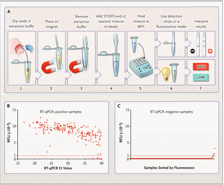
cor-Figure 1. STOPCovid, Version 2 (STOPCovid.v2) Test and Performance Evaluation.
Panel A shows a nasopharyngeal or anterior nasal swab dipped in 400 μl of extraction solution containing lysis buffer and magnetic beads (step 1). After 10 minutes at room temperature, the sample was placed on a magnet (step 2) and extraction buffer was aspirated (step 3). A total of 50 μl of STOPCovid.v2 reaction mixture was added to the beads (step 4), and the sample was heated to 60°C (step 5). For a lateral‑flow readout, after 80 minutes, detection strips were dipped into the reaction mixture (steps 6 and 7, top). After 45 min‑ utes, a fluorescence reader was used to measure the fluorescence of the reaction mixture (steps 6 and 7, bottom). Panel B shows STOP‑ Covid.v2 results for 202 SARS‑CoV‑2–positive nasopharyngeal swab samples obtained from patients and detected by means of a fluores‑ cence readout and measured in relative fluorescence units (RFUs). A swab with 50μl of viral transport medium was dipped into the extraction buffer. Cycle‑threshold (Ct) values were determined with the use of standard reverse‑transcription–quantitative polymerase‑ chain‑reaction (RT‑qPCR) assays. Each dot indicates one sample, and the red dashed line indicates the threshold above which samples were classified as positive. End‑point fluorescence at 45 minutes is shown. Panel C shows STOPCovid.v2 results for 200 SARS‑CoV‑2– negative nasopharyngeal swab samples obtained from patients. The samples were sorted by means of end‑point fluorescence and mea‑ sured in RFUs. Each dot indicates one sample, and the red dashed line indicates the threshold for classifying samples.
A B C B A C 1 2 3 4 5 6 7 HIGH LOW Dip swab in extraction buffer Place on magnet Remove extraction buffer Add STOPCovid.v2 reaction mixture to beads Heat mixture to 60ºC Use detection strips or a fluorescence reader Interpret results RFU ( × 10 –5) RFU ( × 10 –5)
RT-qPCR–positive samples RT-qPCR–negative samples
15 15 10 10 5 1 5 1 0 0 15 20 25 30 35 40
rectly identified 5 positive samples (Ct values, 19 to 36) and 10 negative samples (Fig. S11B through S11E). A detailed protocol for STOPCovid.v2 is provided in the Supplementary Appendix. The simplified format of STOPCovid.V2 is suited for use in low-complexity clinical laboratories. Julia Joung, B.S.
Alim Ladha, B.S.
Massachusetts Institute of Technology Cambridge, MA
Makoto Saito, Ph.D. Broad Institute of MIT and Harvard Cambridge, MA
Nam‑Gyun Kim, Ph.D. University of Washington Seattle, WA
Ann E. Woolley, M.D., M.P.H. Brigham and Women’s Hospital Boston, MA
Michael Segel, Ph.D. Broad Institute of MIT and Harvard Cambridge, MA Robert P.J. Barretto, Ph.D. Kallyope New York, NY Amardeep Ranu, B.S. DynamiCare Health Boston, MA Rhiannon K. Macrae, Ph.D. Guilhem Faure, Ph.D. Broad Institute of MIT and Harvard Cambridge, MA
Eleonora I. Ioannidi, B.S. Rohan N. Krajeski, B.S. Massachusetts Institute of Technology Cambridge, MA Robert Bruneau, B.S. Meei‑Li W. Huang, Ph.D. University of Washington Seattle, WA Xu G. Yu, M.D.
Ragon Institute of MGH, MIT, and Harvard Cambridge, MA
Jonathan Z. Li, M.D. Brigham and Women’s Hospital Boston, MA
Bruce D. Walker, M.D.
Ragon Institute of MGH, MIT, and Harvard Cambridge, MA
Deborah T. Hung, M.D., Ph.D. Broad Institute of MIT and Harvard Cambridge, MA
Alexander L. Greninger, M.D., Ph.D. University of Washington
Seattle, WA
Keith R. Jerome, M.D., Ph.D. Fred Hutchinson Cancer Research Center Seattle, WA
Jonathan S. Gootenberg, Ph.D. Omar O. Abudayyeh, Ph.D. Massachusetts Institute of Technology Cambridge, MA
jgoot@ mit . edu omarabu@ mit . edu Feng Zhang, Ph.D.
Broad Institute of MIT and Harvard Cambridge, MA
zhang@ broadinstitute . org
Ms. Joung and Mr. Ladha and Drs. Gootenberg, Abudayyeh, and Zhang contributed equally to this letter.
Supported by a fellowship (1F31-MH117886, to Ms. Joung) from the National Institutes of Health (NIH); a fellowship (to Dr. Saito) from the Swiss National Science Foundation; a grant (to Drs. Gootenberg and Abudayyeh) from the McGovern Insti-tute for Brain Research at Massachusetts InstiInsti-tute of Technol-ogy; grants (to Drs. Gootenberg, Abudayyeh, and Zhang) from the Patrick J. McGovern Foundation and the Massachusetts Con-sortium on Pathogen Readiness Evergrande Covid-19 Response Fund; and grants (to Dr. Zhang) from the NIH (1R01-MH110049 and 1DP1-HL141201), the Mathers Foundation, the Howard Hughes Medical Institute, the Open Philanthropy Project, and James and Patricia Poitras and Robert Metcalfe.
Disclosure forms provided by the authors are available with the full text of this letter at NEJM.org
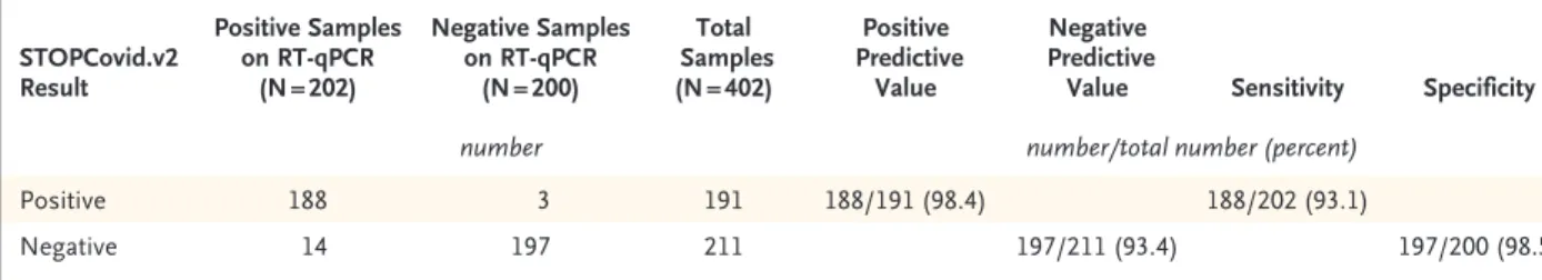
Table 1. Positive and Negative Predictive Values, Sensitivity, and Specificity of STOPCovid.v2 for Detection of SARS-CoV-2 in Nasopharyngeal Samples.* STOPCovid.v2 Result Positive Samples on RT-qPCR (N = 202) Negative Samples on RT-qPCR (N = 200) Total Samples (N = 402) Positive Predictive Value Negative Predictive
Value Sensitivity Specificity
number number/total number (percent)
Positive 188 3 191 188/191 (98.4) 188/202 (93.1)
Negative 14 197 211 197/211 (93.4) 197/200 (98.5)
T h e ne w e ngl a nd jou r na l o f m e dicine
This letter was published on September 16, 2020, at NEJM.org.
1. Gootenberg JS, Abudayyeh OO, Lee JW, et al. Nucleic acid
detection with CRISPR-Cas13a/C2c2. Science 2017; 356: 438-42.
2. Chen JS, Ma E, Harrington LB, et al. CRISPR-Cas12a target
binding unleashes indiscriminate single-stranded DNase activi-ty. Science 2018; 360: 436-9.
3. Broughton JP, Deng X, Yu G, et al. CRISPR-Cas12-based
de-tection of SARS-CoV-2. Nat Biotechnol 2020; 38: 870-4.
4. Notomi T, Okayama H, Masubuchi H, et al. Loop-mediated
isothermal amplification of DNA. Nucleic Acids Res 2000; 28(12): E63.
5. Teng F, Cui T, Feng G, et al. Repurposing CRISPR-Cas12b for
mammalian genome engineering. Cell Discov 2018; 4: 63.
DOI: 10.1056/NEJMc2026172