HAL Id: hal-01188854
https://hal-univ-rennes1.archives-ouvertes.fr/hal-01188854
Submitted on 27 Oct 2015HAL is a multi-disciplinary open access archive for the deposit and dissemination of sci-entific research documents, whether they are pub-lished or not. The documents may come from teaching and research institutions in France or abroad, or from public or private research centers.
L’archive ouverte pluridisciplinaire HAL, est destinée au dépôt et à la diffusion de documents scientifiques de niveau recherche, publiés ou non, émanant des établissements d’enseignement et de recherche français ou étrangers, des laboratoires publics ou privés.
Anti-Porphyromonas gingivalis antibodies titres are
associated with non-smoking status in early rheumatoid
arthritis: Results from the ESPOIR cohort
Raphaèle Seror, Sandrine Le Gall-David, Martine Bonnaure-Mallet, Thierry
Schaeverbeke, Alain Cantagrel, Jacques Minet, Jacques-Eric Gottenberg,
Philippe Chanson, Philippe Ravaud, Xavier Mariette
To cite this version:
Raphaèle Seror, Sandrine Le Gall-David, Martine Bonnaure-Mallet, Thierry Schaeverbeke, Alain Can-tagrel, et al.. Anti-Porphyromonas gingivalis antibodies titres are associated with non-smoking status in early rheumatoid arthritis: Results from the ESPOIR cohort. Arthritis & rheumatology, Wiley, 2015, 67 (7), pp.1729-1737. �10.1002/art.39118�. �hal-01188854�
Anti-Porphyromonas gingivalis antibodies titres are associated with non-smoking status in early rheumatoid arthritis: Results from the ESPOIR cohort
Raphaèle Seror (1) *, Sandrine Le Gall-David (2) *, Martine Bonnaure-Mallet (2), Thierry Schaeverbeke (3), Alain Cantagrel (4), Jacques Minet (2), Jacques-Eric Gottenberg (5), Philippe Chanson (6), Philippe Ravaud (7), Xavier Mariette (1)
* Raphaèle Seror and Sandrine Le Gall-David contributed equally to this work
(1) Department of Rheumatology, Hôpitaux Universitaires Paris-Sud, Assistance Publique–Hôpitaux de Paris (AP-HP), Université Paris-Sud, INSERM U1012, Le Kremlin Bicêtre, France
(2) EA 1254 Equipe, Microbiologie, Université de Rennes 1, Université Européenne de Bretagne, Rennes ;
(3) Department of Rheumatology, Université de Bordeaux, France (4) Department of Rheumatology, Université de Toulouse, France
(5) Rheumatology, Centre National de Référence des Maladies Auto-Immunes Rares, INSERM UMRS_1109, Fédération de Médecine Translationnelle de Strasbourg (FMTS), Strasbourg university Hospital, Université de Strasbourg, Strasbourg, France
(6) Department of Endocrinology, HôpitauxUniversitaires Paris-Sud, Assistance Publique–Hopitaux de Paris (AP-HP), Université Paris-Sud, Le Kremlin Bicêtre, France
(7) Department of Epidemiology, Université Paris-Descartes, Paris, France
Address correspondence and reprint requests to
Dr Raphaèle SEROR, Department of Rheumatology, Hôpital Bicêtre, 78 rue du Général Leclerc, 94275 Le Kremlin Bicêtre. France. e-mail: raphaele.se@gmail.com
And Pr Xavier Mariette, Department of Rheumatology, Hôpital Bicêtre, 78 rue du Général Leclerc, 94275 Le Kremlin Bicêtre. France. e-mail: xavier.mariette@bct.aphp.fr
Word count: 3416
Funding: ESPOIR was created thanks to an unrestricted grant from Merck Sharp and Dohme (MSD) was allocated for the first 5 years. Two additional grants from Institut National de la Santé et de la Recherche Médicale (INSERM) were obtained to support part of the biological database. The French Society of Rheumatology, Pfizer, Abbvie, and Roche-Chugai also supported the ESPOIR cohort study.
ABSTRACT (250 words)
Objectives
To investigate the possible link between Porphyromonas gingivalis (P. gingivalis) infection and RA, according to antibody profile, genetic and environmental factors, and RA severity.
Patients and Methods
For assessing P. gingivalis infection, serum levels of antibodies directed against P. gingivalis LPS were measured in 694 early-RA patients not exposed to steroid or DMARD. Anti-P. gingivalis antibodies titers were compared between early-RA patients and various control groups, and according to various patients characteristics.
Results
The titre of anti-P. gingivalis antibodies did not significantly differ between RA and controls. Anti-P. gingivalis antibody titres did not significantly differ with ACPA, RF, or HLA-shared epitope status. Anti-P. gingivalis antibody titres were significantly higher among never smoker patients compared to ever-smoker (p= 0.0049). Among non-smokers, high anti-P. gingivalis antibody levels were associated with an higher prevalence of erosive change (mSHS erosion subscale ≥1 : 47.5 vs. 33.3%, p=0.0135).
Conclusion
In this large early-RA cohort, we did not detect any association of anti-P. gingivalis antibodies with RA or with ACPA status. These results suggest that the association of periodontitis and RA could be linked to other bacterial species than P. gingivalis or to another mechanism than citrullination. Nevertheless we found higher anti-P. gingivalis antibody titres in non-smokers. In addition, in this population of non-smokers, high anti-P. gingivalis antibody titres were associated with a more severe disease. We hypothesize that the role of tobacco in RA pathogenesis is so high that the effect of P. gingivalis could be revealed only in a population not exposed to tobacco.
Rheumatoid arthritis (RA) is a systemic, inflammatory autoimmune disorder of unknown, complex and multifactorial aetiology. Its pathophysiology relies on an interaction between gene and environment that triggers auto-immunity. One of the most striking examples is the interaction between tobacco and shared epitope carrying in anti-citrillinated peptide antibody (ACPA) positive RA [1]. In this model, tobacco acts as an external agent inducing citrullination of proteins in the lung that triggers auto-immunity and ACPA production in genetically predisposed subjects, leading to emergence of RA.
Among the other potential environmental factors triggering auto-immunity is the oral microbiome. Several epidemiological studies have suggested a link between periodontal disease and RA. The prevalence of periodontal disease is two-fold increased among patients with RA compared to the general population[2-4], and even more in non-smoking RA patients with a 4-fold increased prevalence[5]. In addition, several cohorts have shown that the risk of development of RA was increased in subjects with periodontal disease [6-8].This risk was even more important in non-smokers [4, 8].
One of the main bacteria implicated in chronic periodontal disease is Porphyromas (P) gingivalis [9], a microorganism of the oral cavity located in the sub-gingival tissue. P. gingivalis has been incriminated in RA pathogenesis because it is one of the few microorganisms with a deiminase capable of transforming arginine into citrulline, and is thus suspected of playing a possible role in the production of ACPA. Some recent studies suggest that the presence of P. gingivalis could be associated with development of RA [10-13] and particularly ACPA-positive RA [10, 11, 13-15]. Nevertheless, these results remain controversial [4, 16] and were obtained for most of them from small cohorts.
The objective of this study was to investigate the relationship between P. gingivalis infection (as measured by serum levels of anti-P. gingivalis antibodies) and RA. This study also aimed to investigate the possible link between P. gingivalis infection and RA antibody profile (principally ACPA status), genetic and environmental factors and structural damage in a large cohort of early arthritis patients.
PATIENTS AND METHODS
Patients
The Evaluation et Suivi des POlyarthrites Indifférenciées Récentes (ESPOIR) is a prospective French cohort that included 813 patients with early arthritis. The methodology and the main characteristics of the patients from the ESPOIR cohort have been previously described [17]. The patients were recruited if they had inflammatory arthritis of at least 2 swollen joints lasting for 6 weeks to 6 months and with potential to evolve into RA. Patients were included if they had not received disease-modifying anti-rheumatic drugs (DMARDs) (except within the 15 days before inclusion in the cohort for DMARDs exclusively) or steroids. Patients were excluded if the referring physician considered another defined inflammatory rheumatic disease than RA. The patients were included between December 2002 and March 2005. They have been followed every 6 months during the first 2 years, and every year thereafter. The follow-up is scheduled for at least 15 years from December 2002. Among them, 694 fulfilled the 2010 ACR/EULAR criteria after 2 years of follow-up. Fourteen regional centres in France participated in patients’ inclusion.
Clinical and biological assessment
Clinical variables included total joint count for tenderness and swelling, the Disease Activity Score in 28-joints (DAS28)[18] and the Health Assessment Questionnaire (HAQ)[19]. Laboratory variables included erythrocyte sedimentation rate (ESR, mm/h), C-reactive protein (CRP) level, IgM and IgA rheumatoid factor (RF) (both enzyme-linked immunosorbent assay [ELISA; Menarini], both positive if > 9 UI/ml), ACPA (anti-CCP2, ELISA, DiaSorin, France; positive if > 50 U/ml). All the subjects were genotyped for the HLA-DRB1 shared epitope (SE).
Pro-inflammatory cytokines (Interleukin (IL)-1β, IL-1 receptor antagonist (IL1-Ra), IL-2, IL-4, IL-6, IL-10, IL-17, MCP-1, Tumor Necrosis Factor (TNF) α and interferon (IFN) γ were previously quantified in ESPOIR cohort at baseline using a commercially available multiplex bead immunoassay, based on the Luminex platform (Fluorokine MAP Multiplex Human Cytokine Panel, R&D Systems, Minneapolis, Minnesota, USA) as previously described [20].The corresponding quantifications are expressed in ng/ml. The method of quantification
of serum markers of B cell activation (β2-microglobulin, IgG, IgA, IgM, Free light chain of immunoglobulins [FLCs]) were previously reported [21].
Smoking history
Patients’ smoking habits were evaluated at inclusion. Current smokers were those reporting active smoking. Past-smokers were all patients who had stopped smoking before the first examination at inclusion. Ever-smoker included both current and past-smokers. Non-smokers reported no history of smoking at any time.
Structural assessment
Patients had radiological evaluation every 6 months during the first two years of follow-up allowing studying parameters associated with structural progression. Radiographs of the hands and feet (antero-posterior views) were collected in the radiography coordinating centre. A standardized reading was performed on X-rays obtained at baseline, 1 year and 2 years of follow-up. All sets of X-rays were read by a trained investigator blinded to clinical evaluation (GT) according to the van der Heijde-modified total Sharp score (mSHS). Intra-reader and inter Intra-reader reliability (GT and VD-P) were excellent (Intraclass correlation coefficient = 0.97 and 0.93, respectively). The smallest detectable change (SDC) was 1 [22].
Structural damage at inclusion was assessed qualitatively by the presence of typical RA erosions according to their location and aspect, and was rated according to mSHS[23]. Radiological progression was assessed between baseline and the end of the second year of follow-up. Radiological progression was defined as a progression of more than the SDC (≥1 point of mShS) at 2 years. Rapid Radiological Progression (RRP) was defined as an increase of mShS> 10 within the first two years of follow-up (i.e. 5 points per year).
Controls
Variété is a transversal, non-interventional French national cohort, based on healthy volunteers for establishing normative data for IGF-I and other hormones in the general population (ClinicalTrials.gov identifier: NCT01831648). A total of 974 healthy subjects have been recruited in 10 centres all around France. Subjects with medical conditions and medications that may affect IGF-I measurement have been excluded. A random sample of 79 healthy controls from the Variété cohort was matched, for age- and gender, to a representative sample of 79 RA patients from ESPOIR cohort. Since this cohort focused on healthy subjects with no risk factors, it included only non-current smokers: among them, 83.7% were never-smokers and the remaining 16.2% did not smoke more than 10 cigarettes/day.
Sicca Controls
Sicca controls were 54 patients form the cohort of patients with sicca symptoms referred to the Rheumatology Department of the Bicêtre Hospital for a diagnostic procedure to assess if patients have Sjögren's syndrome. To be considered as controls patients, these subjects were excluded if they had auto-antibodies, lymphocytic sialadenitis on salivary gland biopsy or any features of auto-immunity. Nevertheless, some of them had objective dryness features. These controls were age- and gender-matched to a random sample of RA patients from ESPOIR cohort. Their smoking status was known as never or ever smoker, with no mention of current or past smoking status. This group included 19/51 (37.3%) ever smokers.
Periodontitis controls
The last control group consisted in 61 patients with clinically proven severe periodontitis. In this group, 21 (34.5%) patients were non-smokers, 40 (65.6%) were ever smokers, including 13 (21.3%) current smokers. These patients were not matched with ESPOIR cohort patients, and included a higher proportion of males. We therefore took a matched subsample of this group having a same proportion of male as the RA cohort to perform sensitivity analyses for comparisons with early RA patients. In addition, for this group of patient, Micro-Indent test was performed to detect the presence of P. gingivalis in periodontal tissue (see below).
Bacterial serologic measurement and detection of P. gingivalis
Anti-P. gingivalis antibodies measurement
Immunoglobulin G antibodies specific to LPS of P. gingivalis were measured using a homemade ELISA. The wells of 96-well flat-bottom microtiter plates were coated in triplicate with LPS of P. gingivalis. After washing and blocking the plates, serum samples were added to individual wells and specific human IgG antibodies were detected with an alkaline phosphatase-conjugated antihuman immunoglobulin. The absorbance was read at 405 nm using an ELISA plate reader. The results were expressed as an ELISA index (EI), which was the mean OD 405 nm of a given serum divided by the mean OD 405 nm of the calibrator (reference serum) [11].
Bacteria identification (DNA-DNA hybridation)
Bacteria identification was made in the gingival pockets, with the kit Micro-Ident according to the manufacturer’s instructions (Hain Lifescience, Germany), as follows. DNA–DNA hybridization is a molecular biology technique that measures the degree of genetic similarity between pools of DNA sequences; it can be used to identify different species in a pluribacterial sample. In our case DNA was isolated from gingival pockets, amplified with specific primers (supplied in the kit; HainLifescience, Germany). In the next step, the amplicons were chemically denatured, since detection on the strip was done using single-stranded DNA. The strip was coated with highly specific probes which were complementary to selectively amplified nucleic acid sequences. The single-stranded amplicon binds specifically to the analog probes during hybridization, while non-specifically bound amplicons were removed in subsequent washing steps. During the conjugate reaction, the specifically bound amplicon was marked with the enzyme alkaline phosphatase and was then made visible in a colorimetric detection reaction.
The protocols of the ESPOIR cohort study and of the Variété cohort study were approved by the Ethics Committees of Montpellier University Hospital, and Hôpitaux Paris-Sud, France, respectively. All patients from ESPOIR and Variété Cohorts as well as sicca controls included in the present study gave their written informed consent.
Statistical analyses
All analyses were restricted to the subgroup of 694 patients fulfilling the ACR/EULAR criteria. Categorical variables are reported as numbers (percentages) and were compared using a χ² or, when appropriate, Fischer’s exact test. Quantitative variables are reported as mean (± standard deviation [SD]) and were compared using a Student t-test. For correlations analyses, Spearman’s correlation coefficients were obtained.
Since results were not normally distributed, anti-P. gingivalis antibody titres were log-transformed. Also, anti-P. gingivalis titres were used as dichotomous variable. For that purpose, high titres were defined as a titre above the 75th percentile (upper quartile) of distribution in the RA patients of the ESPOIR cohort.
To assess if anti-P. gingivalis antibodies titres were associated with early RA, titres were compared between RA patients and the different control groups.
To identify parameters associated with anti-P. gingivalis antibodies titres, their serum levels were compared according various demographic, clinical characteristics, biological/biochemical and radiological variables (for categorical variables) and correlated with the levels of clinical, biological and radiological variables (for quantitative parameters).
For all analyses, a p<0.05 was considered statistically significant. Confidence intervals were calculated at the 95% level. Statistical analyses involved use of SAS 9.3 (SAS Inst., Cary, NC).
RESULTS
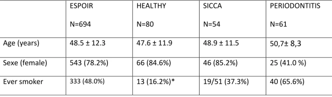
Among patients from the ESPOIR cohort, 694 fulfilled the ACR/EULAR criteria for RA after 2 years of follow-up. Their main characteristics are presented in Table 1. Age, sex and smoking status of early RA and controls groups are reported in Table 2.
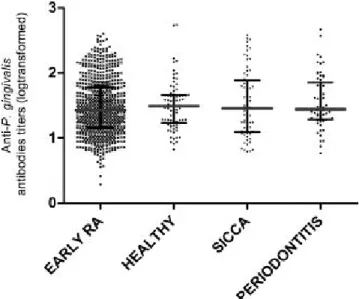
Anti-P. gingivalis antibody titres among controls and early RA patients
Anti-P. gingivalis antibody titres did not significantly differ between early RA patients and healthy controls (1.47 ± 0.42 vs. 1.49 ± 0.33, p=0.66), sicca controls (1.47 ± 0.42 vs. 1.51 ± 0.47, p=0.53) or periodontitis controls (1.47 ± 0.42 vs. 1.55 ± 0.41, p=0.17) (figure 1). Among periodontitis controls, the level of anti-P. gingivalis antibodies tended to be higher among females but this did not reach significance (1.54 ± 0.35 vs. 1.62 ± 0.47, p=0.28). We therefore compared the level of anti- anti-P. gingivalis antibodies between early RA and the sex-matched subsample of periodontitis controls and found a trend to an higher level in periodontitis controls (1.47 ± 0.42 vs. 1.60 ± 0.44, p=0.12). In addition, among periodontitis controls, 24 (39.34%) had P. gingivalis detected in periodontal tissue, these patients had a higher level of P. gingivalis antibodies, compared to periodontitis patients for whom P. gingivalis was not detected (1.74 vs. 1.43; p=0.008, figure 2). Thus, a high titre of anti-P.
gingivalis is a good indirect marker of chronic P. gingivalis infection.
Anti-P. gingivalis antibody titres among different subsets of early RA patients
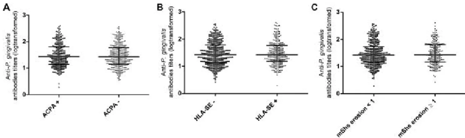
Anti-P. gingivalis antibody titres did not significantly differ with ACPA, RF, or HLA- shared epitope status (figure 3). Among ACPA positive patients, the level of anti-P. gingivalis antibody titres did not correlate with the level of ACPA (rho= 0.042, p=0.46), or RF titres (rho=0.013, p=0.82). Also, they did not correlate with disease activity (DAS28 or its components), pro-inflammatory cytokine levels, serum levels of B cell activation markers (β2-microglobulin, IgG, IgA, IgM, FLCs) or structural damage as measured by the total mShS and its subscales. However, patients with high anti-P.gingivalis antibody levels were more likely to have typical erosion related to RA than those with lower values (35/164 [21.34%] vs. 69/497 [13.88%], p=0.02). Also, 40.1% (65/162) of the patients with high anti-P.gingivalis
antibody titres had erosive RA (as measured by an mShS erosion subscale ≥ 1) compared to 34.9% (173/496) of the patients with lowers titres (p=0.23. figure 4).
Higher anti-P. gingivalis antibody titres in never smokers
Anti-P. gingivalis antibody titres were significantly lower among ever-smoker patients compared to never smoker (1.42 ± 0.43vs. 1.51 ± 0.43, p= 0.0049) and particularly in current smoker patients compared to never smoker (1.37 ± 0.42 vs.1.51 ± 0.43, p= 0.0003), but not past-smokers (1.51 ± 0.43 vs. 1.47 ± 0.43, p= 0.28). Also, patients with high anti-P. gingivalis antibody titres were more likely to be non-smoker than those with low titres (107/175 [61.1%] vs. 254/265 [48.9%], p=0.005).
In the control groups, there was a numerical, but not significant, higher level of anti-P. gingivalis antibodies in never smokers compared to ever smokers in the sicca group (1.56 ± 0.47 vs. 1.46 ± 0.48, p=0.45), and compared to current smokers in the periodontitis group (1.57 ± 0.48 vs. 1.48 ± 0.33, p=0.54).
Among RA patients, smoking status did not alter the level of other antibodies (anti-CCP, IgM-RF, FLC, total IgG, IgA, IgM), except the level of IgA-RF that were found significantly higher in smokers than in non smokers (45.6 UI ± 107.5 vs 29.1 ± 60.4; p=0.014).
Higher anti-P. gingivalis antibody titres in non-smokers were associated with a more severe disease
Among non-smokers, patients with high anti-P. gingivalis antibody levels were more likely to have erosive disease than those with lower titres as demonstrated by a higher proportion of patients having typical erosion related to RA (22/102 [21.6%] vs. 32/241 [13.3%], p=0.054) and having a mShS erosion subscale ≥ 1 at baseline (48/101 [47.5%] vs. 80/240 [33.3%], p=0.014). This trend was not observed in the ever smoker group, neither past nor current smokers (figure 5). In the current smoker group, we observe an inverse but not significant
relationship (p=0.20). However, anti-P. gingivalis antibodies titres were not predictive of structural progression, whatever the definition used.
DISCUSSION
This study is one of the largest that examined the possible link between P. gingivalis infection and RA, with a particular focus on RA-antibody profile (principally ACPA status), genetic and environmental risk factors and structural damage. In this cohort of early RA patients, we could not detect any association between anti-P. gingivalis antibodies and RA whatever the ACPA status. Nevertheless, we found that anti-P. gingivalis antibody titres were increased in never smokers. In this group of never smoker patients, increased levels of anti-P. gingivalis antibody were associated with a more erosive disease.
We did not find higher anti-P. gingivalis antibody titres in RA patients compared to non-RA controls. There is to date no consensual method for dosing anti-P. gingivalis antibodies and each of them may be questionable. We here used an assay that detected antibodies to P. gingivalis LPS [16], rather than lysate or purified proteins used in some previous studies [10, 14, 15]. A method similar to that we used demonstrated such association with RA in a previous study [11]. Moreover, we found higher antibody titres in our positive control group, i.e. patients with P. gingivalis-associated periodontitis. Thus, our method seems to be valid for assessing a long-term exposure to P. gingivalis. Nevertheless, as shown by Scher et al, the presence of anti-P. gingivalis antibody is not necessarily associated with the presence of a currently active P. gingivalis infection. Therefore, none of these methods, including our, is able to detect current P. gingivalis infection, but they assess prior exposure to this pathogen. The only method able to prove P. gingivalis infection is the direct detection of the bacteria. Unfortunately, we did not have the opportunity to assess this in the ESPOIR cohort. At the time of the conception of this cohort, the implication of oral microbiome in RA pathogenesis was not suspected yet. Nevertheless, it is the largest cohort, involving almost 700 patients, exploring exposure to P. gingivalis in early RA. Compared with most of the previous studies, we here included early RA patients and not long standing established RA. Results of the previous studies assessing P. gingivalis infection in early RA are controversial [4, 13] and two
recent studies did not also find any association between anti-P. gingivalis antibodies and RA [4, 16], particularly in the absence of proven periodontitis[16].
This study did not refute the potential role of oral microbiome in RA. First, the association between periodontitis and RA found in many studies, and confirmed in a recent systematic review [24], may be linked to another pathogen than P. gingivalis as recently suggested by Scher et al [4]. Second, tobacco plays such a major role in triggering RA that it may overtake and mask the potential role of P. gingivalis in smoker population. The role of P. gingivalis and other oral bacterial species could be revealed only in a population not exposed to tobacco. Effectively, the increased prevalence of periodontitis in non-smoker RA patients suggested in 2 previous studies, underlined that the link between periodontitis, oral microbiome and RA could be more pronounced in the non-smoking population [4, 8]. Last, even if P. gingivalis plays a major role, it may be by another mechanism than inducing citrullination since in number of studies including ours, there is no difference of anti-P. gingivalis titres in patients with or without ACPA. It could be also linked to the use of an anti-CCP2 kit that detects IgG but not IgA isotypes, the latter being possibly more associated to mucosa-associated antibody production. Nevertheless, other reports which found an association between P. gingivalis and ACPA status used similar assays [10, 15]. Although controversial [25], it has been recently found that auto-citrullination of P. gingivalis peptidylarginine deiminase (PPAD), is probably not the underlying mechanism linking P. gingivalis-associated periodontitis and RA [26]. Moreover, in 2 recent studies, it has been found that patients with periodontitis or with pre symptomatic RA have a higher titre of antibodies against uncitrullinated antigens suggesting that breaking of tolerance to uncitrullinated peptides could be the first event in these patients [27, 28] . Even more interestingly, in patients with periodontitis, the titres of antibodies against uncitrullinated antigens were more elevated in non-smokers than in smokers suggesting that the non-citrulline specific breaking of tolerance associated with periodontitis was even more important in non-smokers [28]. Also, in RA patients with periodontitis, smoking status did not influence ACPA titres, but had an impact on anti-P. gingivalis titres that were found lower in smokers than in non-smokers [29]. This observation clearly fits with our findings of a possible link between P. gingivalis infection and RA and severity of RA only in non-smokers. An alternative hypothesis would be
a protective effect of smoking specifically on anti-P. gingivalis antibody generation, without any general effect on antibody or auto-antibody generation.
In conclusion, we did not detect any association between anti-P. gingivalis antibodies and RA or ACPA status in this large cohort of early-RA. These results suggest that the association of periodonditis and RA could be linked to other bacterial species than P. gingivalis or to another mechanism than citrullination. Nevertheless we found higher anti-P. gingivalis antibody titres in smokers compared to smokers. In addition, in this population of non-smokers, high anti-P. gingivalis antibody titres were associated with a more severe disease. We hypothesize that the role of tobacco in RA pathogenesis is so high that the role of P. gingivalis could be revealed only in a population not exposed to tobacco. Alternatively, smoking might decrease anti-P. gingivalis immune response. Finally, our findings suggest that the association of periodontitis and RA could be linked to other bacterial species than P. gingivalis or to another mechanism than citrullination and reinforce the importance of investigating the potential interest of oral hygiene and supragingival scaling in RA patients, as suggested in a recent meta-analysis [30].
Acknowledgement
We thank N. Rincheval for data management and expert monitoring. We thank the French rheumatologists who referred their patients to the ESPOIR cohort in the following rheumatology departments: Amiens (P. Fardellone, P. Boumier), Bordeaux (T. Schaeverbecke), Brest (A. Saraux), Lille (R. M. Flipo), Paris-Bicêtre (X. Mariette), Paris-Bichat (O. Meyer), Paris-Cochin (M. Dougados), Paris-St. Antoine (F. Berenbaum), Rouen (O. Vittecoq), Strasbourg (J. Sibilia), Toulouse (A. Cantagrel), and Tours (P. Goupille). We are grateful to S. Martin for performing all the centralized assays of CRP level, IgA and IgM RFs, and anti-CCP antibodies. We are grateful to G. Tobon and V. Devauchelle-Pensec for performing all the centralized X-rays reading.
Competing interests
Authors have no competing interest in relation to this study
Contributorship
Conception and design: Raphaèle Seror and Xavier Mariette Data collection: all
Analysis and interpretation: Raphaèle Seror, Xavier Mariette, Sandrine David-Le Gall Drafting and critical reviewing: all
Final approval: all
Ethical approval information
The ESPOIR cohort study was approved in July 2002 by the ethics committee of Montpellier, France (no. 020307), allowing future clinical projects on the database. All patients gave their signed informed consent before inclusion.
Data sharing statement
References
1. Klareskog L, Stolt P, Lundberg K, Kallberg H, Bengtsson C, Grunewald J, et al. A new model for an etiology of rheumatoid arthritis: smoking may trigger HLA-DR (shared epitope)-restricted immune reactions to autoantigens modified by citrullination. Arthritis Rheum 2006;54:38-46.
2. de Pablo P, Dietrich T, McAlindon TE. Association of periodontal disease and tooth loss with rheumatoid arthritis in the US population. J Rheumatol 2008;35:70-6.
3. Chen HH, Huang N, Chen YM, Chen TJ, Chou P, Lee YL, et al. Association between a history of periodontitis and the risk of rheumatoid arthritis: a nationwide, population-based, case-control study. Ann Rheum Dis 2013;72:1206-11.
4. Scher JU, Ubeda C, Equinda M, Khanin R, Buischi Y, Viale A, et al. Periodontal disease and the oral microbiota in new-onset rheumatoid arthritis. Arthritis Rheum 2012;64:3083-94.
5. Potikuri D, Dannana KC, Kanchinadam S, Agrawal S, Kancharla A, Rajasekhar L, et al. Periodontal disease is significantly higher in non-smoking treatment-naive rheumatoid arthritis patients: results from a case-control study. Ann Rheum Dis 2012;71:1541-4.
6. Mercado FB, Marshall RI, Klestov AC, Bartold PM. Relationship between rheumatoid arthritis and periodontitis. J Periodontol 2001;72:779-87.
7. Pischon N, Pischon T, Kroger J, Gulmez E, Kleber BM, Bernimoulin JP, et al. Association among rheumatoid arthritis, oral hygiene, and periodontitis. J Periodontol 2008;79:979-86.
8. Demmer RT, Molitor JA, Jacobs DR, Jr., Michalowicz BS. Periodontal disease, tooth loss and incident rheumatoid arthritis: results from the First National Health and Nutrition Examination Survey and its epidemiological follow-up study. J Clin Periodontol 2011;38:998-1006.
9. Hajishengallis G, Darveau RP, Curtis MA. The keystone-pathogen hypothesis. Nat Rev Microbiol 2012;10:717-25.
10. Mikuls TR, Payne JB, Reinhardt RA, Thiele GM, Maziarz E, Cannella AC, et al. Antibody responses to Porphyromonas gingivalis (P. gingivalis) in subjects with rheumatoid arthritis and periodontitis. Int Immunopharmacol 2009;9:38-42.
11. Hitchon CA, Chandad F, Ferucci ED, Willemze A, Ioan-Facsinay A, van der Woude D, et al. Antibodies to porphyromonas gingivalis are associated with anticitrullinated protein antibodies in patients with rheumatoid arthritis and their relatives. J Rheumatol 2010;37:1105-12.
12. Okada M, Kobayashi T, Ito S, Yokoyama T, Komatsu Y, Abe A, et al. Antibody responses to periodontopathic bacteria in relation to rheumatoid arthritis in Japanese adults. J Periodontol 2011;82:1433-41.
13. Arvikar SL, Collier DS, Fisher MC, Unizony S, Cohen GL, McHugh G, et al. Clinical correlations with Porphyromonas gingivalis antibody responses in patients with early rheumatoid arthritis. Arthritis Res Ther 2013;15:R109.
14. Mikuls TR, Thiele GM, Deane KD, Payne JB, O'Dell JR, Yu F, et al. Porphyromonas gingivalis and disease-related autoantibodies in individuals at increased risk of rheumatoid arthritis. Arthritis Rheum 2012;64:3522-30.
15. Smit MD, Westra J, Vissink A, Doornbos-van der Meer B, Brouwer E, van Winkelhoff AJ. Periodontitis in established rheumatoid arthritis patients: a
cross-sectional clinical, microbiological and serological study. Arthritis Res Ther 2012;14:R222.
16. Mikuls TR, Payne JB, Yu F, Thiele GM, Reynolds RJ, Cannon GW, et al. Periodontitis and Porphyromonas gingivalis in Patients With Rheumatoid Arthritis. Arthritis Rheumatol 2014;66:1090-100.
17. Combe B, Benessiano J, Berenbaum F, Cantagrel A, Daures JP, Dougados M, et al. The ESPOIR cohort: a ten-year follow-up of early arthritis in France: methodology and baseline characteristics of the 813 included patients. Joint Bone Spine 2007;74:440-5.
18. Prevoo ML, van 't Hof MA, Kuper HH, van Leeuwen MA, van de Putte LB, van Riel PL. Modified disease activity scores that include twenty-eight-joint counts. Development and validation in a prospective longitudinal study of patients with rheumatoid arthritis. Arthritis Rheum 1995;38:44-8.
19. Fries JF, Spitz P, Kraines RG, Holman HR. Measurement of patient outcome in arthritis. Arthritis Rheum 1980;23:137-45.
20. Gottenberg JE, Dayer JM, Lukas C, Ducot B, Chiocchia G, Cantagrel A, et al. Serum IL-6 and IL-21 are associated with markers of B cell activation and structural progression in early rheumatoid arthritis: results from the ESPOIR cohort. Ann Rheum Dis 2012;71:1243-8.
21. Gottenberg JE, Miceli-Richard C, Ducot B, Goupille P, Combe B, Mariette X. Markers of B-lymphocyte activation are elevated in patients with early rheumatoid arthritis and correlated with disease activity in the ESPOIR cohort. Arthritis Res Ther 2009;11:R114.
22. Tobon G, Saraux A, Lukas C, Gandjbakhch F, Gottenberg JE, Mariette X, et al. First-year radiographic progression as a predictor of further progression in early arthritis: results of a large national French cohort. Arthritis Care Res (Hoboken) 2013;65:1907-15.
23. van der Heijde DM, van Leeuwen MA, van Riel PL, van de Putte LB. Radiographic progression on radiographs of hands and feet during the first 3 years of rheumatoid arthritis measured according to Sharp's method (van der Heijde modification). J Rheumatol 1995;22:1792-6.
24. Kaur S, White S, Bartold PM. Periodontal disease and rheumatoid arthritis: a systematic review. J Dent Res 2013;92:399-408.
25. Quirke AM, Lugli EB, Wegner N, Hamilton BC, Charles P, Chowdhury M, et al. Heightened immune response to autocitrullinated Porphyromonas gingivalis peptidylarginine deiminase: a potential mechanism for breaching immunologic tolerance in rheumatoid arthritis. Ann Rheum Dis 2014;73:263-9.
26. Konig MF, Paracha AS, Moni M, Bingham CO, 3rd, Andrade F. Defining the role of Porphyromonas gingivalis peptidylarginine deiminase (PPAD) in rheumatoid arthritis through the study of PPAD biology. Ann Rheum Dis 2014;
27. Brink M, Hansson M, Ronnelid J, Klareskog L, Rantapaa Dahlqvist S. The autoantibody repertoire in periodontitis: a role in the induction of autoimmunity to citrullinated proteins in rheumatoid arthritis? Antibodies against uncitrullinated peptides seem to occur prior to the antibodies to the corresponding citrullinated peptides. Ann Rheum Dis 2014;73:e46.
28. de Pablo P, Dietrich T, Chapple IL, Milward M, Chowdhury M, Charles PJ, et al. The autoantibody repertoire in periodontitis: a role in the induction of autoimmunity to citrullinated proteins in rheumatoid arthritis? Ann Rheum Dis 2014;73:580-6.
29. Lappin DF, Apatzidou D, Quirke AM, Oliver-Bell J, Butcher JP, Kinane DF, et al. Influence of periodontal disease, Porphyromonas gingivalis and cigarette smoking on systemic anti-citrullinated peptide antibody titres. J Clin Periodontol 2013;40:907-15. 30. Kaur S, Bright R, Proudman SM, Bartold PM. Does periodontal treatment influence
clinical and biochemical measures for rheumatoid arthritis? A systematic review and meta-analysis. Semin Arthritis Rheum 2014;
Table 1. Characteristics of early-RA patients form the ESPOIR cohort
RA patients N= 694
Sex (female) 543 (78.2%)
Age (years) 48.5 ± 12.3
Symptom duration (days) 74.8 ± 76.6
BMI (kg/m²) 25.1 ± 4.6 Ever smoker 333 (48.0%) Current smokers 151 (21.8%) Past smokers 182 (26.2%) CRP (mg/dl) 22.8 ± 34.7 CRP us (mg/dl) 20.9 ± 33.5 ESR (mm) 29.9 ± 24.8 PGA (/100mm) 61.7 ± 24.6
Tender joint count (/28) 9.4 ± 7.1
Swollen joint count (/28) 7.9 ± 5.4
DAS28 5.3 ± 1.2
Anti-CCP 315 (45.4%)
IgM-RF 372 (53.6%)
Anti-CCP and/or RF (IgM or IgA) 438 (63.1%) Typical RA erosive change 181 (32.9%)
Modified Sharp score 5.2 ± 7.4
Current use of DMARD 48 (6.9%)
Current use of oral NSAIDs 487 (70.2%) Anti-P. gingivalis antibody titre 3.80 ± 2.23 Anti-P. gingivalis antibody titre (logtransfromed) 1.47 ± 0.43
Table 2. Characteristics of early-RA patients and control groups ESPOIR N=694 HEALTHY N=80 SICCA N=54 PERIODONTITIS N=61 Age (years) 48.5 ± 12.3 47.6 ± 11.9 48.9 ± 11.5 50,7± 8,3 Sexe (female) 543 (78.2%) 66 (84.6%) 46 (85.2%) 25 (41.0 %) Ever smoker 333 (48.0%) 13 (16.2%)* 19/51 (37.3%) 40 (65.6%)
Figure 2. Anti-Porphyromonas gingivalis antibody titres in periodontitis controls according to detection of Porphyromonas gingivalis in periodontal tissue
P. gingivalis +: presence of Porphyromonas gingivalis in periodontal tissue samples; P. gingivalis -: absence of Porphyromonas gingivalis in
Figure 3. Anti-Porphyromonas gingivalis antibody titres according to ACPA (A), HLA-SE (B) and erosive change (C) in early-RA patients
Figure 4. Link between anti-Porphyromonas gingivalis antibody titres, erosive changes according to smoking status in early-RA patients
(A): Comparisons of anti-Porphyromonas gingivalis antibody titres according to smoking status
(B): Link between anti-Porphyromonas gingivalis antibody and presence of erosive changes according to smoking status in early-RA patients. Percentage of patients with erosive RA (defined by an mShS erosion subscale ≥ 1) in patients with high anti-P.gingivalis antibody titres ≥75th percentile (dark grey) and in those with low anti-P. gingivalis antibody titres (light grey) according to smoking status.