HAL Id: hal-02959020
https://hal.archives-ouvertes.fr/hal-02959020
Submitted on 6 Nov 2020HAL is a multi-disciplinary open access archive for the deposit and dissemination of sci-entific research documents, whether they are pub-lished or not. The documents may come from teaching and research institutions in France or abroad, or from public or private research centers.
L’archive ouverte pluridisciplinaire HAL, est destinée au dépôt et à la diffusion de documents scientifiques de niveau recherche, publiés ou non, émanant des établissements d’enseignement et de recherche français ou étrangers, des laboratoires publics ou privés.
Cognitive and behavioural inhibition deficits in
neurodegenerative dementias
Raffaella Migliaccio, Delphine Tanguy, Arabella Bouzigues, Idil Sezer, Bruno
Dubois, Isabelle Le Ber, Bénédicte Batrancourt, Valerie Godefroy, Richard
Lévy
To cite this version:
Raffaella Migliaccio, Delphine Tanguy, Arabella Bouzigues, Idil Sezer, Bruno Dubois, et al.. Cogni-tive and behavioural inhibition deficits in neurodegeneraCogni-tive dementias. Cortex, Elsevier, 2020, 131, pp.265-283. �10.1016/j.cortex.2020.08.001�. �hal-02959020�
Cognitive and behavioural inhibition deficits in neurodegenerative dementias
Raffaella Migliaccio a,b,c*, Delphine Tanguy a,d, Arabella Bouzigues a, Idil Sezer a,Bruno
Dubois a,b,c, Isabelle Le Ber a,b,c, Bénédicte Batrancourt a, Valérie Godefroy a,Richard Levy
a,b,c.
a FrontLab, INSERM U1127, Institut du cerveau, Sorbonne Université, Hôpital
Pitié-Salpêtrière, Paris, France
b Centre de Référence des Démences Rares ou Précoces, Hôpital Pitié-Salpêtrière, Assistance
Publique–Hôpitaux de Paris, Paris, France
c Institute of Memory and Alzheimer’s Disease, Centre of Excellence of Neurodegenerative
Disease, Department of Neurology, Hôpital Pitié-Salpêtrière, Assistance Publique–Hôpitaux
de Paris, Paris, France
d Univ Rennes, Inserm, LTSI - UMR 1099, F-35000 Rennes, France
*Corresponding author: Raffaella Migliaccio, Institut du cerveau (ICM), Inserm Unit 1127, 47 bd de l’hôpital, 75013, Paris
Abstract
Disinhibition, mainly caused by damage in frontotemporal brain regions, is one of the
major causes of caregiver distress in neurodegenerative dementias. Behavioural inhibition
deficits are usually described as a loss of social conduct and impulsivity, whereas cognitive
inhibition deficits refer to impairments in the suppression of prepotent verbal responses and
resistance to distractor interference.
In this review, we aim to discuss inhibition deficits in neurodegenerative dementias
through behavioural, cognitive, neuroanatomical and neurophysiological exploration. We also
discuss impulsivity and compulsivity behaviours as related to disinhibition. We will therefore
describe different tests available to assess both behavioural and cognitive disinhibition and
summarise different manifestations of disinhibition across several neurodegenerative diseases
(behavioural variant of frontotemporal dementia, Alzheimer’s disease, Parkinson’s disease,
progressive supranuclear palsy, Huntington’s disease). Finally, we will present the latest
findings about structural, metabolic, functional, neurophysiological and also neuropathological
correlates of inhibition impairments. We will briefly conclude by mentioning some of the latest
pharmacological treatment options available for disinhibition.
Within this framework, we aim to highlight i) the current interests and limits of tests
and questionnaires available to assess behavioural and cognitive inhibition in clinical practice
and in clinical research; ii) the interpretation of impulsivity and compulsivity within the
spectrum of inhibition deficits; and iii) the brain regions and networks involved in such
behaviours.
Keywords: disinhibition; behavioural variant of frontotemporal dementia (bvFTD);
1. Introduction
1.1. Definition
“All the movements of our body are not merely those dictated by impulse or weariness; they are the correct expression of what we consider decorous. Without impulses, we could take no part in social life; on the other hand, without inhibitions, we could not correct, direct, and utilize our impulses”. (Maria Montessori)
Defining cognitive and behavioural inhibition is a complex task. In 2013, Bari and
Robbins (2013) published a large and comprehensive review on inhibition and impulsivity and
they listed 18 types of “inhibition” belonging to different levels of “analysis”. These types
ranged from more fundamental aspects, such as synaptic inhibition like neuronal
hyperpolarisation mediated by GABA fixation for example, to more complex inhibition
mechanisms, such as social inhibition.
From a neuropsychological and clinical point of view, two major types can be defined:
cognitive and behavioural inhibition (Aron, 2007). They are both part of the ‘cognitive control’
system, an umbrella term to define a range of inhibitory mechanisms from the inhibition of
prepotent tendencies, to the updating of working memory contents and consequential shifting
between tasks (Miyake et al., 2000). This division may seem artificial, but it reflects the need
to distinguish the observed phenomena from a clinical standpoint. In agreement with such a
division, in obsessive compulsive disorders, for example, failures in cognitive or behavioural
inhibitory processes lead to different syndromic behaviours: obsessions and compulsions
respectively (Chamberlain et al., 2005). From a neurodevelopmental perspective, behavioural
inhibition, such as the ability to control movements, appears to develop before cognitive
inhibition, which acts on mental processes (Wilson & Kipp, 1998). Both cognitive and
behavioural inhibition appear to then become increasingly efficient with age (Mischel et al.,
In an attempt to keep this distinction, cognitive inhibition is defined as the ability to
resist an exogenous or endogenous interference, inhibit cognitive contents or processes
previously activated and suppress inappropriate or irrelevant responses (Wilson & Kipp, 1998).
It can be intentional and conscious (such as thought suppression), or unintentional and
unconscious (such as the gating of irrelevant information from working memory during
memory processing) (Harnishfeger, 1995). Behavioural inhibition, on the other hand, refers to
the control of emotional and social behaviours in a social context. It is the control of overt
behaviours, the ability to adapt actions to environmental changes, and suppress impulsions
which violate norms (Harnishfeger, 1995).
1.2 Disinhibition, impulsivity and compulsivity: nested concepts related to inhibition deficits
For some authors (e.g., Rascovsky et al., 2011), impulsivity is a subcomponent of the
larger syndrome of disinhibition. Conversely, for other authors (e.g., Rochat et al., 2013),
disinhibition, referring to the lack of control in a specific social context, is a subdimension of
the broader concept of impulsive behaviours. While impulsivity was historically associated
with risk-seeking and compulsivity with harm-avoidance, it is progressively recognised that the
two concepts share neuropsychological mechanisms involving dysfunctional inhibition of
thoughts and behaviours.
Impulsivity is a multidimensional construct defined as “a predisposition toward rapid,
unplanned reactions to internal or external stimuli with diminished regard to the negative
consequences of these reactions” (Chamberlain & Sahakian, 2007). It is measured through
questionnaires as a trait (e.g., Barratt Impulsiveness Scale (BIS-11) (Patton et al., 1995)) or
through tasks (e.g., Go/NoGo task). This reflects the different facets of impulsive behaviour,
which is characterised as the (i) inability to stop automatic responses, (ii) tendency towards
Impulsivity and compulsivity share the feeling of “lack of control” (Fineberg et al., 2014; Stein
& Hollander, 1995). While impulsivity describes quick unplanned actions, compulsivity is
defined as persistent non-goal orientated actions and is typically assessed through tasks
measuring an inability to flexibly adapt behaviours and/or switch attention between stimuli
(Ahearn et al., 2012; Fineberg et al., 2014, 2018).
2. Assessing inhibition deficits
For each test and questionnaire, we have described the instructions, the ability to
identify cognitive or behavioural inhibition disorders and to differentiate dementia syndromes.
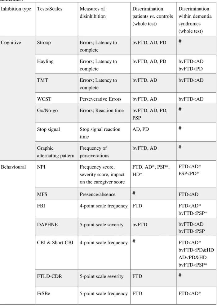
Table 1 summarises this information.
2.1. Cognitive inhibition
In neuropsychological practice, there are a number of tests that are performed routinely.
The Stroop, the Hayling, the Trail Making and the Wisconsin card sorting tests are amongst the
most used (Rabin et al., 2005; O’Callaghan et al., 2013b).
The Stroop Colour Word Interference test (Stroop, 1935), usually named Stroop test, is
a test in which colour words are written in a different coloured ink to the colour they refer to.
Participants are asked to identify and name the colour in which words are written, suppressing
the automatic response to read the words themselves (Matías-Guiu et al., 2018). Behavioural
variant of frontotemporal dementia (bvFTD) and Alzheimer’s disease (AD) patients perform
significantly less well on the Stroop test than controls, but there is a poor differentiation
between these two diseases (Perry&Hodges, 2000; Collette et al., 2007).
One of the most sensitive standard tests for measuring inhibition is the Hayling Sentence
Completion Task (HSCT; Burgess & Shallice 1996; O’Callaghan et al., 2013b), often referred
to as Hayling Test. This test measures response initiation and inhibition of response by a
automatic condition, with an appropriate word (e.g.: for « The rich child attended a public … »,
the correct answer is « school»). In the second part of the test (part B), subjects must complete
a sentence in an inhibition condition, with an inappropriate word, requiring inhibition of the
automatic answer (e.g.: for « London is a very lively… », « city » is considered an incorrect
answer, but « banana » would be considered correct). Errors in part B are recorded and form an
error score, while times to complete part A (initiation time) and part B (initiation + inhibition
time) are also recorded. Together, these measures can be computed to form an overall score of
cognitive inhibition (Burgess & Shallice 1996, Santillo et al., 2016; Matías-Guiu et al., 2018).
Impaired performances are demonstrated in bvFTD and AD patients (Hornberger et al., 2008;
Collette et al., 2009), with poorer performances in bvFTD patients (Hornberger et al., 2010), as
well as in pre-symptomatic C9orf72 carriers with high risk of developing bvFTD (Montembault
et al., 2020).
Both the Stroop and Hayling tests rely on the suppression of prepotent verbal responses
and resistance to distractor interference. However, in healthy subjects, there is a positive
correlation between the Hayling test and the Stroop test for response initiation but not for the
inhibitory component (Jantscher et al., 2011; Santillo et al., 2016). These tests therefore
measure a different dimension of inhibitory capacity (Santillo et al., 2016). It also appears that
the Hayling test could be more discriminative than the Stroop test when comparing bvFTD and
AD (O’Callaghan et al., 2013b).
Another classical but less used test to assess cognitive inhibition specifically is the Trail
Making Test (TMT; 1944). This is a quick and easy test in which participants are asked to
connect numbers from 1 to 25 in ascending order in part A. In part B, participants must perform
the same thing but this time, alternating between 2 series, a number series and a letter series
(1-A-2-B-3-C…). Part B therefore requires inhibitory skills in order to switch from one series to
cognitive inhibition difficulties. A study showed that AD patients made more errors than
healthy subjects, with mostly perseveration errors, due to impairments in flexibility and
inhibition (Amieva et al. 1998).
Finally, the Wisconsin Card Sorting Test (WCST; Berg, 1948) is particularly used in
the clinical field to measure working memory, planning skills and, more particularly, shifting
abilities (Coulacoglou & Saklofske, 2017). Participants are asked to sort cards according to
standard stimuli (their colour, shape or number) one at a time, but without any rules concerning
the sorting criteria. The investigator chooses a classification criteria (sorting the cards according
to their colour, form or number) but this is only revealed to participants by giving them feedback (“right” or “wrong”) on each trial. Regularly, the classification criterion is changed without any warning and the participant must notice according to the feedback received and adapt their
sorting to the new sorting rule (Silva-Filho et al., 2007). Thus, this test assesses the ability to
shift from one task to another and the principal measure of this ability is the number of
perseveration errors, which assesses whether the subject can inhibit a routine response. This
test allows differentiation between bvFTD and AD patients, with a higher number of
perseveration errors in bvFTD patients (Gregory et al., 2002).
Another way to approach inhibition deficits on a more fundamental level is to explore
its motor aspects through motor response inhibition tests. The Go/No-Go and the Stop Signal
tasks were developed to measure motor response inhibition and therefore assess the underlying
process required to cancel an intended movement (O’Callaghan et al., 2013b).
Trials of the Go/No-Go task (Donders, 1969) assess processing speed, in the go trials,
and response inhibition, in the no-go trials. In this task, one or several stimuli are designated as
targets or “go trials” and one stimulus is designated as a nontarget or “no-go trial”. Stimuli can
be visual targets on a screen or motor behaviours performed by an experimenter for example.
button or performing the motor behaviour) but to inhibit their response when it is a no-go trial
(Coulacoglou & Saklofske, 2017). Reaction times and number of errors are recorded.
Depending on the Go/No-Go task used, reaction times have been found to discriminate bvFTD
and AD patients from control subjects, whereas other studies have found that bvFTD patients
show similar (Collette et al., 2007) or even better (Castiglioni et al., 2006) performances
compared with AD patients.
A simple Go/No-Go task is found in the Frontal Assessment Battery (FAB, Dubois et
al., 2000), which is commonly used in clinical practice. In this version of the task, participants
are trained to give a particular response to a specific stimulus (e.g., tapping one time when the
examiner taps twice), followed by learning a new association (e.g., not tapping when the
examiner taps twice) during three more training trials. The Go/No-Go subtest of the FAB has
shown good discrimination of bvFTD patients from age-matched controls. However, it was not
able to discriminate between bvFTD and AD patients (Bertoux et al., 2013; Castiglioni et al.,
2006).
The Stop Signal task, developed by the work of Logan and Cowan (1984), is a variant
of the Go/No-Go task and is widely used to measure inhibition of prepotent responses.
Participants must respond as quickly as possible to predetermined stimuli, the “go trials”. On
some trials, the go stimulus is followed by a stop signal (auditory signal for example) after a
variable Stop Signal Delay (SSD): in that case, participants are asked to inhibit their already
initiated responses. Speed and accuracy on the go trials are measured, as well as the stop signal
reaction time (SSRT), (Friedman & Miyake, 2004; Mc Morris, 2016; Gervais et al., 2017).
Using this test, Amieva and collaborators showed that AD patients show greater impairment in
the Stop Signal task than in the Go/No-Go task (Amieva et al., 2002).
Finally, the Graphic Alternating Patterns test consists in copying graphic alternating
four double loops and a line with alternating peaks and plateaus. Frequency of perseveration in
alternating lines and loops was higher in bvFTD and AD, in comparison to healthy subjects
(Matías-Guiu et al., 2018), as well as the derived rule violations score (Possin et al 2009).
2.2. Behavioural inhibition
Standardised caregiver questionnaires used in the behavioural assessment of
neurodegenerative diseases often contain subscales related to disinhibition. Behavioural
questionnaires to measure disinhibition are an important contribution to the clinical interview
and, in some cases, allow discrimination between neurodegenerative diseases.
The Neuro-Psychiatric Inventory (Cummings et al., 1994)
consists in an interview with the caregiver to assess 12 behavioural disturbances occurring in
dementia patients, including disinhibition, agitation/aggression, aberrant motor activity and
appetite disturbances (Cummings et al., 1994; Boutoleau-Bretonnière et al., 2013). The
frequency, the severity and the impact on the caregiver are determined. This scale is mostly
used in AD (Vercelletto et al., 2006) but is also useful in discriminating between FTD and AD,
with FTD patients showing higher scores on the disinhibition subscale (Levy et al., 1996;
Boutoleau-Bretonnière et al., 2012).
Another clinical and behavioural assessment tool is the Middleheim Frontality Score
(MFS; Deyn et al., 2005) constituted of 10 items including disinhibition, impaired emotional
control or stereotyped behaviours (Deyn et al., 2005). This quick and easy tool reliably
discriminates FTD from AD patients, however, as it only considers the presence or absence of
a behaviour, it does not allow the follow-up of patients (Boutoleau-Bretonnière et al. 2013).
The Frontal Behavioural Inventory (Kertesz et al. 1997) is a questionnaire designed for
FTD patients to capture both behavioural and personality changes and includes 24 items
inhibition such as perseverations, excessive jocularity, poor judgment, inappropriateness,
aggressiveness, impulsivity or hyperorality and each item is scored on a four-point scale
(Kertesz et al., 1997; Santillo et al., 2016). The FBI is more specific for “prefrontal”
disturbances than more general behavioural rating scales such as the NPI (Santillo et al., 2016)
and allows good discrimination between FTD and AD patients (Kertesz et al., 2003; Vercelletto
et al., 2006).
Another and more recent tool specialised in the assessment of bvFTD patients is the
DAPHNE scale, consisting of ten items spread across six domains; disinhibition, apathy,
perseverations, hyperorality, personal neglect and loss of empathy (Boutoleau-Bretonnière et
al., 2015). Within disinhibition, there are 4 items, each scored from 0 to 4 with specific
examples to guide the caregiver. This efficient scale has demonstrated more relevant
psychometric properties than FBI with a specificity and a sensitivity of 92%
(Boutoleau-Bretonnière et al., 2015).
The Cambridge Behavioural Inventory (CBI; Bozeat et al., 2000) is an 81-item
questionnaire completed by a caregiver, divided into 13 subsections scored on a four-point scale
(Wedderburn et al., 2007). This classic tool is useful to distinguish AD and FTD, particularly
using items of disinhibition-related behaviours. The occurrence of behaviours is assessed rather
than the intensity, which is in contrast to the NPI which includes both measures
(Boutoleau-Bretonnière et al., 2013). This scale is very complete but is also lengthy. Wear et al. (2008)
successfully developed a shorter version of 45 items in 2008 which is still able to differentiate
bvFTD and AD from Parkinson’s disease (PD), Huntington Disease (HD) and healthy subjects.
A less used test is the Frontotemporal Lobar Dementia Modified Clinical Dementia
Rating Scale (FTLD-modified CDR scale, Knopman et al., 2008). This scale is a modified
version of the CDR scale (Morris, 1993) but with two additional domains: “Language”, and “Behaviour, Comportment, Personality”. This last domain assesses socially inappropriate
behaviours on a four-point scale (Knopman et al., 2008). This scale is a reliable tool for defining
disease severity in FTD (Johnen & Bertoux, 2019) but the behavioural assessment of
disinhibition remains rather succinct (Boutoleau-Bretonnière et al., 2013).
Finally, the Frontal System Behavioural scale (FrSBe; Grace & Malloy, 2001) is a
46-item behaviour rating scale designed to measure behaviours associated with damage to the
frontal system with subscales measuring apathy, disinhibition and executive dysfunction in
approximately 10 minutes (Grace & Malloy, 2001; Malloy et al., 2007). Behaviours are
assessed before and after the illness, which is useful to quantify changes over time. Fifteen
items are included as representative of disinhibition and are scored on a five-point scale. Some
studies have found different neuroanatomical correlates of disinhibition in FTD by using the
NPI (Rosen et al., 2005; Hornberger et al., 2011) or the FrSBe (Zamboni et al., 2008) (see also
subsection 6 on brain correlates), which may reflect differences in sensitivity between these
two tests for disinhibition measurement (O’Callaghan et al., 2013b).
Overall, studies using questionnaires such as NPI, CBI or FrSBe have found that
behaviours related to disinhibition (especially inappropriate behaviours, impulsive
motor/verbal actions and ritualistic routines) were significantly higher in FTD patients
compared to AD patients (Levy et al., 1996; Bozeat et al., 2000; Wedderburn et al., 2008; O’Callaghan et al; 2013b).
2.3. Current limits in assessing disinhibition deficits in clinical practice and clinical research
The first obvious limitation in assessing disinhibition deficits in clinical practice
concerns the administration of questionnaires to the caregiver. Given the typical anosognosia
in patients, especially those with bvFTD, caregivers are often asked to provide insight into
limitation is the fact that these tools for diagnosis, evaluation, and follow-up assessments are
mostly written tests which lack ecological validity and sometimes are even limited in their
cultural validity in non-western populations (see for example Kohli&Kaur, 2006, on WCST).
There is therefore an important gap within this field of research which requires reliable and
effective methods to assess behavioural symptoms, for example through performance-based
testing (Johnen and Bertoux, 2019). In this vein, we very recently proposed a semi-ecological
paradigm to objectively identify and quantify another behavioural and cognitive symptom
typical of dementia; apathy (Batrancourt et al., 2019). The study of disinhibition could also
benefit from a similar approach, which is undoubtedly less biased in comparison to the
Table 1. Main available tests and scales measuring disinhibition in neurodegenerative
dementias.
Inhibition type Tests/Scales Measures of disinhibition Discrimination patients vs. controls (whole test) Discrimination within dementia syndromes (whole test) Cognitive Stroop Errors; Latency to
complete
bvFTD, AD, PD #
Hayling Errors; Latency to complete bvFTD, AD, PD bvFTD<AD bvFTD<PD TMT Errors; Latency to complete bvFTD, AD bvFTD<AD
WCST Perseverative Errors bvFTD, AD bvFTD<AD
Go/No-go Errors; Reaction time bvFTD, AD, PD, PSP
#
Stop signal Stop signal reaction time AD, PD # Graphic alternating pattern Frequency of perseverations bvFTD, AD #
Behavioural NPI Frequency score,
severity score, impact on the caregiver score
FTD, AD*, PSP*, HD*
FTD<AD* PSP<PD*
MFS Presence/absence # FTD<AD
FBI 4-point scale frequency FTD FTD<AD*
bvFTD<PSP*
DAPHNE 5-point scale severity bvFTD bvFTD<AD
bvFTD<PSP CBI & Short-CBI 4-point scale frequency # FTD<AD*
bvFTD<PD&HD AD<PD&HD bvFTD<PSP*
FTLD-CDR 5-point scale severity FTD #
*: discrimination shown for the disinhibition subscale. #: comparison not specifically assessed
in the publication.
x<y: x patients perform poorer the test /show more abnormalities than y patients.
AD: Alzheimer’s disease; bvFTD: behavioural variant of Frontotemporal dementia; CBI:
Cambridge Behavioural Inventory; FBI: Frontal Behavioural Inventory; FrSBe: Frontal System
Behavioural scale; FTD: frontotemporal dementia; FTLD-CDR: Frontotemporal Lobar
Degeneration modified Clinical Dementia Rating scale; HD: Huntington’s disease; MFS:
Middleheim Frontality Score; NPI: Neuropsychiatric Inventory; PD: Parkinson’s disease; PSP:
Progressive supranuclear palsy; TMT: Trail Making Test; WCST: Wisconsin Card Sorting
Test.
3. Inhibition deficits in behavioural variant frontotemporal dementia and Alzheimer’s
disease
3.1. Behavioural variant frontotemporal dementia (bvFTD)
In bvFTD patients, cognitive and behavioural inhibition is the most frequent and early
impaired domain, reported in 46.2% of cases (Lindau et al., 2000). According to clinical criteria
for bvFTD (Rascovsky et al., 2011), disinhibition, especially behavioural, is a core feature of
the clinical syndrome. Its global frequency ranges from 46 (Lindau et al., 2000) to 76%
(Rascovsky et al., 2011). It results in three main alterations: socially inappropriate behaviour,
loss of manners or decorum, and impulsive, rash or careless actions. Socially inappropriate
behaviour can manifest itself by “staring, inappropriate physical contact with strangers,
inappropriate sexual behaviour, verbal or physical aggression”. Loss of manners or decorum
consists in “lack of social etiquette, insensitive or due comments, preference for crass jokes and
are represented by “new gambling behaviour, driving or investing recklessly, overspending,
gullibility to phishing/Internet scams (Convery et al., 2019).
Some symptoms, including undue familiarity, disorganised behaviours, and sexual
acting out, could be related to impaired mechanisms of cognitive control (Hornberger, Geng,
& Hodges, 2011). In this context, law violations, as a possible surrogate of behavioural
disinhibition, are frequently observed in bvFTD. These consist in “theft, traffic violations,
physical violence, sexual harassment, trespassing, and public urination, thus reflecting mainly
disruptive, impulsive actions” (Diehl-Schmid et al., 2013; Liljegren et al., 2015; Shinagawa et
al., 2017). Patients with bvFTD commit law violations up to five times more often than AD
patients. More rarely, disinhibition can also manifest itself as inappropriate/disinhibited sexual
behaviours (Ahmed et al., 2018; Mendez & Shapira, 2014; Mendez & Shapira, 2013).
Paholpak (2016) suggests that disinhibited behaviours result from at least two
mechanisms, some based on impaired social cognition (e.g. saying embarrassing things) and
others associated with a more generalised impulsivity (e.g. laughing or crying too easily).
A lack of social cognition could lead to disinhibited behaviour in social conditions.
BvFTD patients have impaired emotion recognition (Bertoux et al., 2012), particularly for anger
and disgust (Lough et al., 2006), and are impaired in tests of theory of mind (ToM) (Gregory et
al., 2002). This may partly explain abnormalities in interpersonal behaviour (like offensive
comments or behaviours towards others), and difficulties to identify social violations. Social
inappropriateness is thus often the first clinical sign of a neurodegenerative process (Desmarais
et al., 2017). Finally, disruptive symptoms considered as the result of behavioural disinhibition
are major predictors for caregiver distress in bvFTD (Cheng, 2017).
Behavioural disinhibition is found to worsen until intermediate stages of the disease and
is then followed by a tendency for improvement in later phases, as shown in a recent study on
2020). This is particularly relevant for the evaluation of outcomes in bvFTD therapeutic trials’
design: behavioural disinhibition cannot be a good marker of therapeutic efficacy given its
progression.
Patients with bvFTD often present with deficits in executive functions, a term that
encompasses various complex cognitive functions including cognitive inhibition (Perry and
Hodges, 2000; Slachevsky et al., 2004; Hornberger et al., 2008; Krueger et al., 2009; Torralva
et al., 2009 a,b). Errors in different cognitive tests (e.g. perseveration or rule violations) are
considered representative of cognitive disinhibition and such performance assessments can aid
in the diagnosis of bvFTD (Kramer et al., 2003; Thompson et al., 2005; Libon et al., 2007;
Carey et al., 2008). Generally, decreased error sensitivity or insensitivity (frequency of
uncorrected errors) in cognitive inhibition tasks is considered a highly sensitive index of bvFTD
(Ranasinghe et al., 2016).
However, if we look for clear discriminative abilities, most tests used in clinical
practice, such as the Stroop test, are not good enough (Hutchinson & Mathias, 2007; Perry &
Hodges, 2000; Matías-Guiu et al., 2018). Only the Hayling test seems to be a good candidate
to reliably distinguish between bvFTD and early AD patients, at least at the individual level
(Hornberger et al., 2008; Hornberger et al., 2010; Ramanan et al., 2017). Among the different
cognitive tests available, the Hayling Test is more fitted to real-life inhibitory demands, with
the ability to suppress inappropriate words being a part of many social interactions. In bvFTD
patients, it is found to be highly sensitive to cognitive inhibition impairments (Hornberger et
al., 2011; Santillo et al., 2016; Matías-Guiu et al., 2018).
Very recently, using the Hayling test, we demonstrated that cognitive inhibition was
amongst the first cognitive functions showing subtle changes in non-symptomatic individuals
at risk for bvFTD due to carrying a C9orf72 mutation (C9+) (Montembeault et al., 2020). We
equivalent completion time (part B - part A), compared to controls. C9+ individuals older than
40 years had both higher error scores and longer completion times. Completion time
significantly predicted the estimated number of years to clinical conversion from
pre-symptomatic to pre-symptomatic phase in C9+ individuals (based on the average age at onset of
affected relatives in the family).
3.2. Alzheimer’s disease (AD)
Previous studies have reported that behavioural disinhibition is more characteristic of
bvFTD than of AD (Levy et al., 1996; Mendez et al., 1998; Hirono et al., 1999; Kertesz et al.
2000; Bathgate et al., 2001; Nagahama et al., 2006). However, neuropsychiatric symptoms such
as apathy, anxiety, and disinhibition can be core aspects of AD patients (Kumar et al., 1988),
especially at middle/late stages (Kumfor et al., 2014). Approximately 30% of patients with AD
show inappropriate social behaviours typically within 30–36 months of diagnosis (Craig et al.,
2005). Disinhibited behaviours range from impulsive decisions and hypersexual comments or
actions, to disproportionate jocularity and incongruous approach of strangers. Although
uncommon as first symptoms, among alterations of social behaviour, disinhibition has been
reported in 6.9% of cases, compared to 5% and 2% of social awkwardness and apathy at the
beginning of the disease (Lindau et al., 2000). Social disinhibition seen in AD appears to be
multifactorial and secondary to impaired social cognitive abilities (Desmarais et al., 2017), as
already discussed for bvFTD (see subsection 4.1). Although AD patients are not impaired in
tests of ToM (Gregory et al., 2002), abnormalities in recognition of basic emotions are
frequently reported (Weiss et al., 2008), which could contribute to abnormal and inappropriate
behaviours.
More rarely and at very early stages, AD patients may present with an atypical,
behavioural profile than in bvFTD, with a high co-occurrence of memory dysfunction and
dysexecutive abnormalities (Mendez et al., 2013), typical AD atrophy-pattern mainly centred
around temporoparietal regions (Ossenkoppele et al., 2015) and cerebrospinal fluid biomarkers
or post-mortem confirmation of AD pathology.
Regarding the association of such symptoms with AD severity, one of the most common
tools for social/behavioural evaluation in dementia, the NPI (Cummings et al., 1994), has been
used in combination with the CDR scale (Morris, 1993), which is a disease severity scale.
Several NPI dimensions, but in particular apathy and disinhibition, are found to correlate with
the CDR score (Kazui et al., 2016).
Inhibitory mechanisms play a crucial role in various domains of cognition, such as
working memory, selective attention and shifting abilities, usually impaired in AD. In AD, a
discrepancy in performance amongst different tests can be found, according to the type of
inhibitory mechanisms affected. For example, controlled inhibition processes measured by the
Stroop task appear to be affected, while the more automatic inhibition of return is relatively
preserved, suggesting that inhibitory deficits are not the result of a general breakdown of
inhibitory function (Amieva et al., 2004). Substantial effects of AD on tasks such as negative
priming (Sullivan et al., 1995; Amieva et al., 2002), which are not cognitively complex but do
require controlled inhibition, support this hypothesis.
More recently, Kaiser and collaborators (2018) conducted a meta-analysis of 64 studies
to quantify the magnitude of impairment of inhibitory control in patients with AD compared
with healthy subjects on different commonly used tasks (trail making, Stroop, stop signal,
negative priming, inhibition of return, Hayling, Go/No-Go and antisaccade tests). Comparing
two indexes, the response time and error rates, they found large differences between AD
patients and controls in the basic inhibition conditions, with AD patients being slower and
baseline control-conditions and a derived inhibition score (i.e., control-condition score -
inhibition-condition score) suggested only a small difference for errors and not for the response
time, with high variability across tasks and AD severity. Inhibition tasks (especially the error
rate) can discriminate AD patients from controls well, suggesting a specific deterioration of
inhibitory-control skills. However, further processes such as a general reduction in processing
speed and other attentional processes contribute to AD patients' performance deficits observed
on a variety of inhibitory-control tasks.
The Hayling test shows a great potential to discriminate patients, including AD patients,
from controls (Nash et al., 2007). Another commonly used clinical test, the Trail-Making-Test
part B, is able to show cognitive inhibition deficits and can discriminate between AD and
bvFTD patients (Ranasinghe et al., 2016).
In summary, disinhibition is frequent in both bvFTD and AD, with greater social
disinhibition in bvFTD and comparable generalised impulsivity in both diseases (Paholpak et
al., 2016). However, using the Barratt Impulsivness Scale (BIS-11), Mariano and collaborators
(2019) showed higher scores of impulsive behaviour in bvFTD patients than in AD patients and
controls, but not with neuropsychological tests. Dealing with these behaviours is a hard and
heavy challenge for caregivers, with cases of personal neglect or reduced self-care (Diogenes
syndrome) (Finney & Mendez, 2017), sexists comments, overt aggression, and in more extreme
situations, they can result in criminal charges or pathological gambling (Tondo et al., 2017).
3.3. Anatomopathology of disinhibition in AD and bvFTD
Few studies have analysed the occurrence of disinhibition in autopsy-confirmed cases.
alone or a combination of apathy and disinhibition symptoms (Mendez et al., 2013; Léger &
Banks, 2014).
Some authors highlighted the occurrence of disinhibition as a clinical presentation of
bvFTD, but also of some AD cases and “phenocopy” patients with psychiatric disorders (e.g.,
addictive disorders, gambling disorder and kleptomania) similar to bvFTD (Miki et al., 2016).
The “in vivo” differentiation of “true” bvFTD cases with FTLD pathology from mimicking
cases is not always easy. Thus, these authors proposed the following features (among others)
as markers of underlying FTLD pathology:
a) “socially inappropriate behaviours can be frequently interpreted as contextually
inappropriate behaviours prompted by environmental visual and auditory stimuli, in the
framework of environmental dependency syndrome (Lhermitte, 1986). Taking a detailed
history usually reveals various kinds of such behaviours in various situations in everyday life
rather than the repetition of a single kind of behaviour (e.g., repeated shoplifting);
b) a correlation between the distribution of cerebral atrophy and neurological and behavioural
symptoms is usually observed, and the proportion of FTLD cases with right side-predominant
cerebral atrophy may be higher in a psychiatric setting (“behavioural patients”) than a
neurological setting (“cognitive patients”)”.
This last element highlights the so-called “right hemisphere bias” (see specific subsection 6.4,
page 30) in the generation of disinhibited behaviours.
A recent study investigated whether different types of post-mortem neuropathology in
887 patients with clinical diagnosis of AD or bvFTD were differentially related to the two main
neuropsychiatric symptoms of apathy and disinhibition. They identified that the combination
of apathy and disinhibition was largely associated with FTLD neuropathology, irrespective of
clinical diagnosis. Disinhibition alone occurred less frequently in either AD or FTLD
associated with bvFTD. Finally, the frequency of disinhibition over disease progression
remained low for those with either AD or FTLD neuropathology (Borges et al., 2019).
4. Inhibition deficits in other neurodegenerative diseases
4.1. Parkinson's disease
In Parkinson’s disease (PD), disinhibition is often reported under the term of “impulsivity” (see also section 5) and occurs in 13.6% of treated patients (Weintraub et al., 2010). It can manifest itself by pathological gambling, hypersexuality, compulsive shopping
and binge eating, which have significant implications for patients and their families (Potenza et
al., 2007; Voon and Fox, 2007). Some studies have also reported social disinhibition in PD
patients who show difficulties in recognition of emotions and ToM impairments (Bodden et al.,
2010; Saltzman et al., 2000; Desmarais et al., 2017). However, in contrast to AD and bvFTD,
deficits in social cognition occur later in the course of the disease and early PD patients perform
comparably to healthy controls (Bodden et al., 2010). This social impairment could have a
multifactorial origin as the disease evolves, with advanced PD patients presenting executive
dysfunction and psychiatric comorbidities which could explain the severe functional
impairments observed (Desmarais et al., 2017).
Cognitive tasks also show inhibition deficits as PD patients are found to make riskier
choices in response to monetary rewards (Voon et al., 2011) and have impaired tolerance for
delayed gratification (Voon et al., 2010). PD patients also show impulsivity on both verbal and
action-response measures of inhibitory functioning, such as the Hayling Test and Go/No-Go
tasks (Cooper et al., 1994; Obeso et al., 2011).
However, compared with bvFTD patients, PD patients show a milder degree of
inhibitory impairment, both behaviourally (Barrett Impulsiveness Scale) and cognitively
4.2. Progressive supranuclear palsy
In Progressive Supranuclear Palsy (PSP), the pathology affecting the brainstem and
basal ganglia can extend to the prefrontal cortex, resulting in a prefrontal-subcortical syndrome
(Rosen et al., 2005; Donker Kaat et al., 2007; Williams&Lees, 2009). This results in an
apathetic phenotype mostly, but can include disinhibited behaviours, albeit less frequently as
only described in a third of patients (Litvan et al., 1996; Bak et al., 2010). Assessment using
the NPI reported that behavioural disinhibition in PSP was significantly higher than in PD, with
both groups of patients under dopaminergic treatment (Aarsland et al., 2001). Another study
showed that PSP and PD patients had impaired performance on the Go/No-Go task compared
to healthy subjects, but there were no differences between patients (Zhang et al., 2016). Finally,
in a very recent study, PSP patients performed similarly to controls on the Hayling test, but they
presented “positive” disinhibition-related symptoms on the FBI which were less severe than in
bvFTD (Santillo et al., 2016). This finding is in agreement with previous results using the CBI
(Bak et al., 2010).
4.3. Huntington's disease
Inhibition impairments are also reported in 34.6% of patients suffering from Huntington’s disease (HD; Paulsen et al., 2001), manifesting as impulsivity, hyperactivity, “acting out” and emotional lability (Duff et al., 2010). These behaviours can
include speaking out of turn, embarrassing remarks (like inappropriate sexual remarks) and
childish behaviours (Eddy et al., 2016), which are particularly troubling for caregivers.
Sometimes, as mentioned above in previous diseases, disinhibition can also lead to illegal
behaviours such as stealing (Johnson&Paulsen, 2015). HD patients are also impaired in social
et al., 2008; Snowden et al., 2003; Bodden et al. 2010). These are found to lead to disinhibited
behaviours in social situations (Eddy et al., 2016). Some studies showed that disinhibition is
negatively correlated with age (Paulsen et al., 2001), and more severe in younger mutated gene
carriers (Duff et al., 2007).
Basal ganglia and caudate nucleus represent the epicentre of brain damage. The
occurrence of inhibition disorders in these patients underlines the role of such structures in the
genesis of such pathological behaviours (see also section 6).
5. Impulsivity and compulsivity across neurodegenerative diseases
Excessive impulsivity and compulsivity are common in neurodegenerative diseases, in
particular in bvFTD for which they are among the discriminative diagnostic criteria (Rascovsky
et al., 2007, 2011). In AD, impulsivity (including agitation, irritability, aggressivity,
disinhibition) (Rochat et al., 2013) tends to increase with the severity and progression of the
disease (Bidzan et al., 2012; Rochat et al., 2008) but both impulsive and compulsive behaviours
are generally less frequent than in bvFTD (Miller et al., 1995; Rascovsky et al., 2011). It
therefore appears that in these neurodegenerative diseases, at least, impulsive and compulsive
behaviours co-occur with disinhibition.
6. Structural, metabolic and functional correlates of disinhibition
Here, we report major imaging and physiological results available in the literature of
the considered neurodegenerative dementias. Figure 1 summarises the main hemispheric areas.
6.1. Structural studies: grey matter
As mentioned above, disinhibition, both cognitive and behavioural, is a symptom of
has been made in relating brain and neurodegeneration to disinhibition and its severity, often
combining cognition and behaviour.
One of the first voxel-based morphometry studies conducted by Rosen and collaborators
in 2005 correlated subscores of the NPI with atrophy in 148 patients with various
neurodegenerative dementias, including patients with FTD, semantic (sv-PPA) and non-fluent
variant (nf-PPA) of primary progressive aphasia, corticobasal degeneration, PSP and AD. The
study found that the NPI subscore of disinhibition correlated with atrophy in the subgenual
cingulate cortex, but only in the FTLD patient group (Rosen et al. 2005). Later, Possin and
collaborators assessed executive function, in a large sample of patients (FTD, AD, PSP,
sv-PPA, corticobasal syndrome, amyotrophic lateral sclerosis, Mild Cognitive Impairment-MCI)
and controls by using various tests (e.g. TMT, the Design Fluency, etc) (Possin et al. 2009).
Rule violation scores, considered a proxy measure of cognitive disinhibition, correlated with
right inferior and middle frontal gyri atrophy.
In more recent studies targeting FTD patients and using more specific measures of
disinhibition as well as advanced imaging analysis methods, the orbitofrontal cortex seems to
be the most important region implicated in disinhibition. In particular, atrophy of the medial
part of orbitofrontal cortex is found to be associated with scores on disinhibition items of the
NPI (Massimo et al., 2009). In a study comparing subgroups of bvFTD patients having as first
symptom either apathy or disinhibition, it was found that the latter group showed greater
atrophy in frontotemporal regions compared with the former (Santamaria Garcia et al., 2016).
Multivariate analyses confirmed that brain areas including right orbitofrontal, but also right
dorsolateral prefrontal, and left caudate were enough to distinguish these patient subgroups
The importance of orbitofrontal cortex in disinhibition has also been corroborated in
other neurodegenerative diseases. A study spanning a large cohort of several neurodegenerative
diseases (including AD, bvFTD, sv-PPA, nf-PPA, PSP, CBD and MCI) found that although
NPI-measured disinhibition correlated with various brains regions, only the orbitofrontal cortex
significantly predicted disinhibition when entered into a hierarchical regression (Krueger et al.,
2011). The frontal lobes are also associated with NPI-measured disinhibition in AD patients
alone, in particular with the right frontal pole cortical thickness (Finger et al., 2017).
However, studies have demonstrated that disinhibition neural correlates also extend to
the temporal lobe. In a study of bvFTD and AD patients, atrophy in orbitofrontal/subgenual,
medial prefrontal cortex and anterior temporal lobe areas covaried with the total error score of
the Hayling Test of disinhibition (Hornberger et al., 2011). Similarly, atrophy in both
orbitofrontal cortex and temporal pole brain regions correlated with the NPI disinhibition
frequency score in this same study (Hornberger et al., 2011). This convergent evidence from
two different behavioural measures suggests that behavioural disinhibition involves the
orbitofrontal but also temporal cortices.
Temporal lobe involvement has been corroborated in studies using other measures of
behavioural disinhibition. In a recent study comparing subgroups of FTD patients ranging from “primary severe apathy” to “primary severe disinhibition” measured on the CBI-Revised questionnaire, authors found that those having isolated or additional behavioural disinhibition
presented with a specific atrophy of right temporal gyri (O’Connor et al., 2017). This is in
agreement with a pioneering study on anatomical correlates of disinhibition which found a
correlation between the severity of disinhibition, quantified by using the FSBD subscale, and
atrophy in the right superior temporal sulcus, right mediotemporal limbic structures and right
nucleus accumbens (Zamboni et al., 2008). The relationship between disinhibition and right
patients having the behavioural disinhibition as first symptoms were more atrophic in
mediotemporal limbic structures, compared with those presenting with apathy (Santamaria
Garcia et al., 2016). These findings support the view that successful execution of complex social
behaviours also relies on the integration of reward attribution and emotional processing,
represented in mesolimbic structures.
Taken together, these findings suggest that behavioural disinhibition stems from the
breaking down of connections between the temporal lobe and orbitofrontal regions rather than
a consequence of the loss of specific functions represented separately in the temporal or frontal
lobes (Bakchine, 2000). It has been suggested that temporolimbic structures and the nucleus
accumbens are part of the same circuit including the orbitofrontal cortices and that behavioural
disinhibition may be due to a loss of inhibition by the frontal monitoring system on the limbic
system (Zamboni et al., 2008). This is enhanced by the findings that atrophy of insula (part of
the limbic system) correlates with behavioural disinhibition, and that in humans the anterior
insula integrates emotional and visceral information into representations of present moment
context that guide socially appropriate behaviours (Wiech et al., 2010). Moreover, atrophy in
the anterior insula is one of the earliest structural biomarkers of behavioural symptoms in FTD
(Seeley, 2010). Frontolimbic disconnection through the anterior insula is therefore a strong
candidate mechanism for explaining symptoms of behavioural disinhibition in FTD.
Finally, a recent paper, using statistical classification approaches, identified four
subtypes of bvFTD based on distinctive patterns of atrophy defined by selective vulnerability
of specific networks (Ranasinghe et al., 2016). The frequency of disinhibition and obsessive
behaviours were the highest in the group which showed semantic appraisal
network-predominant degeneration which included the anterior temporal lobe and subgenual cingulate
(Ranasinghe et al., 2016). Thus, it may be possible to divide disinhibition into different
to build a consensus and refine disinhibition definitions and measures will be required in order
to investigate the underlying neural correlates more precisely.
In addition to the hemispheric areas, recently we have found that performance on the
Hayling test was correlated with grey matter volume in the cerebellum (i.e. lower cerebellar
volumes are associated with lower performance) in individuals carrying a C9orf72 mutation
(Montembault et al., 2020). Connectivity studies have shown that the cerebellum is extensively
connected with the prefrontal cortex via the thalamus (Behrens et al., 2003), which are both key
regions in FTD. The neural connectivity between these brain regions might explain the
correlation between cerebellum integrity and cognitive inhibition. More specifically,
brain-behaviour relationships between specific posterior cerebellar regions and cognitive inhibition,
or executive tasks suggestive of cognitive inhibition, have also been reported, especially in
patients affected by spinocerbellar ataxia type 2. Thus, Stroop test performance has been
associated with grey matter volume in the vermis crus II (Olivito et al., 2018), Go/No-Go task
performance with grey matter volume in the vermis lobule VI (Lupo et al., 2018) and finally,
Wisconsin Card Sorting task performance with grey matter volume in the vermis VIIb (Olivito
et al., 2018).
6.2. Structural studies: white matter
The relationship between white matter fibers integrity and behavioural disinhibition has
been investigated in recent years using diffusion tensor based imaging methods.
Powers and collaborators (2014) found a correlation between fractional anisotropy in
the right corona radiata and NPI-measured disinhibition in bvFTD. However, this could be
and may therefore also reflect the overall pathology severity rather than specific severity of
behavioural disinhibition.
A more detailed study in bvFTD and AD patients found that fractional anisotropy of the
uncinate fasciculus (connecting the frontal with the superior and middle temporal gyri) and of
the forceps minor (from the genu of the corpus callosum to the frontal pole) correlated with
scores on the Hayling Test (Hornberger et al., 2011). As these tracts all enable the connection
among frontal subregions and between frontal and temporal regions, this study points to
networks within these regions being implicated in inhibitory functioning. The role of right
uncinate fasciculus in disinhibition has been confirmed in another study on bvFTD and PSP
patients, using a different measure, that of the Hayling total error score (Santillo et al., 2016),
along with the right anterior cingulum and forceps minor. The authors concluded that their
results support an associative model of inhibitory control, within a distributed network
including medial temporal lobe, insula and orbitofrontal cortex, and connected by the
intercommunicating white matter tracts.
Another white matter tract related to disinhibition is the superior longitudinal fasciculus
(SLF) which connects the frontal, occipital, and temporal lobes. Bilateral SLF integrity is
associated with disinhibition scores measured on the FrSBe in bvFTD patients (Sheelakumari
et al., 2019). However, another study found a relationship among bilateral SLF and a large
battery of cognitive and neuropsychological tests which also included tests of disinhibition such
as the TMT (Borroni et al., 2007). These latter findings therefore suggest that SLF may not in
fact be a specific neural correlate of disinhibition but of overall cognitive abilities.
The white matter tracts seemingly involved in disinhibition are numerous. This may be
due to the fact that disinhibition is a complex behaviour which reflects the disconnection of
multiple areas of the brain within the frontal, temporal and limbic regions, but also with the
“Neurophysiological correlates” for a complementary interpretation). The interpretation of these findings is also limited by the diverse measures used to assess disinhibition in different
studies, with scales focusing more on cognitive disinhibition while others focus on behavioural
disinhibition. Finally, although bvFTD patients’ disinhibition can appear as a prominent and
dominant behavioural dysfunction, it is not often isolated from other behavioural dysfunctions,
such as apathy for instance. This makes the investigation of the neural correlates of disinhibition
more difficult and studies should consider including measures of other behavioural symptoms
as covariates in their analysis.
6.3. Metabolic studies
The study of brain metabolism has provided further evidence in the investigation of the
neural correlates of disinhibition in neurodegenerative diseases. A strong correlation between
behavioural disinhibition, measured by the NPI, and reduced metabolic activity in the posterior
orbitofrontal cortex in bvFTD patients has been demonstrated (Peters et al., 2006). However,
other studies with similar results in orbitofrontal cortex also found a relationship with other
limbic structures including hippocampus, amygdala, caudate, accumbens and insula in patients
with AD, FTLD and subjective cognitive impairments, in agreement with structural studies
(Schroeter et al., 2011; Franceschi et al., 2005). This was also the case in a study which used a
clustering approach to identify two major variants of cerebral hypometabolism in bvFTD
patients, “frontal” and “temporo-limbic”, and which found that isolated disinhibition assessed
on the NPI and FBI was only present in the latter sub-group (Cerami et al., 2016). However,
this neuropsychological data was available for very few participants.
More recently, a study found hypometabolism in the bilateral medial and basal frontal
cortex to be related to disinhibition in bvFTD patients presenting behavioural disinhibition
of both extension and statistical significance in all comparisons. Disinhibition in these patients
was assessed according to clinical examination and unstructured interviews and so is difficult
to define precisely.
6.4. Right hemisphere bias?
Some of these studies paved the way for discussion about a potential right hemisphere
bias with respect to behavioural disinhibition especially. Previously, this has been attributed to
the involvement of the right hemisphere in complex social behaviours.
In a recent study this point was addressed dividing disinhibition items from the FrSBe
into person-based versus non-person-based items. Person-based items were related to social
aspects, and non-person-based items were related to generalised impulsivity (Paholpak et al.,
2016). Using grey matter volumes from tensor-based morphometry, Paholpak and colleagues
(2016) found that severity of person-based disinhibition in both bvFTD and early-onset AD
patients significantly correlated with the left anterior superior temporal sulcus, whereas
generalised-impulsivity correlated with the right orbitofrontal cortex (OFC) and the left anterior
temporal lobe. This study therefore suggests that behavioural disinhibition can be dissected into
subcategories corresponding to different brain areas highlighting the need for further defining
of behavioural disinhibition.
6.5. Functional studies
Not many studies have used functional approaches to relate behavioural and cognitive
disinhibition with functional networks in neurodegenerative diseases. One study published in
2013 found that prefrontal hyperactivity unique to bvFTD was marginally associated with their
behavioural disinhibition scores on the FBI (Farb et al., 2013). Moreover, frontolimbic
stereotypy rating inventory) was correlated with elevated default-mode network connectivity
in particular within the right angular gyrus node. The authors suggested that in FTD patients,
observed frontolimbic disconnection may lead to unconstrained prefrontal cortex
hyperconnectivity which may represent a compensatory response to the absence of affective
feedback during the planning and execution of behaviour. Conversely, hyperconnectivity can
also be interpreted as in some psychiatric conditions. In schizophrenia, for example,
disease-induced impaired connectivity may lead to isolation of some brain systems, which can then
demonstrate hyperconnectivity because they become less susceptible to influence from other
systems (van den Heuvel et al., 2013). It is increasingly clear that more studies investigating
networks functionally involved in behavioural disinhibition are required in order to confirm
Figure 1. Hemispheric brain regions (left and right hemisphere, medial/limbic regions)
associated with different measures of disinhibition in patients with neurodegenerative diseases.
In red, atrophied regions associated with disinhibition, as such left and right orbitofrontal
cortex, right inferior and middle frontal cortex, right inferior, middle and superior gyri, temporal
pole and subgenual cingulate. In bleu, regions showing different functional activity in
disinhibited patients, as such the right angular cortex and prefrontal cortex. In yellow, regions
found to show metabolic differences in disinhibited patients, as such posterior orbitofrontal
cortex, medial frontal cortex and limbic structures including the hippocampus, amygdala,
caudate, nucleus accumbens and insula (insula not shown here). In violet, overlapping regions
involved in both structural and functional studies, such as the right lateral prefrontal cortex and
left inferior frontopolar regions.
6.6 Structural, metabolic and functional correlates of impulsivity and compulsivity
Lesion and neuroimaging (structural, functional and PET) studies indicate that the main
regions involved in the circuits modulating impulsivity, compulsivity and related disorders in
neurodegenerative diseases are the ventromedial prefrontal cortex (VMPFC), the OFC, the
ACC, the amygdala, dorsal and ventral striatum (Averbeck et al., 2014; Brown et al., 2006;
Matsuo et al., 2009; Paholpak et al., 2016). Impulsive and compulsive processes are thus
thought to be related to cortico-striatal circuits modulated by different neurotransmitters
(Robbins, 2007), with one striatal component (ventral striatum for impulsion/dorsal striatum
for compulsion) driving impulsive and compulsive behaviours and one prefrontal component
(VMPFC for impulsion/OFC for compulsion) restraining them (Fineberg et al., 2014). More
research will clarify to what extent these regions may ressemble those underlying disinhibition
7. Neurophysiological correlates
In 2018, Hughes and colleagues found changes in cortical oscillatory dynamics as well
as in frontal connectivity (specifically, cross-frequency coupling changes at the inferior frontal
gyrus, pre-supplementary motor area, and motor cortex) in patients with bvFTD during a well
characterised response inhibition task (Hughes et al., 2018). More interestingly, these brain
measures of motor inhibition were correlated with everyday disinhibited behaviours in patients,
as measured by Go/No-Go response inhibition paradigm.
Other authors have investigated recursive social decision-making behaviour which
requires flexible, context-sensitive long-term strategies for negotiation which is highly sensitive
to inhibition deficits (Melloni et al., 2016). They found that oscillatory measures could track
the subtle social negotiation impairments in bvFTD. Risky offers during an ultimatum game
moderated classic anticipatory alpha/beta activity in controls, but these effects were reduced in
bvFTD patients. Source analysis demonstrated fronto-temporo-parietal involvement in
long-term social negotiation that was also affected in bvFTD. This neurophysiological evidence is
in agreement with evidence from structural and functional imaging pointing to similar regions.
In brief, both studies suggest that brain oscillation patterns associated with behavioural control
provide a neuropathological pathway of the different sources of impaired control in bvFTD.
Within this framework, Agustín Ibáñez has proposed three alternative neuroanatomical
frameworks of inhibitory control and social-behavioural inappropriateness (2018). The first one
is the canonical response inhibition network which includes motor, pre-motor and the inferior
frontal gyrus. The second one, termed “bvFTD general disinhibition model”, is a network which
takes into account multiple dimensions of non-adaptive behaviour and includes lateral temporal
cortex, posterior and dorsal-anterior cingulate cortex, and posterior parietal cortex (Lansdall et
of some measures of disinhibition with the SLF, a major white matter bundle connecting
parietal and frontal cortices. This bvFTD general disinhibition model also includes other
components associated with impulse control depending on the inferior frontal region, anterior
cingulate cortex but also with basal-temporal involvement (Zamboni et al., 2008). This is in
line with the occurrence of disinhibition in neurodegenerative diseases primarily affecting basal
ganglia as PD and HD. Finally, the third framework is the “social context network model”
(SCNM) (Ibañez and Manes, 2012; Baez et al., 2017; Ibáñez and García, 2018) which explains
the bvFTD deficit as a general contextual deficit of social and cognitive processes and involves
several brain regions, including the frontal pole, the orbitofrontal and temporal cortices, the
insula, and the anterior cingulate cortex. Within these regions, for example, reduced
fronto-posterior oscillations in bvFTD are associated with impaired social coordination (Melloni et al.,
2016; Ibáñez et al., 2017), and complex social emotions (exacerbated counter-empathic
dispositions) are related with disinhibition and extended fronto-temporo-parietal atrophy
(Santamaria-Garcia et al., 2017). These proposed neurophysiological models of inhibition
deficits mirror very closely structural, metabolic and functional correlates reported (Figure 1).
8. Pharmacological interventions of disinhibition
The discussion of treatment strategies to diminish general behavioural disinhibition or
impulsive behaviours in dementia patients is mainly based on bvFTD. No work has investigated
this in other patients, such as AD (Keszycki et al., 2019).
Trieu and collaborators (2020) have just published a complete and systematic review of
the literature through searching on PubMed, Embase, and PsycINFO databases for reports on
pharmacological interventions for individuals with bvFTD (Trieu et al., 2020). Studies were
included only if the efficacy of the intervention in alleviating bvFTD symptoms was provided
or the FBI (Milan et al., 2008). The authors collapsed several clinical signs and symptoms under
the term of “disinhibition”, such as general disinhibition, agitation, aggression, irritability, and
abnormal risk-taking behaviours. In this study, consistently with our review approach,
interesting results for general disinhibition and abnormal risk-taking behaviour were reported.
It was found that five studies reported a positive effect of therapeutic intervention on general
disinhibition. The most interesting were those administrating dextroamphetamine (Huey et al,
2008), or citalopram (30 mg, followed by a 6-week treatment with a daily dose of citalopram
of 40 mg; Herrmann et al, 2012). Moreover, positive effects were also obtained with
yokukansan (an Asian herbal medicine; Kimura et al, 2010), and with Souvenaid (Pardini et al,
2015), a patented combination of nutrients which include omega-3 fatty acids, choline, uridine
monophosphate and a mixture of antioxidants and vitamin B. Finally, abnormal risk-taking
behaviour was improved by a single dose of methylphenidate (40 mg) (Rahman et al 2006).
Other studies have shown positive effects on disinhibition behaviours by using diverse
selective serotonin reuptake inhibitors (SSRIs-namely, paroxetine, sertraline, and fluoxetine)
(Swartz et al., 1997; Lebert and Pasquier, 1999; Herrmann et al., 2012). Some SSRIs, such as
citalopram, seem to reduce sexual disinhibition in patients with dementia (Tosto et al., 2008).
Generally speaking, neurochemistry of bvFTD is characterised by serotoninergic network
disruption, with decreased serotonin levels and corresponding receptors in frontotemporal
regions and neuronal loss in the raphe nuclei (Franceschi et al., 2005; Huey et al., 2006).
Also, the use of antipsychotic medications for the treatment of severe neurobehavioural
symptoms has been suggested, particularly when SSRIs are not successful (Miller & Llibre
Guerra, 2019). Within this category and concerning behavioural disinhibition, aripiprazole has
shown good control of sexual disinhibition in a patient with bvFTD (Nomoto et al., 2017). In
general, olanzapine, quetiapine, and risperidone have inconsistent evidence of benefit. Overall,