HAL Id: inserm-02492142
https://www.hal.inserm.fr/inserm-02492142
Submitted on 26 Feb 2020
HAL is a multi-disciplinary open access archive for the deposit and dissemination of sci-entific research documents, whether they are pub-lished or not. The documents may come from teaching and research institutions in France or abroad, or from public or private research centers.
L’archive ouverte pluridisciplinaire HAL, est destinée au dépôt et à la diffusion de documents scientifiques de niveau recherche, publiés ou non, émanant des établissements d’enseignement et de recherche français ou étrangers, des laboratoires publics ou privés.
Sonic Hedgehog in the mouse brain
Linda Tirou, Mariagiovanna Russo, Hélène Faure, Giuliana Pellegrino, Ariane
Sharif, Martial Ruat
To cite this version:
Linda Tirou, Mariagiovanna Russo, Hélène Faure, Giuliana Pellegrino, Ariane Sharif, et al.. C9C5 pos-itive mature oligodendrocytes are a source of Sonic Hedgehog in the mouse brain. PLoS ONE, Public Library of Science, 2020, 15 (2), pp.e0229362. �10.1371/journal.pone.0229362�. �inserm-02492142�
C9C5 positive mature oligodendrocytes are a
source of Sonic Hedgehog in the mouse brain
Linda Tirou1, Mariagiovanna Russo1, Helene Faure1, Giuliana Pellegrino1, Ariane Sharif2,Martial RuatID1*
1 UMR-9197, Neuroscience Paris-Saclay Institute, CNRS, Paris Saclay University, Gif-sur-Yvette, France, 2 UMR-S Inserm 1172/Lille University, France
*ruat@inaf.cnrs-gif.fr
Abstract
In the mature rodent brain, Sonic Hedgehog (Shh) signaling regulates stem and progenitor cell maintenance, neuronal and glial circuitry and brain repair. However, the sources and distribution of Shh mediating these effects are still poorly characterized. Here, we report in the adult mouse brain, a broad expression pattern of Shh recognized by the specific mono-clonal C9C5 antibody in a subset (11–12%) of CC1+mature oligodendrocytes that do not express carbonic anhydrase II. These cells express also Olig2 and Sox10, two oligodendro-cyte lineage-specific markers, but not PDGFRα, a marker of oligodendrocyte progenitors. In agreement with oligodendroglial cells being a source of Shh in the adult mouse brain, we identify Shh transcripts by single molecule fluorescent in situ hybridization in a subset of cells expressing Olig2 and Sox10 mRNAs. These findings also reveal that Shh expression is more extensive than originally reported. The Shh-C9C5-associated signal labels the oligo-dendroglial cell body and decorates by intense puncta the processes. C9C5+cells are dis-tributed in a grid-like manner. They constitute small units that could deliver locally Shh to its receptor Patched expressed in GFAP+and S100β+astrocytes, and in HuC/D+neurons as
shown in PtcLacZ/+reporter mice. Postnatally, C9C5 immunoreactivity overlaps the myelina-tion peak that occurs between P10 and P20 and is down regulated during ageing. Thus, our data suggest that C9C5+CC1+oligodendroglial cells are a source of Shh in the mouse post-natal brain.
Introduction
Sonic Hedgehog (Shh) is a secreted molecule implicated in neural patterning during embryo-genesis. It acts at short- and long-range through its release as soluble multimers and its trans-port in extracellular particles or along filopodia [1]. In the mature rodent brain, the protein is implicated in stem cell maintenance and brain repair, in the plasticity of neuronal circuits and in mediating communication between neurons and astrocytes. Shh is autoproteolytically cleaved to an amino-terminal active fragment (ShhN), which is widely distributed in the rodent brain. Its axonal transport suggests that the protein, which is synthetized by a restricted population of GABAergic, cholinergic, and layer V corticofugal neurons, can signal at the
a1111111111 a1111111111 a1111111111 a1111111111 a1111111111 OPEN ACCESS
Citation: Tirou L, Russo M, Faure H, Pellegrino G, Sharif A, Ruat M (2020) C9C5 positive mature oligodendrocytes are a source of Sonic Hedgehog in the mouse brain. PLoS ONE 15(2): e0229362.https://doi.org/10.1371/journal. pone.0229362
Editor: Ken Arai, Massachusetts General Hospital/ Harvard Medical School, UNITED STATES Received: October 24, 2019
Accepted: February 4, 2020 Published: February 20, 2020
Copyright:© 2020 Tirou et al. This is an open access article distributed under the terms of the
Creative Commons Attribution License, which permits unrestricted use, distribution, and reproduction in any medium, provided the original author and source are credited.
Data Availability Statement: All relevant data are within the manuscript and its Supporting Information files.
Funding: M. R. and L. T. are the recipient of a doctoral fellowship from the Ministère de la Recherche and L.T. from Fondation pour l’Aideà la Recherche sur la Scle´rose En Plaque (ARSEP), respectively. This work was supported by grants from ARSEP, Association pour la Recherche contre le Cancer (ARC) and CNRS to M. R�. The funders
nerve terminals [2,3]. Shh binds to the transmembrane receptor Patched (Ptc), which relieves the repression exerted on the G-protein coupled receptor Smoothened. This initiates signal transduction implicating the activation of the transcription factors of the Gli family (Gli1-3) and the transcription of targets genes [1]. However, several non-canonical pathways not linked to gene transcription have been described [4].
Activation of Shh signaling in astrocytes has been proposed to promote neuroprotection [5], to reduce neuronal activity [6,7] and to keep their physiology [8] whereas its attenuation leads to reactive astrocytosis [9]. Shh from neuronal sources [10,11,12,13,14,15] regulates adult mouse neurogenic niches providing olfactory bulb interneurons and granule neurons in the dentate gyrus of the hippocampus. Shh present in the cerebrospinal fluid [16] may regulate the neurogenic niche in the subventricular zone of the lateral ventricle and the balance between quiescence and activation of neural stem cells [17]. Shh signaling also participates in the embryonic and postnatal production of oligodendroglial cells and is of interest for remyeli-nation [18].
Here, we investigated the distribution pattern of Shh in the mouse brain using the mono-clonal C9C5 antibody directed against human SHHN. We further characterized a broad distri-bution of C9C5 immunoreactive signal in brain tissues, identified a subset of mature
oligodendrocytes as a source of Shh and showed that these cells constitute Shh-expressing domains in the brain.
Materials and methods
Animals
Post-natal and adult C57BL/6J male mice were from Janvier Labs (Genest-Saint-Isle, France). Glast-CreERT2R26R-YFP and Ptc+/LacZmouse lines have been described [19]. Adult mice (10 weeks or older as indicated) were used. Animal experiments were performed in accordance with the Council Directive 2010/63EU of the European Parliament and approved (project n˚ 4558) by the French ethic committee (C2EA– 59 Comite Paris Centre et Sud).
Cell culture and transfection
HEK293 (ATCC) cells were cultured, transfected using X-tremeGENE9 (Sigma) with
pRK5-mShh for the expression of mouse Sonic Hedgehog (mShh) (provided by Dr P. Beachy) or empty vector and harvested 48h later as described [10].
Western blotting
Adult brain tissues from four mice were dissected, homogenized in RIPA Buffer (Thermo) and solubilized (1h30). Twentyμg of the cleared tissues supernatants and 1 μg of mock or mShh transfected HEK293 homogenates were subjected to SDS-PAGE and Western blotting [10]. Protein concentrations were determined using the BCA protein assay (Thermo, #23225). Anti-SHHN C9C5 rabbit monoclonal antibody (1/1000, #2207, Cell Signaling, epitope sur-rounding Glu53 of human SHH), and anti-Actin AC-40 mouse monoclonal antibody (1/2000, #A4700, Sigma) were probed overnight and chemiluminescence was acquired with Chemidoc apparatus (BIORAD). The specificity of the C9C5 antibody was assessed by Western blot in three independent blocking experiments: C9C5 antibody was pre-incubated overnight at 4˚C with 1 or 30μg/ml of mouse Shh (residues 25–198) fused to glutathione-S-transferase (GST-mShhN) or of GST alone [10] for brain extracts and cell homogenates, respectively. Specificity of the C9C5 antibody for ShhN in brain extracts is also provided inS1 Fig.
analysis, decision to publish, or preparation of the manuscript.
Competing interests: The authors have declared that no competing interests exist.
Immunofluorescence
Mice were deeply anesthetized with intraperitoneal injection of Xylazine (Rompun1, 10 mg/ kg) and Ketamine (Imalgène1, 100 mg/kg) and were perfused with 4% paraformaldehyde (PFA) in 0.2 M sodium phosphate, pH 7.4. After dissection, brains were post-fixed for 2 h in 4% PFA and cryoprotected in 10% sucrose in PBS until tissue sank (6–12 hrs) and then 30% sucrose in PBS until tissue sank. The tissues were embedded in Optimal Cutting Temperature (O.C.T.) compound Tissue-Tek (Sakura Finetek), frozen in liquid nitrogen cooled isopentane and kept at -80˚C until use. Tissues were allowed to equilibrate to –20˚C in a cryostat (Leica, CM3050) for ~30 min and 14μm coronal sections were prepared and mounted onto Super-Frost1 ultra Plus slides (Thermo Scientific). For immunostaining, sections were incubated 1h in PBS, 0.25% Triton, 1% BSA and 1h with donkey anti-mouse Fab fragment (1/50, 715-007-003, Jackson IR). The primary antibodies were incubated overnight at 4˚C: rabbit anti-SHHN (1/300, C9C5, #2207, Cell Signaling), mouse anti-GFAP (1/400, MAB360, Millipore), goat anti-Olig2 (1/400, AF2418, R&D Systems), mouse anti-S100β (1/500, S2532, Sigma), mouse adenomatous polyposis coli (APC) (1/600, clone CC1, OP80, Millipore), chicken anti-βgalactosidase (1/200, ab9361, Abcam), goat anti-Sox10 (1/100, AF2864-SP, R&D Systems), rat anti-PDGFRα (1/300, 558774, BD Pharmingen), sheep anti-Carbonic Anhydrase II (CAII) (1/200, AHP206, BIORAD), rabbit Iba1 (1/500, 019–19741, Fujifilm Wako), mouse anti-HuC/D (1/400, A-21271, Molecular Probes). Sections were incubated with the appropriate sec-ondary antibody (1/200 to 1/400, Millipore, Jackson IR) for 2h at room temperature. Images were acquired with a 40X objective (N.A. 0.75) using fluorescence (Leica DM2000) or confocal (Leica TCS-SP8) microscope. For the later, Z-stacks (9.6μm), containing twelve Z-sections images were analyzed using LASAF (Bitplane AG) and reconstructed in ImageJ 1.39t (Free-ware, NIH) and Photoshop-CS3 (Adobe). C9C5 immunostaining was performed from the brain of twenty adult mice samples used in this study with similar results. The specificity of C9C5 antibody was assessed on adult mouse brain sections in three independent blocking experiments: C9C5 antibody was pre-incubated overnight at 4˚C with 400μg/ml of recombi-nant mouse ShhN fragment (residues 25–198) fused to glutathione-S-transferase (GST-mShhN) or of GST alone as control [10]. Single molecule fluorescentin situ hybridization was performed on frozen brain sections of three adult mice using the RNAscope1 Multiplex Fluo-rescent Kit-v2 according to the manufacturer’s protocols (Advanced Cell Diagnostics, New-ark). Specific probes were used to detect Shh (#314361), Olig2 (#447091-C2), Sox10 (#435931-C3) and GAD (#400951-C3) mouse mRNAs.
Cell counting and statistical analysis
Quantification of C9C5+cells in the hypothalamic parenchyma was performed on sections obtained at the level of the median eminence from three animals at P4, P10 and 9 months stainings, or four animals at P20, 2 and 12 months, and was expressed as a number of cells per mm2. The number of C9C5+counted cells per animal ranged between 0 and 4 at P4, 45 and 57 at P10, 41 and 70 at P20, 39 and 61 at 2 months, 4 and 8 at 9 months, and 0 and 3 at 12 months. Quantification of C9C5+CC1+Olig2+, C9C5+CC1+Sox10+, C9C5+CAII+and C9C5+PDGFRα+
co-staining in the cerebral cortex and hypothalamic parenchyma was performed on sections obtained at the level of the median eminence from three adult mice except for Sox10 staining (n = 4). The number of counted cells per region and per animal ranged between 200 and 350 for CC1+cells, 300 and 700 for Olig2+cells, 300 and 600 for Sox10+cells, 100 and 250 for CAII+cells, 50 and 200 for PDGFRα+cells. Quantification ofβgalactosidase+S100β+ co-stain-ing was performed in the hypothalamic parenchyma at the level of the median eminence from four adult mice. The number of S100β+
cells. Quantification ofShh+Olig2+andShh+Sox10+co-stainings in the cerebral cortex and in the hypothalamic parenchyma was done on sections obtained at the level of the median emi-nence from three adult mice. The number of counted cells per region and per animal ranged between 120 to 300Olig2+cells and from 100 to 550Sox10+cells. Countings of co-localised stainings were done using ROI and multi-point ImageJ tools. Quantitative data are expressed as the mean± standard error of the mean (SEM). Comparisons between two experimental groups were made using unpaired Student’s t-tests. A value of p < 0.05 was considered statisti-cally significant. Graphs were drawn using GraphPad Prism 5.2 (GraphPad Software, Inc).
Results
The Sonic Hedgehog antibody ‘C9C5’ labels CC1 positive oligodendrocytes
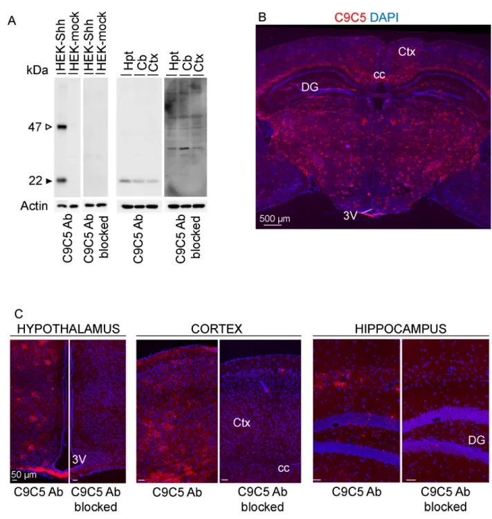
We sought to identify in the adult mouse brain, the protein expression and cell distribution of Sonic Hedgehog (Shh) using the monoclonal ‘C9C5’ antibody directed against the amino-ter-minal fragment of human SHH. From Western blot experiment analysis (Fig 1A,S1 Fig), we observed that the C9C5 antibody detected a 22 kDa signal corresponding to the expected size for the cleaved amino-terminal fragment of Shh (ShhN) in both HEK293 cells expressing mouse Shh and brain tissues lysates from adult mice. This signal was eliminated in blocking experiments (Fig 1A) consistent with this signal being related to ShhN.
Immunofluorescent labeling with C9C5 revealed high immunoreactivity throughout most brain regions including the cerebral cortex, the hippocampus, in fiber tracts, in thalamic nuclei, in the tuberal region of the hypothalamus including the median eminence (ME) (Fig 1B and 1C) and in the cerebellum (S2A and S2B Fig). Specificity of C9C5 binding was pro-vided by the absence of these signals in blocking experiments (Fig 1C). Interestingly, C9C5-immunoreactive cells displayed a stellate morphology with intense signal in their cell bodies and their processes that were decorated by immunoreactive puncta (Figs2A–2C,3A, 3B,5E,6B–6E;S2B,S3A and S3BFigs). These cells were distributed in almost all brain regions analyzed (Figs1,2A–2C,3A,3B,5A,5Eand6D;S2A,S2B,S3A and S3BFigs). In the cerebel-lum, Purkinje cells are expressing Shh [2,3] but were C9C5 negative (S2A and S2B Fig) sug-gesting the antibody does not recognize neuronal Shh.
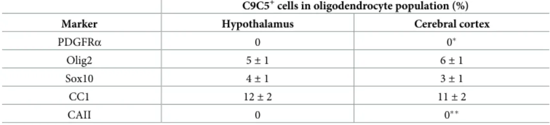
All C9C5+cells were also immunoreactive for the three oligodendroglial markers Olig2, Sox10 and CC1 (Fig 2A–2C;Fig 3A,S3A Fig). C9C5+cells were not immunoreactive for Plate-let-derived growth factor receptor-alpha (PDGFRα), a marker of oligodendrocyte progenitor cells (OPCs) [20] and for carbonic anhydrase (CA) II, a marker of a subpopulation of mature oligodendrocytes [21] (Fig 3B,S3B Fig). Quantification of the distribution of C9C5+cells in these various oligodendroglial cell populations in the hypothalamus and in the cerebral cortex are presented inTable 1. Thus, in these two regions, C9C5+cells represented 11–12% of CC1+ cells and 3–6% of Sox10+and Olig2+cells. These data suggest that C9C5 labels a subpopulation of mature oligodendrocytes as shown in hypothalamic nuclei, the ME, the hippocampal and cerebral cortical layers and in the white matter of the corpus callosum (Figs2A–2Cand3A; S3A Fig). In the hippocampus, these oligodendroglial cells were scattered over the pyramidal cell populations (Fig 2B, red square), were present in the hilus region and the neurogenic niche associated to the granule cell layer of the dentate gyrus (Fig 2B, yellow square;S3A and S3B Fig).
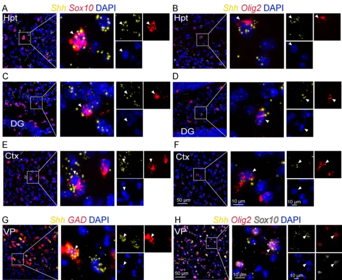
To further support that oligodendroglial cells are a source of Shh in the adult brain, we used single molecule multiplex fluorescentin situ hybridization to investigate Shh mRNA distribu-tion. We successfully detected robust expression of Shh transcripts using RNAscope in a subset of scattered cells expressing the transcripts of Olig2 and Sox10 in the brain regions described above (Fig 4A–4F,Table 2). In forebrain regions, Shh mRNAs were detected in glutamic acid
decarboxylase (GAD)+GABAergic neurons (Fig 4G) as reported previously [10] and in Sox10+Olig2+oligodendroglial cells (Fig 4H).
Fig 1. Characterization of the C9C5 anti-SHHN antibody expression pattern. (A) Western blot of protein homogenates from HEK293 cells transfected with a mouse Shh (HEK-Shh) or control vector (HEK-mock) and from hypothalamic (Hpt), cerebellar (Cb) or cerebrocortical (Ctx) tissues of an adult mouse with the C9C5 antibody, and was reproduced with tissues from three other animals. A 22 kDa band, the expected size for the aminoterminal fragment of Shh, was detected in HEK-Shh cells and in tissues but was absent in HEK-mock cells and in blocking experiments (n = 3). A 47 kDa signal corresponding to the uncleaved Shh protein was detected in HEK-Shh cells but not in tissues. Actin served as a loading control. (B-C) Low-magnification photomontages of coronal brain sections of adult mice showing strong C9C5 immunoreactivity in almost all brain regions (B), which was absent in blocking experiments (n = 3) (C). Staining was replicated in the twenty adult mice used in this study. Nuclei stained with DAPI. 3V, third ventricle; Hpt, Hypothalamus; Ctx, cerebral cortex; Cb, cerebellum; cc, corpus callosum; DG, dentate gyrus of the hippocampus.
Shh from C9C5
+cells is localized next to Ptc-expressing cells
The distribution of the C9C5 signal in the oligodendroglial cell body and processes suggested that the associated Shh protein could be secreted to act locally on cells expressing its receptor Patched (Ptc). In the tuberal region of the hypothalamus of PtcLacZ/+knock-in reporter mice, βgalactosidase (βgal) was strongly expressed in GFAP+
(Fig 5A–5C) or S100β+(79.3± 3.1%, n = 4 animals) (Fig 5D) astrocyte nuclei.βgalactosidase was also detected with a lower intensity in cells positive for the neuronal marker HuC/D but was not identified in cells expressing the microglial marker Iba1 (Fig 5D). Using confocal microscopy, we showed that C9C5+cells and their processes were in close contact with cells showing an intenseβgal staining (Fig 5E). These observations suggest that Shh secreted from the oligodendroglial cell could act on sev-eral neighboring astrocytes or neurons expressing Ptc.
Fig 2. C9C5+cells express the oligodendroglial markers CC1 and Olig2. (A-C) Immunostaining of coronal brain sections from an adult mouse of the hypothalamic median eminence (A), the hippocampus (B), the cerebral cortex and corpus callosum (C) with the C9C5 antibody (yellow) and the oligodendroglial markers CC1 (green) and Olig2 (red). Yellow and red squares highlight magnifications of the ventromedial hypothalamic nucleus and the median eminence (A), the dentate gyrus and CA3 pyramidal layer (B) of the hippocampus, the cerebral cortex and the corpus callosum (C). White boxes show C9C5/CC1/Olig2 triple positive cells with stellate morphology presented in merge and single channels with the nuclear marker DAPI. Staining was replicated at least on three mice. 3V, third ventricle; Ctx, cerebral cortex; cc, corpus callosum; DG, dentate gyrus of the hippocampus.
https://doi.org/10.1371/journal.pone.0229362.g002
Fig 3. Characterization of C9C5+cells at different stages of the oligodendroglial lineage. (A-B) Immunostaining of coronal brain sections from an adult mouse of the cerebral cortex and corpus callosum with the C9C5 antibody (yellow) and the oligodendroglial markers (A) CC1 (green) and Sox10 (red) or (B) CAII (green) and PDGFRα (red). White boxes (A, B) highlight magnifications of the cerebral cortex and the corpus callosum. (A) C9C5/CC1/Sox10 triple positive cells (white arrowhead) with stellate morphology are presented in merge and single channels with the nuclear marker DAPI. (B) C9C5+(white arrowhead), CAII+(orange arrow), and PDGFRα+(white arrow) cells are presented in merge and single channels with the nuclear marker
DAPI. Note that C9C5 positive cells are CAII and PDGFRα negative. Staining was replicated at least on three mice. Ctx, cerebral cortex; cc, corpus callosum.
Distribution of C9C5
+cells varies with age
Then, we investigated the presence of C9C5+cells in the brain during the postnatal period and in aged animals (Fig 6A–6G). In hypothalamic nuclei, C9C5 immunoreactivity was present in cells displaying stellate morphology starting at P10 with a pattern of cell distribution at P20 that mimicked the adult distribution (Fig 6B–6E). The number of C9C5+cells strongly decreased during ageing in the hypothalamus (Fig 6D–6G). In the ME, C9C5+cells were already observed at P4 (Fig 6A) while intense signal was present in adults and persisted in old animals (Fig 6D–6F), suggesting a different regulation of the signal over time compared to the nearby hypothalamic parenchyma. Quantification of the distribution of C9C5+cells with age in hypothalamic nuclei are presented inFig 6G.
Fig 4. Shh mRNA expression in oligodendroglial cells. Multiplexin situ hybridization for Shh and for the oligodendroglial markers Olig2 and
Sox10 as indicated, at the level of the tuberal region of the hypothalamus (Hpt) (A-B), the CA3 region of the hippocampus (C-D), the cerebral cortex (Ctx) (E-F), the ventral pallidum (VP) (H), and for the GABAergic marker GAD in the ventral pallidum (G). White boxes highlight magnifications of double Shh/Sox10 (A, C, E), Shh/Olig2 (B, D, F), Shh/GAD (G) or triple Shh/Sox10/Olig2 (H) positive cells (white arrowhead) presented in merge and single channels with the nuclear marker DAPI. Staining was replicated on three mice. DG, dentate gyrus of the hippocampus.
Fig 5. C9C5+cells delineate Shh expression domains containing Ptc-expressing cells. (A-E) Fluorescence confocal (A-C, E) imaging ofβgalactosidase (βgal), C9C5, GFAP, S100β, HuC/D, and Iba1 immunostaining on coronal brain sections of adult
Discussion
Here, we report the widespread distribution pattern of the C9C5 immunoreactivity in the adult mouse brain and show that C9C5 antibody marks a subset of CC1+(11–12%), Olig2+ and Sox10+(3–6%), CAII-mature oligodendrocytes as quantified in the cerebral cortex and in the hypothalamus. These cells do not express the PDGFRα signal implicated in OPCs prolifer-ation and in the inhibition of the expression of oligodendroglial maturprolifer-ation markers such as CC1 [18,20]. Thus, these mature oligodendroglial cells constitute a source for delivering Shh to brain cells. Our experiments show that the C9C5 antibody recognizes ShhN in rodent brain extracts as previously reported [22,23], and on postnatal mouse brain sections. These data are also in agreement with previous reports describing that C9C5 detects specifically Shh in ventral midbrain progenitor domains and in the diencephalic organizer zona limitans intrathalamica of mouse embryo [24,25], and in adult mouse sensory neurons [26].
Interestingly, we also identify for the first time Shh mRNAs in Olig2+and Sox10+cells in the adult mouse brain using RNAscope which indicates that Shh is more widely expressed than originally reported [10,14], and suggests novel physiological functions for this molecule. Thus, altogether, our data support that C9C5 recognizes Shh produced by a small population of mature oligodendrocytes. It is not the case for Shh from Purkinje cells of the cerebellar cor-tex [27,28] reflecting possible difference in Shh post-translational modifications [1]. We can-not exclude that C9C5 may recognize acan-nother yet unknown protein showing SHHN
homology, restricted to a subset of oligodendrocytes and which is not Desert Hedgehog or Indian Hedgehog that are not expressed in the rodent brain [2,27].
Studies using other Shh antibodies [13,14,29] or reporter mouse lines [9,14,30] did not report Shh expression in oligodendroglial cells in normal adult mouse brain. However, a dynamic expression of Shh in PDGFRα+
and Olig2+oligodendrocyte progenitors occurs dur-ing remyelination [29]. This apparent discrepancy with our data may reflect different sensitiv-ity of the reporters, of the mouse line constructs or of the antibodies used. For the latter, it may indicate different lipid modifications of ShhN [1] that might occur in oligodendrocyte precur-sor cells and mature oligodendrocytes.
Interestingly, the Shh signal labels the oligodendroglial cell body and decorates by intense puncta the processes extending at several cell diameters. Thus, Shh could be secreted to act locally on its receptor Ptc expressed by neighboring astrocytes and neurons. It is noteworthy that C9C5+CC1+cells are distributed in a grid-like manner with cells residing mostly in non-superposed domains. They constitute small Shh-expressing units that could distribute locally the protein in almost all brain regions and could represent a source for spreading Shh. Thus, our data suggest that besides neurons in the adult forebrain and cerebral cortex [9,31] and in the cerebellar cortex [8], oligodendroglial cells are a potential source of Shh for signaling in astrocytes and neurons. The appearance of C9C5+and CC1+cells in the postnatal mouse brain overlaps the myelination peak that occurs between P10 and P20 [18] further arguing for a role of mature oligodendrocytes in postnatal Shh pathway regulation.
PtcLacZ/+mouse hypothalamus at the level of the median eminence. (A, E) Detection ofβgal signal (yellow) alone and together
with C9C5 signal (red) indicated wide distribution of Ptc-expressing cells in hypothalamic nuclei. (B-C) Orthogonal views of the magnified white square (A, left) showing co-localization ofβgal and the astroglial marker GFAP. (D) High magnifications of the hypothalamic parenchyma from independent experiments showing colocalization ofβgal (white arrowheads) with the astroglial marker S100β or with the neuronal marker HuC/D, but not with the microglial marker Iba1 (white arrow). (E) White square magnification (from A, right) showing C9C5+cell bodies (white arrows) delineating Shh expression domains in the parenchyma and next to the ependymal layer (Ep) of the third ventricle (3V). White boxes highlight magnifications with orthogonal views of C9C5+processes contactingβgal+Ptc-expressing cells (white arrowheads). Staining was replicated on four mice. Nuclei stained
with DAPI.
Recently, an unsuspected oligodendrocyte heterogeneity has been reported by RNA sequencing of oligodendrocyte lineage cells both in the juvenile and adult mouse brain [32, 33]. Further studies of human brain from patients with multiple sclerosis or from spinal cord of mouse models of multiple sclerosis, revealed several clusters of oligodendroglial cells that could be further classified according to their gene expression pattern [34,35]. It would be important to identify to which clusters C9C5+CC1+cells belong and to characterize their prop-erties in CNS inflammation and during remyelination [36]. Thus, the oligodendroglial C9C5+Shh-associated signal should undoubtedly be taken into consideration when analyzing brain disorders associated to Shh signaling including brain tumors, neurodevelopmental dis-orders and neurodegenerative diseases [2].
Supporting information
S1 Fig. Western blot analysis of Shh protein with C9C5 with short and long exposure time. Twentyμg of RIPA solubilized samples from cerebellum (Cb) and hypothalamus (Hpt) and 1μg of mock or mShh transfected HEK293 homogenates were subjected to Western blot and immunodetection with C9C5 antibody (1/1000). Chemiluminescence was acquired with Chemidoc apparatus. The exposure time is indicated below. The 22 kDa (ShhN, filled arrow-head) and 47 kDa (uncleaved Shh; empty arrowarrow-head) bands are indicated. A single band
Fig 6. C9C5 immunoreactivity in the mouse hypothalamus across life. (A-F) Immunostaining with the C9C5 antibody on coronal sections of the hypothalamus at P4 (A) (n = 3), P10 (B) (n = 3), P20 (C) (n = 4), and at two (D) (n = 4), nine (E) (n = 3) and twelve (F) (n = 4) months. C9C5+cells are detected in hypothalamic nuclei at P10 until
adulthood but not at P4. An age-associated decrease of C9C5+cells in the parenchyma but not in the median eminence
(ME) was observed (D-F). Magnifications of white boxes show C9C5+cells with a stellate morphology, which were absent at P4 (A) and in twelve month-old animals (F). C9C5+cells are present in the ME at P4 (A). (G) Quantification of C9C5+cells across life (n = 3–4 per age). Means
± SEM.�p < 0.05;��p < 0.01;���p < 0.001. Nuclei stained with
DAPI. 3V, third ventricle; mo, months.
https://doi.org/10.1371/journal.pone.0229362.g006
Table 1. Quantification of C9C5+cells in oligodendrocyte populations in the adult mouse brain. C9C5+cells in oligodendrocyte population (%) Marker Hypothalamus Cerebral cortex
PDGFRα 0 0�
Olig2 5± 1 6± 1
Sox10 4± 1 3± 1
CC1 12± 2 11± 2
CAII 0 0��
Data are from experiments presented in Figs2and3andS3 Figand are means± SEM from 3–4 mice.
�One C9C5+cell / 364 PDGFRα+cells quantified ��one C9C5+cell / 982 CAII+cells quantified.
https://doi.org/10.1371/journal.pone.0229362.t001
Table 2. Analysis of Shh mRNA expression in Olig2- and Sox10-expressing cells in the adult mouse brain. Region Shh+Olig2+/Olig2+(% cells) Shh+Sox10+/Sox10+(% cells)
Hypothalamus 20.1± 3.1 32.3± 0.6
Cerebral cortex 12.0± 1.3 13.5± 0.9
Data are from RNAscope1experiments (means
± SEM, n = 3 mice)
corresponding to ShhN is detected in brain extracts after short and long time exposure, further supporting specificity of the C9C5 antibody.
(TIF)
S2 Fig. Characterization of C9C5 immunoreactivity in the adult mouse cerebellum. (A-B) Immunostaining with C9C5 and CC1 antibodies on a coronal section of the cerebellum. (B) Magnification of red box indicates that Purkinje cell bodies (white arrowheads), and their pro-jections in the molecular layer are not labeled by the C9C5 antibody. Magnification of white box highlights a C9C5 positive cell expressing the oligodendroglial marker CC1 (white arrow) within the granular cell layer. Staining was replicated on three mice Mol, molecular cell layer; Gr, granular cell layer; Px, Purkinje cells. Scale bar: A, 100μm. B, 50 μm.
(TIF)
S3 Fig. Characterization of C9C5+cells at different stages of the oligodendroglial lineage. (A-B) Immunostaining of coronal brain sections from an adult mouse of the hypothalamus at the level of the median eminence and of the dentate gyrus of the hippocampus with the C9C5 antibody (yellow) and the oligodendroglial markers (A) CC1 (green) and Sox10 (red) or (B) CAII (green) and PDGFRα (red). White boxes (A, B) highlight magnifications of the hypotha-lamic parenchyma and the granule cell layer of the dentate gyrus. (A) C9C5/CC1/Sox10 triple positive cells (white arrowhead) with stellate morphology are presented in merge and single channels with the nuclear marker DAPI. (B) C9C5+(white arrowhead), CAII+(orange arrow),
PDGFRα+
(white arrow) cells are presented in merge and single channels with the nuclear marker DAPI. Note that C9C5 positive cells are CAII and PDGFRα negative. Staining was rep-licated on three mice. Hpt, hypothalamus; DG, dentate gyrus of the hippocampus.
(TIF)
S1 Raw images. (PDF)
Author Contributions
Conceptualization: Martial Ruat.
Data curation: Linda Tirou, Mariagiovanna Russo, Helene Faure, Giuliana Pellegrino, Ariane Sharif.
Formal analysis: Linda Tirou, Mariagiovanna Russo, Helene Faure, Giuliana Pellegrino, Ariane Sharif.
Methodology: Martial Ruat. Supervision: Martial Ruat.
Writing – original draft: Martial Ruat.
References
1. Briscoe J, Therond PP. The mechanisms of Hedgehog signalling and its roles in development and dis-ease. Nat Rev Mol Cell Biol. 2013; 14(7):416–29.https://doi.org/10.1038/nrm3598PMID:23719536. 2. Ruat M, Faure H, Daynac M. Smoothened, stem cell maintenance and brain diseases. Top Med Chem
2015; 16:147–71.https://doi.org/10.1007/7355_2014_83
3. Garcia ADR, Han YG, Triplett JW, Farmer WT, Harwell CC, Ihrie RA. The elegance of Sonic hedgehog: emerging novel functions for a classic morphogen. J Neurosci. 2018; 38(44):9338–45.https://doi.org/ 10.1523/JNEUROSCI.1662-18.2018PMID:30381425.
4. Ruat M, Hoch L, Faure H, Rognan D. Targeting of Smoothened for therapeutic gain. Trends Pharmacol Sci. 2014; 35(5):237–46.https://doi.org/10.1016/j.tips.2014.03.002PMID:24703627.
5. Allahyari RV, Clark KL, Shepard KA, Garcia ADR. Sonic hedgehog signaling is negatively regulated in reactive astrocytes after forebrain stab injury. Scientific reports. 2019; 9(1):565.https://doi.org/10.1038/ s41598-018-37555-xPMID:30679745.
6. Bezard E, Baufreton J, Owens G, Crossman AR, Dudek H, Taupignon A, et al. Sonic hedgehog is a neuromodulator in the adult subthalamic nucleus. Faseb J. 2003; 17(15):2337–8.https://doi.org/10. 1096/fj.03-0291fjePMID:14525941.
7. Pascual O, Traiffort E, Baker DP, Galdes A, Ruat M, Champagnat J. Sonic hedgehog signalling in neu-rons of adult ventrolateral nucleus tractus solitarius. Eur J Neurosci. 2005; 22(2):389–96.https://doi. org/10.1111/j.1460-9568.2005.04223.xPMID:16045492.
8. Farmer WT, Abrahamsson T, Chierzi S, Lui C, Zaelzer C, Jones EV, et al. Neurons diversify astrocytes in the adult brain through sonic hedgehog signaling. Science. 2016; 351(6275):849–54.https://doi.org/ 10.1126/science.aab3103PMID:26912893.
9. Garcia AD, Petrova R, Eng L, Joyner AL. Sonic hedgehog regulates discrete populations of astrocytes in the adult mouse forebrain. J Neurosci. 2010; 30(41):13597–608.https://doi.org/10.1523/
JNEUROSCI.0830-10.2010PMID:20943901.
10. Traiffort E, Moya KL, Faure H, Hassig R, Ruat M. High expression and anterograde axonal transport of aminoterminal sonic hedgehog in the adult hamster brain. Eur J Neurosci. 2001; 14(5):839–50.https:// doi.org/10.1046/j.0953-816x.2001.01708.xPMID:11576188.
11. Charytoniuk D, Porcel B, Rodriguez Gomez J, Faure H, Ruat M, Traiffort E. Sonic Hedgehog signalling in the developing and adult brain. J Physiol Paris. 2002; 96(1–2):9–16. https://doi.org/10.1016/s0928-4257(01)00075-4PMID:11755778.
12. Lai K, Kaspar BK, Gage FH, Schaffer DV. Sonic hedgehog regulates adult neural progenitor prolifera-tion in vitro and in vivo. Nat Neurosci. 2003; 6(1):21–7.https://doi.org/10.1038/nn983PMID:12469128. 13. Machold R, Hayashi S, Rutlin M, Muzumdar MD, Nery S, Corbin JG, et al. Sonic hedgehog is required
for progenitor cell maintenance in telencephalic stem cell niches. Neuron. 2003; 39(6):937–50.https:// doi.org/10.1016/s0896-6273(03)00561-0PMID:12971894.
14. Ihrie RA, Shah JK, Harwell CC, Levine JH, Guinto CD, Lezameta M, et al. Persistent sonic hedgehog signaling in adult brain determines neural stem cell positional identity. Neuron. 2011; 71(2):250–62.
https://doi.org/10.1016/j.neuron.2011.05.018PMID:21791285.
15. Gonzalez-Reyes LE, Chiang CC, Zhang M, Johnson J, Arrillaga-Tamez M, Couturier NH, et al. Sonic Hedgehog is expressed by hilar mossy cells and regulates cellular survival and neurogenesis in the adult hippocampus. Scientific reports. 2019; 9(1):17402.https://doi.org/10.1038/s41598-019-53192-4
PMID:31758070.
16. Angot E, Loulier K, Nguyen-Ba-Charvet KT, Gadeau AP, Ruat M, Traiffort E. Chemoattractive activity of sonic hedgehog in the adult subventricular zone modulates the number of neural precursors reaching the olfactory bulb. Stem Cells. 2008; 26(9):2311–20.https://doi.org/10.1634/stemcells.2008-0297
PMID:18617686.
17. Daynac M, Tirou L, Faure H, Mouthon MA, Gauthier LR, Hahn H, et al. Hedgehog Controls Quiescence and Activation of Neural Stem Cells in the Adult Ventricular-Subventricular Zone. Stem Cell Reports. 2016; 7(4):735–48.https://doi.org/10.1016/j.stemcr.2016.08.016PMID:27666792.
18. Laouarem Y, Traiffort E. Developmental and repairing production of myelin: the role of Hedgehog sig-naling. Front Cell Neurosci. 2018; 12:305.https://doi.org/10.3389/fncel.2018.00305PMID:30237763. 19. Ferent J, Cochard L, Faure H, Taddei M, Hahn H, Ruat M, et al. Genetic activation of hedgehog
signal-ing unbalances the rate of neural stem cell renewal by increassignal-ing symmetric divisions. Stem Cell Reports. 2014; 3(2):312–23.https://doi.org/10.1016/j.stemcr.2014.05.016PMID:25254344. 20. Dang TC, Ishii Y, Nguyen V, Yamamoto S, Hamashima T, Okuno N, et al. Powerful Homeostatic
Con-trol of Oligodendroglial Lineage by PDGFRalpha in Adult Brain. Cell reports. 2019; 27(4):1073–89 e5.
https://doi.org/10.1016/j.celrep.2019.03.084PMID:31018125.
21. Matsusue Y, Horii-Hayashi N, Kirita T, Nishi M. Distribution of corticosteroid receptors in mature oligo-dendrocytes and oligodendrocyte progenitors of the adult mouse brain. J Histochem Cytochem. 2014; 62(3):211–26.https://doi.org/10.1369/0022155413517700PMID:24309510.
22. Eitan E, Petralia RS, Wang YX, Indig FE, Mattson MP, Yao PJ. Probing extracellular Sonic hedgehog in neurons. Biol Open. 2016; 5(8):1086–92.https://doi.org/10.1242/bio.019422PMID:27387534. 23. Rivell A, Petralia RS, Wang YX, Clawson E, Moehl K, Mattson MP, et al. Sonic hedgehog expression in
24. Tang M, Villaescusa JC, Luo SX, Guitarte C, Lei S, Miyamoto Y, et al. Interactions of Wnt/beta-catenin signaling and sonic hedgehog regulate the neurogenesis of ventral midbrain dopamine neurons. J Neu-rosci. 2010; 30(27):9280–91.https://doi.org/10.1523/JNEUROSCI.0860-10.2010PMID:20610763. 25. Andreu-Cervera A, Anselme I, Karam A, Laclef C, Catala M, Schneider-Maunoury S. The ciliopathy
gene Ftm/Rpgrip1l controls mouse forebrain patterning via region-specific modulation of Hedgehog/Gli signaling. J Neurosci. 2019; 39(13):2398–415.https://doi.org/10.1523/JNEUROSCI.2199-18.2019
PMID:30692221.
26. Lu WJ, Mann RK, Nguyen A, Bi T, Silverstein M, Tang JY, et al. Neuronal delivery of Hedgehog directs spatial patterning of taste organ regeneration. Proc Natl Acad Sci U S A. 2017; 115(2):E200–E9.https:// doi.org/10.1073/pnas.1719109115PMID:29279401.
27. Traiffort E, Charytoniuk DA, Faure H, Ruat M. Regional distribution of sonic hedgehog, patched, and smoothened mRNA in the adult rat brain. J Neurochem. 1998; 70(3):1327–30.https://doi.org/10.1046/j. 1471-4159.1998.70031327.xPMID:9489757
28. Wallace VA. Purkinje-cell-derived Sonic hedgehog regulates granule neuron precursor cell proliferation in the developing mouse cerebellum. Curr Biol. 1999; 9(8):445–8.https://doi.org/10.1016/s0960-9822 (99)80195-xPMID:10226030
29. Ferent J, Zimmer C, Durbec P, Ruat M, Traiffort E. Sonic Hedgehog Signaling Is a Positive Oligoden-drocyte Regulator during Demyelination. J Neurosci. 2013; 33(5):1759–72.https://doi.org/10.1523/ JNEUROSCI.3334-12.2013PMID:23365216.
30. Sanchez MA, Sullivan GM, Armstrong RC. Genetic detection of Sonic hedgehog (Shh) expression and cellular response in the progression of acute through chronic demyelination and remyelination. Neuro-biol Dis. 2018; 115:145–56.https://doi.org/10.1016/j.nbd.2018.04.003PMID:29627579.
31. Hill SA, Blaeser AS, Coley AA, Xie Y, Shepard KA, Harwell CC, et al. Sonic hedgehog signaling in astro-cytes mediates cell-type-specific synaptic organization. Elife. 2019; 8.https://doi.org/10.7554/eLife. 45545PMID:31194676.
32. Marques S, Zeisel A, Codeluppi S, van Bruggen D, Mendanha Falcao A, Xiao L, et al. Oligodendrocyte heterogeneity in the mouse juvenile and adult central nervous system. Science. 2016; 352(6291):1326– 9.https://doi.org/10.1126/science.aaf6463PMID:27284195.
33. Chen R, Wu X, Jiang L, Zhang Y. Single-Cell RNA-Seq Reveals Hypothalamic Cell Diversity. Cell reports. 2017; 18(13):3227–41.https://doi.org/10.1016/j.celrep.2017.03.004PMID:28355573. 34. Falcao AM, van Bruggen D, Marques S, Meijer M, Jakel S, Agirre E, et al. Disease-specific
oligodendro-cyte lineage cells arise in multiple sclerosis. Nat Med. 2018; 24(12):1837–44.https://doi.org/10.1038/ s41591-018-0236-yPMID:30420755.
35. Jakel S, Agirre E, Mendanha Falcao A, van Bruggen D, Lee KW, Knuesel I, et al. Altered human oligo-dendrocyte heterogeneity in multiple sclerosis. Nature. 2019; 566(7745):543–7.https://doi.org/10.1038/ s41586-019-0903-2PMID:30747918.
36. Niu J, Tsai HH, Hoi KK, Huang N, Yu G, Kim K, et al. Aberrant oligodendroglial-vascular interactions dis-rupt the blood-brain barrier, triggering CNS inflammation. Nat Neurosci. 2019; 22(5):709–18.https://doi. org/10.1038/s41593-019-0369-4PMID:30988524.