HAL Id: hal-02711883
https://hal.inrae.fr/hal-02711883
Submitted on 1 Jun 2020
HAL is a multi-disciplinary open access
archive for the deposit and dissemination of
sci-entific research documents, whether they are
pub-lished or not. The documents may come from
teaching and research institutions in France or
abroad, or from public or private research centers.
L’archive ouverte pluridisciplinaire HAL, est
destinée au dépôt et à la diffusion de documents
scientifiques de niveau recherche, publiés ou non,
émanant des établissements d’enseignement et de
recherche français ou étrangers, des laboratoires
publics ou privés.
The FGF6 gene within the FGF multigene family
François Coulier, Vincent Ollendorff, Irène Marics, Olivier Rosnet, Michèle
Batoz, Jacqueline Planche, Sylvie Marchetto, Marie-Josephe Pébusque, Odile
Delapeyrière, Daniel Birnbaum
To cite this version:
François Coulier, Vincent Ollendorff, Irène Marics, Olivier Rosnet, Michèle Batoz, et al.. The FGF6
gene within the FGF multigene family. Annals of the New York Academy of Sciences, Wiley, 1991,
638, pp.53-61. �hal-02711883�
The
FGF6 Gene within the
FGF
Multigene Family"
FRANCOIS COULIER, VINCENT OLLENDORFF, JACQUELINE PLANCHE, SYLVIE MARCHEmO,
AND DANIEL BIRNBAUM
U. 119 INSERM 27 Boulevard Lei' Roure 13009, Marseille, France
IRENE MARKS, OLIVIER ROSNET, MICHELE BATOZ, MARIE-JOSEPHE PEBUSQUE, ODILE DELAPEYRIERE,
Cell growth and differentiation are controlled at multiple levels through various pathways. External signals and their specific receptors represent important protago- nists of this control. Among the signaling molecules are peptide regulatory factors including numerous neuropeptides, cytokines, and growth factors, often grouped in families. Growth factors and their receptors are involved in various processes including cell proliferation, cell differentiation, cell-cell interactions and develop- ment.
The FGF (historically meaning fibroblast growth factor) family presently com- prises seven related members (hereafter designated FGFl to FGF7) but the actual size of the family remains unknown. Like most of the signaling molecules, they have various known properties as well as suspected functions in basic processes as diverse as cell proliferation, angiogenesis, tissue differentiation and regeneration, and embryogenesis.' Due to these activities, FGFs may also play an important role in carcinogenesis. This involvement in carcinogenesis is strongly suspected but is still, in early 1991, a matter of discussion. The FGFs can behave as oncogene products or potent angiogenic and mitogenic factors in in vitro cell cultures and in animal models.'-l Moreover, amplifications of some of the FGF (IN72 and HSTIFGFK) and
FGF receptor (FLG and BEK) genes are observed in human carcinomas.s" In
cancer, each member of the couple growth factor-growth factor receptor represents a particular potential therapeutical target aimed at blocking the specific interaction. Therefore, it is paramount to characterize every member of the FGF and FGFR families.
The sixth member of the FGF family, the FGF6 gene, was isolated a couple of years ago" by virtue of its sequence similarities with FGF4 (HSTIFGFK). The human'" and murine'" genes have been characterized, mapped on their respective chromosome, and their expression and transforming capacity studied. These results are the object of this report.
'This work was supported by grants from INSERM and from the following organisms: Association FranGaise contre la Myopathie, Association pour la Recherche sur le Cancer, Cornites Departementaux de Bouches-du-RhBne et du Var de la Ligue Contre le Cancer, FNCLCC, MGEN.
54 ANNALS NEW YORK ACADEMY OF SCIENCES
STRUCTURE OF THE FGF6 GENE AND ENCODED PROTEINS
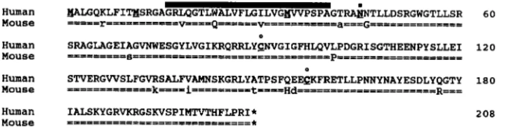
The FGF6 gene is localized on chromosome 12, band p12, in the human and on chromosome 6, region F3-G1, in the mouse. It is composed of three coding exons separated by large introns. Human and mouse deduced amino acid sequences are shown in FIGURE 1. There is 93% amino acid sequence identity between the human and mouse gene products. At the nucleotide level (not shown), the calculated rate of nonsynonymous substitutions is 0.17 x lo-’ per site per year (assuming a time of divergence between human and rodent genes of 80 x 10 years ago). This is well below average” and indicates a strong conservation.
The localization of the splice junctions is similar to that of the other members of the family. There are four stop codons located immediately upstream of the beginning of the coding sequence, followed by three in-frame ATG codons. With the first ATG starts an open reading frame of 624 nucleotides, encoding a putative protein of 208 amino acids. A potential signal peptide, constituted of a hydrophobic region of 25 amino acids, starts shortly after the methionine residue corresponding to the second ATG, extends from position 16 (considering the first ATG as position 1) to position 40. Two potential proteolytic cleavage sites are located towards the end of the signal peptide. In vitro translation experiments (not shown) seem to indicate that it is the first site that is used. A potential asparagine-linked glycosylation site is immediately adjacent, at position 45. This site is functional in in vitro translation experiments (not shown).
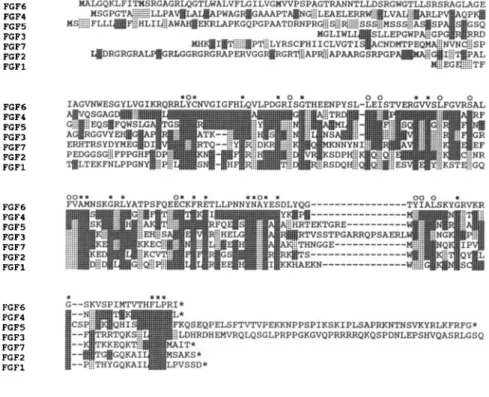
The comparison of the deduced amino acid sequence of the human FGF6 gene product with those of the other FGFs (FIGURE 2) shows that the closest relative is the FGF4 gene product, as was expected considering the cross-reactivity of the respective molecular probes. FGFs have sequence similarities in a “core” portion coded by the 3‘ half of exon 1, exon 2, and exon 3, but differ greatly in the portion coded by the 5’ half of exon 1. In addition, some FGF proteins present specific insertions and extensions. Paralogous FGF6 proteins have more similarities than FGF6 and FGF4 proteins of the same species; this is especially true when consider- ing the portion coded by the 5’ part of exon 1. It is possible that exon 1 corresponds to a merger between two ancestral exons, created as a result of the loss of an intron. The structural features of the FGF6 gene product are summarized in FIGURE 3.
Human 60
208
FIGURE 1. Conservation between human and mouse FGF6 deduced amino acid sequences. The human FGF6 amino acid sequence is shown using the single-letter code. The mouse corresponding sequence is shown beneath using an = symbol for identical amino acids. Different but conserved amino acids are in lowercase letters. Specific features are indicated as follows: signal peptide, black bar; three potential methionine initiation codons, bold and underlined; N-glycosylation site and cysteine residues, bold, underlined with a symbol. The overall amino acid identity is 93.3% (97.6% when considering conservative changes).
COULIER ef al.: THE FGF6 GENE 55 FGFC FGF4 FGFS FGF3 FGF7 FGFZ F G F l FGFL FGF4 FGFS FGF3 FGF7 FGFZ F G F l FGFL FGF4 FGFS FGF3 FGF7 FGFZ F G F l FGFL FGF4 FGFS FGF3 FGF7 FGFZ F G F l FGF3 LEASAH*
FIGURE 2. Comparison between seven human FGF amino acid sequences. Sequences are aligned, using the single-letter code, to allow comparison with the FGF6 amino acid sequence. Identical amino acids are shown in dark shaded area and conserved amino acids in light shaded area. Initiation methionines are in bold, stops in asterisks. Black squares indicate seven identical residues; and circles, seven identical and conserved residues. FGFs are designated 1 to 7. The correspondence with commonly used names is as follows: FGFl = aFGF or FGFA or
HBGF1; FGF2 = bFGF or FGFB or HBGF2; FGF3 = INT2; FGF4 = HST or HSTFl or
K-FGF or FGFK FGF7 = KGF. A 5 ’ extension for INT2 has been demonstrated in mouse and probably also exists in human but was not represented. Sequences were taken from References 10 and 27-32.
The structure of the FGF genes is very similar in human and mouse. The overall
phylogcnetic conservation is however difficult to evaluate. A member of the FGF
gene family has been cloned in frog.” We have looked for evidence of such a conservation in lower species using zoo blot hybridizations (FIGURE 4). A murine Fgf6 probe hybridizes well with the human FGF6 gene as well a s with the human FGF4 (HSTIFGFK ) gene [lighter bands of 6 kilobases (kb) and 2.7 kb]. It lights up clear bands in chicken and frog DNAs. Bands are also present in the Drosophilu lane.
Except for human, it is not known presently whether these cross-reacting bands correspond to homologous genes.
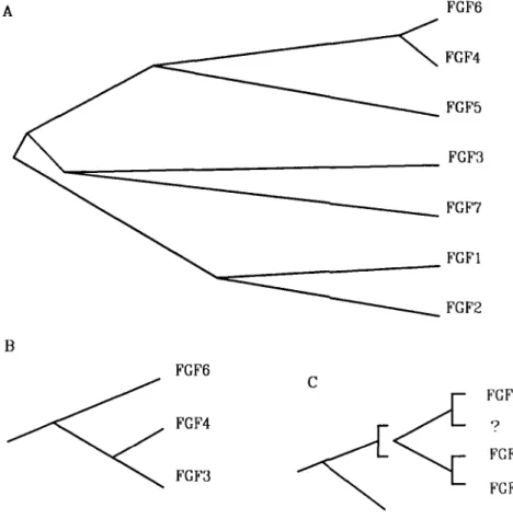
In view of the high degree of sequence similarities existing between them, we can readily surmise that the FGF genes are homologues. A phylogenetic tree based o n sequencc data is shown in FIGURE 5A. FGF genes have probably evolved by
56 ANNALS NEW YORK ACADEMY OF SCIENCES successive duplications but the evolution may have followed complex schemes at certain stages. Among many possibilities, two such schemes could be hypothesized. In the first, a recent duplication is at the origin of FGF3 (“2) and FGF4
(HSTIFGFK), both located on chromosomal band llq13.3, separated by less than 40 kb.” Then, FGF3 evolved separately and rapidly diverged from FGF4, which remained very similar to FGF6. The second scheme hypothesizes a putative gene similar to FGF3 on human chromosome 12. In FIGURE 5, we have tentatively depicted these hypotheses (note that proteins and not gene sequences were used in this figure).
TRANSFORMING CAPACITY OF CLONED FGF6 GENE
The normal FGF6 gene has a transforming capacity comparable to that of
FGF4.”16 This was tested by bioassays involving the transfection of human and murine cloned genes into cultured murine NIH 3T3 fibroblasts followed by either a tumorigenicity assay or a focus assay. Clones of the normal FGF6 gene were either genomic cosmid clones or cDNA clones inserted into mammalian expression vectors. It is noteworthy that the sequence encoding the signal peptide is necessary for the transforming activity of the transfected FGF6 gene (Coulier et al., in press). Supernatants from mass culture of the transformed cells and proteins from in v i m
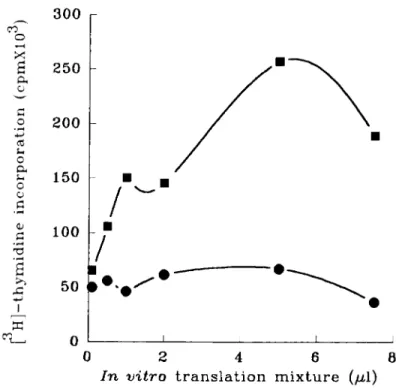
translated FGF6 gene are mitogenic for fibroblasts (FIGURE 6 ) . Moreover, FGF6- transformed cells are tumorigenic when injected into nude mice.
Despite this intrinsic transforming capacity, the FGF6 gene has never been detected so far in transfection assays, in contrast to FGF4 which has been frequently identified in these assays.”~’* Furthermore, the gene has never been found altered in human tumors. The intrinsic oncogenic capacity of the FGF genes must be repressed
in viva However, the mechanisms of this repression are not known.
M M
M i
C C P o t e n t i a l cleavage1
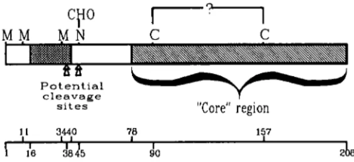
s i t e s “Core” region 1 1 3440 78 157 I I 16 dl345 90 2FIGURE 3. Structure of the FGF6 protein. The structure of the FGF6 protein was predicted
from cDNA sequence and in vitro translation experiments. A long open reading frame starting with three in-frame ATG codons is able to code for a protein of 208 residues. Only the two first ATG codons seem to be used for translation initiation. A stretch of 25 hydrophobic residues, indicated by a hatched box (a bar in FIGURE 1). may act as a signal peptide. Two potential
cleavage sites are indicated. A unique N-linked glycosylation site, which appears to be used in
vifro, is indicated by N. Two cysteine residues, conserved among the seven FGFs described to date, are indicated; they may be involved in a disulfide bridge. A “core” region (hatched) represents a region having sequence similarities in all members. The corresponding amino acid positions are indicated below.
COULIER el al.: THE FGF6 GENE
H M C F D
5723-
9.4
-
6.6-
4.4-
2.3-
2.0,
mu.FGF6 probe
FIGURE 4. Conservation of the FGF6 gene. Southern “ZOO blot” hybridization was carried out
at 4TC, in 35% formamide, 5 x SSC, 5 x Denhardt’s, on 10 pg of D N A from various species as follows: H, human; M, mouse; C, chicken; F, frog (Xenopus luevis); D, fly (Drosophila rnelunoguster). The probe used (probe PN) was a murine Fgf6 cDNA spanning the three coding
exons.
EXPRESSION OF THE FGF6 GENE IN
ADULT
AND EMBRYONIC TISSUESFGF genes are expressed in a wide variety of cells, in the adult as well as in the embryo. FGF genes present various patterns of expression in the developing embryo, where they could play an important physiological role as inducers of cell determina- tion and migration.”.”
58 ANNALS NEW YORK ACADEMY OF SCIENCES the embryonic mouse.'" In adult tissues the Fgf6 gene is expressed in skeletal muscle and heart, but not in smooth muscle. An Fgf6 transcript was also detected in adult testis. Use of northern blot hybridization with sense and antisense RNA probes showed that this testis-specific expression corresponds to an antisense transcript from the Fgf6 locus (FIGURE 7). Presently, nothing is known of its structure and role.
FGF6 FGF4 FGF5 FGF3 FGF7 FGFl FGF2
B
FGF6 ? F G M FGF3 CFIGURE 5. Phylogenetic FGF protein trees. A Unrooted phylogenetic tree inferred from the amino acid sequences of the FGFs. The operational units (OU) present at the external nodes are the seven FGF proteins with names used in FIGURE 2. The tree shows one topology only (out of 954 theoretically possible given 7 OU; Reference 11). Branches are scaled, i.e., their lengths are proportional to the number of substitutions. B and C portions of unrooted trees inferred from sequence and chromosomal localization data, with focus on three OU; FGF3 and
FGF4 are clustered on chromosomal band llq13," while FGF6 lies on band 12pter.l" In these particular trees, a specific rapid divergence of FGF3 (B) or a cluster duplication (C) is hypothesized.
The presence of an antisense transcript originating from an FGF locus has already
been noted in the case of FGF2 in Xenopus luevis.
In mouse embryos, Fgf6 is expressed during middle and late gestation with a peak at day 15. The transcript has a size of 4.8 kb. As a rule, the expression level is low, necessitating use of poly(A)' RNA to be detected.
COULIER et a/.: THE FGF6 GENE 59
300
250 200 150 100 50 0i
0 2 4 6 8In
v i t r o translation mixture (p1)FIGURE 6. Mitogenic activity of in virro translated FGF6 peptides. RNAs transcribed in vitro
were used in a rabbit reticulocyte lysate system, in the presence of canine pancreatic microso- ma1 membranes, to translate FGF6 peptides. Increasing amounts ofin vitro translation mixtures were tested for stimulation of ['Hlthymidine incorporation of Balb/c 3T3 cells. Samples tested included translation products from antisense (circles) or sense (squares) transcripts.
M
AS
S
Q
0.85
Kb
FIGURE 7. Detection of an antisense transcript from the FGF6 locus by northern blot hybridization. Poly ( A + ) RNA was isolated from 2-month-old mouse testis and hybridized with ["PICTP-labeled RNA, transcribed either in the Fgf6 sense orientation by T 7 (lane S) or in the antisense orientation by T3 (lane AS) polymerase from the appropriately linearized c D N A clone p173. This clone represents the mouse FKf6 gene first exon. Markers on the left represent Hae 111 DNA fragments from phage PhiX 174. The sense probe reveals an antisense transcript of 0.8.5 kilobase pair (kbp) (arrowhead).
60 ANNALS NEW YORK ACADEMY OF SCIENCES
CONCLUSION
When the number of the known FGFs suddenly rose from two to seven, it became important to define their specific role and how they assume it. Characterization of the FGF receptors could have helped in resolving this question. However, the analyses of ligand-receptor interactions reveal a peculiar pattern characterized by an ambiguity in the interacting FGF-FGFR couples; for the time being this contributes to darkening the picture a little more. How the cells discriminate between the various FGFs remains unknown. Furthermore, nothing is known about possible interactions between FGFs themselves. Is there such a thing as a coordinate regulation of FGF gene expression, at least during certain periods of development, under certain circumstances, or in certain areas? Study of the FGF6 gene has so far only introduced an element of complexity into these problems. It has shown that some members of the family can be more closely related than the average. It has confirmed that some FGF genes do present an intrinsic transforming activity even if their role in
carcinogenesis-especially of those having this capacity-is far from being estab- lished. Finally, the restricted pattern of expression of the gene points to a specific activity of FGF6 in the embryo and in cardiac and skeletal muscle cells. Regulating muscular activity or development appears to be an important function of the FGFs, and members of the family seem to exert a control on myoblasts.2'-26 The precise role of FGF6 with respect to muscle function will have to be defined.
ACKNOWLEDGMENTS
We thank C. Mawas for critical reading of the manuscript, and M. Marillet and S.
Kerridge for the gift of frog and Drosophila D N A , respectively.
REFERENCES 1. 2. 3. 4. 5. 6. 7. 8. 9. 10. 11. 12.
GOLDFARB, M. 1990. Cell Growth Diff. I: 439445.
DICKSON, C., R. SMITH, S. BROOKES & G. PETERS. 1984. Cell 37: 529-536.
MULLER, W., F. LEE, C. DICKSON, G. PETERS, P. PAITENGALE & P. LEDER. 1990. EMBO J. HALABAN, R., R. LANGDON, N. BIRCHALL, C. CUONO, A. BAIRD, G. Scorn, G. MOELLMAN
& J. MCGUIRE. 1988. J. Cell. Biol. 107: 1611-1619.
LIDEREAU, R., R. CALLAHAN, C. DICKSON, G. PETERS, C. ESCOT & I. U. &I. 1988. Oncogene Res. 2: 285-291.
FANTL, V., M. RICHARDS, R. SMITH, G. LAMMIE, G. JOHNSTONE, D. ALLEN, W. GREGORY, G. PETERS, C. DICKSON & D. BARNES. 1990. Eur. J. Cancer 2 6 423-429.
TSUDA, T., E. TAHARA, G. KAJIYAMA, H. SAKAMOTO, M. TERADA & T. SUGIMURA. 1989. Cancer Res. 4 9 5505-5508.
ADNANE, J., P. GAUDRAY, C. DIONNE, G. CRUMLEY, M. JAYE, J. SCHLESSINGER, P. JEAMEUR, D. BIRNBAUM & C. THEILLET. 1991. Oncogene 6 659-663.
MARICS, I., J. ADELAIDE, F. RAYBAUD, M.-G. MATTEI, F. COULIER, J. PLANCHE, 0. DELAPEYRIERE & D. BIRNBAUM. 1989. Oncogene 4 335-340.
DELAPEYRIERE, O., 0. ROSNET, D. BENHARROCH, F. RAYBAUD, S. MARCHEITO, J. PLANCHE, F. GALLAND, M. G. MAITEI, N. G. COPELAND, N. A. JENKINS, F. COULIER &
D. BIRNBAUM. 1990. Oncogene 5: 823-831.
LI, W-H. & D. GRAUR. 1991. Fundamentals in Molecular Evolution. Sinauer Assoc. Sunderland, Mass.
KIMELMAN, D., J. ABRAHAM, T. HAAPARANTA, T. PALISI & M. KIRSCHNER. 1988. Science 9 907-913.
COULIER et al.: THE FGF6 GENE 61 13 14. 15. 16. 17. 18. 19. 20. 21. 22. 23. 24. 25. 26. 27. 28. 29. 30. 31. 32.
NGUYLN, C., D. Roux, M. G. MATI-EI, 0. DELAPEYRIERE, M. GOLDFAKB, D. BIRNBAUM &
B. JORDAN. 1988. Oncogene 3: 703-708.
SAKAMOTO, H., T. YOSHIDA, M. NAKAKUKI, H. ODAGIRI, K. MIYAGAWA, T. SUGIMURA &
M. TERADA. 1988. Biochem. Biophys. Res. Commun. 151: 965-972.
MIYAGAWA, K., H. SAKAMOTO, T. YOSHIDA, Y. YAMASHITA, Y. MITSUI, M. FURUSAWA, S. MAEDA, F. TAKAKU, T. SUGIMURA & M. TERADA. 1988. Oncogene 3: 383-389.
DELLI Bow, P., A. M. CURATOLA, K. NEWMAN, Y. SATO, D. MOSCATELLI, R. HEWICK, D. RIFKIN & C. BASILICO. 1988. Mol. Cell. Biol. 8: 2933-2941.
SAKAMOTO, H., M. MORI, M. TAIKA, T. YOSHIDA, S. MATSUKAWA, K. SHIMIZU, M. SEKIGUCHI, M. TERADA & T. SUGIMURA. 1986. Proc. Nat. Acad. Sci. USA 83: 3997- 4001.
DELLI Bow, P. & C. BASILICO. 1987. Proc. Nat. Acad. Sci. USA 84: 5660-5664. WILKINSON, D., G. PETERS, C. DICKSON & A. MCMAHON. 1988. EMBO J. 7: 691495. VOLK, R., M. KOSTER, A. POTING, L. HARTMANN & W. KNOCHEL. 1989. EMBO J. GOSPODAROWICZ, D., J. WESEMAN, J. MORAN & J. LINDSTKOM. 1976. J. Cell. Biol. ALLEN, R., M. DODSON & L. LUITEN. 1984. Exp. Cell. Res. 152: 154-160.
LATHKOP, B., K. THOMAS & L. GLASEK. 1985. J. Cell. Biol. 101: 2194-2198. SEED, J. & S. HAUSCHKA. 1988. Dev. Biol. 128 40-49.
VAIDYA, T., S. RHODES, E. TAPAKOWSKY & S. KONIECZNY. 1989. Mol. Cell. Biol.
9 3576-3579.
ALTEKIO, J., Y. COURTOIS, J. ROBELIN, D. BECHET & I. MARTELLY. 1990. Biochem. Biophys. Res. Commun. 166: 1205-1212.
JAYE, M., R. HOWK, W. BURGESS, G. RICCA, I. M. CHIU, M. RAVERA, S. O’BRIEN, W. MODI, T. MACIAG & W. DROHAN. 1986. Science 233: 541-544.
PRATS, H., M. KAGHAD, A. C. PRATS, M. KLAGSBRUN, J. M. LELIAS, P. LIAUZUN, P. CHALON, J. P. TAUBEK, F. AMALRIC, J. SMITH & D. CAPUT. 1989. Proc. Nat. Acad. Sci. USA. 8 6 1836-1840.
BROOKES, S., R. SMITH, G. CASEY, C. DICKSON & G. PETERS. 1989. Oncogene 4 429436. TAIRA, M., T. YOSHIDA, K. MIYAGAWA, H. SAKAMOTO, M. TERADA & T. SUGIMURA. 1987. ZHAN, X., B. BATES, X. Hu & M. GOLDFARB. 1988. Mol. Cell. Biol. 8 3487-3495. FINCH, P., J. RUBIN, T. MIKI, D. RON & S. AARONSON. 1989. Science 245: 752-755.
8 2983-2988.
7 0 395405.