HAL Id: inserm-00273825
https://www.hal.inserm.fr/inserm-00273825
Submitted on 16 Apr 2008
HAL is a multi-disciplinary open access archive for the deposit and dissemination of sci-entific research documents, whether they are pub-lished or not. The documents may come from teaching and research institutions in France or abroad, or from public or private research centers.
L’archive ouverte pluridisciplinaire HAL, est destinée au dépôt et à la diffusion de documents scientifiques de niveau recherche, publiés ou non, émanant des établissements d’enseignement et de recherche français ou étrangers, des laboratoires publics ou privés.
Matthieu Roustit, Grant Simmons, Patrick Carpentier, Jean-Luc Cracowski
To cite this version:
Matthieu Roustit, Grant Simmons, Patrick Carpentier, Jean-Luc Cracowski. Abnormal digital neu-rovascular response to local heating in systemic sclerosis.. Rheumatology, Oxford University Press (OUP), 2008, 47 (6), pp.860-4. �10.1093/rheumatology/ken065�. �inserm-00273825�
For Peer Review
Abnormal Digital Neurovascular Response to Local Heating
in Systemic Sclerosis
M. Roustit1,2, G.H. Simmons3, P. Carpentier4, J.L. Cracowski1,2
1Inserm CIC3, Grenoble Clinical Research Center, Grenoble University Hospital, France 2Inserm ERI17, Grenoble Medical School, France, 3Department of Human Physiology,
University of Oregon, Eugene, Oregon, USA, 4Vascular Medicine Department, Grenoble University Hospital, France.
Correspondence: Jean-Luc Cracowski, M.D., Ph. D., Inserm CIC03 – Centre d'Investigation Clinique, CHU de Grenoble, 38043 Grenoble Cedex 09, France.
Tel: +33 476 769 260 Fax: +33 476 769 262
E-mail : [email protected]
Short title: Abnormal neurovascular response in SSc
3 4 5 6 7 8 9 10 11 12 13 14 15 16 17 18 19 20 21 22 23 24 25 26 27 28 29 30 31 32 33 34 35 36 37 38 39 40 41 42 43 44 45 46 47 48 49 50 51 52 53 54 55 56 57 58 59 60
For Peer Review
Abstract
Objectives. To investigate neurovascular dysfunction using the axon reflex dependent
hyperaemia (initial peak of skin local heating response) in fingers of patients with systemic sclerosis (SSc) or primary Raynaud’s phenomenon (RP).
Methods. Ten healthy subjects were initially enrolled to compare axon reflex dependent
thermal hyperaemia between the finger and forearm cutaneous circulations. Then, 10 patients with primary RP and 16 patients with SSc participated in a similar protocol focusing on the finger circulation only. Lidocaine/prilocaine cream was applied for 1 hour to produce local blockade of cutaneous sensory nerves. After lidocaine/prilocaine pre-treatment, laser Doppler probes were heated from skin temperature to 42°C for 30 min, and 44°C for 5 min to achieve maximal skin blood flow. Data were expressed as a percentage of maximal cutaneous vascular conductance.
Results. In healthy volunteers, we observed a significantly higher initial peak on the finger
compared to the forearm, with both responses blunted following topical anesthesia. In primary RP patients, we observed a decreased initial peak following lidocaine/prilocaine pre-treatment in the finger circulation (96.7% (33.4) versus 75.9% (29.5) with anesthesia, P=0.02). In contrast, pre-treatment did not alter the initial peak in patients with SSc. A min by min analysis showed no delay of the initial peak.
Conclusions. We show an abnormal digital neurovascular response to local heating in SSc.
Thermal hyperaemia could be monitored as a clinical test for neurovascular function in SSc. Further studies are required to test whether the abnormal digital neurovascular response correlates to the degree of peripheral vascular involvement.
Key Words: Systemic sclerosis; Raynaud; Microcirculation; Thermal hyperaemia; Neurovascular. 3 4 5 6 7 8 9 10 11 12 13 14 15 16 17 18 19 20 21 22 23 24 25 26 27 28 29 30 31 32 33 34 35 36 37 38 39 40 41 42 43 44 45 46 47 48 49 50 51 52 53 54 55 56 57 58 59 60
For Peer Review
Introduction
Microvascular dysfunction is an early event in the pathogenesis of systemic sclerosis (SSc). SSc microvascular dysfunction involves a decreased endothelium-dependent dilation, and an alteration in neurovascular control [1].
Microvascular function can routinely be studied in humans using non invasive laser Doppler flowmetry of the skin [2]. The hyperaemic response to local heating of the skin provides an integrated index of neurovascular and nitric oxide-dependent cutaneous blood flow regulation [2,3]. In healthy subjects, local thermal hyperaemia is characterized by an initial peak within the first 5 min, a subsequent nadir, and finally a sustained plateau. The initial peak is axon reflex dependent. This axon reflex is mediated by activation of peripheral C-fibers, which induce antidromic impulse conduction, ultimately resulting in the release of vasodilating neuropeptides [4]. Available data suggest that the neuropeptides involved in the initial peak response are calcitonin gene-related peptide (CGRP) and substance P, whereas the plateau phase is nitric oxide-dependent [5].
We, and others, have recently used local thermal hyperaemia as an integrated test to study microvascular function in SSc [6-9]. We have previously shown that local thermal hyperaemia is impaired in patients with secondary Raynaud’s phenomenon (RP) compared to patients with primary RP [6]. In patients with SSc, the initial peak and the late plateau phase of the response are both impaired. These alterations do not relate to skin fibrosis or to associated macroangiopathy in most cases [7]. In addition, both RP and SSc have been linked to a reduction in the number of CGRP neurons in the finger skin [10]. However, few data are available on dynamic neurovascular regulation in the circulation to the finger pads, which are rich in arterio-venous anastomoses. Indeed, most studies in this area have focused on the non-glabrous skin of the forearm.
3 4 5 6 7 8 9 10 11 12 13 14 15 16 17 18 19 20 21 22 23 24 25 26 27 28 29 30 31 32 33 34 35 36 37 38 39 40 41 42 43 44 45 46 47 48 49 50 51 52 53 54 55 56 57 58 59 60
For Peer Review
Lidocaine/prilocaine, when applied as a cream over the skin of healthy subjects, induces a time-dependent decrease in cutaneous sensitivity that is associated with a blunted axon reflex-mediated vasodilation [11]. As such, lidocaine/prilocaine cream can be used as a pharmacological tool to inhibit the axon reflex. As thermal hyperaemia data on finger pads is lacking, we used this pharmacological tool in the first study to compare the axon reflex-dependent thermal hyperaemia on fingers and forearm of healthy volunteers. Then, we performed a second study to assess whether the axon reflex-dependent thermal hyperaemia is altered in the fingers of patients with SSc or primary RP.
Methods
First study
Study population
Ten healthy subjects were recruited through local newspaper advertisements. Inclusion criteria included an age of 18 years or older, and no significant medical history. For all subjects, non-inclusion criteria included any allergies to local anaesthetics and cigarette smoking. Grenoble Institutional Review Board approval was obtained and each subject gave written informed consent before participation.
Study design
This was an open labelled physiology study performed in a temperature-controlled room (23 °C +/-1). Upon arrival of the subject to the laboratory, a subject medical history was taken. Two sites were chosen on the ventral side of the left upper forearm and two sites on finger pads (randomly chosen between index, middle and ring finger). One hour before starting thermal hyperaemia, 1 g of lidocaine/prilocaine cream (5 g tubes containing 125 mg lidocaine and 125 mg prilocaine) was placed on one skin site of the forearm and on one fingertip. The initial application of lidocaine/prilocaine cream covered 1 cm2of skin surface.
3 4 5 6 7 8 9 10 11 12 13 14 15 16 17 18 19 20 21 22 23 24 25 26 27 28 29 30 31 32 33 34 35 36 37 38 39 40 41 42 43 44 45 46 47 48 49 50 51 52 53 54 55 56 57 58 59 60
For Peer Review
Subsequently, an occlusive transparent dressing was placed over the cream on both sites to enhance cutaneous diffusion and cover a larger skin area. The anaesthetized area of skin was larger than the size of the local heating devices. No cream was placed on the control sites. In order to avoid bias, each site was at least 3 cm apart from the other on the forearm. One hour later, the lidocaine/prilocaine cream was removed with a cotton swab. The subject laid supine for the duration of the whole experiment, and blood pressure was taken manually.
In order to test whether lidocaine/prilocaine application induced a similar anaesthesia on finger pads and forearm, we quantified skin sensitivity of the forearm and the finger tips pre-treated for 1 hour with lidocaine/prilocaine. We used 6 different Semmes-Weinstein monofilament sensory evaluators (0.07, 0.4, 2, 4, 10, 300 g Touch-Test®, Biomedix IT&M, France). We observed a similar loss of sensitivity between healthy controls and patients with SSc on the finger tips (0.07 g before versus 0.4 g after lidocaine/prilocaine application). However, on the forearm, the effect was more pronounced (0.4 g before versus 10 g after).
Laser Doppler measurements
All the skin sites were instrumented for measurement of skin blood flow using laser Doppler flowmetry (Periflux System 5000, Perimed, Järfälla, Sweden) with integrated local heaters (Probe 457, Perimed, Järfälla, Sweden). A five minute baseline was recorded prior to thermal hyperaemia.
Thermal hyperaemia
As previously described, the local heating units were heated from skin temperature to 42°C over 15 to 22 s and maintained at this temperature for 30 min. After 30 min, the skin sites were heated to 44°C for 5 min to achieve maximal skin blood flow [6].
Second study Study population 3 4 5 6 7 8 9 10 11 12 13 14 15 16 17 18 19 20 21 22 23 24 25 26 27 28 29 30 31 32 33 34 35 36 37 38 39 40 41 42 43 44 45 46 47 48 49 50 51 52 53 54 55 56 57 58 59 60
For Peer Review
Patients with primary RP and SSc were recruited through local newspaper advertisements and from the Vascular Medicine Department, respectively. Primary RP were diagnosed according to the criteria of Leroy and Medsger [12]. SSc was classified as limited cutaneous (lcSSc) or diffuse cutaneous SSc (dcSSc) with the criteria of LeRoy and Medsger [13]. Inclusion criteria included an age of 18 years or older. Exclusion criteria included any allergies to local anaesthetics, cigarette smoking, diabetes mellitus, hypercholesterolemia or any associated severe disease (cancer, cardiac and pulmonary failure, myocardial infarction, angina pectoris). Grenoble Institutional Review Board approval was obtained and each subject gave written informed consent before participation.
Study design
Two sites on the finger pads were randomly chosen between the index, middle finger and ring finger. For patients with SSc, fingers with active tip ulcerations were excluded. As in the first study, lidocaine/prilocaine cream was randomly placed on one of the finger pads with an occlusive transparent dressing for 1 hour. No cream was placed on the control site. The lidocaine/prilocaine cream was removed with a cotton swab.
Laser Doppler measurements and thermal hyperaemia
The same protocol used in the first study was performed to assess skin blood flow response to local heating.
Data analysis
Data were digitized and stored on a computer and analyzed off-line with signal processing software (PeriSoft 2.5.5, Perimed, Järfälla, Sweden). The amplitude of the thermal hyperaemia was determined by the thermal peak, 10–30 min 42°C thermal plateau and 44°C thermal plateau, expressed as cutaneous vascular conductance (flux in mV divided by mean arterial pressure) and scaled to maximal vasodilatation (44°C thermal plateau). Indeed,
3 4 5 6 7 8 9 10 11 12 13 14 15 16 17 18 19 20 21 22 23 24 25 26 27 28 29 30 31 32 33 34 35 36 37 38 39 40 41 42 43 44 45 46 47 48 49 50 51 52 53 54 55 56 57 58 59 60
For Peer Review
expressing data as conductance is a more physiological approach as it takes into account differences and variations in blood pressure [2].
Conductance values for the initial peak were averaged over 1 min between the 150th and 210th seconds, as 3 min is the consistent mean time to peak [6,7]. Conductance values for the thermal 42°C plateau phase, and the 44°C plateau phase, were averaged over a 3 min period. The day-to-day reproducibility of the thermal hyperaemia has been demonstrated previously [6,7].
Statistical analysis
Quantitative data are expressed as the median and interquartile in parenthesis. Qualitative data are expressed as numerical values and percentage in parenthesis. Quantitative data were analyzed with the Wilcoxon test for paired analyses, or Mann-Whitney test. The kinetics of the thermal hyperaemia was assessed by performing repeated measures ANOVA on the minute average data for cutaneous vascular conductance. P-values less than 0.05 were considered statistically significant.
Results
First study: regional variation of the axon reflex induced by thermal hyperaemia in healthy controls
The median age of the 10 healthy volunteers was 51 (3) and median body mass index was 21.7 (7).
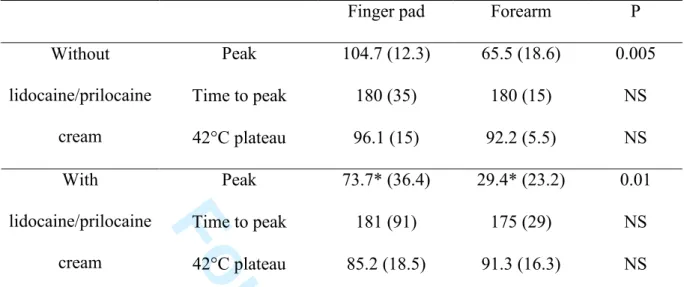
Typical local thermal hyperaemia patterns, including an early initial peak, a nadir, and a late plateau, were found on the finger tip as is usually seen on the forearm. However, as shown on Table 1 and figure 1, we observed a significantly higher initial peak on the finger
3 4 5 6 7 8 9 10 11 12 13 14 15 16 17 18 19 20 21 22 23 24 25 26 27 28 29 30 31 32 33 34 35 36 37 38 39 40 41 42 43 44 45 46 47 48 49 50 51 52 53 54 55 56 57 58 59 60
For Peer Review
pad compared to the forearm. Indeed, the initial peak is higher than the 44°C plateau on the fingertip, whereas it is much lower on the forearm.
Lidocaine/prilocaine cream significantly decreased the initial peak on both finger and forearm sites (table 1, fig 1). However, the decrease was more pronounced on the forearm in comparison with the finger.
Baseline cutaneous conductances without and with lidocaine/prilocaine cream were not significantly different on the finger pad (10.3 (13.3) and 7.9 (1.8) mV/mmHg, respectively) and on the forearm (1.1 (0.4) and 1.4 (2.9) mV/mmHg). Median maximum conductances (44°C plateau) were not significantly different without and with lidocaine/prilocaine cream on the finger pad (41 (39.6) and 32.6 (11.7) mV/mmHg, respectively) and on the forearm (15.3 (4) and 20.5 (13.9) mV/mmHg).
Second study: effect of lidocaine/prilocaine cream on the axon reflex induced by thermal hyperaemia in primary RP and SSc.
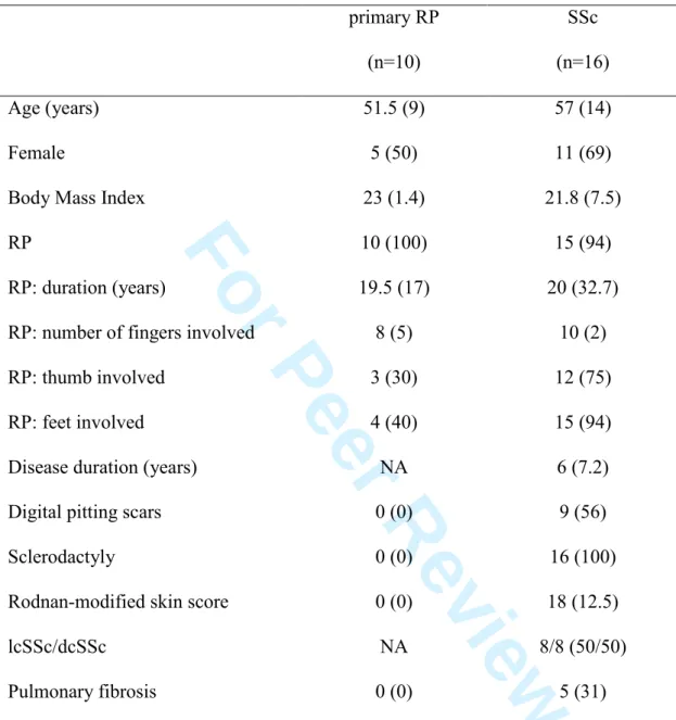
The demographic and clinical characteristics of the 26 patients enrolled in the second study are listed in Table 2.
Among the 16 patients with SSc, 8 were on calcium channel blockers, 3 on colchicine, 3 on angiotensin-converting enzyme inhibitors, and 2 on angiotensin II receptor blockers. Among the 10 patients with primary RP, 1 was on calcium channel blockers.
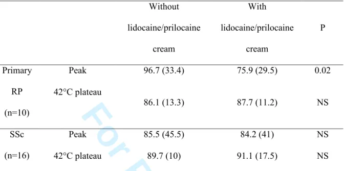
Thermal hyperaemia data are shown in table 3. We observed a decreased initial peak following lidocaine/prilocaine cream application in patients with primary RP, similar to healthy subjects. In contrast, lidocaine/prilocaine cream application did not alter the initial peak in patients with SSc (table 3, fig 2).
We further tested whether the effect of lidocaine/prilocaine cream could be delayed in SSc patients. The kinetic assessment of median conductance (minute averages), with and
3 4 5 6 7 8 9 10 11 12 13 14 15 16 17 18 19 20 21 22 23 24 25 26 27 28 29 30 31 32 33 34 35 36 37 38 39 40 41 42 43 44 45 46 47 48 49 50 51 52 53 54 55 56 57 58 59 60
For Peer Review
without lidocaine/prilocaine cream, is shown in figure 3. We observed no significant difference between sites.
Baseline conductances without and with lidocaine/prilocaine cream on the finger pad were not significantly different in patients SSc (3.3 (6.6) mV/mmHg and 3.7 (8) mmHg, respectively) but statistically different in patients with primary RP (11.5 (7.9) and 1.7 (6) mV/mmHg).
Median maximum conductances (44°C plateau) without and with lidocaine/prilocaine cream on the finger pad were not significantly different in patients with primary RP (38.9 (14.9) mV/mmHg and 37.7 (22) mmHg, respectively) and in patients with SSc (14.6 (20.4) and 12.6 (13.1) mV/mmHg).
Discussion
Our study demonstrates that the axon-reflex dependent thermal hyperaemia is blunted in the finger pads of patients with SSc, but not in those with primary RP.
In the current studies, laser Doppler flowmetry was used to monitor cutaneous perfusion rather than laser Doppler imaging. Indeed, laser Doppler imaging is less well suited for temporal monitoring of cutaneous perfusion whereas laser Doppler flowmetry enables the evaluation of cutaneous blood flow over time (continuous assessment). This aspect makes laser Doppler flowmetry especially useful in monitoring the kinetics of the cutaneous thermal hyperaemia (e.g., initial peak) [2,3]. Indeed, using our heating protocol, the initial peak in cutaneous perfusion is consistently reached 3 min after the onset of the local heating to 42°C. It is followed by a nadir, and a secondary plateau is reached at 20 to 30 min. In healthy subjects, the initial peak is mediated by a CGRP dependent axon reflex whereas the plateau is primarily mediated by NO [5]. We have previously shown a blunted digital vascular response to local heating in SSc [6,7]. In the current studies, the use of laser Doppler flowmetry
3 4 5 6 7 8 9 10 11 12 13 14 15 16 17 18 19 20 21 22 23 24 25 26 27 28 29 30 31 32 33 34 35 36 37 38 39 40 41 42 43 44 45 46 47 48 49 50 51 52 53 54 55 56 57 58 59 60
For Peer Review
enabled us to further explore this phenomenon, focusing specifically on the initial peak of the response.
Previously, using laser Doppler imaging, Gunawardena et al. [9] demonstrated a blunted local response after 6 min of heating at 44°C on the dorsum of the fingers, suggesting an abnormal digital microvascular regulatory response in patients with SSc. Murray et al. used a slower heating protocol (34°C to 40°C in 3 min, then 40°C during 3 min) and showed no significant difference between patients and controls on the dorsum of the hand but did observe an altered digital response to post-ischemia [8]. Taken together with our results, these data are consistent with an abnormal early digital vascular response to local heating in SSc.
In order to assess the involvement of the axon-reflex in the decreased initial peak previously observed in SSc after local heating, we used topical lidocaine/prilocaine to block sensory nerves in a localized region of skin. Indeed, we and others have shown that these topical anaesthetics can be used as pharmacological tools when local heating is started right after cream removal on the forearm [11,14,15]. In healthy volunteers, we showed that the amplitude of the initial peak was higher on the finger pad in comparison with the forearm. Indeed, such a peak was even higher than the 44°C late plateau usually used as an index of maximal vasodilation on the forearm [2]. We also observed less of a lidocaine/prilocaine cream effect on the finger pad in comparison with the forearm. This was not due to some intrinsic difference in axon reflex-mediated vasodilatation between skin regions. Rather, there was a decreased aesthetic effect of topical lidocaine/prilocaine on the finger pads, as suggested by the Semmes-Weinstein monofilament sensory test results. We cannot rule out the hypothesis that the effect of topical anaesthesia applied for a longer period of time would have been more pronounced. However, the application time was similar in all primary RP and SSc patients. A potential bias between groups is the fact that most of the SSc patients were under vasoactive medication, whereas this was less frequent in patients with primary RP.
3 4 5 6 7 8 9 10 11 12 13 14 15 16 17 18 19 20 21 22 23 24 25 26 27 28 29 30 31 32 33 34 35 36 37 38 39 40 41 42 43 44 45 46 47 48 49 50 51 52 53 54 55 56 57 58 59 60
For Peer Review
However, this does not influence the main objective, given the fact that each subject was its own control, and the drug would affect all sites. Furthermore, while calcium channel blockers may influence baseline blood conductance, there is no pharmacological evidence that they would interfere with the neurogenic response, which was the objective in the present study. It must also be highlighted that skin fibrosis is unlikely to explain our results, as we previously showed that patients with SSc without skin thickening still present an abnormal thermal hyperaemia on the finger pad [7].
The current findings demonstrate a blunted axon reflex-dependent early vasodilatation during local heating in patients with SSc, for whom topical anaesthesia had less effect than in patients with primary RP and healthy controls. This was not due to a delayed response, as shown by our analysis of response kinetics (no effect of topical lidocaine/prilocaine). Similarly to what was studied in an aging population, these findings could be consecutive to a decreased sensory component to the change in temperature, or to a reduction in the release and/or effect of neurotransmitters [16]. However, sensory neurological involvement is uncommon in SSc. In contrast, the crucial role of the peripheral nervous system in the control of vascular tone in SSc has been demonstrated previously, suggesting a functional impairment of the perivascular neurofibers [17]. Decreased substance P has been observed in late-stage SSc, while decreased CGRP has been observed both in patients with early and late-stage SSc [10,17]. As CGRP is the most likely neurotransmitter involved in this axon reflex[18], this suggests a dysfunction of the CGRP neurovascular axis in the pathophysiology of SSc. The current findings indicate that axon reflex-mediated vasodilatation is normal in the digits of patients with primary RP, but not in those with SSc.
Abnormal neurovascular control in SSc may be associated with both functional and structural disorders [17]. Indeed, several studies have described changes in the neural network of affected and non-affected skin in patients with SSc [17,19]. When using local heating as a
3 4 5 6 7 8 9 10 11 12 13 14 15 16 17 18 19 20 21 22 23 24 25 26 27 28 29 30 31 32 33 34 35 36 37 38 39 40 41 42 43 44 45 46 47 48 49 50 51 52 53 54 55 56 57 58 59 60
For Peer Review
test for axon reflex-mediated vasodilation, one cannot discriminate functional from structural changes. Blockade of the cutaneous nerves proximal to the site of local heating does not alter the thermal hyperaemic response, while local anaesthesia does. This shows that the initial thermal hyperaemia is dependent on a local neurovascular mechanism [5].
Our data suggest that monitoring the initial rise in digital skin blood flow following local heating could be a new and easy tool to assess neurovascular control in SSc in a clinical setting. In diabetes, others have used local anaesthesia as a pharmacological tool to assess neurovascular regulation in the skin of the feet [20]. They showed that blockade of C-fibres with lidocaine/prilocaine cream impaired axon reflex-mediated vasodilation in healthy volunteers and nonneuropathic diabetic patients, whereas local anaesthesia had no effect in neuropathic diabetic patients [20]. These findings suggest that neurovascular assessment in diabetic patients can serve as a specific tool to evaluate small fibre integrity which allows a reliable detection of subclinical neuropathy [21]. However, the evaluation of axon reflex-dependant vasodilation to assess neurovascular function in diabetes is not standardized. The current study takes a similar approach in patients with SSc and highlights the need for a standardized tool to assess neurovascular regulation in this patient population. Local heating could serve as an appropriate tool for this purpose because protocols are relatively short (median time to peak is 3 min after the onset of probe heating) and non-invasive. Larger studies are needed to assess the link between neurovascular control (as assessed by local heating) and the pathogenesis and severity of SSc.
In conclusion, we show an abnormal digital neurovascular response to local heating in SSc. The initial peak of the cutaneous vascular response to local heating (i.e., thermal hyperaemia) could be monitored as a clinical test for neurovascular function in SSc. Further studies are required to test whether abnormal digital neurovascular response correlate with the degree of peripheral vascular involvement.
3 4 5 6 7 8 9 10 11 12 13 14 15 16 17 18 19 20 21 22 23 24 25 26 27 28 29 30 31 32 33 34 35 36 37 38 39 40 41 42 43 44 45 46 47 48 49 50 51 52 53 54 55 56 57 58 59 60
For Peer Review
Acknowledgements
We thank the patient association ‘Association des Sclérodermiques de France’ for patient participation and financial support, the ‘Groupe Français de Recherche sur la Sclérodermie’ and the Délégation Régionale à la Recherche Clinique of Grenoble University Hospital for financial support. We thank the Clinical Research Center of Grenoble University Hospital for reviewing the protocols corresponding to these studies. We also thank Dr Muriel Salvat-Melis, Mrs Dominique Abry and Nicole Henquin for assisting with the laser Doppler measurements. 3 4 5 6 7 8 9 10 11 12 13 14 15 16 17 18 19 20 21 22 23 24 25 26 27 28 29 30 31 32 33 34 35 36 37 38 39 40 41 42 43 44 45 46 47 48 49 50 51 52 53 54 55 56 57 58 59 60
For Peer Review
References
1. Herrick AL. Vascular function in systemic sclerosis. Curr Opin Rheumatol 2000;12:527-33.
2. Cracowski JL, Minson CT, Salvat-Melis M, Halliwill JR. Methodological issues in the assessment of skin microvascular endothelial function in humans. Trends Pharmacol Sci 2006;27:503-8.
3. Charkoudian N. Skin blood flow in adult human thermoregulation: How it works, when it does not, and why. Mayo Clin Proc 2003;78:603-12.
4. Lisney S, Bharali L. The axon reflex: An outdated idea or a valid hypothesis? . New Physiol Sci 1989:45–8.
5. Minson CT, Berry LT, Joyner MJ. Nitric oxide and neurally mediated regulation of skin blood flow during local heating. J Appl Physiol 2001;91:1619-26.
6. Boignard A, Salvat-Melis M, Carpentier PH, et al. Local hyperemia to heating is impaired in secondary raynaud's phenomenon. Arthritis Res Ther 2005;7:R1103-12. 7. Salvat-Melis M, Carpentier PH, Minson CT, et al. Digital thermal hyperaemia
impairment does not relate to skin fibrosis or macrovascular disease in systemic sclerosis. Rheumatology (Oxford) 2006;45:1490-6.
8. Murray AK, Moore TL, King TA, Herrick AL. Abnormal microvascular response is localized to the digits in patients with systemic sclerosis. Arthritis Rheum
2006;54:1952-60.
9. Gunawardena H, Harris ND, Carmichael C, McHugh NJ. Maximum blood flow and microvascular regulatory responses in systemic sclerosis. Rheumatology (Oxford) 2007;46:1079-82.
10. Bunker CB, Terenghi G, Springall DR, Polak JM, Dowd PM. Deficiency of calcitonin gene-related peptide in raynaud's phenomenon. Lancet 1990;336:1530-3.
3 4 5 6 7 8 9 10 11 12 13 14 15 16 17 18 19 20 21 22 23 24 25 26 27 28 29 30 31 32 33 34 35 36 37 38 39 40 41 42 43 44 45 46 47 48 49 50 51 52 53 54 55 56 57 58 59 60
For Peer Review
11. Cracowski JL, Lorenzo S, Minson CT. Effects of local anaesthesia on subdermal needle insertion pain and subsequent tests of microvascular function in human. Eur J Pharmacol 2007;559:150-4.
12. LeRoy EC, Medsger TA, Jr. Raynaud's phenomenon: A proposal for classification. Clin Exp Rheumatol 1992;10:485-8.
13. LeRoy EC, Medsger TA, Jr. Criteria for the classification of early systemic sclerosis. J Rheumatol 2001;28:1573-6.
14. Krishnan ST, Rayman G. The ldiflare: A novel test of c-fiber function demonstrates early neuropathy in type 2 diabetes. Diabetes Care 2004;27:2930-5.
15. Caselli A, Uccioli L, Khaodhiar L, Veves A. Local anesthesia reduces the maximal skin vasodilation during iontophoresis of sodium nitroprusside and heating. Microvasc Res 2003;66:134-9.
16. Holowatz LA, Thompson-Torgerson CS, Kenney WL. Altered mechanisms of vasodilation in aged human skin. Exerc Sport Sci Rev 2007;35:119-25.
17. Kahaleh B, Matucci-Cerinic M. Raynaud's phenomenon and scleroderma.
Dysregulated neuroendothelial control of vascular tone. Arthritis Rheum 1995;38:1-4. 18. Schmelz M, Michael K, Weidner C, Schmidt R, Torebjork HE, Handwerker HO.
Which nerve fibers mediate the axon reflex flare in human skin? Neuroreport 2000;11:645-8.
19. Terenghi G, Bunker CB, Liu YF, et al. Image analysis quantification of peptide-immunoreactive nerves in the skin of patients with raynaud's phenomenon and systemic sclerosis. J Pathol 1991;164:245-52.
20. Caselli A, Rich J, Hanane T, Uccioli L, Veves A. Role of c-nociceptive fibers in the nerve axon reflex-related vasodilation in diabetes. Neurology 2003;60:297-300.
3 4 5 6 7 8 9 10 11 12 13 14 15 16 17 18 19 20 21 22 23 24 25 26 27 28 29 30 31 32 33 34 35 36 37 38 39 40 41 42 43 44 45 46 47 48 49 50 51 52 53 54 55 56 57 58 59 60
For Peer Review
21. Caselli A, Spallone V, Marfia GA, et al. Validation of the nerve axon reflex for the assessment of small nerve fibre dysfunction. J Neurol Neurosurg Psychiatry
2006;77:927-32. 3 4 5 6 7 8 9 10 11 12 13 14 15 16 17 18 19 20 21 22 23 24 25 26 27 28 29 30 31 32 33 34 35 36 37 38 39 40 41 42 43 44 45 46 47 48 49 50 51 52 53 54 55 56 57 58 59 60
For Peer Review
Table 1. Effects of lidocaine/prilocaine cream on the hyperaemia to local heating on the
finger pad and the forearm of healthy subjects (n=10).
Finger pad Forearm P Peak 104.7 (12.3) 65.5 (18.6) 0.005 Time to peak 180 (35) 180 (15) NS Without lidocaine/prilocaine cream 42°C plateau 96.1 (15) 92.2 (5.5) NS Peak 73.7* (36.4) 29.4* (23.2) 0.01 Time to peak 181 (91) 175 (29) NS With lidocaine/prilocaine cream 42°C plateau 85.2 (18.5) 91.3 (16.3) NS Data are expressed as median percentage (interquartile) of maximum conductance (44°C plateau) except for the time to peak, expressed as median time in seconds (interquartile). * P < 0.05 versus peak without lidocaine/prilocaine cream, Wilcoxon test. NS: non- significant. 3 4 5 6 7 8 9 10 11 12 13 14 15 16 17 18 19 20 21 22 23 24 25 26 27 28 29 30 31 32 33 34 35 36 37 38 39 40 41 42 43 44 45 46 47 48 49 50 51 52 53 54 55 56 57 58 59 60
For Peer Review
Table 2. Demographic and clinical characteristics of patients with primary Raynaud’s
phenomenon (RP) and systemic sclerosis (SSc)
primary RP (n=10) SSc (n=16) Age (years) 51.5 (9) 57 (14) Female 5 (50) 11 (69) Body Mass Index 23 (1.4) 21.8 (7.5) RP 10 (100) 15 (94) RP: duration (years) 19.5 (17) 20 (32.7) RP: number of fingers involved 8 (5) 10 (2) RP: thumb involved 3 (30) 12 (75) RP: feet involved 4 (40) 15 (94) Disease duration (years) NA 6 (7.2) Digital pitting scars 0 (0) 9 (56) Sclerodactyly 0 (0) 16 (100) Rodnan-modified skin score 0 (0) 18 (12.5)
lcSSc/dcSSc NA 8/8 (50/50) Pulmonary fibrosis 0 (0) 5 (31)
Quantitative data were expressed as median (interquartile). Qualitative data were expressed as number (percentage). RP: Raynaud’s phenomenon. NA: not applicable. lSSc: limited systemic sclerosis. lcSSc: limited cutaneous systemic sclerosis. dcSSc: diffuse cutaneous systemic sclerosis. 3 4 5 6 7 8 9 10 11 12 13 14 15 16 17 18 19 20 21 22 23 24 25 26 27 28 29 30 31 32 33 34 35 36 37 38 39 40 41 42 43 44 45 46 47 48 49 50 51 52 53 54 55 56 57 58 59 60
For Peer Review
Table 3. Effects of lidocaine/prilocaine cream on the hyperaemia to local heating on the
finger pad of patients with primary Raynaud’s phenomenon (RP) or systemic sclerosis (SSc). Without lidocaine/prilocaine cream With lidocaine/prilocaine cream P Peak 96.7 (33.4) 75.9 (29.5) 0.02 Primary RP (n=10) 42°C plateau 86.1 (13.3) 87.7 (11.2) NS Peak 85.5 (45.5) 84.2 (41) NS SSc (n=16) 42°C plateau 89.7 (10) 91.1 (17.5) NS Data are expressed as median percentage (interquartile) of maximum conductance (44°C plateau). NS: non-significant. 3 4 5 6 7 8 9 10 11 12 13 14 15 16 17 18 19 20 21 22 23 24 25 26 27 28 29 30 31 32 33 34 35 36 37 38 39 40 41 42 43 44 45 46 47 48 49 50 51 52 53 54 55 56 57 58 59 60
For Peer Review
Figure legends
Figure 1. Tracings of the thermal hyperaemia recorded on the finger pad and forearm of a
healthy subject, with and without 1 h topical lidocaine/prilocaine pre-treatment. The initial peak, but not the late plateau, was decreased by the pre-treatment at both sites. PU: perfusion units.
Figure 2. Tracings of the thermal hyperaemia recorded on the finger pad of a patient with
limited cutaneous systemic sclerosis, with and without 1 h topical lidocaine/prilocaine pre-treatment. The pre-treatment had no effect on the tracing. PU: perfusion units.
Figure 3. Median conductance of digital blood flow in patients with SSc averaged over 1 min
periods, with and without 1 h topical lidocaine/prilocaine pre-treatment.
Conflict of interest
No conflict of interest has been declared by the authors.
Key Messages
• The axon-reflex dependent peak is blunted in the finger pad of patients with SSc compared to primary RP, suggesting an abnormal neurovascular response to local heating in SSc.
• Digital thermal hyperaemia could be monitored as a clinical test for neurovascular function in SSc. 3 4 5 6 7 8 9 10 11 12 13 14 15 16 17 18 19 20 21 22 23 24 25 26 27 28 29 30 31 32 33 34 35 36 37 38 39 40 41 42 43 44 45 46 47 48 49 50 51 52 53 54 55 56 57 58 59 60
For Peer Review
Tracings of the thermal hyperaemia recorded on the finger pad and forearm of a healthy subject, with and without 1 h topical lidocaine/prilocaine pre-treatment. The initial peak, but not the late plateau, was decreased by the pre-treatment at both sites. PU: perfusion
units. 420x327mm (72 x 72 DPI) 3 4 5 6 7 8 9 10 11 12 13 14 15 16 17 18 19 20 21 22 23 24 25 26 27 28 29 30 31 32 33 34 35 36 37 38 39 40 41 42 43 44 45 46 47 48 49 50 51 52 53 54 55 56 57 58 59 60
For Peer Review
Tracings of the thermal hyperaemia recorded on the finger pad of a patient with limited cutaneous systemic sclerosis, with and without 1 h topical lidocaine/prilocaine
pre-treatment. The pre-treatment had no effect on the tracing. PU: perfusion units. 412x249mm (72 x 72 DPI) 3 4 5 6 7 8 9 10 11 12 13 14 15 16 17 18 19 20 21 22 23 24 25 26 27 28 29 30 31 32 33 34 35 36 37 38 39 40 41 42 43 44 45 46 47 48 49 50 51 52 53 54 55 56 57 58 59 60
For Peer Review
Median conductance of digital blood flow in patients with SSc averaged over 1 min periods, with and without 1 h topical lidocaine/prilocaine pre-treatment.
240x107mm (150 x 150 DPI) 3 4 5 6 7 8 9 10 11 12 13 14 15 16 17 18 19 20 21 22 23 24 25 26 27 28 29 30 31 32 33 34 35 36 37 38 39 40 41 42 43 44 45 46 47 48 49 50 51 52 53 54 55 56 57 58 59 60