HAL Id: hal-02618217
https://hal.inrae.fr/hal-02618217
Submitted on 25 May 2020
HAL is a multi-disciplinary open access
archive for the deposit and dissemination of
sci-entific research documents, whether they are
pub-lished or not. The documents may come from
teaching and research institutions in France or
abroad, or from public or private research centers.
L’archive ouverte pluridisciplinaire HAL, est
destinée au dépôt et à la diffusion de documents
scientifiques de niveau recherche, publiés ou non,
émanant des établissements d’enseignement et de
recherche français ou étrangers, des laboratoires
publics ou privés.
Distributed under a Creative Commons Attribution| 4.0 International License
The Escherichia coli colibactin resistance protein ClbS is
a novel DNA binding protein that protects DNA from
nucleolytic degradation
Katja Molan, Zdravko Podlesek, Vesna Hodnik, Matej Butala, Eric Oswald
To cite this version:
Katja Molan, Zdravko Podlesek, Vesna Hodnik, Matej Butala, Eric Oswald. The Escherichia coli
colibactin resistance protein ClbS is a novel DNA binding protein that protects DNA from
nucle-olytic degradation. DNA Repair, Elsevier, 2019, 79, pp.50-54. �10.1016/j.dnarep.2019.05.003�.
�hal-02618217�
Contents lists available atScienceDirect
DNA Repair
journal homepage:www.elsevier.com/locate/dnarepair
The Escherichia coli colibactin resistance protein ClbS is a novel DNA binding
protein that protects DNA from nucleolytic degradation
Katja Molan
a, Zdravko Podlesek
a, Vesna Hodnik
a,1, Matej Butala
a, Eric Oswald
b,
Darja
Žgur Bertok
a,⁎aDepartment of Biology, Biotechnical Faculty, University of Ljubljana, Večna pot 111, Ljubljana, Slovenia bIRSD, Université de Toulouse, INSERM, INRA, ENVT, UPS, Toulouse, France
A R T I C L E I N F O
Keywords: ClbS
DNA-binding protein DNA damage protection Pks genomic island Escherichia coli
A B S T R A C T
Cells employ specific and nonspecific mechanisms to protect their genome integrity against exogenous and endogenous factors. The clbS gene is part of the polyketide synthase machinery (pks genomic island) encoding colibactin, a genotoxin implicated in promoting colorectal cancer. The pks is found among the Enterobacteriaceae, in particular Escherichia coli strains of the B2 phylogenetic group. Several resistance mechanisms protect toxin producers against toxicity of their products. ClbS, a cyclopropane hydrolase, was shown to confer colibactin resistance by opening its electrophilic cyclopropane ring. Here we report that ClbS sustained viability and en-abled growth also of E. coli expressing another genotoxin, the Usp nuclease. The recA::gfp reporter system showed that ClbS protects against Usp induced DNA damage. To elucidate the mechanism of ClbS mediated protection, we studied the DNA binding ability of the ClbS protein. We show that ClbS directly interacts with single-stranded DNA (ssDNA) and double-stranded DNA (dsDNA), whereas ssDNA seems to be the preferred substrate. Thus, the ClbS DNA-binding characteristics may serve bacteria to protect their genomes against DNA degradation.
1. Introduction
To protect the integrity of their genomes against exogenous and endogenous assault, cells employ specific and as well as nonspecific mechanisms. Exogenous factors are radiation, chemical agents and others, while endogenous factors are reactive oxygen species, metabolic products [1] and in a number of bacterial species potentially also genotoxins. Genotoxins such as colibactin are significant bacterial virulence factors that provoke DNA damage in eukaryotic cells. How-ever, the genotoxin producing cell must be protected against its cognate toxin.
Colibactin is encoded by the polyketide synthase (pks) genomic is-land present among the Enterobacteriaceae, particularly pathogenic and commensal Escherichia coli strains of the B2 phylogenetic group [2]. The prevalence of these strains has in developed countries increased sig-nificantly [3,4]. Colibactin generates DNA cross-links that are, during DNA damage repair, converted to double strand breaks [5]. Recently, alkylation of DNA by colibactin has been demonstrated [6]. It has also been shown to induce colorectal cancer [7–11].
Colibactin is initially produced as an inactive prodrug that is
activated by ClbP-mediated cleavage upon export to the periplasm [12,13]. Further, it was postulated that ClbS, also encoded by the pks island, is a colibactin resistance protein [14] that cleaves the colibactin cyclopropane ring, essential for genotoxicity, via its cyclopropane hy-drolase activity [15].
As we previously demonstrated that immunity to the Usp genotoxin is assured by the Imu3 protein via nonspecific DNA binding [16], we postulated that ClbS might also provide direct genome protection. We therefore investigated whether ClbS exhibits DNA binding ability. Our results show that ClbS is a multifunctional protein, that besides directly inactivating colibactin, as shown previously, also associates with DNA to presumably protect the genome against certain genotoxic agents. 2. Material and methods
2.1. Bacterial strains and plasmid construction
Laboratory E. coli strains and plasmids used in this study are pre-sented inTable 1. The clbS gene was PCR amplified from BAC pks+
plasmid [2] with ClbS-F (5`- TGGATCCGCTGTTCCATCATCAAAAGAA
https://doi.org/10.1016/j.dnarep.2019.05.003
Received 2 December 2018; Received in revised form 29 March 2019; Accepted 18 May 2019
⁎Corresponding author at: Department of Biology, Biotechnical Faculty, University of Ljubljana, Večna pot 111, 1000 Ljubljana, Slovenia.
E-mail address:darja.zgur.bertok@bf.uni-lj.si(D.Žgur Bertok).
1Present address: Sandoz, Kolodvorska 27, SI-1234 Mengeš, Slovenia.
Available online 19 May 2019
1568-7864/ © 2019 The Authors. Published by Elsevier B.V. This is an open access article under the CC BY license (http://creativecommons.org/licenses/BY/4.0/).
GAG-3`) and ClbS-R (5`-TTACGCGTTATTCTGCAAGACATTTCTGCAG-3`) primers (Sigma-Aldrich) at 55 °C annealing temperature. The PCR product, digested with BamHI and MluI, was cloned into the expression vector pET8c, in a fusion with a thrombin-cleavable N-terminal hex-ahistidine tag (His6-tag). This plasmid, designated pCLB-his, was
sub-sequently used for ClbS protein isolation and for the construction of vectors, used for in vivo studies. Sequencing of the inserted clbS frag-ment was performed to check for potential PCR introduced mutations. For the construction of vectors, the BamHI and EcoRV digested and blunted fragment of the low copy number pLysS plasmid was employed. This backbone fragment contained only the replication region and the chloramphenicol resistance gene, with the T7 lysozyme gene removed. The source of the usp gene for subcloning into the pLysS fragment was plasmid pUSP4 [17]. Two plasmids were thus constructed, pLUBI, carrying the usp gene but without its immunity genes, and pLCUBI harbouring usp and clbS, and were transformed into E. coli BL21 (DE3). Both genes were controlled by independent T7 polymerase promoters. To follow the SOS response, plasmid pSC201 with a recA::gfp fusion was used [18], transformed into either E. coli BL21 (DE3) with pLCUBI, or E. coli BL21 (DE3) with pLUBI.
2.2. Expression, Isolation, purification and detection of the ClbS protein ClbS was expressed in E. coli strain BL21 (DE3) pCLB-his. An over-night culture was used to inoculate liquid Luria Bertani medium sup-plemented with 25μg/mL chloramphenicol and grown at 37 °C to an OD6000.5 when ClbS production was induced by the addition of
iso-propylβ-D-1-thiogalactopyranoside (IPTG) at a final concentration of 0.8 mM. The culture was subsequently grown for an additional 4 h when biomass was collected by 10 min centrifugation at 4.000 × g. His6-tagged ClbS was purified by Ni-NTA metal affinity
chromato-graphy (Qiagen) and stored in elution buffer (50 mM NaH2PO4,
300 mM NaCl, 250 mM imidazole, pH 8) at 4 °C. Further, overnight dialysis was performed against a final buffer (50 mM NaH2PO4·H2O,
200 mM NaCl, 20 mM β-mercaptoethanol, pH 8) at 4 °C. The con-centration of the purified ClbS protein was determined using a NanoDrop 1000 (Thermo Fisher Scientific) with the extinction coeffi-cient at 280 nm of 37530 M−1cm−1and by Pierce BCA protein assay kit, using Bovine Serum Albumin (BSA) (Sigma-Aldrich) as the stan-dard. Additionally, the His6-tag was removed from His6-ClbS with the
Thrombin cleavage capture kit (Novagen) and the buffer exchanged by overnight dialysis as described above. To assay the removal of the af-finity tag, the thrombin-digested or undigested His6-ClbS samples were
resolved on a 12% acrylamide gel (Fig. S1). 2.3. Electrophoretic mobility shift assays (EMSA)
Various concentrations of ClbS carrying or lacking the affinity His6
-tag (0.15μM – 2.4 μM) and 60 ng/μL of DNA (Table 2) were used to establish the nucleic acid-binding ability of ClbS. ClbS was incubated with DNA in 1 X FastDigest Green Buffer (Fermentas) at 37 °C for
15 min, prior to the analysis of the protein-DNA complexes by EMSA analysis on 1.5–2 % agarose gels. As a negative control BSA was in-cluded at afinal concentration of 1.6 μM. Additionally, mixtures of pUC19/EcoRI (60 ng/μL) and ClbS (0.15 μM – 2.4 μM) were pre-incubated at 37 °C for 5–15 minutes when colicin E7 (ColE7), isolated from E. coli [19] or commercial DNase I (Sigma-Aldrich) were added in final concentrations 0.42 mg/ml and 0.5 mg/mL, respectively, followed by an additional 30 min incubation. As a control, DNA without ClbS was incubated with ColE7 or commercial DNase I. Results were ana-lyzed employing 2% agarose gels.
2.4. Surface plasmon resonance assays (SPR)
Measurements were performed at 25 °C on a Biacore T100 (GE Healthcare, UK) at the Infrastructural Centre for Analysis of Molecular Interactions (University of Ljubljana, Slovenia). To prepare biotinylated DNA for immobilization on the streptavidin sensor chip, plasmid pJBPL-A was used as a matrix DNA to amplify the 403 bp long DNA sequence (from Salmonella bongori). Annealing temperature of 55 °C was used to amplify with primers Bong-biotin (5` -CATAAAACCTCGTGTA TGGTGCGC-3 and Bong-R (5`-GAATTCCATTTTACGACATCCGAATAT AAATTAAACGAA-3`) (Integrated DNA Technology). Approximately 50 response units (RU) of the 3′-biotinylated 403 bp long PCR product were immobilized on the second flow cell of the streptavidin (SA) sensor chip (GE Healthcare) in SPR buffer (20 mM Tris (pH 7.4), 140 mM NaCl, 2 mM DTT, 0.005% surfactant P20 and 0.1 mg/ml BSA). Thefirst flow cell was left empty and used to control any non-specific binding to the sensor surface. The interaction between ClbS and chip-immobilized DNA was studied by injecting various concentrations of ClbS in SPR buffer. Injections of ClbS over the immobilized DNA frag-ment were performed at a flow rate of 10 μl min−1for 2 min with
dsDNA and 4 min with ssDNA. The sensor chip with bound DNA was regenerated by injection of SPR buffer containing 0.03% SDS for 15 s at a flow rate of 30 μl min−1. Further, chip-immobilized dsDNA
dena-turation was applied with 2 pulses of 50 mM NaOH for 30 s to obtain ssDNA. The response after NaOH treatment dropped for approximately 20 RU, indicating that one DNA chain was removed. Injections of var-ious concentrations of ClbS in SPR buffer was applied at a flow rate of 10μl min−1for 4 min. SPR experiments were performed in triplicate and representative experiments are shown. The data were analyzed with the Biacore T100 Evaluation Software and equilibrium dissocia-tion constants (KD) were determined byfitting the data to the steady
state affinity model.
2.5. Quenching of intrinsic ClbS tryptophanfluorescence by ss- and dsDNA Intrinsic tryptophan fluorescence of ClbS (40 μg/mL in 50 mM phosphate buffer, 200 mM NaCl, β-mercaptoethanol, pH 8.0) was measured. DNA used for titrations are presented in Table 2. After adding DNA, a 10 s incubation followed when measurements were made in a constantly stirred 1 cm path length quartz cuvette with a scan Table 1
Bacterial strains and plasmids used in this study.
Bacterial strain/plasmid Description Source
DH5α (DE3) Expression host, harboring the lambda DE3. Thermo Fisher Scientific E. coli BL21(DE3) Expression host, harboring the lambda DE3; CmR. Thermo Fisher Scientific
BAC pks+ The source of the clbS gene. (2)
pUSP4 The source of the usp gene; ApR. (17) pET8c E. coli T7 expression vector, containing an N-terminal hexahistidine tag (His6) and thrombin cleavage site. Novagen
pCLB-his pET8c with clbS gene. This study
pLysS pLys with removed lysozyme gene; CmR. Thermo Fisher Scientific pLCUBI pLysS with clbS and usp genes; CmRand ApR. This study
pLUBI pLysS with usp gene; CmRand ApR. This study pSC201 Plasmid with recA::gfp fusion; KnR. (18)
K. Molan, et al. DNA Repair 79 (2019) 50–54
rate of 250 nm/min (37 °C, excitation and emission slits at 5 nm each). The fluorescence emission spectra were recorded in the wavelength range of 300–450 nm after excitation at 295 nm, specifically monitoring the tryptophan fluorescence emission. Fluorescent emissions of the buffer titrated with DNA (background intensities) were subtracted. 2.6. Growth of E. coli clbS+usp+and E. coli usp+strains and cell
morphology analysis
The two E. coli BL21 (DE3) strains were cultivated at 37 °C in liquid BHI medium with ampicillin and chloramphenicol atfinal concentra-tions 100 and 20μg/ml, respectively. To suppress basal level expression of the T7 polymerase promoter, glucose was added at a final con-centration of 0.4%. When cultures reached OD6000.5, protein synthesis
was induced by addition of IPTG atfinal concentration 0.8 mM. One ml of both cultures was sampled before induction and each hour following induction (at least three hours). The OD600was measured and plotted in
time.
Prior to microscopy, the E. coli BL21 (DE3) clbS+usp+and E. coli
usp+strains were transformed with a plasmid carrying a recA::gfp gene
fusion and cultured as described above in media supplemented with 30μg/ml kanamycin. Both cultures were sampled before and every hour following induction. Cells were washed twice with NaCl and at-tached to glass slides coated with 0.1% (w/v) poly-L-lysine (Sigma). Cell morphology was observed under light and recA::gfp expression under fluorescence microscopy, using a Zeiss AxioImager Z1 micro-scope equipped with a HRc digital camera.
3. Results and discussion
3.1. ClbS binds ss- and dsDNA in a non-specific manner
Initially, EMSA was applied and showed that ClbS causes circular and linear DNA retardation (Fig. 1A) and, that it interacts with ss- or dsDNAs of various lengths and source (Table 2, Fig. S2). Thus, EMSA experiments implied that ClbS binds DNA in a non-specific manner. It is of note that ClbS binding activity was not affected by the His6tag (Fig.
S3). SPR spectrometry measurements of ClbS binding to ssDNA (Fig. 1B) or dsDNA (Fig. 1C) revealed an approximately 40-times higher affinity of ClbS for ssDNA exhibiting an apparent equilibrium dis-sociation constant (KD) of 24.5 ± 1.2 nM compared to dsDNA
ex-hibiting a KDof 936.8 ± 12.3 nM (Fig. 1D). Additionally, to confirm
that ClbS interacts with DNA we assayed the tryptophanfluorescence spectra of ClbS, which carriesfive tryptophan residues, in the presence or in the absence of DNA. We observed quenching of intrinsic ClbS tryptophan fluorescence by various ss- and dsDNAs. The most pro-nounced decrease in intensity of emission maximum was observed upon titration with ssDNA (Fig. 1E). Our results implied that the five ClbS tryptophan residues behave as two populations of fluorophores, one accessible to quenchers and the other buried in the protein structure [21], presented as downward curvature in the Stern-Volmer plot (Fig. 1F).
3.2. ClbS protects DNA from deoxyribonucleases
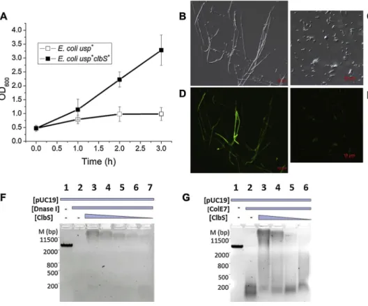
DNA protection by ClbS was examined by measuring growth of the E. coli strain carrying a plasmid encoded usp gene and its cognate im-munity genes replaced with clbS, in comparison to a strain harbouring only the usp gene (Table 1). Expression of usp and clbS was controlled by the T7 polymerase promoter. Following IPTG induction of usp and clbS the strain encoding only usp (lacking its immunity genes or clbS), exhibited dramatically reduced growth in comparison to the strain with both usp and clbS (Fig. 2A). DNA damage induces the SOS response involved in DNA repair. The principal regulators of the SOS response are the LexA repressor and RecA activator. Upon DNA damage, RecA is activated by binding to exposed ssDNA which stimulates LexA auto-cleavage, derepressing SOS genes. SOS induction inhibits cell division, resulting in cellfilamentation [22]. Light microscopy of IPTG-induced cells expressing usp revealed pronouncedfilamentation (Fig. 2B), while typical cell shape and size were detected among E. coli expressing both usp and clbS genes (Fig. 2C). The "two examined strains also carried a plasmid encoded recA::gfp, a DNA damage SOS reporter gene fusion. Fluorescent microscopy revealed that usp+ cells without clbS,
ad-ditionally exhibited elevated expression of recA::gfp (Fig. 2D) when compared to the usp+clbS+cells (Fig. 2E). Filamentation and elevated expression of recA are hallmarks of induction of the DNA damage in-ducible SOS response. Thus, typical cell shape and size, as well as basal level expression of recA in usp+clbS+cells indicates, that ClbS protects cells from Usp nuclease activity.
We subsequently hypothesized that binding of ClbS could provide DNA protection against other deoxyribonucleases. Co-incubation of DNA and ClbS protected plasmid DNA against DNase I (Fig. 2F) and the ColE7 nuclease (Fig. 2G) in a concentration dependent manner, con-firming that ClbS provides protection against various nucleases by presumably interacting non-specifically with DNA. ClbS binding to DNA is evident as DNA retardation (Fig. 2G, and F). Other examples of protective bacterial proteins have been characterized previously namely, two E. coli nucleoid associated proteins, the multifunctional Dps and CbpA, that non-specifically bind DNA, to drive nucleoid compactation and DNA protection [23–26]. Further, theα/β type small acid-soluble spore proteins that are synthesized during sporulation, bind DNA changing its conformation to an A-like helix, that is more resilient against damage [27,28].
In conclusion, our results imply that ClbS, besides directly inter-acting with colibactin is also a novel DNA binding protein. Thus, be-yond colibactin, ClbS also protects DNA against degradation/genotoxic activity presumably providing a selective advantage for bacterial strains encountering stressful conditions also within the host.
Conflict of interest
The authors have no conflict of interest. Footnotes
Data accessibility available via Mendeley Data:
https://data.mendeley.com/datasets/4y3wpkrfzc/draft?a=
Table 2
DNA employed in this study.
DNA Length
(bp or bases)
Application Reference
pUC19 2686 EMSA Fermentas
pJBPL-A 3377 Matrix DNA for PCR reaction. This laboratory PCR amplicon 403 SPR (ds- and ssDNA), EMSA,
Trpfluorescence quenching This study ds- and ssDNA phage genome M13 6407 EMSA, Trpfluorescence quenching [20]
0843f073-1b07-4239-83ad-28f474895ca9
Acknowledgements
We thank dr. Miloš Vittori for assistance with microscopy and prof.
dr. Kristina Sepčič with interpretation of results obtained from intrinsic ClbS tryptophanfluorescence quenching. This work was supported by grant P1-0198 and J1-8150 from the Slovene Research Agency. Katja Molan is a recipient of a PhD grant from the Slovene Research Agency. Fig. 1. ClbS is a DNA binding protein. (A) EMSA of ClbS interacting with the DNA. M designates marker (bp). Lane 1 contains pUC19 plasmid DNA, lane 2 pUC19 with BSA and lane 3 pUC19 with ClbS. Lane 4 contains pUC19 linearized with EcoRI, lane 5 pUC19/EcoRI with BSA and lane 6 pUC19/EcoRI with ClbS. (B) SPR sensorgrams showing the interaction of ClbS with 403 bp long chip-immobilized ssDNA or (C) dsDNA. (D) The apparent equilibrium dissociation constant (KD) values
of ClbS interacting with ssDNA, red or with dsDNA, black. The average KDs and standard deviations were determined from three titrations of each protein. (E)
Quenching of intrinsic tryptophanfluorescence of ClbS titrated with ssDNA. The legend denotes ssDNA concentrations preincubated with ClbS. (F) Stern-Volmer plot of tryptophan quenching within ClbS due to interaction with ssDNA or dsDNA.
Fig. 2. ClbS protects DNA from deoxyr-ibonucleases. (A) Growth curves of E. coli usp+clbS+ (black squares) and E. coli usp+
strains (white squares). Induction of proteins at time 0 h. The experiment was performed seven times, and the means ± standard errors of the means (error bars) are shown. (B) Light and (D)fluorescent microscopy of the E. coli usp+
strain following 3 h - induction. (C) Light and (E) fluorescent microscopy of the E. coli usp+clbS+strain following 3 h - induction. (F
and G) ClbS protect DNA against DNase de-gradation. (F) M designates marker (bp). Lane 1 contains pUC19/EcoRI plasmid DNA, lane 2 DNA with DNase I (0.5 mg/mL), lanes 3–7 DNA with ClbS (2.4-0.15μM) and DNase I. (G) M designates marker (bp). Lane 1 contains pUC19/EcoRI DNA, lane 2 DNA with ColE7 (0.42 mg/ml) and lanes 3–6 DNA with ClbS (2.4-0.15μM) and ColE7.
K. Molan, et al. DNA Repair 79 (2019) 50–54
Appendix A. Supplementary data
Supplementary material related to this article can be found, in the online version, at doi:https://doi.org/10.1016/j.dnarep.2019.05.003. References
[1] N. Chatterjee, G.C. Walker, Mechanisms of DNA damage, repair and mutagenesis, Environ. Mol. Mutagen. 58 (2017) 235–263,https://doi.org/10.1002/em.22087. [2] J.P. Nougayrède, S. Homburg, F. Taieb, M. Boury, E. Brzuszkiewicz, G. Gottschalk,
C. Buchrieser, J. Hacker, U. Dobrindt, E. Oswald, Escherichia coli induces DNA double-strand breaks in eukaryotic cells, Science 313 (2006) 848–851,https://doi. org/10.1126/science.1127059.
[3] P. Escobar-Páramo, K. Grenet, A. Le Menac’h, L. Rode, E. Salgado, C. Amorin, S. Gouriou, B. Picard, M.C. Rahimy, A. Andremont, E. Denamur, R. Ruimy, Large-scale population structure of human commensal Escherichia coli isolates, Appl. Environ. Microbiol. 70 (2004) 5698–5700,https://doi.org/10.1128/AEM.70.9. 5698-5700.2004.
[4] F.L. Nowrouzian, A.E. Wold, I. Adlerberth, Escherichia coli strains belonging to phylogenetic group B2 have superior capacity to persist in the intestinal microflora of infants, J. Infect. Dis. 191 (2005) 1078–1083,https://doi.org/10.1086/427996. [5] N. Bossuet-Greif, J. Vignard, F. Taieb, G. Mirey, D. Dubois, C. Petit, E. Oswald,
J.P. Nougayrède, The colibactin genotoxin generates DNA interstrand cross-links in infected cells, MBio 9 (2018) e02393–17,https://doi.org/10.1128/mBio.02393-17. [6] M.R. Wilson, Y. Jiang, P.W. Villalta, A. Stornetta, P.D. Boudreau, A. Carrá,
C.A. Brennan, E. Chun, L. Ngo, L.D. Samson, B.P. Engelward, W.S. Garrett, S. Balbo, E.P. Balskus, The human gut bacterial genotoxin colibactin alkylates DNA, Science 363 (2019),https://doi.org/10.1126/science.aar7785.
[7] T. Secher, A. Samba-Louaka, E. Oswald, J.P. Nougayrède, Escherichia coli producing colibactin triggers premature and transmissible senescence in mammalian cells, PLoS One 8 (2013) e77157, ,https://doi.org/10.1371/journal.pone.0077157. [8] J.C. Arthur, E. Perez-Chanona, M. Mühlbauer, S. Tomkovich, J.M. Uronis, T.-J. Fan,
B.J. Campbell, T. Abujamel, B. Dogan, A.B. Rogers, J.M. Rhodes, A. Stintzi, K.W. Simpson, J.J. Hansen, T.O. Keku, A.A. Fodor, C. Jobin, Intestinal inflammation targets cancer-inducing activity of the microbiota, Science 338 (2012) 120–123,
https://doi.org/10.1126/science.1224820.
[9] G. Dalmasso, A. Cougnoux, J. Delmas, A. Darfeuille-Michaud, R. Bonnet, The bac-terial genotoxin colibactin promotes colon tumor growth by modifying the tumor microenvironment, Gut Microbes 5 (2014) 675–680,https://doi.org/10.4161/ 19490976.2014.969989.
[10] A. Cougnoux, G. Dalmasso, R. Martinez, E. Buc, J. Delmas, L. Gibold, P. Sauvanet, C. Darcha, P. Déchelotte, M. Bonnet, D. Pezet, H. Wodrich, A. Darfeuille-Michaud, R, Bonnet. Bacterial genotoxin colibactin promotes colon tumour growth by indu-cing a senescence-associated secretory phenotype, Gut 63 (2014) 1932–1942,
https://doi.org/10.1136/gutjnl-2013-305257.
[11] C.M. Dejea, P. Fathi, J.M. Craig, A. Boleij, R. Taddese, A.L. Geis, X. Wu, C.E. DeStefano Shields, E.M. Hechenbleikner, D.L. Huso, R.A. Anders,
F.M. Giardiello, E.C. Wick, H. Wang, S. Wu, D.M. Pardoll, F. Housseau, C.L. Sears, Patients with familial adenomatous polyposis harbor colonic biofilms containing tumorigenic bacteria, Science 359 (2018) 592–597,https://doi.org/10.1126/ science.aah3648.
[12] D. Dubois, O. Baron, A. Cougnoux, J. Delmas, N. Pradel, M. Boury, B. Bouchon, M.A. Bringer, J.P. Nougayrède, E. Oswald, R. Bonnet, ClbP is a prototype of a
peptidase subgroup involved in biosynthesis of nonribosomal peptides, J. Biol. Chem. 286 (2011) 35562–35570,https://doi.org/10.1074/jbc.M111.221960. [13] C.A. Brotherton, E.P. Balskus, A prodrug resistance mechanism is involved in
co-libactin biosynthesis and cytotoxicity, J. Am. Chem. Soc. 135 (2011) 3359–3362,
https://doi.org/10.1021/ja312154m.
[14] N. Bossuet-Greif, D. Dubois, C. Petit, S. Tronnet, P. Martin, R. Bonnet, E. Oswald, J.P. Nougayrède, Escherichia coli ClbS is a colibactin resistance protein, Mol. Microbiol. 99 (2016) 897–908,https://doi.org/10.1111/mmi.13272. [15] P. Tripathi, E.E. Shine, A.R. Healy, C.S. Kim, S.B. Herzon, S.D. Bruner,
J.M. Crawford, ClbS is a cyclopropane hydrolase that confers colibactin resistance, J. Am. Chem. Soc. 139 (2017) 17719–17722,https://doi.org/10.1021/jacs. 7b09971.
[16] M.Črnigoj, Z. Podlesek, M. Budič, D. Žgur Bertok, The Escherichia coli uropathogenicspecificproteinassociated immunity protein 3 (Imu3) has nucleic acid -binding activity, BMC Microbiol. 14 (2014) 16, https://doi.org/10.1186/1471-2180-14-16.
[17] D. Nipič, Z. Podlesek, M. Budič, M. Črnigoj, D. Žgur Bertok, Escherichia coli ur-opathogenic-specific protein, Usp, is a bacteriocin-like genotoxin, J. Infect. Dis. 208 (2013) 1545–1552,https://doi.org/10.1093/infdis/jit480.
[18] M. Ronen, R. Rosenberg, B.I. Shraiman, U. Alon, Assigning numbers to the arrows: parameterizing a gene regulation network by using accurate expression kinetics, Proc. Natl. Acad. Sci. 16 (2002) 10555–10560,https://doi.org/10.1073/pnas. 152046799.
[19] M. Budič, M. Rijavec, Ž. Petkovšek, D. Žgur Bertok, Escherichia coli bacteriocins: antimicrobial efficacy and prevalence among isolates from patients with bacter-aemia, PLoS One 6 (2011) e28769, ,https://doi.org/10.1371/journal.pone. 0028769.
[20] P.M. van Wezenbeek, T.J. Hulsebos, J.G. Schoenmakers, Nucleotide sequence of the filamentous bacteriophage M13 DNA genome: comparison with phage fd, Gene 11 (1980) 129–148,https://doi.org/10.1016/0378-1119(80)90093-1.
[21] J.R. Lakowicz, Quenching offluorescence, Principles of Fluorescence Spectroscopy, Springer, Boston, MA, 2006, pp. 277–330, https://doi.org/10.1007/978-0-387-46312-4_8.
[22] R. D’Ari, O. Huisman, Novel mechanism of cell division inhibition associated with the SOS response in Escherichia coli, J. Bacteriol. 156 (1983) 243–250 https:// www.ncbi.nlm.nih.gov/pmc/articles/PMC215076/#reference-sec.
[23] J.G. Bird, S. Sharma, S.C. Roshwalb, J.R. Hoskins, S. Wickner, Functional analysis of CbpA, a DnaJ homolog and nucleoid-associated DNA-binding protein, J. Biol. Chem. 281 (2006) 34349–34356,https://doi.org/10.1074/jbc.M603365200. [24] S. Cosgriff, K. Chintakayala, Y.T. Chim, X. Chen, S. Allen, A.L. Lovering,
D.C. Grainger, Dimerization and DNA-dependent aggregation of the Escherichia coli nucleoid protein and chaperone CbpA, Mol. Microbiol. 77 (2010) 1289–1300,
https://doi.org/10.1111/j.1365-2958.2010.07292.x.
[25] M. Almirón, A.J. Link, D. Furlong, R. Kolter, A novel DNA-binding protein with regulatory and protective roles in starved Escherichia coli, Genes Dev. 6 (1992) 2646–2654,https://doi.org/10.1101/gad.6.12b.2646.
[26] A. Martinez, R. Kolter, Protection of DNA during oxidative stress by the nonspecific DNA-binding protein Dps, J. Bacteriol. 179 (1997) 5188–5194,https://doi.org/10. 1128/jb.179.16.5188-5194.1997.
[27] P. Setlow, Mechanisms which contribute to the long-term survival of spores of Bacillus species, J. Appl. Bacteriol. 76 (1994) 49S–60S.
[28] W.L. Nicholson, N. Munakata, G. Horneck, H.J. Melosh, P. Setlow, Resistance of Bacillus endospores to extreme terrestrial and extraterrestrial environments, Microbiol. Mol. Biol. Rev. 64 (2000) 548–572.