HAL Id: hal-01329474
https://hal.archives-ouvertes.fr/hal-01329474
Submitted on 26 May 2021
HAL is a multi-disciplinary open access
archive for the deposit and dissemination of
sci-entific research documents, whether they are
pub-lished or not. The documents may come from
teaching and research institutions in France or
abroad, or from public or private research centers.
L’archive ouverte pluridisciplinaire HAL, est
destinée au dépôt et à la diffusion de documents
scientifiques de niveau recherche, publiés ou non,
émanant des établissements d’enseignement et de
recherche français ou étrangers, des laboratoires
publics ou privés.
Distributed under a Creative Commons Attribution| 4.0 International License
Béatrice Gleize, Franck Tourniaire, Laurence Depezay, Romain Bott, Marion
Nowicki, Lionel Albino, Denis Lairon, Emmanuelle Kesse-Guyot, Pilar Galan,
Serge Hercberg, et al.
To cite this version:
Béatrice Gleize, Franck Tourniaire, Laurence Depezay, Romain Bott, Marion Nowicki, et al.. Effect
of type of TAG fatty acids on lutein and zeaxanthin bioavailability. British Journal of Nutrition,
Cambridge University Press (CUP), 2013, 110, pp.1-10. �10.1017/S0007114512004813�. �hal-01329474�
Effect of type of TAG fatty acids on lutein and zeaxanthin bioavailability
Be´atrice Gleize
1,2,3, Franck Tourniaire
1,2,3, Laurence Depezay
4, Romain Bott
1,2,3, Marion Nowicki
1,2,3,
Lionel Albino
4, Denis Lairon
1,2,3, Emmanuelle Kesse-Guyot
5, Pilar Galan
5, Serge Hercberg
5and
Patrick Borel
1,2,3*
1INRA, UMR1260, Research Unit in Nutrition, Obesity and Risk of Thrombosis, Faculte´ de Me´decine, 27 Boulevard
Jean-Moulin, F-13385 Marseille, Cedex 5, France
2INSERM, UMR1062, Marseille F-13385, France
3Aix-Marseille Universite´, Marseille F-13385, France
4Bonduelle, Villeneuve d’Ascq F-59653, France
5Nutritional Epidemiology Research Unit (UREN), INSERM U557, INRA U1125, CNAM, Universite´ Paris 13, Sorbonne Paris
Cite´, F-93017 Bobigny Cedex, France
(Submitted 23 May 2012 – Final revision received 17 September 2012 – Accepted 22 September 2012 – First published online 11 December 2012)
Abstract
The xanthophylls lutein and zeaxanthin probably play a role in visual function and may participate in the prevention of age-related eye diseases. Although a minimum amount of TAG is required for an optimal bioavailability of these carotenoids, the effect of the type of TAG fatty acids (FA) is less clear. The aim was to assess the effect of the type of TAG FA on bioavailability of these xanthophylls. A total of three complementary models were used: an in vitro digestion model to study bioaccessibility, Caco-2 cells to study uptake efficiency and orally administered rats to study in vivo bioavailability. Results showed that lutein and zeaxanthin bioaccessibility was greater (about 20 – 30 %, P , 0·05) with butter and palm oil than with olive and fish oils. Mixed micelle size, which was significantly lower (about 8 %, P, 0·05) with SFA than with unsaturated FA, was inversely related to lutein and zeaxanthin bioaccessibility. There was no significant effect of the type of TAG FA on xanthophyll uptake by Caco-2 cells, but some compounds present in natural oils significantly affected xanthophyll uptake. Oral administration of rats with spinach and butter over 3 d led to a higher fasting plasma lutein concentration than oral administration with olive or fish oils. In conclusion, dietary fats rich in SFA lead to a higher bioavailability of lutein and zeaxanthin, as compared with fats rich in MUFA and PUFA. This is due partly to the higher bioaccessibility of these xanthophylls in the smaller mixed micelles produced when SFA are incorporated into mixed micelles.
Key words:Xanthophyll: Bioavailability: SFA: Unsaturated fatty acids
Carotenoids are pigments responsible for the yellow, orange and red colours found in fruit, vegetables, algae and fungi. More than 650 carotenoids have been described and isolated from natural sources; however, only about fifty carotenoids
can be detected in human blood and tissues(1). Carotenoids
are classified as hydrocarbon carotenoids or carotenes (e.g. b-carotene, a-carotene and lycopene) and oxy-carotenoids or xanthophylls (e.g. lutein, zeaxanthin and b-cryptoxanthin). The consumption of foods rich in carotenoids has been associ-ated with a decrease in the incidence of some human
degen-erative and chronic diseases(2,3). Lutein and zeaxanthin are
gaining increasing interest as they selectively accumulate in
the human retina(4), where they apparently protect
photo-receptors against light-initiated oxidative damage(5,6), and
because they have been associated with a lower incidence
of age-related macular degeneration(7,8).
In order to achieve their potential beneficial health effects, xanthophylls must be efficiently absorbed and carried to their target tissues. Absorption of xanthophylls, like other lipo-philic compounds, is ensured through three key limiting steps: (1) release from the food matrix (usually a vegetable matrix) and transfer into the mixed micelles during digestion, termed ‘bioaccessibility’; (2) uptake by the enterocyte; (3) enterocyte transport and packaging into the chylomicrons for secretion into the lymph. Several dietary factors have been shown to affect these key steps in xanthophyll absorption, e.g.
the food matrix(9)and the interactions between xanthophylls
and other food components like fibre, lipids, phytosterols
* Corresponding author: P. Borel, fax þ 33 4 91 78 21 01, email [email protected]
Abbreviations: BHT, butylated hydroxytoluene; DMEM, Dulbecco’s modified Eagle’s medium; FA, fatty acid; SU.VI.MAX, Supple´mentation en Vitamines et Mine´raux AntioXydants.
British Journal of Nutrition (2013), 110, 1–10 doi:10.1017/S0007114512004813
qThe Authors 2012
British
Journal
of
Nutrition
https://www.cambridge.org/core . IP address: 78.202.154.114 , on 26 May 2021 at 12:20:55, subject to the Cambridge Core terms of use, available at
https://www.cambridge.org/core/terms
.
and other carotenoids(10 – 12). Among all these dietary factors, it is assumed that co-ingestion of xanthophylls with dietary lipids has the greatest positive impact on xanthophyll tion. This assumption is supported by the fact that the absorp-tion efficiency of xanthophylls increases when the amount of
fat in the diet increases(13,14). Two studies have further
suggested that the type of fatty acid (FA) that constitutes the oil TAG can affect xanthophyll absorption. First, a study in rats has shown that plasma lutein and zeaxanthin concen-trations were markedly higher when test meals contained olive oil (rich in MUFA) compared with sunflower and
groundnut oils, which are rich in PUFA(15). Second, an
in vitro study has shown that xanthophyll bioavailability was higher in the presence of coconut oil, which is rich in SFA, than in presence of rapeseed oil, which is rich in unsaturated
FA(16). Nevertheless, natural dietary fat can contain fat-soluble
micronutrients, e.g. vitamin E, b-carotene and phytosterols, and it is not known whether the observed effects were due to these micronutrients or to the different FA compositions of the sources of TAG. Thus, the mechanism(s) that can explain the effect of the source of dietary fat on xanthophyll bioavailability remains to be elucidated.
We performed the present work to allow for a firm statement on the effect of natural dietary fat on lutein/zeaxanthin bioavail-ability, to identify the mechanism(s) involved and to assess the effect of this dietary factor on long-term xanthophyll status in human subjects. We used three complementary models to attain our objectives: an in vitro digestion model to study bio-accessibility, Caco-2 cell monolayers to study uptake efficiency and orally administered rats to study in vivo bioavailability. We also determined, in a cohort of 622 subjects who took part in the Supple´mentation en Vitamines et Mine´raux AntioXydants (SU.VI.MAX) study, whether SFA intake modulated the expected association between blood lutein and zeaxanthin concentrations and fruit and vegetable intake.
Materials and methods Chemicals
Lutein, zeaxanthin and echinenone (. 95 % pure) were gene-rously provided by DSM Limited (formerly F. Hoffmann-La Roche). Neoxanthin (97 % pure) was purchased from Carote-nature. Porcine pepsin, porcine pancreatin, porcine bile extract, butylated hydroxytoluene (BHT), palm oil (refined) and fish oil (from menhaden) were purchased from Sigma-Aldrich. Potatoes, minced beef (with 5 % of fat), olive oil, sunflower oil and butter were purchased from a local super-market. Canned sweetcorn and frozen chopped spinach were provided by Bonduelle.
Preparation of meals used in in vitro digestion experiments Meal composition is given in Table 1. Potatoes and minced beef were included in the meals to align macronutrient (carbo-hydrate, lipid and protein) proportions to current RDA. Indeed, as macronutrients can affect carotenoid bioavailabil-ity, it was important to use a meal that mimics the proportions
of each macronutrient found in regular meals. The test meal without the xanthophyll source contained 53·6 % energy as carbohydrates, 28·4 % as fat and 18·0 % as proteins. These proportions are close to the US dietary reference intakes (DRI), i.e. 45 – 65 % carbohydrates, 20 – 35 % fat and 10 – 35 % proteins. Potatoes were boiled in tap water, peeled and hand pure´ed. Meat was medium fried in a frying pan without added fat. Potato pure´e and fried meat were divided into ali-quots and frozen at 2 208C. The sweetcorn source that was used in all experiments was preliminarily masticated by eight adult volunteers from the laboratory to mimic at best the in vivo conditions. Then, it was divided into aliquots and frozen at 2 208C. The chopped spinach (30 g frozen) was cooked in a microwave oven, for 1 min at 750 W, on the days of the experiments. Just before the experiments, BHT was added into the different sources of dietary fat (1 % in weight), either natural dietary fats or pure TAG mixtures (Table 2), in order to prevent oxidation that could degrade carotenoids during the in vitro digestion experiments. To accurately assess the amount of carotenoids present in the meals, the meal components were mixed with 32 ml of NaCl 0·9 % with an ultra-turrax disperser (Ika) to obtain a homo-geneous pure´e. Aliquots of this pure´e were stored at 2 808C
under a N2 atmosphere until carotenoid analysis by HPLC
(see Carotenoid analysis section).
In vitro digestion experiments
The in vitro digestion model was adapted from Reboul et al.(9)
with minor modifications. Meal components, which contained either masticated sweetcorn or chopped spinach, were mixed with 32 ml of 0·9 % NaCl (ultra-turrax disperser (Ika), 30 s at 6000 rpm), and 2 ml of artificial saliva was added to the
mixture(17). The samples were incubated for 10 min in a
shaking water-bath at 378C. The pH was then adjusted to
4 ^ 0·02 with about 500 ml of 1M-HCl. Then, 2 ml of porcine
pepsin (40 mg/ml in 0·1M-HCl) was added and the mixture
was incubated at 378C in a shaking water-bath for 30 min to mimic the gastric phase of digestion. The pH of the partially digested mixture was raised to 6 ^ 0·02 by adding about
800 ml of 0·9M-sodium bicarbonate (pH 9 – 10). Then,
9 ml of a mixture of porcine bile extract and pancreatin (con-taining 2 mg/ml pancreatin and 12 mg/ml bile extract in
Table 1. Composition of meals used in in vitro digestion experiments
Component Amount (g) Preparation
Pure´ed potatoes 6·7 Boiled
Minced beef meat (5 % fat) 1·2 Fried
Dietary fat* or TAG mixture 0·2 Incorporation of 1 % BHT
Canned sweetcorn† 4·0 Masticated by adults
volunteers
Frozen chopped spinach† 4·0 Cooked with microwave
oven
BHT, butylated hydroxytoluene.
* The studied dietary fats were butter, palm, olive, sunflower and fish oils. For the butter condition, the amount of butter added was calculated to provide 0·2 g of fat. † Digestion experiments contained either sweetcorn or chopped spinach. B. Gleize et al. 2
British
Journal
of
Nutrition
https://www.cambridge.org/core . IP address: 78.202.154.114 , on 26 May 2021 at 12:20:55, subject to the Cambridge Core terms of use, available at
https://www.cambridge.org/core/terms
.
100 mM-trisodium citrate (pH 6·0)) and 4 ml of porcine bile extract at 0·1 g/ml were added. Samples were further incu-bated in a shaking water-bath at 378C for 30 min to mimic digestion within the duodenum.
The aqueous fraction, which contains the mixed micelles that were produced upon in vitro digestion, was separated from oil droplets and food particles by centrifugation (2200 g for 1 h at 108C). The aqueous fraction was collected and was passed through 0·8 and 0·22 mm filters (Millipore) in order to discard remaining small food particles and to obtain a clear solution of mixed micelles. Aliquots were stored at 2 808C
under a N2atmosphere until carotenoid analysis.
Experiments on uptake efficiency of micellar xanthophylls by intestinal cells
Caco-2 clone TC-7 cells(18,19)were a gift from Dr M. Rousset
(UMR_S 872). Cells were cultured in the presence of Dulbecco’s modified Eagle’s medium (DMEM) supplemented with 20 % heat-inactivated fetal bovine serum, 1 % non-essential amino acid and 1 % antibiotics (complete medium). Cells were incubated at 378C in a humidified atmosphere of
air – CO2 (90:10, v/v) and the medium was changed every
48 h, as described previously(20).
For each experiment, cells were seeded at a density of
25 £ 104 cells/well and grown on transwell membrane
(six-well plate, 1 mm pore size polycarbonate membrane; Becton
Dickinson), and were cultured as described previously(20).
At the beginning of each experiment, cell monolayers were washed twice with 1 ml of PBS. The apical side of the cell mono-layers received 1 ml of mixed micelles that came from the in vitro digestion experiments (diluted at 1/3 in DMEM). Cell mono-layers were incubated at 378C for 2 h 30 min. The incubation time has been chosen after a preliminary experiment to obtain sufficient amounts of absorbed carotenoids for accurate measurements. After the incubation period, media from each side of the membrane were harvested. Cell monolayers were washed twice with 1 ml of ice-cold PBS, scraped and collected
in 500 ml of PBS. All samples were stored at 2 808C under a N2
atmosphere until carotenoid analysis.
Determination of spinach lutein bioavailability in rats The animal model chosen to study lutein bioavailability was multiple-dose oral administration over 3 d in young male albino Wistar rats (6 weeks old) of normal weight (about 200 g). Experiments were conducted according to animal ethics policies and were approved by the experimental animal ethics committee of Aix-Marseille University. All rats were fed the same standard diet ad libitum throughout the experiment. The rats were randomly divided into three groups (n 10 per group). The different groups of rats were orally administered with 1 ml of spinach/dietary fat emulsion (spinach – dietary fat – water; 1:1:1 by weight for 1 ml). We used spinach instead of sweetcorn as the source of xantho-phylls in the present animal study because it was not possible to have homogeneous administration with sweetcorn, which contains a hull that is difficult to grind homogeneously. Never-theless, it should be reminded that there was no effect of the vegetable matrix on xanthophyll bioaccessibility.
The dietary fats used in the present study were butter, olive and fish oils. Oral administration was performed every eve-ning for three consecutive days. At 16 h after the last adminis-tration, an intra-cardiac venepuncture was performed on anaesthetised rats. The plasma samples were obtained after centrifugation (500 g, 10 min, 108C) of blood samples and stored at 2 808C until carotenoid analysis.
Before the protocol, a blood sample of eight rats randomly chosen was collected by intra-cardiac venepuncture to check baseline plasma lutein concentration, which was expected and found to be null because there was no lutein in the rat diet.
Subject sample
In order to assess whether long-term FA consumption, by affecting xanthophyll bioavailability, may in turn affect blood concentrations of xanthophylls in human subjects, we
Table 2. Fatty acid composition of dietary fat and TAG mixtures (percentage in weight) used in in vitro digestion experiments
Butter Palm oil Olive oil Sunflower oil Fish oil
Fat TAG mix Fat TAG mix Fat TAG mix Fat TAG mix Fat TAG mix
SCFA* 25·4 26 0·8 9 Lauric acid 3·5† 26† Myristic acid 10† 0·8† 9 SFA 42·5 42 46·4 47 14·3 14 10·6 10 22·7 31 Palmitic acid 29·1† 42† 41·8† 47† 10·8† 14† 6·4† 10† 18·9† 31† Stearic acid 13·4† 4·5† 3·4† 4·2† 3·7† MUFA 28·0 28 42·5 43 79·0 80 27·2 26 24·6 25 Palmitoleic acid 1·3† 0·1† 0·9† 6·4† 13·1† 25† Oleic acid 26·7† 28† 42·4† 43† 78·1† 80† 26·2† 26† 11·5† 25† PUFA 4·1 4 10·3 10 6·7 6 63·1 64 43·8 44 Linoleic acid 3·7† 4† 10·1† 10† 6·1† 6† 62·3† 64† 1·9† 0† EPA 16·1† 22† DHA 17·1† 22†
* SCFA: fatty acids containing from four to fourteen carbons. TAG mixes were achieved with trilaurin, tripalmitin, triolein, trilinolein, trieicosapentaenoin and tridocosahexaenoin. Butylated hydroxytoluene (1 % in weight) was added into the mixtures in order to preserve the xanthophylls from degradation (oxidation). The fatty acid composition was determined by GC according to the method described previously(41).
† Main individual fatty acids in each fatty acid family.
Fatty acid and xanthophyll bioavailability 3
British
Journal
of
Nutrition
https://www.cambridge.org/core . IP address: 78.202.154.114 , on 26 May 2021 at 12:20:55, subject to the Cambridge Core terms of use, available at
https://www.cambridge.org/core/terms
.
decided to determine whether FA intake modulated the expected association between blood lutein and zeaxanthin concentrations and fruit and vegetable intake. To do that, we used data of the ‘SU.VI.MAX’ study. This study was designed to test the benefits of a multivitamin – mineral
sup-plementation on cancer and CVD in France(21). Eligibility
cri-teria were lack of disease likely to hinder active participation or threatened 5-year survival; lack of previous regular plementation with any of the vitamins or minerals in the sup-plement provided; and absence of extreme beliefs or behaviour regarding diet. Further exclusion criteria after an examination by a physician were: suffering from cancer; diabetes; severe kidney, liver, lung diseases or CVD; mental or physical disability; neurological and psychiatric diseases; and inborn errors of metabolism. In 2007 – 09, a total of 6850 subjects who agreed to participate in a post-supplementation follow-up were included in the SU.VI.MAX 2 study, which sought to investigate the impact of nutrition on quality of ageing. Clinical and biological measurements were assessed. A subsample of 622 subjects was then selected, for whom plasma carotenoids were measured (i.e. the sample studied in the present work). The characteristics of this subsample
are given in detail in previous publications(22,23). During the
SU.VI.MAX 2 study, participants were asked to complete a
validated FFQ including 240 items(23). Nutrient intakes were
estimated from FFQ using a food composition table(24).
Socio-demogaphic data and lifestyles were self-reported in questionnaires.
Xanthophyll analysis
Xanthophylls were extracted as described previously(20). The
procedure was as follows: 500 ml of sample were added to 500 ml of ethanol-containing internal standard (echinenone). The mixture was extracted twice with two volumes of hexane. The hexane phases obtained after centrifugation
(500 g, 5 min, 108C) were evaporated to dryness under N2,
and the dried extract was dissolved in 200 ml of methanol – dichloromethane (65:35, v/v). A final volume of 50 ml for the pure´e sample, 100 ml for the mixed micelle sample, 160 ml for cellular and rat plasma samples and 20 ml for the human plasma sample were used for HPLC analysis.
Xanthophylls were separated as recently described(25). The
HPLC system comprised a Dionex separation module (P680 HPLC Pump and ASI-100 Automated Sample Injector) and a Dionex UVD340U photodiode array detector (Dionex SA). Analyses were performed on a 250 £ 4·6 nm RP C30, 5 mm YMC column coupled with a 20 £ 4·6 mm inner diameter C18, 5 mm Zorbax guard column (Interchim) and kept at 358C. Carotenoids were detected at 450 nm and identified by retention time and spectral analysis (300 – 500 nm) com-pared with pure standards of xanthophylls and echinenone. Quantification was performed using Chromeleon software (version 6.8.SR7, Dionex) by comparing peak area with standard reference curves (1 – 100 mg/ml) and corrected by extraction efficiency based on the recovery of internal stan-dard (95 % in mean).
Measurement of mixed micelle size and zeta potential The intensity-weighted mean hydrodynamic radius and the zeta potential (which reflects the electric potential on the surface of particles) of the mixed micelles were determined by photon correlation spectroscopy at 258C (Zetasizer Nano Zs, Malvern Instruments) directly following the preparation of mixed micelles. The samples were stored at room tempera-ture prior to analysis.
Calculation and statistical analysis
Bioaccessibility was defined as the percentage of xanthophylls recovered in the micellar fraction after in vitro digestion, in relation to the amount of xanthophylls measured in the diges-tive medium just before the addition of artificial saliva. Cellular uptake efficiency was defined as the percentage of carotenoids, added in micelles in the apical chamber of the cell culture, which was recovered in the scraped cells. We defined the in vitro bioavailability of xanthophylls as the product of their bioaccessibility by their cellular uptake efficiency. The degree of FA unsaturation of TAG was calculated as the sum of the molar percentage multiplied by the number of double bond content of each FA in the fat studied. Results are expressed as means with their standard errors. Mean values obtained in both the in vitro experiments and in the rat study were compared by the non-parametric Kruskal – Wallis test, followed by the Mann – Whitney U test when the Kruskal – Wallis test found significant (P, 0·05) differences. Correlations between data obtained in the in vitro experiments were tested using non-parametric regression analyses (Spearman’s rank tests). In the human study, the association between fasting plasma xanthophyll concentrations and fruit and vegetable intakes were estimated through covariance analysis according to SFA intake. All dietary and nutrient intake were adjusted for
energy intake using the residual method(26). Sex-specific tertiles
of fruit and vegetable consumption were calculated. Models were adjusted for age, sex, education, tobacco status, sup-plementation group during the trial phase, energy and alcohol intake, BMI, season of biological data collection and
cholester-olaemia(27 – 29). Probability values of less than 0·05 were
con-sidered significant. Statistical analyses were performed using Statview and SAS softwares (SAS Institute).
Results
Size and zeta potential of mixed micelles recovered after in vitro digestion of meals containing sweetcorn and different types of dietary fat
The size and zeta potential of mixed micelles produced during the in vitro digestions were modified by the type of dietary fat present in the meal (Table 3). More precisely, the presence of butter or palm oil induced a mean radius of mixed micelles sig-nificantly smaller than the presence of sunflower oil (2 6 %) and olive or fish oils (2 9 %). The zeta potential of mixed micelles was always negative and the electrical charge of mixed micelles from butter and palm oil was significantly lower than those from
B. Gleize et al. 4
British
Journal
of
Nutrition
https://www.cambridge.org/core . IP address: 78.202.154.114 , on 26 May 2021 at 12:20:55, subject to the Cambridge Core terms of use, available at
https://www.cambridge.org/core/terms
.
sunflower and fish oils (12 % on average). The size and zeta potential of mixed micelles were positively correlated (r ¼ 0·43, P¼ 0·033).
Effect of source and type of TAG fatty acid on xanthophyll bioaccessibility
It is worth mentioning that when no BHT (2,6-di-tert-butyl-4-methylphenol, a commonly used antioxidant) was incor-porated in the tested fat sources (see the Materials and methods section) in the in vitro digestions, we observed a significant degradation of xanthophylls, particularly in the presence of fish oil (source of PUFA very sensitive to oxidation; data not shown). Because this oxidation generated a confounding variable, which did not allow to correctly assess the effect of the type of fat on xanthophyll bioaccessibility, we added BHT in all subsequent digestion experiments. A control study (results not shown) showed that in the presence of BHT, xanthophylls and FA were not degraded.
Effect of source of natural dietary fat on sweetcorn xanthophyll bioaccessibility
The first noteworthy observation made was that the bio-accessibility of zeaxanthin, i.e. the proportion of the sweetcorn zeaxanthin that was incorporated into mixed micelles, was sys-tematically lower (about 10 % on average) than that of lutein (Fig. 1). The second observation was that the bioaccessibility of the xanthophylls was different when the source of dietary fat incorporated in the test meal was different. More precisely, xanthophyll bioaccessibility was significantly greater with butter, palm and sunflower oils than with olive and fish oils (20 % on average for lutein and 23 % for zeaxanthin). Besides, as displayed in Fig. 2, there was a significant negative corre-lation between xanthophyll bioaccessibility and mixed micelle size (r ¼ 2 0·40, P¼ 0·048 for lutein and r ¼ 2 0·46, P¼ 0·022 for zeaxanthin). Conversely, there was no significant correlation between xanthophyll bioaccessibility and mixed micelle zeta potential (data not shown).
Effect of pure TAG mixtures that mimicked the fatty acid compositions of natural fat on sweetcorn xanthophyll bioaccessibility
Natural dietary fats contain fat-soluble microconstituents, e.g. phytosterols (sunflower oil), polyphenol (olive oil) and b-carotene (butter), which can affect xanthophyll bioaccessibility. Therefore, to eliminate these interferences and to be able to accurately assess the effect of the type of TAG FA, we measured xanthophyll bioaccessibility when sweetcorn was digested with mixtures of pure TAG that mimicked the FA compositions of the studied natural dietary fat sources (Table 2). Results showed that
Table 3. Size and zeta potential of mixed micelles* recov-ered after in vitro digestion of meals containing different dietary fats
(Mean values with their standard errors of five independent experiments)
Size (mean
radius, nm) Zeta potential (mV)
Mean SEM Mean SEM
Butter 2·85a 0·07 220·00a,c 0·62
Palm oil 2·88a,c 0·04 220·14a 0·41
Olive oil 3·12b 0·07 220·34a,b 1·09
Sunflower oil 3·05b 0·05 217·74b 0·32
Fish oil 3·12b,c 0·10 217·82b,c 0·75
a,b,cMean values within a column with unlike superscript letters are
significantly different (P, 0·05).
* The intensity-weighted mean radius (size) and the zeta potential of the lipid particles of micellar fraction were determined by photon correlation spectroscopy at 258C. 40 30 a x a x a x b y b y 20 Butter Xanthoph yll bioaccessibility (%)
Palm oil Olive oil Sunflower oil Fish oil 10
0
Fig. 1. Effect of the type of dietary fat on the bioaccessibility of lutein ( ) and zeaxanthin ( ) from sweetcorn. Bioaccessibility refers to the percentage of sweetcorn xanthophylls found in the micelle fraction after the in vitro digestion experiments. Values are means of five independent experiments for lutein and zeaxanthin, with their standard errors represented by vertical bars.a,bMean values with unlike letters were significantly different (P, 0·05). x,yMean values with unlike letters were significantly different (P, 0·05).
0
2·4 2·6 (A)
2·8 3·0
Mixed micelle mean radius (nm)
Lutein bioaccessibility (%) 3·2 3·4 3·6 10 20 30 40 50 0 2·4 2·6 (B) 2·8 3·0
Mixed micelle mean radius (nm)
Zeaxanthin bioaccessibility (%) 3·2 3·4 3·6 10 20 30 40 50
Fig. 2. Relationship between the size of mixed micelles found in the medium of the in vitro digestion experiments and the bioaccessibility of sweetcorn xanthophylls. (A) Correlation (Spearman’s rank test) between the size of mixed micelles and lutein bioaccessibility (r ¼ 2 0·404, P¼ 0·048). (B) Corre-lation between the size of mixed micelles and zeaxanthin bioaccessibility (r ¼ 2 0·466, P¼ 0·022).
Fatty acid and xanthophyll bioavailability 5
British
Journal
of
Nutrition
https://www.cambridge.org/core . IP address: 78.202.154.114 , on 26 May 2021 at 12:20:55, subject to the Cambridge Core terms of use, available at
https://www.cambridge.org/core/terms
.
xanthophyll bioaccessibility was significantly higher with the butter-like TAG mix than with the palm oil-like TAG mix and olive oil-like TAG mix (data not shown). Finally, the sunflower oil-like TAG and fish oil-like TAG mixes exhibited comparable and significantly lower xanthophyll bioaccessibility, as com-pared with the other TAG mixes. Correlation analyses showed a negative relationship between the degree of FA unsaturation of a TAG mix and the bioaccessibility of lutein (r ¼ 2 0·80, P¼ 0·0004) and zeaxanthin (r ¼ 2 0·77, P¼ 0·0008). They also showed a negative correlation between the TAG FA chain length and lutein (r ¼ 2 0·80, P¼ 0·0004) and zeaxanthin (r ¼ 2 0·77, P¼ 0·0008) bioaccessibility. Finally, we found that mixed micelle size, which was significantly affected by the type of a TAG mix added in the in vitro digestion experiments (data not shown), was inversely related to xanthophyll bioac-cessibility (r ¼ 2 0·404, P¼ 0·0479 for lutein and r ¼ 2 0·466, P¼ 0·0223 for zeaxanthin).
Effect of source of natural dietary fat on spinach xanthophyll bioaccessibility
In order to establish that the observed effects of dietary fat on xanthophyll bioaccessibility were not specific to the sweet-corn matrix, we measured the bioaccessibility of the main spinach xanthophylls, i.e. lutein and neoxanthin, under the same experimental conditions. Results showed once again that butter led to a higher bioaccessibility of the xanthophylls than olive, sunflower and fish oils (Table S1, available online). The positive effect of butter on xanthophyll bioaccessibility was therefore similar for the three studied xanthophylls, i.e. lutein, zeaxanthin and neoxanthin, either when they were in sweetcorn or in spinach.
Effect of source and type of TAG fatty acids on xanthophyll uptake by intestinal Caco-2 cells
Cellular uptake efficiency of micellarised zeaxanthin was sys-tematically higher than that of micellarised lutein (about 19 % on average). Furthermore, uptake efficiency of sweetcorn xanthophylls was significantly affected by the source of dietary fat incorporated into the meal (Fig. 3). More precisely, sweet-corn xanthophyll uptake efficiency was significantly higher in the presence of palm and olive oils than in the presence of sun-flower and fish oils (24 % in mean for lutein and 22 % for zeax-anthin). Butter showed a high/intermediate uptake efficiency. There was no significant correlation between xanthophyll cellu-lar uptake level and mixed micelle size (data not shown).
We performed similar cellular uptake experiments with mixed micelles that were issued from digestion of mixtures of pure TAG in order to avoid the potential effects of fat-soluble microconstituents present in natural fats. The data obtained showed no significant effect of the nature of TAG mix on xanthophyll uptake efficiency by Caco-2 cells (data not shown). Finally, lutein- and neoxanthin-rich micelles, as issued upon in vitro digestion of spinach with different sources of dietary fat, were taken up with a similar efficiency, whatever the fat used: butter, palm or fish oil (Table S2, available online).
Effect of source and type of TAG fatty acids on in vitro bioavailability of xanthophylls
The data obtained after calculation of the in vitro ity (Fig. 4) showed: (1) that sweetcorn zeaxanthin bioavailabil-ity was generally higher (about 10 % on average) than that of sweetcorn lutein and (2) that the bioavailability of lutein and zeaxanthin was significantly greater in the presence of butter and palm oil than in the presence of the other unsaturated fats. More precisely, other fats showed a lower bioavailability of lutein, as compared with butter and palm oil (2 21 % for olive oil, 2 28 % for sunflower oil and 2 40 % for fish oil) and of zeaxanthin (2 30 % for olive oil, 2 32 % for sunflower oil and 2 39 % for fish oil).
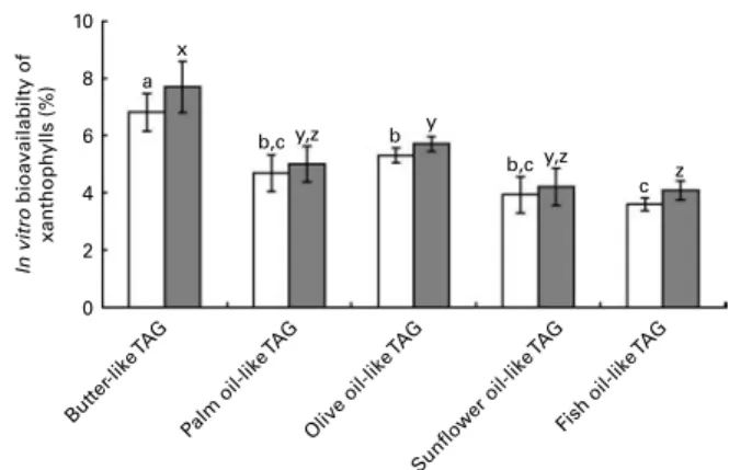
The data of in vitro bioavailability with the mixtures of pure TAG are presented in Fig. 5. The butter-like TAG mix induced a significantly higher in vitro bioavailability of xanthophylls, as compared with the other TAG mixtures. Conversely, the fish oil-like TAG mix elicited a significantly lower in vitro xanthophyll bioavailability. 30 a,b x a x b y a x b y 20 Butter Xanthoph yll uptak e ef ficiency (%)
Palm oil Olive oil Sunflower oil Fish oil 10
0
Fig. 3. Effect of the type of fat used in the in vitro digestion experiments on the uptake efficiency of sweetcorn xanthophylls by Caco-2 cell monolayers. Mixed micelles recovered after digestion of meals containing sweetcorn and different dietary fats were added on Caco-2 monolayers to measure xanthophyll cellu-lar uptake. Uptake efficiency refers to the percentage of added micelcellu-larised sweetcorn xanthophylls recovered in the Caco-2 cells after 2 h 30 min incu-bation. Values are means of five independent experiments for lutein ( ) and zeaxanthin ( ), with their standard errors represented by vertical bars.
a,bMean values with unlike letters were significantly different (P, 0·05). x,yMean values with unlike letters were significantly different (P, 0·05).
10 a,b x a x c y b,c y d y 6 8 Butter In vitro bioavailabilty of xanthoph ylls (%)
Palm oil Olive oil Sunflower oil Fish oil 4
2 0
Fig. 4. Effect of the type of dietary fat on the in vitro bioavailability of sweet-corn xanthophylls. In vitro bioavailability refers to the percentage of sweetsweet-corn xanthophylls transferred from the food matrix to the Caco-2 cells. It was calcu-lated as the product of bioaccessibility (%) by uptake efficiency (%). Values are means of five independent experiments for lutein ( ) and zeaxanthin ( ), with their standard errors represented by vertical bars.a,b,c,dMean values with
unlike letters were significantly different (P, 0·05).x,yMean values with unlike
letters were significantly different (P, 0·05).
B. Gleize et al. 6
British
Journal
of
Nutrition
https://www.cambridge.org/core . IP address: 78.202.154.114 , on 26 May 2021 at 12:20:55, subject to the Cambridge Core terms of use, available at
https://www.cambridge.org/core/terms
.
Moreover, we calculated in vitro bioavailability after the diges-tion and cellular uptake of spinach xanthophylls. The results obtained were in agreement with those obtained with the sweetcorn matrix, with a significantly higher in vitro bio-availability of the xanthophylls with butter as compared with olive, sunflower and fish oils (Table S3, available online).
Effect of source of dietary fat on spinach lutein bioavailability in rats
In order to check whether the in vitro data could predict in vivo bioavailability, we determined the effect of three diet-ary fats on lutein bioavailability in an in vivo model. Adult rats were orally administered with a xanthophyll source (spinach) together with three different TAG emulsions made with butter, olive or fish oil for three consecutive days. The butter emul-sion led to a significantly higher fasting plasma concentration of lutein than the olive oil emulsion, and even more markedly than the fish oil emulsion (Fig. 6).
Effect of dietary intake of SFA on associations between plasma xanthophyll concentrations and fruit and vegetable intake
As shown in Table 4, both lutein ^ zeaxanthin and b-cryptox-anthin plasma concentrations were significantly positively associated with fruit and vegetable intake among subjects with a low (, 34 – 37 g/d) dietary intake of SFA. Conversely, this association was not observed for lutein ^ zeaxanthin in subjects with a high (. 34 – 37 g/d) dietary intake of SFA.
Discussion
In order to understand how the type of FA that constitutes dietary TAG can affect xanthophyll absorption efficiency, we used two complementary models to study the key steps of xanthophyll absorption: an in vitro digestion model, to study carotenoid bioaccessibility, and intestinal Caco-2 mono-layers, to study uptake by intestinal cells. In vivo data in rats and association studies using data from a human study were obtained to substantiate these in vitro observations.
Data obtained with the in vitro digestion model showed that the dietary fat sources that had the higher proportions of SFA, i.e. butter and palm oil, led to a higher bioaccessibility of the xanthophylls than the fat sources that were rich in long-chain MUFA and PUFA. This result did not depend on the vegetable matrix in which the xanthophylls were incorporated because comparable data were obtained with either sweetcorn or spinach. This finding is in agreement with previous studies showing that lutein and zeaxanthin were more bioaccessible in the presence of coconut or peanut oil (rich in SFA) than in
the presence of rapeseed oil (rich in unsaturated FA)(16,30).
The present result raises the following question: does the effect of the source of dietary fat on xanthophyll bioaccessibility depends on its FA composition or does it depend on the presence of fat-soluble microconstituents in some sources of dietary fats (e.g. phytosterols and vitamin E in sunflower oil,
b-carotene in butter)(31,32)? To answer this question, we
per-formed digestion experiments in which we replaced the natural sources of dietary TAG by mixtures of pure TAG that mimicked the FA composition of the natural studied fats. These experi-ments provided similar findings to those using natural TAG sources, i.e. butter- and palm oil-like TAG mixtures led to a higher xanthophyll bioaccessibility than sunflower and fish oils-like TAG. We therefore conclude that the microconstituents present in the studied natural sources of dietary TAG have no major effect on xanthophyll bioaccessibility.
In order to understand the mechanism(s) involved,
we determined correlations between FA characteristics
and xanthophyll bioaccessibility. These calculations showed that there was a significant (P, 0·05) negative relationship between the degree of unsaturation of TAG FA and xantho-phyll bioaccessibility. We also found a significant (P, 0·05) negative relationship between xanthophyll bioaccessibility and the mean length of FA present in the TAG. These observed relationships thus support the hypothesis that the physico-chemical properties of TAG FA play a key role in xanthophyll bioaccessibility. To go further into the mechanism(s) involved, 6 5 a b c 3 4 Butter Plasma lutein ( µ mol/l)
Olive oil Fish oil 2
1 0
Fig. 6. Effect of different types of dietary fat used in oral administration experiments in rats on fasting plasma lutein concentration. Rats (n 10 per group) were orally administered for three consecutive days with 1 ml spinach/dietary fat emulsions (for more details, see the Materials and methods section). Values are means, with their standard errors represented by vertical bars.a,b,cMean values with unlike letters are significantly different
(P, 0·05). 10 a x b,c y,z b,c y,z b y c z 6 8 But ter-lik e TA G In vitro bioavailabilty of xanthoph ylls (%) Palm oil-lik e TA G Olive oil-lik e TA G Sunflower oil-lik e TA G Fish oil-lik e TA G 4 2 0
Fig. 5. Effect of different TAG mixtures on the in vitro bioavailability of sweet-corn xanthophylls. The mixtures of TAG were elaborated to mimic the fatty acids composition of the studied dietary fat sources. In vitro bioavailability refers to the percentage of sweetcorn xanthophylls transferred from the food matrix to the Caco-2 cells. It was calculated as the product of bioaccessibility (%) by uptake efficiency (%). Values are means of five independent exper-iments for lutein ( ) and zeaxanthin ( ), with their standard errors rep-resented by vertical bars. a,b,cMean values with unlike letters were
significantly different (P, 0·05).x,y,zMean values with unlike letters were
sig-nificantly different (P, 0·05).
Fatty acid and xanthophyll bioavailability 7
British
Journal
of
Nutrition
https://www.cambridge.org/core . IP address: 78.202.154.114 , on 26 May 2021 at 12:20:55, subject to the Cambridge Core terms of use, available at
https://www.cambridge.org/core/terms
.
we measured the size and zeta potential of mixed micelles. Results showed that the size of the mixed micelles produced on in vitro digestion of the fats rich in SFA were lower than those produced from the fats rich in unsaturated FA. In fact, micelle mean radius was positively related (P, 0·005) to the degree of unsaturation of FA that constituted the fat sources. This is in agreement with a previous publication that has shown that addition of long-chain FA to bile salt solutions
increased the micellar size(33) but, to our knowledge, this is
the first demonstration of an effect of the unsaturation degree of FA on mixed micelle size. However, the most interesting find-ing of these association studies, with regard to our research question, was the significant (P, 0·05) inverse relationship
between micelle size and xanthophyll bioaccessibility.
Indeed, the lower the unsaturation degree of FA, the lower the size of mixed micelles and the higher their ability to solubil-ise the xanthophylls. To explain this finding, we hypothessolubil-ise that lutein and zeaxanthin, which possess two polar groups, have these groups located at the micelle surface. As reducing the size of a fixed amount of lipid particles increases its specific
surface area(34), we hypothesise that the lower size of micelles
rich in SFA increases the amount of xanthophylls that can be incorporated per amount unit of micelle lipids.
The second mechanism by which TAG FA could have an effect on xanthophyll absorption is an effect on xanthophyll uptake by intestinal cells. Nevertheless, we did not find any sig-nificant effect of the type of TAG FA on xanthophyll uptake by Caco-2 cells when pure TAG mixtures were used. Thus, we con-clude that the type of FA does not have an important effect on this step of xanthophyll absorption. However, the significant differences observed on xanthophyll uptake when mixed micelles produced on digestion of natural fats suggest that some compounds in natural fats, probably fat-soluble micronu-trients, may affect xanthophyll uptake by Caco-2 cells. This
hypothesis is supported by previous publications(20,35).
To validate the earlier in vitro findings, we performed a study in a rat model, i.e. rats that were orally administered with a mix-ture of spinach plus different fat sources for 3 d. The results obtained supported well the ones obtained with the in vitro models, with a higher in vivo xanthophyll bioavailability in the presence of butter compared with unsaturated dietary fats. The significantly lower xanthophyll bioavailability observed when spinach was orally administered with fish oil, as com-pared with olive oil, was noteworthy given there was no signifi-cant difference between these two oils with regard to in vitro xanthophyll bioaccessibility (Fig. 1). A first hypothesis to explain this finding is that the observed difference between these two oils with regard to xanthophyll uptake by intestinal cells (Fig. 3) may have a significant effect on xanthophyll bioa-vailability in vivo. A second hypothesis is that the difference in plasma lutein concentration between rats orally administered with olive and fish oils was due to a partial degradation of the xanthophyll in the rat gastrointestinal tract in the presence of
the highly oxidisable fish oil FA(36,37). Indeed, we did not add
BHT in the fat sources used in the rat study to be closer to dietary conditions, but we found that xanthophylls were partially degraded in the in vitro digestion experiments when no BHT was added. A third hypothesis is that, as has previously been
Table 4 . Epidemiological associations between fasting plasma xanthophyll concentrations and dietary intake of fruit and (in tertiles) according to SFA in the Supple ´mentation e n V itamines e t M ine ´raux AntioXydants study v egetables* (Mean values and 9 5 % confidence intervals) Low SFA (, 34/37 g p er d† in men/women) High SFA ($ 34/37 g p er d in m en/women) Tertiles o f fruit and v egetable intakes Tertiles of fruit and vegetable intakes T1 T2 T3 T1 T2 T3 Mean 9 5 % CI Mean 95 % C I Mean 9 5 % CI P Mean 95 % C I Mean 9 5 % CI Mean 9 5 % CI PP for interaction Lutein þ zeaxanthin 0 ·47 0·42, 0 ·53 0 ·54 0·48, 0 ·60 0 ·54 0 ·49, 0 ·60 0 ·04 0 ·48 0 ·43, 0 ·53 0 ·48 0 ·43, 0 ·53 0 ·49 0·43, 0 ·55 0·87 0 ·19 b -Cryptoxanthin 0 ·20 0·17, 0 ·24 0 ·23 0·19, 0 ·27 0 ·26 0 ·22, 0 ·31 0 ·03 0 ·23 0 ·20, 0 ·27 0 ·25 0 ·21, 0 ·30 0 ·24 0·20, 0 ·29 0·77 0 ·02 * D ietary intakes were estimated from a va lidated FFQ includ ing 2 40 items (23) and using a food composition table (24) . † Median daily intake for men and women. The median is us ually used to divid e a popula tion in to two groups in order to test in teractions. Cut-offs for ter tiles o f fruit and v e g etable intakes were 415/594 g per d in men and 4 71/64 3 g per d in women. Values are adjusted me ans and 95 % C I estimated using ANCOVA. P for linea r trend across sex-sp ecific energy-adjusted tertiles o f fruit an d v egetable intake s. Model was ad justed for age, se x, education, tobacco status, supplementatio n g roup during the trial phase, energy and alcohol intake, BMI, sea son of biolo g ical data col lection and cholesterol aem ia. B. Gleize et al. 8
British
Journal
of
Nutrition
https://www.cambridge.org/core . IP address: 78.202.154.114 , on 26 May 2021 at 12:20:55, subject to the Cambridge Core terms of use, available at
https://www.cambridge.org/core/terms
.
reported(38 – 40), MUFA in olive oil had induced a higher secretion of chylomicrons, and thus a higher bioavailability of lutein, as compared with PUFA present in fish oil. In conclusion, we suggest that the in vivo bioavailability of xanthophylls can be negatively affected by fish oils by three mechanisms: (1) by intrinsic physical properties of fish oil FA that lead to bigger micelles that solubilise less xanthophylls, (2) by an inhibitory effect of a micronutrient present in fish oil on xanthophyll uptake by intestinal cells and (3) by the oxidation of fish oil FA in the gastrointestinal tract, which in turn may degrade a frac-tion of the antioxidant xanthophylls.
Overall, the results obtained with the complementary models suggest that butter, and probably palm oil, enhances xantho-phyll bioavailability, as compared with olive, sunflower and fish oils. This effect is apparently due to the higher bioaccessibil-ity of xanthophylls induced by the smallest micelles produced in the presence of SFA. However, other mechanisms are appar-ently involved, such as a possible inhibitory effect of some fat-soluble micronutrients, which may impair xanthophyll uptake by intestinal cells, and partial degradation of the xanthophylls in the gastrointestinal tract in the presence of highly oxidisable FA. Finally, it should be reminded that the type of TAG FA might also affect the intracellular transport of xanthophylls in the enterocyte and their packaging in the chylomicrons, mechan-isms that were not assessed in the present study.
The last question was to determine whether long-term SFA consumption, by affecting xanthophyll bioavailability, may in turn affect blood concentrations of xanthophylls in human subjects. In order to verify this hypothesis, we determined, in a cohort of 622 subjects who took part in the SU.VI.MAX
study(21), whether SFA intake modulated the expected
associ-ation between blood lutein and zeaxanthin concentrassoci-ations and fruit and vegetable intake. The answer was yes. Indeed, there was a positive significant association (P¼ 0·04) between fruit and vegetable intake and plasma lutein ^ zeaxanthin levels in subjects with low SFA intake, while there was no significant association (P¼ 0·87) in subjects with a high SFA intake. Nevertheless, the suggested effect of SFA intake on the relationship between fasting plasma concentrations of lutein and zeaxanthin and fruit and vegetable intake observed in the epidemiological association study was apparently not explained by the positive effect of SFA on lutein and zeax-anthin bioavailability suggested by the results of the present in vitro and rat studies. Indeed, the high intake of SFA was associated with lower plasma lutein and zeaxanthin concen-trations in the second and third tertiles of fruit and vegetable intake. This apparent discrepancy may be due to potential limitations of the SU.VI.MAX study. Indeed, it is possible that there were some inaccuracies regarding dietary intake of the subjects that may have occurred as a result of the use of a self-administered FFQ. This apparent discrepancy can be also due to the fact that other mechanisms, as well as the posi-tive effect of SFA on lutein and zeaxanthin bioavailability, are involved in the effect of SFA consumption on fasting blood lutein and zeaxanthin concentrations. For example, there might be an effect of blood SFA on uptake, or release, of xanthophylls by storage tissues, thus affecting blood con-centrations of xanthophylls.
In conclusion, the results obtained from both in vitro and the rat studies suggest that SFA improve lutein and zeaxanthin bioavailability, mainly by increasing their bioaccessibility. Epi-demiological associations, performed in a sample of the SU.VI.MAX study, support an interaction between SFA intake and fasting blood concentrations of lutein and zeaxanthin. However, higher intakes of SFA were not associated with higher fasting blood concentrations of these xanthophylls. This discrepancy may be, at least partly, ascribed to residual confounding in the human study. Thus, further experiments are necessary to conclude on the long-term effect of SFA con-sumption on lutein and zeaxanthin status.
Supplementary material
To view supplementary material for this article, please visit http://dx.doi.org/10.1017/S0007114512004813
Acknowledgements
The authors are grateful to Maria-Erica Lopez and Jean-Marc Feuerstein (C.F.R.E.M., Marseille, France), Jean-Franc¸ois Landr-ier, Aure´lie Goncalves and Charlotte Sy (UMR INRA 1260, Mar-seille, France) for their technical help for the rat study. Thanks also to Marie-Jose`phe Amiot-Carlin who put our team in touch with Bonduelle who funded the in vitro and rat experiments of the present study. The SU.VI.MAX study was funded by DGS (Ministry of Health) and supported by Me´de´ric, Ipsen, MGEN, SODEXHO and Pierre Fabre. Work on the subset cohort of SU.VI.MAX was granted by ANR (no. ANR-05-PNRA-010). The authors’ contributions to the study were as follows: B. G., F. T., L. D., L. A., D. L. and P. B. designed the research; B. G., R. B. and M. N. conducted the research; E. K.-G., P. G. and S. H. constituted the SU.VI.MAX 2 investigators; B. G. and P. B. analysed data and performed statistical analysis; B. G. and P. B. wrote the paper and had primary responsibility for final content. All authors read and approved the final manu-script. No authors have any conflict of interest to declare.
References
1. Khachik F, Beecher GR, Goli M, et al. (1992) Separation and identification of carotenoids and their oxidation products in the extracts of human plasma. Anal Chem 64, 2111 – 2122. 2. Rao AV & Rao LG (2007) Carotenoids and human health.
Pharmacol Res 55, 207 – 216.
3. Maiani G, Caston MJ, Catasta G, et al. (2009) Carotenoids: actual knowledge on food sources, intakes, stability and bioavailability and their protective role in humans. Mol Nutr Food Res 53, Suppl. 2, S194 – S218.
4. Bone RA, Landrum JT, Friedes LM, et al. (1997) Distribution of lutein and zeaxanthin stereoisomers in the human retina. Exp Eye Res 64, 211 – 218.
5. Junghans A, Sies H & Stahl W (2001) Macular pigments lutein and zeaxanthin as blue light filters studied in liposomes. Arch Biochem Biophys 391, 160 – 164.
6. Krinsky NI (2002) Possible biologic mechanisms for a protec-tive role of xanthophylls. J Nutr 132, 540S – 542S.
Fatty acid and xanthophyll bioavailability 9
British
Journal
of
Nutrition
https://www.cambridge.org/core . IP address: 78.202.154.114 , on 26 May 2021 at 12:20:55, subject to the Cambridge Core terms of use, available at
https://www.cambridge.org/core/terms
.
7. Snodderly DM (1995) Evidence for protection against age-related macular degeneration by carotenoids and antioxidant vitamins. Am J Clin Nutr 62, 1448S – 1461S.
8. Richer S, Stiles W, Statkute L, et al. (2004) Double-masked, placebo-controlled, randomized trial of lutein and antioxi-dant supplementation in the intervention of atrophic age-related macular degeneration: the Veterans LAST study (Lutein Antioxidant Supplementation Trial). Optometry 75, 216 – 230.
9. Reboul E, Richelle M, Perrot E, et al. (2006) Bioaccessibility of carotenoids and vitamin E from their main dietary sources. J Agric Food Chem 54, 8749 – 8755.
10. van Het Hof KH, West CE, Weststrate JA, et al. (2000) Dietary factors that affect the bioavailability of carotenoids. J Nutr 130, 503 – 506.
11. Borel P (2003) Factors affecting intestinal absorption of highly lipophilic food microconstituents (fat-soluble vitamins, caro-tenoids and phytosterols). Clin Chem Lab Med 41, 979 – 994. 12. Yonekura L & Nagao A (2007) Intestinal absorption of dietary
carotenoids. Mol Nutr Food Res 51, 107 – 115.
13. Roodenburg AJ, Leenen R, van Het Hof KH, et al. (2000) Amount of fat in the diet affects bioavailability of lutein esters but not of alpha-carotene, beta-carotene, and vitamin E in humans. Am J Clin Nutr 71, 1187 – 1193.
14. Unlu NZ, Bohn T, Clinton SK, et al. (2005) Carotenoid absorption from salad and salsa by humans is enhanced by the addition of avocado or avocado oil. J Nutr 135, 431 – 436. 15. Lakshminarayana R, Raju M, Krishnakantha TP, et al. (2007) Lutein and zeaxanthin in leafy greens and their bioavailabil-ity: olive oil influences the absorption of dietary lutein and its accumulation in adult rats. J Agric Food Chem 55, 6395 – 6400.
16. Huo T, Ferruzzi MG, Schwartz SJ, et al. (2007) Impact of fatty acyl composition and quantity of triglycerides on bioaccessi-bility of dietary carotenoids. J Agric Food Chem 55, 8950 – 8957. 17. Arvisenet G, Billy L, Poinot P, et al. (2008) Effect of apple particle state on the release of volatile compounds in a new artificial mouth device. J Agric Food Chem 56, 3245 – 3253.
18. Chantret I, Rodolosse A, Barbat A, et al. (1994) Differential expression of sucrase-isomaltase in clones isolated from early and late passages of the cell line Caco-2: evidence for glucose-dependent negative regulation. J Cell Sci 107, 213 – 225.
19. Salvini S, Charbonnier M, Defoort C, et al. (2002) Functional characterization of three clones of the human intestinal Caco-2 cell line for dietary lipid processing. Br J Nutr 87, 211 – 217.
20. Reboul E, Abou L, Mikail C, et al. (2005) Lutein transport by Caco-2 TC-7 cells occurs partly by a facilitated process invol-ving the scavenger receptor class B type I (SR-BI). Biochem J 387, 455 – 461.
21. Hercberg S, Galan P, Preziosi P, et al. (2004) The SU.VI.MAX Study: a randomized, placebo-controlled trial of the health effects of antioxidant vitamins and minerals. Arch Intern Med 164, 2335 – 2342.
22. Borel P, de Edelenyi FS, Vincent-Baudry S, et al. (2010) Genetic variants in BCMO1 and CD36 are associated with plasma lutein concentrations and macular pigment optical density in humans. Ann Med 43, 47 – 59.
23. Kesse-Guyot E, Castetbon K, Touvier M, et al. (2010) Relative validity and reproducibility of a food frequency question-naire designed for French adults. Ann Nutr Metab 57, 153 – 162.
24. Hercberg S (2005) Tables de composition des aliments SU.VI.-MAX (Tables of Food Composition SU.VI.SU.VI.-MAX) (in French). Paris: Economica.
25. Gleize B, Steib M, Andre´ M, et al. (2012) Simple and fast HPLC method for simultaneous determination of retinol, tocopherols, coenzyme Q10 and carotenoids in complex samples. Food Chem 134, 2560 – 2564.
26. Willet W, (1998) Nutritional Epidemiology, 2nd ed. New York: Oxford University Press.
27. Ito Y, Sasaki R, Suzuki S, et al. (1991) Relationship between serum xanthophyll levels and the consumption of cigarettes, alcohol or foods in healthy inhabitants of Japan. Int J Epide-miol 20, 615 – 620.
28. Rock CL, Thornquist MD, Neuhouser ML, et al. (2002) Diet and lifestyle correlates of lutein in the blood and diet. J Nutr 132, 525S – 530S.
29. Gruber M, Chappell R, Millen A, et al. (2004) Correlates of serum lutein þ zeaxanthin: findings from the Third National Health and Nutrition Examination Survey. J Nutr 134, 2387–2394. 30. O’Connell O, Ryan L, O’Sullivan L, et al. (2008) Carotenoid
micellarization varies greatly between individual and mixed vegetables with or without the addition of fat and fiber. Int J Vitam Nutr Res 78, 238 – 246.
31. Tyssandier V, Cardinault N, Caris-Veyrat C, et al. (2002) Veg-etable-borne lutein, lycopene, and beta-carotene compete for incorporation into chylomicrons, with no adverse effect on the medium-term (3-wk) plasma status of carotenoids in humans. Am J Clin Nutr 75, 526 – 534.
32. Reboul E, Thap S, Perrot E, et al. (2007) Effect of the main dietary antioxidants (carotenoids, gamma-tocopherol, poly-phenols, and vitamin C) on alpha-tocopherol absorption. Eur J Clin Nutr 61, 1167 – 1173.
33. Carey MC & Small DM (1970) The characteristics of mixed micellar solutions with particular reference to bile. Am J Med 49, 590 – 608.
34. Borel P, Armand M, Ythier P, et al. (1994) Hydrolysis of emul-sions with different triglycerides and droplet sizes by gastric lipase in vitro – effect on pancreatic lipase activity. J Nutr Biochem 5, 124 – 133.
35. Reboul E, Thap S, Tourniaire F, et al. (2007) Differential effect of dietary antioxidant classes (carotenoids, polyphe-nols, vitamins C and E) on lutein absorption. Br J Nutr 97, 440 – 446.
36. Kanner J & Lapidot T (2001) The stomach as a bioreactor: dietary lipid peroxidation in the gastric fluid and the effects of plant-derived antioxidants. Free Radic Biol Med 31, 1388 – 1395.
37. Lorrain B, Dangles O, Genot C, et al. (2010) Chemical mod-eling of heme-induced lipid oxidation in gastric conditions and inhibition by dietary polyphenols. J Agric Food Chem 58, 676 – 683.
38. Harris WS & Muzio F (1993) Fish oil reduces postprandial tri-glyceride concentrations without accelerating lipid-emulsion removal rates. Am J Clin Nutr 58, 68 – 74.
39. van Greevenbroek MM, van Meer G, Erkelens DW, et al. (1996) Effects of saturated, mono-, and polyunsaturated fatty acids on the secretion of apo B containing lipoproteins by Caco-2 cells. Atherosclerosis 121, 139 – 150.
40. Sauvant P, Mekki N, Charbonnier M, et al. (2003) Amounts and types of fatty acids in meals affect the pattern of retinoids secreted in human chylomicrons after a high-dose preformed vitamin A intake. Metabolism 52, 514 – 519. 41. Gleize B, Payet M, Esmail MH, et al. (2004)
Docosahexae-noic acid-enriched egg phospholipids supplementation induces accretion of arachidonic acid in rat blood lipids. Cell Mol Biol 50, 861 – 867.
B. Gleize et al. 10
British
Journal
of
Nutrition
https://www.cambridge.org/core . IP address: 78.202.154.114 , on 26 May 2021 at 12:20:55, subject to the Cambridge Core terms of use, available at
https://www.cambridge.org/core/terms
.