ANALYSIS OF ANAPLASMA MARGINALE
STRAINS GROWN IN VITRO
Thèse présentée à la faculté des Sciences Institut de Biologie
Université de Neuchâtel
Pour l’obtention du grade de docteur ès Sciences (Ph.D.)
Par
Katarzyna Lis
Jury
Prof. Dr. Kurt Pfister, Directeur de thèse (Université de Neuchâtel, Switzerland) Prof. Dr. Lygia Passos, Supervisor (Ludwig-Maximilians-Universität, Germany) Prof. hon. Bruno Betschart (Université de Neuchâtel, Switzerland)
Prof. Dr. Lise Gern (Université de Neuchâtel, Switzerland) Prof. Dr. Patrick Guerin (Université de Neuchâtel, Switzerland) Prof. Dr. Steffen Rehbein (Merial GmbH, Germany)
Université de Neuchâtel - 2014 -
P
REFACE
This PhD project was conducted at the Institute of Comparative Tropical Medicine and Parasitology at Ludwig-Maximilians-Universität, München, Germany.
This work of research was a part of the POSTICK ITN (Post-graduate training network for capacity building to control ticks and tick-borne diseases), an integrated Project of the European Union’s FP7- PEOPLE – ITN programme (2009-2013), (EU Grant No. 238511).
Key words:
Anaplasma marginale, tick cell cultures, Percoll gradients, MajorSurface Proteins, 2D-DIGE, IDE8
Mots clés:
Anaplasma marginale, culture cellulaire tique, Percoll gradients, MajorSurface Proteins, 2D-DIGE, IDE8
Abstract
Anaplasma marginale is a tick-borne pathogen that affects ruminants worldwide,
causing a disease called anaplasmosis. The disease is endemic in tropical and subtropical regions of the New World, Europe, Africa, Asia and Australia where it causes large economic losses in the cattle industry.
A. marginale is an obligatory intracellular bacterium that multiplies only within tick
cells or ruminants' erythrocytes. Many differences among A. marginale strains have emerged, which were probably driven by continuous exposure to different host immune systems during the transition of bacteria between ticks and vertebrates. The vast majority of studies aiming at elucidating differences between strains were conducted on the genomic level, and little is known about protein expression. Thus, this thesis investigates differences in protein regulation among A. marginale strains.
A. marginale cultivated in vitro are in general an excellent source of organisms for
experimentation. Furthermore, culture-derived organisms offer an alternative to the use of experimental animals.
Many studies require intracellular organisms free from host cell debris. Therefore the use of Percoll gradients for the separation of A. marginale was evaluated. Bacteria isolated in this way contained only minimal amounts of IDE8 cell stroma but most importantly they retained their viability. A. marginale purified this way can be used directly for proteomic studies or for vaccination trials.
In this thesis three geographical A. marginale strains grown in vitro have been partially characterized by gene and serological analyses. The differences on the proteomic level have been assessed by the 2D-DIGE technique, indicating that many antigenic membrane proteins are differentially regulated among the strains examined. Some of these proteins are also known to be virulence-associated.
Increasing the number of strains in continuous in vitro cultivation, and improving purification methods for rickettsia, allow researchers to investigate differences in protein expression between A. marginale strains, and therefore identify proteins which could be incorporated into an improved vaccine against anaplasmosis.
Résumé
Anaplasma marginale est un agent pathogène issu des tiques qui affectent les
ruminants dans le monde entier, causant une maladie appelée anaplasmosis. La maladie est endémique dans les régions tropicales et subtropicales du Nouveau Monde, d’Europe, d’Afrique, d’Asie et d’Australie où elle cause d’importantes pertes économiques dans l’industrie du bétail.
A. marginale est une bactérie obligatoirement intracellulaire qui ne se multiplie que
dans les cellules des tiques ou des érythrocytes des ruminants. De nombreuses différences parmi les souches de A. marginale sont apparues, probablement à cause d’une exposition continue à différents systèmes immunitaires d’hôtes lors du passage de la bactérie des tiques aux vertébrés. Un grand nombre d’études génétiques ayant pour but d’élucider les différences entre les souches ont été réalisées.
Malheureusement, peu de résultats concernant l’expression des protéines d’A. marginale ont été obtenus. Alors que cette thèse prouve des différences dans la régulation des protéines parmi les souches de A. marginale.
A. marginale s’étant reproduites in vitro sont en général une excellente source
d’organismes pour les expérimentations. De plus, les organismes issus de ces cultures sont une excellente alternative aux animaux de laboratoire.
De nombreuses études cliniques requièrent des organismes intracellulaires débarrassés de tout débris de la cellule hôte. Pour ce faire, l’utilisation de gradients Percoll pour la séparation de A. marginale a été pratiquée. Les bactéries isolées de cette manière ne contenaient qu’une quantité infime de IDE8 cellules, mais plus intéressant elles conservaient leur viabilité. A. marginale purifiées de cette manière peuvent être utilisées directement pour des études protéomiques ou pour des études ayant pour but le développement de nouveaux vaccins.
Trois souches séparées géographiquement de A. marginale élevées in vitro ont été partiellement caractérisées par des analyses génétiques et sérologiques. Les différences au niveau protéomique ont été mesurées au moyen de la technique 2D-DIGE, indiquant que de nombreuses membranes protéines antigéniques sont
différemment dosées dans les souches étudiées. Certaines de ces protéines sont aussi connues pour avoir un facteur de virulence.
L’augmentation du nombre de souches dans de continuelles cultures in vitro et l’amélioration des méthodes de purification des bactéries ont permis aux
scientifiques de rechercher les différences dans l’expression protéine des souches
A. marginale et ainsi d’identifier quelles protéines pourraient être utilisées pour un
11
Table of content
1. LITERATURE REVIEW 17
1.1. Anaplasma marginale - historical background 17
1.2. Epidemiology 17
1.3. Classification 18
1.4. Life cycle 19
1.5. Transmission 22
1.6. Pathogenesis 24
1.7. Differences within strains 27
1.7.1. Morphology 27
1.7.2. Major Surface Proteins 29
1.7.3. Tick transmission 31
1.7.4. Virulence 32
1.8. Anaplasmosis control methods 34
1.8.1. Antibiotics 34 1.8.2. Arthropod control 34 1.8.3. Vaccination 35 1.8.3.1. Live vaccines 35 1.8.3.2. Inactivated vaccines 36 1.8.3.3. Culture-derived vaccines 37
1.9. CELL CULTURE SYSTEMS 37
1.10. RECENT INTERESTS IN ANAPLASMOSIS RESEARCH 39
1.11. PROJECT OUTLINE 41
2. USE OF PERCOLL GRADIENTS TO PURIFY ANAPLASMA MARGINALE
(RICKETTSIALES: ANAPLASMATACEAE) FROM TICK CELL CULTURES 45
2.1. Abstract 45
2.2. Introduction 46
2.3. Materials and methods 47
2.3.1. Cell cultures 47
2.3.2. A. marginale strains 47
2.3.3. Preparation of Percoll density gradient 48
2.3.4. Cryopreservation and viability of purified bacteria 48 2.3.5. Quantification of A. marginale DNA by real-time PCR 49
2.4. Results 50
2.4.1. Percoll density gradients and quantifications 50
12
2.5. Discussion 51
Rerferences 55
3. FUNCTIONAL AND IMMUNOLOGICAL RELEVANCE OF ANAPLASMA MARGINALE MAJOR SURFACE PROTEIN 1A SEQUENCE AND STRUCTURAL ANALYSIS 61
3.1. Abstract 61
3.2. Introduction 61
3.3. Results and Discussion 65
3.3.1. Classification of A. marginale strains using MSP1a sequence data 65 3.3.1. The biological implications of sequence variation of MSP1a tandem repeats. 66
3.3.2.1. O-glycosylation 67
3.3.2.2. Relevance of amino acids at position 20 for binding to tick cell extract (TCE) 68
3.3.2.1. Protein conformation 69
3.3.2.1. Pathogen-environmental relationships 73
3.3.2.2. Influence of a combination of factors 75
3.3.2.1. Analysis of B cell epitope in MSP1a tandem repeats 76
3.4. Methods 79
3.4.1. Anaplasma marginale strains classification 79
3.4.2. Amino acid variability within MSP1a tandem repeats 80 3.4.3. Correlation analysis between MSP1a tandem repeats and world ecological regions 80
3.4.4. Bioinformatics 81
Supporting Information: Table 3.S1. Classification of A. marginale strains based on the proposed
nomenclature. 83
References 91
4. MOLECULAR AND IMMUNOLOGICAL CHARACTERIZATION OF THREE STRAINS
OF ANAPLASMA MARGINALE GROWN IN CULTURED TICK CELLS 97
4.1. Abstract 97
4.2. Introduction 98
4.3. Materials and methods 100
4.3.1. Anaplasma marginale strains 100
4.3.2. Bacteria purification and protein extraction 100
4.1.1. Antibodies 101
4.1.2. Western blot analysis 102
4.1.3. DNA isolation and amplification 102
4.1.1. Sequencing of PCR products 103
4.1.2. Phylogenetic analysis 103
4.1. Results and Discussion 105
4.1.1. Propagation of A. marginale strains in tick cell culture 105 4.1.2. Molecular and immunological characterization of HSP70 protein from the A. marginale strains.
105
4.1.1. Molecular characterization of sodb gene and putative proteins from different isolates of
A. marginale 107
4.1.1. MSP1a genotype and tick cells infection phenotype 110 4.1.2. Molecular and immunological characterization of MSP4 and MSP5 proteins from the three A.
13
marginale strains 111
4.1. Conclusions 113
References 114
5. IDENTIFICATION OF DIFFERENTIALLY EXPRESSED PROTEINS AMONG
GEOGRAPHICAL A. MARGINALE STRAINS GROWN IN VITRO BY 2D- DIGE 121
5.1. Introduction 121
5.2. Materials and methods 122
5.2.1. Anaplasma marginale strains 122
5.2.2. Bacteria purification and protein extraction 122
5.2.3. 2D-DIGE 123
5.2.4. Image acquisition and data analysis 124
5.2.5. In-gel digestion and mass spectrometry 124
5.2.6. Protein analysis 125
5.2.7. In silico analysis of hypothetical proteins 125
5.3. Results and Discussion 126
5.3.1. Proteome analysis 126
5.3.2. Outer membrane proteins 126
5.3.3. Major Surface Proteins 127
5.3.3.1. MSP2 127
5.3.3.2. MSP4 127
5.3.3.3. Type IV secretion system proteins 129
5.3.4. Other outer membrane proteins 129
5.3.5. Protein biosynthesis 130
5.3.6. Stress induced proteins 133
5.3.7. Hypothetical proteins 134
5.3.8. Intermediary metabolism 138
5.4. Conclusions 142
6. CONCLUSIONS AND RECOMMENDATIONS 143
LIST OF ABBREVIATIONS 145
REFERENCES 147
17
1. Literature review
1.1.
Anaplasma marginale - historical background
Anaplasma marginale was first described in the early 1900s by Sir Arnold Theiler who
observed “marginal points” in erythrocytes of cattle suffering from gallsickness
(galsiekte) (Theiler, 1910, 1911, 1912). Although two decades earlier the
microorganism had already been discovered by other investigators, it had been erroneously considered as a part of the Babesia bigemina life cycle (Smith and Kilborne, 1893). Yet, Theiler demonstrated that babesiosis and anaplasmosis can often co-exist in the same animal; he then succeeded in separating the two agents and produced a “pure infection” with only A. marginale. He indicated that the “marginal points” differ from any known blood parasite and named the new pathogen Anaplasma
marginale.
The scientific name proposed by Theiler was based on the microscopic observation
of the pathogen in infected blood smears. “Anaplasma” stands for the absence of a
stained cytoplasm, and “marginale” for its marginal localization in infected erythrocytes. Furthermore, Theiler (1911) also described a subspecies of A. marginale: located in the centre of erythrocytes. Anaplasma centrale, is less pathogenic than A.
marginale and causes only a slight attack of the disease. An A. centrale-based,
blood-derived, live vaccine was exported from South Africa to other parts of the world, i.e. Australia, Israel and Latin America, where it has been in use for over a century (Kocan et al., 2010b).
1.2.
Epidemiology
A. marginale is one of the most prevalent tick-borne pathogens of cattle in tropical
and subtropical areas worldwide (~40° N to ~32° S) (Aubry and Geale, 2011). It is endemic in the New World, Central and South America, Australia and some regions of Asia and Africa (Kocan et al., 2010b). In the USA anaplasmosis is enzootic throughout the southern states, but due to the movement of cattle, anaplasmosis has now been reported in almost every state (Kocan et al., 2010a). In some countries it is
18
considered to be a foreign animal disease e.g. in Canada, where an outbreak of anaplasmosis resulted from mechanical transmission of the organism from imported cattle (Boulanger et al., 1971). In Europe, it is found mainly in Mediterranean countries like Italy (de la Fuente et al., 2005e; de la Fuente et al., 2005f; Torina et al., 2008) and Spain (de la Fuente et al., 2005d), although few isolated cases have also been reported in Hungary (Hornok et al., 2012) and Austria (Baumgartner et al., 1992).
The wider distribution and increase in outbreaks of the disease result from transport of asymptomatic carrier animals, which are reservoirs for subsequent mechanical or biological transmission to susceptible cattle in non-endemic areas. Wildlife may be possible reservoir hosts, and represent a source of infection for free-ranging cattle (Kocan et al., 2010a; Kocan et al., 2010b). Furthermore, factors such as climate, host abundance, tick-host diversity and topography have been all shown to have an impact on the epidemiology of A. marginale (Estrada-Pena et al., 2008). Changes in climate influence the distribution, physiology and behavior of many different arthropod vectors (Jonsson and Reid, 2000). The possible introduction of new tick species into areas where they did not exist before may complicate the control and prevention of tick-borne diseases.
1.3.
Classification
In 2001 Dumler et al. proposed a reclassification of Rickettsiales based upon genetic analysis of 16S rRNA and groESL genes. Organisms of this taxon were then assigned to one of the two families: Rickettsiaceae and Anaplasmataceae. All bacteria classified within these families are intracellular pathogens. However, unlike the Rickettsiaceae, which grow freely within the host cytoplasm or nucleus, members of Anaplasmataceae are found exclusively within membrane-bound vacuoles in the cytoplasm of the host cells. Moreover, almost all organisms assigned to the family Anaplasmataceae multiply in both vertebrate and invertebrate hosts (Kocan et al., 2010a).
Following the phylogenetic analysis, four genera were formed within the Anaplasmataceae family, namely: Ehrlichia, Neorickettsia, Anaplasma and Wolbachia. All are gram-negative bacteria, demonstrating two morphological structures: large
19
reticulate forms, or smaller dense forms with condensed protoplasm. Anaplasmataceae infect canids, humans, ruminants and rodents. Formerly the genus Anaplasma consisted of A. ovis, A. marginale (Table 1.1) and a less pathogenic subspecies of A. marginale, A. centrale (A. marginale ss. centrale). Following the
reclassification, A. bovis (formerly Ehrlichia bovis), A. phagocytophilum (formerly
Ehrlichia phagocytophila, E. equi and the human granulocytic ehrlichiosis (HGE)
agent), A. platys (formerly Ehrlichia platys) and Aegyptianella (genus incertae sedis due to the lack of sequence information) have also been included into the Anaplasma genus (Dumler et al., 2001).
Table 1.1. Anaplasma marginale classification according to Dumler et al. (2001).
Class: Alphaproteobacteria Order: Rickettsiales
Family: Anaplasmataceae Genus: Anaplasma
Species: Anaplasma marginale
1.4.
Life cycle
The life cycle of A. marginale is coordinated with the tick feeding cycle (Figure 1.1) (Kocan et al., 2004; Kocan et al., 1992a). Ticks become infected when feeding on A.
marginale-infected animals.
The likelihood of ticks acquiring at least one organism is higher in the acute phase (95–100 %) of infection when compared to the chronic phase (27–84 %) (Eriks et al., 1993). Infected erythrocytes are ingested by ticks with the blood meal, providing the source of A. marginale infection for tick gut cells (Figure 1.2.A). Afterwards extensive replication occurs within other tick tissues, including the salivary glands (Figure
20
Figure 1.1. Developmental cycle of A. marginale in cattle and ticks (taken from
Kocan et al. 2003).
The level of A. marginale organisms in adult male Dermacentor andersoni ticks can
reach approximately 105 organisms per salivary gland (Kocan et al., 1992a)
regardless of the rickettsemia level in the blood during acquisition feeding (Eriks et al., 1993). During subsequent feeding rickettsiae are transmitted via the salivary glands of the tick to vertebrate hosts (Kocan et al., 1992a).
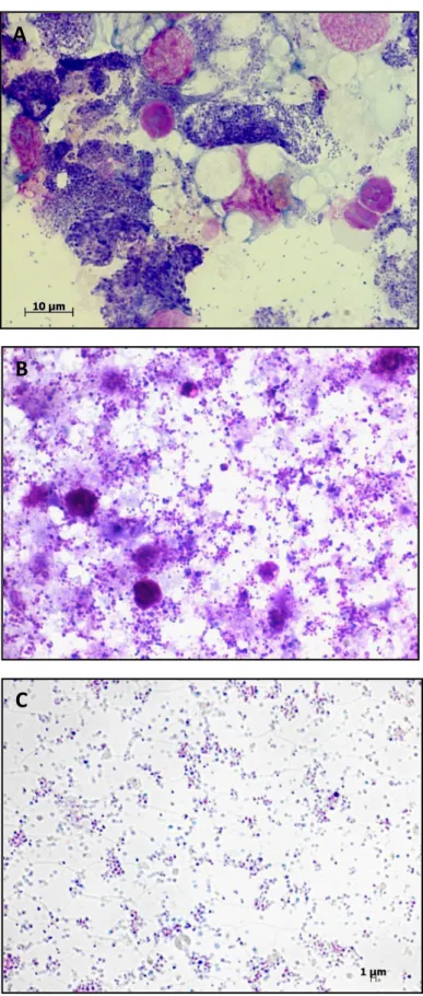
At each infection site within the tick, A. marginale develops within membrane-bound vacuoles, forming colonies. The first form seen within A. marginale colonies is the reticulated (vegetative) form, which divides by binary fission (Figure 1.3, asterisk) and results in the formation of large colonies containing hundreds of organisms. The reticulated forms are then transformed into dense forms (0.5-0.8 µm) (Figure 1.3, arrow), which are the infective forms. They can survive for a short time outside cells (Kocan et al., 2008).
21
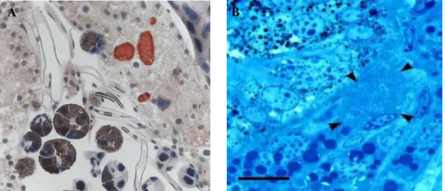
Figure 1.2. A. marginale development within tick tissues. A). Colonies (red) in D. andersoni midgut cells, (taken from “Livelihood hazards” M. Sebaihia, N. R.
Thomson, L. Crossman and J. Parkhill); B). A. marginale colonies (arrowheads) in
Dermacentor reticulatus salivary gland cell (taken from Zivkovic et al. 2007).
Cattle become infected when the dense form is transmitted during tick feeding via the salivary glands (Kocan et al., 2004). The tick cell culture model has been used for studying entry and exit mechanisms of the rickettsia from IDE8 cells (Blouin and Kocan, 1998). Host cell invasion is initiated by the adhesion of the dense form to the tick cell membrane, and the rickettsia subsequently enters into cells by endocytosis.
While leaving the cell, colony and cell membranes fuse, allowing the rickettsia exit without host cell injury. Blouin and Kocan (1998) suggested that the same mechanism occurs within naturally infected tick cells, which may facilitate high infection rates without pathological changes in ticks.
Figure 1.3. Electron micrograph of the
developmental stages of A. marginale within colonies in tick cells. Reticulated forms within a colony divide by binary fission (asterisk), dense forms (arrow). Bar =1 mm (taken from Kocan et al. 2004).
22
1.5.
Transmission
Transmission of A. marginale to vertebrate hosts occurs in two main ways: biologically by ticks, and mechanically by biting flies or by blood contaminated fomites. Transplacental transmission to the calf fetus has also been reported (Grau et al., 2013; Maldonado et al., 2012; Rey Valeiron et al., 2003; Zaugg, 1985).
Various tick species have been reported to be vectors of A. marginale in different regions of the world (Table 1.2) (Kocan et al., 2004). A. marginale DNA has been identified in many tick species or in ticks which transmitted the disease experimentally. However, this does not necessarily imply that they are able to transmit the organisms under natural conditions (Shkap et al., 2009; Zivkovic et al., 2007). In addition, recent analysis suggests that in some regions tick species which have not previously been considered as vectors may also transmit A. marginale (de la Fuente et al., 2005d; Fyumagwa et al., 2009; Zahang et al., 2013). Above all,
Rhipicephalus (Boophilus) spp. are the most prevalent vectors of anaplasmosis in
most tropical and subtropical countries. In the United States, however, Dermacentor spp., including D. variabilis, D. andersoni and D. albipictus, are the major vectors of anaplasmosis (de la Fuente et al., 2001c; Kocan et al., 1981), probably because a compulsory acaricide-treatment program in the 1940s (Stiller et al., 1989) led to the eradication of the R. (B.) microplus tick.
T ransmission occurs by one stage (intrastadial) or from stage to stage (inter- or transstadial). Intrastadial transmission is effectuated mainly by male ticks (Kocan et al., 2010a). Serial transmission by male D. andersoni ticks to five consecutive cattle has been demonstrated (Kocan et al., 1992a). However, it has been shown that A.
marginale was not transmitted from infected to uninfected adult Dermacentor spp.
ticks during co-feeding on the same cattle (Kocan and de la Fuente, 2003). Interstadial transmission e.g. ingestion by nymphs and inoculation by adults has been demonstrated by R.(B.) annulatus, a single-host tick (Shkap et al., 2009), and by D.
23
Table 1.2. Tick species transmitting Anaplasma marginale (modified after Kocan et al.
(2004).
Tick species References
Ixodid Ticks
Amblyomma gemma* (Fyumagwa et al., 2009)
Dermacentor albipictus (Lankester et al., 2007)
Dermacentor andersoni (Anthony and Roby, 1966; Kocan et al., 1992a;
Kocan et al., 1981; Lankester et al., 2007)
Dermacentor hunteri (Stiller et al., 1999)
Dermacentor occidentalis (Anthony & Roby, 1966)
Dermacentor reticulatus (Zivkovic et al., 2007)
Dermacentor variabilis (Anthony & Roby, 1966; Kocan et al., 1981;
Lankester et al., 2007; Stich et al., 1989)
Hyalomma asiaticum (Zahang et al., 2013)
Hyalomma excavatum (Shkap et al., 2009)
Hyalomma marginatum rufipes (Potgieter, 1979)
Ixodes scapularis (Rees, 1934)
Ixodes ricinus (Helm, 1924)
Rhipicephalus (Boophilus) annulatus (Samish et al., 1993)
Rhipicephalus appendiculatus (Fyumagwa et al., 2009)
Rhipicephalus bursa (Sergent et al., 1945)
Rhipicephalus (Boophilus) calcaratus (Sergent et al., 1945)
Rhipicephalus compositus* (Fyumagwa et al., 2009)
Rhipicephalus (Boophilus) decoloratus (Potgieter, 1979; Theiler, 1912)
Rhipicephalus (Boophilus) microplus (Futse et al., 2003)
Rhipicephalus praetextatus* (Fyumagwa et al., 2009)
Rhipicephalus pulchellus* (Fyumagwa et al., 2009)
Rhipicephalus sanguineus (Shkap et al., 2009)
Argasid Ticks
Argas persicus (Howell et al., 1941)
24
The occurrence of transovarial transmission of few tick-borne pathogens e.g. Babesia spp. by single-host R. Boophilus spp. is well known (Howell et al., 2007). Yet, transmission of A. marginale from one tick generation to the other remains controversial, although Theiler (1912) and few other authors have suggested that this type of transmission does occur (Anthony and Roby, 1962; Rees and Avery, 1939; Stich et al., 1989). Interestingly, multiplication of A. marginale within the tissues of engorged R.(B.) microplus females has been confirmed (Ribeiro and Lima, 1996). Moreover, in eggs and larvae derived from R. B. microplus ticks collected from infected cattle, A. marginale specific DNA fragments have been amplified. Yet, the transmission of A. marginale by these larvae to animals has never been proven (Moura et al., 2003). Some authors suggested that Ehrlichia spp. and Anaplasma spp. are not transmitted transovarially due to the lack of the aldolase/adducing domain protein (Dunning Hotopp et al., 2006).
Mechanical transmission of the pathogen occurs when infected blood is transferred to susceptible animals by contaminated fomites: needles, dehorning saws, nose tongs, tattooing instruments, ear tagging devices and castration instruments (Kocan et al., 2004) . Additionally, different species of hematophagous diptera e.g. Tabanus spp. flies (Hawkins et al., 1982), Stomoxys calcitrans (stable fly) (Potgieter et al., 1981) or mosquitoes have been demonstrated to have the ability to disseminate A. marginale. Although biological transmission has been shown to be more efficient (Scoles et al., 2008), mechanical transmission is the major route of infection in areas where the strains are not tick-transmissible or appropriate tick vectors do not occur (de la Fuente et al., 2001c).
1.6.
Pathogenesis
A. marginale is very host specific and under natural conditions infects only ruminants.
Although clinical anaplasmosis occurs most often in cattle, other ruminants like water buffalo (Bubalus bubalis), American bison (Bison bison), white-tailed deer (Odocoileus virginianus), black-tailed deer (Odocoileus hemionus columbianus), Rocky Mountain elk (Cervus elaphus nelsoni), black wildebeest (Connochaetes gnou), blesbuck (Damaliscus pygargus phillipsi) and duiker (Sylvicapra grimmia) can also become infected (Aubry and Geale, 2011; Kocan et al., 2010b; Kuttler, 1984).
25
The only known site of A. marginale development in cattle is within erythrocytes (Figure 1.4.A). Interestingly, because bacteria can be propagated in a bovine endothelial cell line (Munderloh et al., 2004), it has been suggested that endothelial cells may serve as a site of initial replication after tick attachment, or as a reservoir for A. marginale during persistent infection. After experimental infection of calves with the A. marginale St. Maries strain, Carreno et al. (2007) observed the infection of endothelial cells by dual fluorescence microscopy. In contrast, Wamsley et al. (2011) did not detect A. marginale within endothelial cells after tick-feeding transmission to immunocompetent cattle either in dermal samples of tick attachment sites or in post- mortem tissues. In addition, they also did not observe seroconversion or clinical anaplasmosis in calves, when A. marginale grown in the endothelial cell line (RF/6A) was used for the experiments. At the moment in vivo infection of endothelial cells remains controversial.
A. marginale enters erythrocytes by endocytosis and resides within small membrane-
bound inclusions, referred to as initial bodies, where it divides by binary fission (Kocan et al., 1978b). The membrane-bound vacuole derives from the erythrocyte membrane and can contain four to eight organisms (Figure 1.4.B). In acute anaplasmosis multiple infections of single erythrocytes are observed. A. marginale has rarely been observed free of erythrocytes. Interestingly treatment of cells with a calcium ionophore induced bacteria exit, suggesting a mechanism that is dependent on the mobilization of calcium (Brown et al., 2006).
Clinical disease in cattle is directly related to the number of infected erythrocytes. During the initial infection, there is a geometric increase phase when the number of infected red blood cells doubles nearly every 24 h (Miller, 1956). In the acute phase up to 70 % of erythrocytes can be infected (Kieser et al., 1990; Kocan et al., 2010a), although the first symptoms can occur as soon as only 15 % of erythrocytes are infected. The incubation period varies with the number of organisms in the infective dose and ranges from 7 to 60 days (Kocan and de la Fuente, 2003).
26
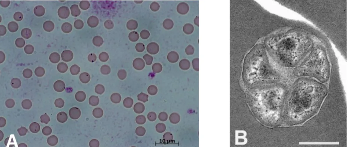
Figure 1.4. Bovine erythrocytes infected with A. marginale. A). Organisms are seen
as black, irregular shaped dots, usually at the edge of infected red blood cells, Giemsa staining, B). An infected erythrocyte with five A. marginale inclusion bodies. Electron microphotograph, bar 0,5μm (taken from Kocan et al. 2004).
During the course of infection, erythrocytes become chemically altered by the
bacteria. Subsequently, „marked‟ erythrocytes are recognized by reticuloendothelial
cells and removed from the circulation, which results in anemia and icterus (Kocan et al., 2003).
The acute phase of the disease includes symptoms such as fever, weight loss, icterus, abortion, lowered milk production and even death (Kuttler, 1984). Differences in virulence between Anaplasma strains and the level and duration of the rickettsemia play a role in the severity of clinical manifestations. Although cattle of all ages can become infected with A. marginale, the severity of disease is age dependent. Calves under 6 months of age are much more resistant to disease (although not infection) than older cattle. In older calves mild or acute, but rarely fatal disease develops, while in cattle over 2 years of age, the disease often is fatal (Kocan et al., 2003). Cattle that recover from anaplasmosis remain lifelong carriers serving as a reservoir of the rickettsia (Kieser et al., 1990). During the carrier state, the rickettsemic cycles occur at approximately seven weeks intervals, with peaks of 107 rickettsia per ml of blood ( Eriks et al., 1993; French et al., 1998; Kocan et al., 2010b). The chronically infected cattle are generally immune to further clinical disease; however, they can relapse to anaplasmosis, for example when infected with other pathogens.
27
1.7.
Differences within strains
Initially a small number of A. marginale strains was recognized on the basis of morphological characteristics, geographical origin, whether they were cross- protective in cattle or infectious and transmissible by ticks. Presently strains are characterized not only on the basis of the above characteristics, but additionally by either level of virulence or variation in membrane surface proteins (MSPs).
1.7.1. Morphology
Two morphological forms of A. marginale are known, one with (Table 1.3) and one without an inclusion appendage. Inclusion appendages, are also called "tails", "bands" or "filaments". They usually occur in the form of a tapering tail, a loop, a disk or a ring, and can only be visualized through immunological or ultrastructural techniques.
With traditional staining, only the “head portion” of the tailed Anaplasma is visible
(Carson et al., 1974).
The inclusion appendages observed under the electron microscope are not directly attached to the bacterium and are not surrounded by an inclusion membrane (Figure
1.5) (Kocan et al., 1984). In cattle erythrocytes, the tailed strain appears as a
spherical marginal body, or as a “comet – shaped” organism, which contains a head,
body and tail. In D. andersoni nymphs the inclusion appendages were observed in midgut tissues till 10 days after repletion from infected cows. Following day 15, appendages were found free in the midgut lumen or attached to the cell membrane of midgut epithelial cells (Kocan et al., 1984).
The tails are composed of polymerized F-actin filaments with a diameter of 7-10 nm and contain no parasite DNA (Stich et al., 1997). Ferritin-conjugated anti-A. marginale sera react with bacterial-specific antigens, indicating that appendages are recognized by the host’s immune system (Kocan et al., 1978a; Kocan et al., 1978b). Stich et al. (1997) have also shown that the inclusion appendage contains host actin filaments. Interestingly, unlike the classic pattern in which actin is assembled on the bacterial surface, the A .marginale-associated appendage assembles on the external vacuolar
28
surface and does not have to be secreted across the bacterial membrane and the membrane surrounding the parasitophorous vacuole. A new polymorphic appendage-associated protein has been identified, designated as A. marginale appendage associated protein (AAAP), however, its role in invasion or replication within the host cell is still unknown (Stich et al., 2004).
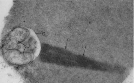
Figure 1.5. Electron micrograph of tailed Anaplasma marginale in bovine
erythrocytes containing a comet-like (arrows) inclusion appendage (C) containing two subunits (U) (taken from Simpson et al. 1965).
The function of the A. marginale appendage in the infection of ticks and erythrocytes is unclear. At first, Kocan et al. (1984) suggested that the appendage may play a role in infection of tick gut cells, as they observed that only the tailed Virginia isolate, and not the Florida isolate (without appendage), infected D. andersoni ticks. Two years later, Smith et al. (1986) verified that an inclusion appendage is not responsible for infectivity, as only one of the two tailed Anaplasma strains tested, was readily transmitted by D. variabilis ticks. In some viruses and bacteria, the appendage has been shown to influence motility, which improve their propagation and enhance the spread of infection (Cossart and Lecuit, 1998). Stich et al. (1997) suggested that appendage increase A. marginale motility enhancing contact with the tick gut epithelium or bovine erythrocytes.
29
Table 1.3. Anaplasma marginale isolates with an inclusion appendage.
A. marginale isolate Reference
Illinois (Smith et al., 1986)
Virginia (Smith et al., 1986)
UFMG1 (Brazil) (Ruiz et al., 2005)
California (Potgieter et al., 1981)
Texas (Franklin and Redmond, 1958 )
Oregon (Pilcher et al., 1961)
Mexico (Simpson et al., 1965)
Oklahoma (Kocan et al., 1978b)
Washington (Barbet et al., 1983)
Israeli T (Palmer et al., 1988)
Interestingly, A. marginale with an appendage was initially named Anaplasma
caudatum (caudatum - tailed) (Boone et al., 2005; Kreier and Ristic, 1963). Moreover,
in 1974 the creation of a new genus Paranaplasma has been proposed, due to serological and immunological differences between isolates with and without an appendage (Kreier and Ristic, 1974). Nonetheless, according to the work of Smith et al. (1986) this classification has been abandoned and presently A. marginale with an inclusion is not considered a separate species.
1.7.2. Major Surface Proteins
The surface of tick-borne intracellular bacteria consists of many proteins which are remodeled during the transmission of the pathogen between vertebrate and invertebrate hosts. They mediate functions which are necessary for survival, replication and transmission. Their expression changes, in order to facilitate bacterial survival in different hosts.
Six major surface proteins (MSPs) namely: MSP1a, MSP1b, MSP2, MSP3, MSP4 and MSP5 have been identified on A. marginale (Alleman et al., 1997; Barbet and Allred, 1991; McGarey and Allred, 1994; McGuire et al., 1994; Palmer et al., 1994;
30
Palmer et al., 1985) and are being used for phylogenetic analyses of A. marginale strains (as reviewed by de la Fuente et al., 2005b). These MSPs are involved in host– pathogen interactions and may evolve more rapidly than other nuclear genes because of selective pressure exerted by the host’s immune system.
The A. marginale MSP1 complex is composed of a heterodimer of two structurally unrelated polypeptides: MSP1a and MSP1b, linked by disulfide bonds (Vidotto et al.,
1994). The MSP1a is encoded by the single-copy gene, msp1α and varies in size
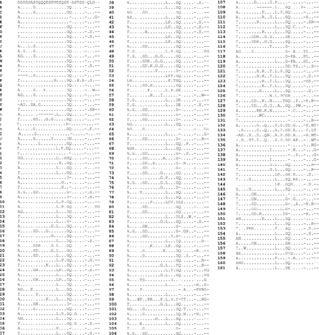
among different geographic isolates due to the changing number of 23-31 amino acid tandem repeat peptides in the N-terminal part of the protein (Allred et al., 1990; as reviewed by Cabezas-Cruz et al., 2013; de la Fuente et al., 2003; de la Fuente et al., 2001b; de la Fuente et al., 2002c). The N-terminal part of MSP1a is highly glycosylated (Garcia-Garcia et al., 2004a). The expression of MSP1a in A. marginale from tick cell cultures is downregulated in comparison with bacteria derived from bovine erythrocytes (Garcia-Garcia et al., 2004b).
Formerly, the msp1α gene was widely used for phylogenetic studies, as it did not
appear to undergo antigenic variation in cattle or ticks (Bowie et al., 2002). However, while phylogenetic studies of MSP1a repeat sequences provided evidence of
A. marginale-tick coevolution, they could not provide phylogeographic information on
a global scale because of the high level of MSP1a genetic diversity among geographic strains (Estrada-Pena et al., 2009).
MSP1b is encoded by members of the msp1 β multigene family (Barbet and Allred,
1991), which are polymorphic between different isolates of A. marginale. In contrast to MSP1a which has been shown to be an adhesin for bovine erythrocytes and tick cells (cultured and native), MSP1b is an adhesin only for bovine erythrocytes (de la Fuente et al., 2001a; McGarey and Allred, 1994).
MSP2 and MSP3 unlike other outer membrane proteins in A. marginale, have a single expression site but multiple alleles distributed throughout the chromosome (Brayton et al., 2003).
MSP2 is an immunodominant outer membrane protein, encoded by a polymorphic gene family (Palmer et al., 1994). The MSP2 expression is under the control of a single operon consisting of a promoter and four open reading frames. The msp2
31
gene consists of nine pseudogenes, which play a substantial role in achieving multiple antigenic variations (Brown et al., 2003).
MSP3 is also an immunodominant antigen, encoded by a polymorphic multigene family whose exact function is unknown (Alleman et al., 1997). Recently, it has been shown that simple variants of MSP3 are expressed in early mammalian infection and within the tick vector, and multiple antigenic variants emerge only under selective immune pressure during persistent infection (Palmer and Brayton, 2013).
Antigenic variation of MSP2 and MSP3 has been proposed as a likely mechanism by which A. marginale evades the host immune system, resulting in lifelong persistence in the mammalian host (French et al., 1999; French et al., 1998; Palmer et al., 2000).
MSP5 and MSP4 are immunodominant proteins, encoded by single gene copies, which remain conserved in different A. marginale strains, as well as in A. centrale (Molad et al., 2004; Oberle et al., 1993; Visser et al., 1992). At present, the role of these proteins is not well defined, however; the fact that they remain conserved suggests that they are important in the Anaplasma life cycle. Phylogenetic analysis indicated that MSP4 is not a good genetic marker for global analysis, but it can provide some information about strain differences within geographic regions (de la Fuente et al., 2005b).
The recombinant MSP5 protein and monoclonal antibodies against it are used for detection of anti-Anaplasma-specific antibodies by ELISA (de Echaide et al., 2005; Ewing et al., 1997). Although the msp5 gene is widely used for the detection of
A. marginale carrier cattle by nested PCR (Bock and de Vos, 2001), the MSP5 ELISA
is the recommended method for confirming infection, as rickettsemia can drop below PCR-detectable levels.
1.7.3. Tick transmission
A. marginale strains differing in their tick transmissibility and in general infectivity for
ticks may serve as useful tools to identify the genetic requirements for tick transmission. I t has been hypothesized that differences in tick transmission
32
efficiency are due to genetic variability within A. marginale strains, which confer a tick transmission phenotype. Nevertheless, after comparison of genomes of five strains differing in tick transmissibility, no specific genes were determined (Dark et al., 2009). Therefore Dark et al. (2009) suggested that the differences exist most likely in shared genetic elements: either in coding or regulatory regions.
More promising results have been obtained with proteomic approaches which aimed at elucidating proteins responsible for colonization of tick cells. When proteomes of A.
marginale strain from tick cell culture and erythrocytes were compared, a set of
up-regulated proteins was identified (Noh et al., 2008; Ramabu et al., 2011). Although the functions of most of these proteins are still unclear, of particular interest is the ankyrin-repeat containing protein, Am638, as ankyrin-ankyrin-repeat motifs are thought to mediate protein-protein interactions (Ramabu et al., 2011). Further proteomic analysis comparing more A. marginale strains from naturally infected cattle and ticks are required in order to find key proteins involved in tick transmissibility.
The MSP1a tandem repeats were shown to be necessary for adhesion of A. marginale to tick and mammalian cells, which was attributed to differences in amino acid sequences of individual repeats. The negatively charged amino acids, aspartic acid (D) and glutamic acid (E) at position 20 were shown to be essential for binding of MSP1a to tick cell extracts. When glycine (G) was located at position 20, binding was not observed (de la Fuente et al., 2003). Recently, it has been confirmed that the 2-D conformation of MSP1a protein also correlates with tick transmissibility
(Cabezas-Cruz et al., 2013). In most cases the α-helix conformation was found in abundance
in strains transmitted by ticks.
1.7.4. Virulence
Several isolates of A. marginale have been identified which differ in virulence (Bastos et al., 2010; Rodriguez Camarillo et al., 2008). The identification of strains with low pathogenicity is very important as they could be used as live vaccines. Live vaccines result in persistent, life-long infections, providing protection against homologous and heterologous strains.
33
There are many reasons for differences in pathogenicity, one of which is protein glycosylation. Several glycoproteins of Gram-negative bacteria have already been shown to play a role in adhesion, invasion and pathogenesis. Most bacterial glycoproteins appear to be either associated with the surface of the organism or to be secreted into the environment, suggesting their role in the interaction with the host. Although the function of protein glycosylation is unclear in many cases, it has been proven to be essential for the attachment and infectivity of Chlamydia trachomatis elementary bodies and also to be responsible for the binding of blocking antibodies in
Neisseria meningitidis (as reviewed by Benz and Schmidt, 2002). Remarkably, the
MSP1a protein of A. marginale, which serves as an adhesin for host cells, is highly glycosylated (Garcia-Garcia et al., 2004a). The exact role of MSP1a glycosylation in invasion of host cells or pathogenicity has not been entirely explained. Most likely, since it is a surface protein and is directly exposed to the host cells, the bacteria may modulate MSP1a expression/glycosylation in order to evade the host’s immune response.
Besides glycosylation, A. marginale inclusion appendages may also play a role in pathogenicity. Appendages have been associated with serological or immunological variances (Kuttler and Winward, 1984). Differences in pathogenicity have been observed between two Brazilian A. marginale strains, which correlated with the presence of the inclusion appendage (Bastos et al., 2010). The low pathogenic, tailed UFMG1 strain (Ribeiro et al., 1997), provided protection against the highly pathogenic non-tailed UFMG2 strain (Bastos et al., 2010).However in vaccination trials using a heterologous geographical strain from Israel as a challenge, cattle vaccinated with UFMG1 were not protected from the disease (Kenneil et al., 2013). Interesting results have been obtained with two U.S. strains: the tailed Virginia and non-tailed Florida isolates (Kuttler et al., 1984). Kuttler at al. (1984) observed that cattle vaccinated with the commercial killed vaccine were resistant to a challenge with the Virginia isolate, whereas a 47 % mortality has been observed when the cattle were challenged with the Florida isolate. However, there was no cross-protection when cattle were vaccinated with Virginia strain and afterwards cross-challenged with Florida, or the other way round.
34
provided immunity against challenge with other Mexican strains (Rodriguez Camarillo et al., 2008). However, experiments with different geographical strains are required, to confirm the low virulence of this strain.
Although immense progress has been made in molecular biology and in in vitro cultivation of A. marginale, at present animal experimentation to measure the severity of the disease is the only way of assessing strain pathogenicity.
1.8.
Anaplasmosis control methods
Presently used control methods, consisting of antibiotic treatment, vaccination and arthropod control, have not significantly changed for many years and depend on the geographical area, availability, cost and feasibility of application (Kocan et al., 2000).
1.8.1. Antibiotics
Three types of antibiotics are used for the treatment of anaplasmosis: tetracyclines,
fluoroquinolones and imidocarb dipropionate (Aubry and Geale, 2011).
Chemotherapeutic treatment is effective in decreasing bacterial numbers, but the efficacy in clearing infection and thus preventing the establishment of a pathogen reservoir is variable (Coetzee et al., 2005; Reinbold et al., 2010; Wallace et al., 2007). Recently, treatment of persistently infected steers with oral chlortetracycline for 80 days cleared A. marginale infections (Reinbold et al., 2010). Nevertheless, treatment of clinically affected animals is expensive. Moreover, it is becoming less acceptable, as antibiotic resistance rises in pathogens. In addition, some countries restricted the use of imidocarb due to its prolonged retention in the edible tissues of animals for slaughter (Kocan et al., 2010b).
1.8.2. Arthropod control
Treatment of animals with acaricides reduces the number of ticks, thus indirectly decreasing anaplasmosis transmission. However, the use of acaricides for vector
35
control is becoming a concern due to increasing acaricide-resistance among tick populations (George et al., 2004), the pollution of the environment and the contamination of milk and meat products (Graf et al., 2004).
The modern approach for arthropod control is based on the use of anti-tick vaccines,
which have the benefits of being cost-effective, reducing environmental
contamination and preventing the development of acaricide-resistant ticks. Two vaccines Gavac (Vargas et al., 2010) and TickGARD (Odongo et al., 2007) containing recombinant R. (B.) microplus gut antigens Bm86 and Bm95 are currently being used.
Another promising antigenic protein involved in the modulation of tick feeding and reproduction, subolesin, has been tested (de la Fuente et al., 2005a). Preliminary experiments have shown, that the number of ticks infected with Anaplasma spp. were reduced when ticks were injected with subolesin double- stranded RNA before being fed on cattle with ascending rickettsemia (de la Fuente et al., 2006). Furthermore, cattle immunized with recombinant subolesin were protected against R.
(B.) microplus infestations due to a decrease in tick survival and reproduction rates
(Almazan et al., 2003; Merino et al., 2011).
1.8.3. Vaccination
A long-lasting immunity induced by vaccination is an economical and effective way to prevent and control bovine anaplasmosis. Mass vaccination programs can significantly reduce the use of acaricides and antibiotics thus preventing an emergence of resistant ticks or pathogens. At present, two types of vaccines are used and are the vaccines of choice: live vaccines and inactivated formulations. They induce protection from severe clinical symptoms, but do not prevent infection, so that cattle after infection may remain carriers of A. marginale (Kocan et al., 2003).
1.8.3.1. Live vaccines
36
isolation of A. centrale (Theiler, 1911). They consist of less pathogenic A. centrale or attenuated strains of A. marginale.
The immune response induced by such vaccines is similar to a natural infection and
animals develop persistent infections with the vaccine strain. However,
preimmunization with one strain has been shown not to provide cross-protection in widely separated geographic areas (Kenneil et al., 2013; Kuttler et al., 1984). Live vaccines consist of infected blood, taken from splenectomized, quarantined calves inoculated with the selected vaccine strain. These vaccines carry the risk of
transmitting other “silent” pathogens and despite the global impact of anaplasmosis,
their use is forbidden in the US (Rogers et al., 1988).
Immunization of cattle with less pathogenic subspecies i.e. A. centrale is in routine use in several countries: South Africa, Zimbabwe, Malawi, Australia, Israel, Uruguay and Argentina (Shkap et al., 2009).
It is noteworthy that some African and Latin American isolates of A. marginale can overcome an A. centrale induced immunity (Bock et al., 2003; Brizuela et al., 1998).
The second type of live vaccine consists of attenuated A. marginale strains. These vaccines were used in South America and California, although severe reactions have been observed in adult cattle after vaccination (Henry et al., 1983). In calves, however, these vaccines produce mild infections and lead to immunity against clinical anaplasmosis, although not in widely separated geographic areas (Kocan et al., 2003). The attenuation of A. marginale can be achieved by two methods. The first involves irradiation and subsequent multiple passages through deer and sheep (Ristic and Carson, 1977). The second consists of numerous passages through splenectomized calves followed by passages through splenectomized sheep (Jorgensen et al., 1993). Yet, it has been reported that attenuated A. marginale vaccines reverted to virulence after successive passages through cattle or ticks (Kocan et al., 2000).
1.8.3.2. Inactivated vaccines
An inactivated vaccine comprising o f non-living A. marginale was developed in the United States and was used effectively till its withdrawal from the market in 1999
37
(Kocan et al., 2003). Although inactivated vaccines are also produced in splenectomized animals, it is less likely that any other pathogens contaminating the vaccine will remain viable and infectious after the inactivation process. However, extensive purification is required to remove bovine cell stroma as only partial purification resulted in the development of erythrocytic isoantibodies in vaccinated cattle. The inactivated vaccines reduced clinical disease and mortality, yet did not always provide cross-protection (Kuttler and Winward, 1984). For this reason inactivated vaccines are most likely to be useful when produced from locally isolated strains.
1.8.3.3. Culture-derived vaccines
Bacteria grown in tick cell cultures are being investigated as an alternative source of
A. marginale for live vaccine production. This technique has the advantage of allowing
the inclusion of multiple strains, ease of standardization, freedom from bovine red blood cells and pathogens and does not require the use of expensive, splenectomized calves (Kocan et al., 2003). Cattle immunized with a cell culture- derived A. marginale strains develop protective immunity and do not develop clinical signs of anaplasmosis after challenge. However, as with most anaplasmosis vaccines, infection with the challenge strain is not prevented (Bastos et al., 2010; de la Fuente et al., 2002b; Kocan et al., 2001).
1.9.
Cell culture systems
For a long time the lack of an in vitro culture system has been the major impediment to anaplasmosis research and infected cattle served as the only source of
A. marginale. Although bovine erythrocytes can be used for maintaining bacteria in
culture, they are not suitable for continuous propagation (Blouin et al., 2002a; Waghela et al., 1997). Since the establishment of the first tick cell line in 1975 (Varma et al., 1975), the number of continuous tick cell lines has increased to over 50 derived from both ixodid and argasid species (Bell-Sakyi et al., 2007; as reviewed by Passos, 2012). Currently, most of the available tick cell lines have been deposited in the Tick Cell Biobank (pirbright.ac.uk/research/tickcell/Default.aspx).
38
More than 200 A. marginale isolates have been reported worldwide (as reviewed by Cabezas-Cruz et al., 2013), but only few of them have been propagated in tick cell cultures, namely: from Brazil UFMG1 and UFMG2 (Bastos et al., 2010; Bastos et al., 2009), from the USA Virginia, Oklahoma (Blouin et al., 2000; Munderloh et al., 1996), St. Maries (Hammac et al., 2013) and Oregon (Kocan et al., 2004) and an isolate from Israel (unpublished work).
Bacteria are propagated mostly in cell lines derived from Ixodes scapularis IDE8 and ISE6 (Munderloh et al., 1994) although some strains grow also in cell lines from
R. (B). microplus or D. andersoni ticks (Oliva Chavez et al., 2012). Anaplasma
colonies from tick cell cultures are similar to those observed in ticks (Blouin and Kocan, 1998) and remain infective for ticks and cattle after continuous passages in tick cell cultures (Blouin et al., 2000).
In addition to tick cell lines, A. marginale infects and grows in several mammalian cell lines: Vero (kidney epithelial) and RF/6A (retina choroid endothelium), as well as in primary cultures of bovine vascular endothelial cells (Munderloh et al., 2004; Oliva Chavez et al., 2012; Wamsley et al., 2011).
A. centrale used as a live vaccine is still produced in cattle, because all attempts
to propagate this vital subspecies in vitro have failed.
In vitro cultures provide an excellent source of A. marginale organisms which can be
used for serological diagnosis (Saliki et al., 1998), screening of antibiotics (Blouin et al., 2002b), vaccines development (de la Fuente et al., 2002b; Hammac et al., 2013) or proteome profiling (Noh et al., 2008). Furthermore, culture systems allow the study of pathogen–host cell interactions and pathogen variations in response to a changing host cell environment (Blouin et al., 2002a). Additionally great quantities of bacteria can be obtained in less time at reduced costs, and most importantly without the use of experimental animals. Therefore, more effort should be put into establishing additional A. marginale strains in vitro.
39
1.10. Recent interests in anaplasmosis research
While research carried out in the last two decades has contributed greatly to our knowledge of the antigenic composition of A. marginale, it did not lead to the development of effective vaccines which could provide cross-protection worldwide. The availability of in vitro grown bacteria together with novel genomic, transcriptomic and proteomic techniques, has great potential for vaccine development. Currently, two trends can be observed in anaplasmosis research. The first is developing effective tools to induce immunity in cattle through vaccination. The second is preventing transmission of A. marginale, by yet undiscovered methods of blocking tick transmission of pathogens. In general, new approaches for anaplasmosis control focus on the use of outer membrane proteins (OMPs) in novel vaccines. OMPs are not only essential for the bacterium but also serve as major targets for the immune system of the host.
Kocan et al. (1996) suggested that the vaccination of cattle with A. marginale OMPs, should not only aim at preventing the bacterial infection in animals but also at preventing the transmission of A. marginale by infected ticks. The rationale of this idea was the fact that some bovine immunoglobulins can cross the tick midgut epithelium and enter the hemolymph. Feeding ticks are exposed to antibodies present in host serum for a relatively long time. Therefore, cattle immunized against ticks and/or against stages of hemoparasites within ticks would produce antibodies which would be taken up by a tick with the bloodmeal, thus affecting the vector and/or the parasite. Preliminary experiments, however, have not shown any difference in development or transmission of A. marginale in ticks fed on vaccinated cattle.
Selected epitopes of immunogenic sub-dominant proteins are being tested in vaccination trials. Subdominant antigens tend to be less variable, as parasites allow the host to mount an immune response against them, therefore they are likely irrelevant for the survival of the organism ( B r o w n e t a l . , 2 0 0 6 ) . It has been shown, that cattle immunized with the A. marginale MSP1 protein complex presented a protective humoral immune response, however, its efficacy was variable. Similarly, mice vaccinated with chemically synthesized critical motifs of MSP1a functional epitope (Santos et al., 2012) essential for antibody recognition,
40
were protected against A. marginale challenge (Santos et al., 2013). Although the protective immunity in cattle vaccinated with subdominant OMP AM779 alone was not sufficient to induce protection, slightly greater T-cell responses were observed when compared to animals vaccinated with OMPs (Albarrak et al., 2012). Therefore Albarrak et al. (2012) suggested that subdominant antigens should still be considered individually and collectively for vaccine development.
Using sera from cattle vaccinated with A. marginale OMP complexes, Lopez et al. (2005) identified 24 immunodominant A. marginale proteins, resolved on 2- dimensional gels. As expected all identified proteins were membrane-associated. These included the well characterized surface-exposed OMPs like MSP2, MSP3 and MSP5, as well as recently identified appendage-associated proteins. Additionally, among the 21 newly described antigenic proteins, type IV secretion system proteins and members of the MSP2 superfamily were identified. Similar antigens with some additional OMPs were detected in an experiment when sera from A. centrale- immunized cattle were used (Agnes et al., 2011).
Two sets of individual OMPs were tested by Noh et al. (2013) as immunogens: a complex of OMPs linked by covalent bonds or treated with dithiothreitol (DTT) which reduces disulfide bonds. Although both immunogens induced protective immunity, the antibody response induced by the linked immunogen was much better. These findings suggest that cross-linking enhances immunogenicity and could minimize the dose of antigen required for the induction of protective immunity.
Parallel to proteomics, comparative genomic analyses are also being run. They similarly aim at elucidating immunodominant proteins, which are identical within strains and so may be used as vaccine antigens (Dark et al., 2011; Dark et al., 2012; Palmer et al., 2012) Notably, the results are similar to those obtained with proteomic techniques i.e. comparing the genome of 10 U.S. A. marginale strains Dark et al. (2011) selected 19 conserved antigens, mostly OMPs and Type 4 Secretion Proteins.
41
1.11. Project outline
Anaplasma marginale affects ruminants worldwide, causing anaplasmosis in tropical
and subtropical regions. Considerable effort is made to control the disease, yet strategies have only minimally advanced over the past few decades even though our knowledge of A. marginale has increased considerably within this time.
On the one hand, the problem arises from A. marginale itself, as differences in isolates, mainly in protein expression lead to a lack of cross-protection among geographically separated strains. On the other hand, drug resistance increases not only in bacteria, but also in the tick vectors due to the ungoverned use of acaricides. Furthermore, the only vaccine on the market, A. centrale, is forbidden in many countries due to the risk of transmitting other blood-borne pathogens.
This thesis aims to shed light onto differences within geographical A. marginale strains grown in the IDE8 tick cell line. In order to address this question the main objectives of the project were:
to develop a practical and effective approach for the purification of intact
and viable A. marginale from infected tick cell cultures
to check if there are differences in genes/proteins within geographical
A. marginale strains propagated in vitro, differing in morphology, protein
sequence and pathogenicity
Few A. marginale strains can be propagated in tick cell cultures, yet, due to its obligatory intracellular nature, separation of bacteria from host cell materials is problematic. In Chapter 2 various purification methods are reviewed. The use of Percoll gradients for separation of intact and viable A. marginale from IDE8 cells is examined, with the focus on the easiness, reproducibility and lack of toxicity of the method.
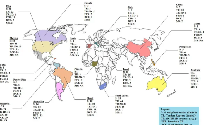
The functional and immunological relevance of MSP1a protein from 224 A. marginale strains were analyzed. Additionally, the consistent nomenclature based on the MSP1a structure was proposed, is covered in Chapter 3.
42
In Chapter 4 some molecular and immunological characteristics of different geographical A. marginale strains grown in IDE8 tick cell culture are depicted with the particular focus on MSPs and proteins participating in response to stress conditions.
In Chapter 5 differences in protein expression among these strains are evaluated with the use of 2D-DIGE technique and Mass Spectrometry. The possibility of inclusion of selected proteins into novel vaccines is also discussed.
43
C
HAPTER
2
Use of Percoll gradients to purify Anaplasma marginale
(Rickettsiales: Anaplasmataceae) from tick cell cultures
K. Lis, N. Najm, J. de la Fuente, I. Fernández de Mera, E. Zweygarth, K. Pfister,
L. M. Passos
45
2. Use of Percoll gradients to purify Anaplasma marginale
(Rickettsiales: Anaplasmataceae) from tick cell cultures
2.1.
Abstract
Anaplasma marginale (Rickettsiales: Anaplasmataceae) is an obligate intracellular
bacterium that multiplies exclusively within membrane-bound vacuoles in the cytoplasm of the host cells. A number of A. marginale isolates can be propagated in the Ixodes scapularis IDE8 tick cell line, which provides a reliable source of antigens for a wide variety of studies. However, because of its intracellular nature, separation of bacteria from host cell materials remains an important constraint for researchers. In the present study we evaluated the use of Percoll gradients for purification of two Brazilian strains of A. marginale grown in IDE8 tick cells. The purified A. marginale monitored in Giemsa-stained smears contained only minimal amounts of IDE8 cell stroma. The total protein yields were 1.2 mg and 1.7 mg, while the DNA titres
quantified with real-time PCR were 6.4 x 10⁹ for UFMG1 and 4.87 x 10⁹ for UFMG2
copies in the purified material, respectively. Additionally, we confirmed the viability of purified bacteria by infecting tick cells after being freshly purified and after retrieval from long-term storage. Importantly, the viability of the organisms is preserved after use of this separation method and therefore the purified organisms can be used in enzymatic assays and other research approaches where living organisms would be preferred.