HAL Id: hal-02348603
https://hal.archives-ouvertes.fr/hal-02348603
Preprint submitted on 5 Nov 2019
HAL is a multi-disciplinary open access
archive for the deposit and dissemination of sci-entific research documents, whether they are pub-lished or not. The documents may come from teaching and research institutions in France or abroad, or from public or private research centers.
L’archive ouverte pluridisciplinaire HAL, est destinée au dépôt et à la diffusion de documents scientifiques de niveau recherche, publiés ou non, émanant des établissements d’enseignement et de recherche français ou étrangers, des laboratoires publics ou privés.
and efficacy of the RoxS riboregulator of central
metabolism in Bacillus subtilis
Sylvain Durand, Adam Callan-Sidat, Josie Mckeown, Stephen Li, Gergana
Kostova, Juan R. Hernandez-Fernaud, Mohammad Tauqeer Alam, Andrew
Millard, Chrystala Constantinidou, Ciaran Condon, et al.
To cite this version:
Sylvain Durand, Adam Callan-Sidat, Josie Mckeown, Stephen Li, Gergana Kostova, et al.. Novel regulation from novel interactions: Identification of an RNA sponge that controls the levels, processing and efficacy of the RoxS riboregulator of central metabolism in Bacillus subtilis. 2019. �hal-02348603�
Novel regulation from novel interactions: Identification of an RNA sponge that controls the
1
levels, processing and efficacy of the RoxS riboregulator of central metabolism in Bacillus
2
subtilis
3
4
Sylvain Durand*1, Adam Callan-Sidat†2, Josie McKeown†2, Stephen Li†2,Gergana Kostova1, Juan R.
5
Hernandez-Fernaud3,4, Mohammad Tauqeer Alam2, Andrew Millard2,5, Chrystala Constantinidou2,
6
Ciarán Condon1, Emma L. Denham*2,6
7
8
1 - UMR8261, CNRS, Université de Paris, Institut de Biologie Physico-Chimique, 13 rue Pierre et
9
Marie Curie, 75005 Paris, France
10
2 - Division of Biomedical Sciences, Warwick Medical School, University of Warwick, Gibbet Hill Road,
11
Coventry, UK
12
3 – School of Life Sciences, Proteomics Research Technology Platform, University of Warwick, Gibbet
13
Hill Road, Coventry, UK
14
4 – Present address - Unidad de Investigacion del Hospital Universitario de Canarias. Calle Ofra, s/n.
15
38320. La Laguna. Canary Islands. Spain.
16
5 – Present address - Department of Genetics and Genome Biology, University of Leicester, Leicester,
17
UK
18
6 – Present address - Department of Biology and Biochemistry, University of Bath, Claverton Down,
19
Bath, UK
20
21
* To whom correspondence should be addressed. Tel +44 1225 383424; Email:
22
[email protected] Correspondence may also be addressed to Tel +33 1 58 41 50 31; Email:
23
24
25
† These authors contributed equally to this work.
ABSTRACT
27
28
Small RNAs (sRNAs) are a taxonomically-restricted but transcriptomically-abundant class of
post-29
transcriptional regulators. While potentially of importance, we know the function of few. This is in no
30
small part because we lack global-scale methodology enabling target identification, this being especially
31
acute in species without known RNA meeting point proteins (e.g. Hfq). We apply a combination of
32
psoralen RNA cross-linking and Illumina-sequencing to identify RNA-RNA interacting pairs in vivo in
33
Bacillus subtilis, resolving previously well-described interactants. Although sRNA-sRNA pairings are
34
rare (compared with sRNA/mRNA), we identify a robust example involving the unusually conserved
35
sRNA (RoxS/RsaE) and an unstudied sRNA that we term Regulator of small RNA A (RosA). This
36
interaction is found in independent samples across multiple conditions. Given the possibility of a novel
37
associated regulatory mechanism, and the rarity of well-characterised bacterial sRNA-sRNA
38
interactions, we mechanistically dissect RosA and its interactants. RosA we show to be a sponge RNA,
39
the first to be described in a Gram-positive bacterium. RosA interacts with at least two sRNAs, RoxS
40
and FsrA. Unexpectedly, it acts differently on each. As expected of a sponge RNA, FsrA is sequestered
41
by RosA. The RosA/RoxS interaction is more complex affecting not only the level of RoxS but also its
42
processing and efficacy. Importantly, RosA provides the condition-dependent intermediary between
43
CcpA, the key regulator of carbon metabolism, and RoxS. This not only provides evidence for a novel,
44
and functionally important, regulatory mechanism, but in addition, provides the missing link between
45
transcriptional and post-transcriptional regulation of central metabolism.
46
47
INTRODUCTION
48
To adapt to changing environments and survive exposure to harsh conditions, organisms have evolved
49
complicated metabolic and genetic regulatory networks to ensure that a homeostatic balance is
50
maintained 1,2. At the RNA synthesis level, gene expression can be modulated through combinations of
51
transcription factors controlling genes required for growth and survival under specific conditions 3-5. At
52
the post-transcriptional level, small regulatory RNAs (sRNAs) act to temper gene expression by short
53
imperfect base pairing with their mRNA targets, altering the level of protein production by increasing or
54
decreasing access to the ribosome-binding site, or by facilitating or blocking the access to the mRNA
55
by ribonucleases (RNases) 6,7. Most regulatory RNAs are independently expressed under the control of
56
specific transcription factors. However, more recently, it has been shown that sRNAs can also be
57
produced by processing RNAs that have other functions in the cell, such as tRNAs 8 and mRNAs 9.
58
59
Regulation by RNA is an important mechanism for fine-tuning gene expression in the Gram-positive
60
model bacterium Bacillus subtilis, recently reviewed in 10. Over 150 potential sRNAs have been identified
61
in B. subtilis and shown to be expressed in a condition-dependent fashion 11-13. To date the roles of very
62
few of these putative sRNAs have been determined. However, where targets have been identified, they
63
have been shown to play key roles in stress adaptation. B. subtilis notably expresses three sRNAs with
64
C-rich regions (CRRs) with similar predicted secondary structure; RoxS/S415 (Related to oxidative
65
stress) 14, FsrA/S512 (Fur regulated small RNA) 15 and CsfG/S547 (Controlled by F and
sigma-66
G) 16, (S numbers relate to transcriptionally active segments identified by Nicolas et al. 12). The RoxS
67
sRNA is one of the best characterised sRNAs in Gram-positive bacteria 14,17,18 and is conserved among
68
Bacilli and Staphylococci, where it is named RsaE 19,20. RoxS has been shown to be upregulated in
69
response to nitric oxide (NO) in B. subtilis and S. aureus, by the two component system ResDE, and its
70
homolog SsrAB, respectively 14. RoxS expression is also activated when malate is supplied as a carbon
71
source. This control is mediated by the transcription factor Rex, that is known to sense the NAD/NADH
72
ratio of the cell. Indeed, this ratio is perturbed by the conversion of malate to pyruvate by the three
73
malate dehydrogenases of B. subtilis that reduce NAD+ to NADH, and by its cycling through the TCA
74
pathway. It has been proposed that one role of RoxS is to re-equilibrate the NAD/NADH ratio of the cell
75
by inhibiting the expression of enzymes leading to the production of NADH. FsrA is regulated by the
76
transcription factor Fur and acts as part of the iron-sparing response 15. Fur down-regulates mRNAs
77
whose protein products contain iron as part of their structures, but are not essential for growth, therefore
78
ensuring iron availability for essential iron-containing proteins 15,21. Interestingly, both RoxS and FsrA
79
down-regulate several genes encoding enzymes of the TCA cycle that produce NADH. CsfG is highly
80
expressed during sporulation, anaerobic growth and after glucose exhaustion 12. During sporulation,
81
expression of this sRNA is controlled by the sigma factors F and G which are restricted to the forespore
82
16. However, to date no mRNA target or physiological role for CsfG has been identified. Durand et al.
83
have hypothesised that its similar sequence motifs and structure to RoxS and FsrA suggests these three
84
sRNAs may have overlapping targets and play similar roles under different growth conditions 14.
85
86
The lack of well resolved pathways through which sRNAs act in no small parts reflects the difficulty of
87
global scale target identification, this being more acute in some bacteria than others. In many
88
enterobacteria, such as Escherichia coli and Salmonella typhimurium, the RNA chaperone Hfq plays a
89
key role as a mediator of sRNA-mRNA interactions and has greatly enabled the identification of mRNA
90
targets through pull-down studies 22,23. Although Hfq is conserved in Gram-positive bacteria, it does not
91
appear to play a global role in RNA-mediated regulation of gene expression 24,25. Hfq-dependent
92
regulation by only one sRNA in Listeria and a handful in Clostridium are the only known exceptions. It
93
is therefore generally accepted that sRNA regulation in the Firmicutes either depends on different RNA
94
chaperones or can occur in the absence of any protein factors. A number of groups have used in vivo
95
RNA cross-linking with the psoralen AMT, followed by ligation to form chimeras and RNAseq to identify
96
RNA-RNA interactions in eukaryotic cells 26-28. Here then we employed LIGR-seq 26 to identify sRNA
97
targets in B. subtilis. In addition to identifying many known members of the FsrA and RoxS regulons and
98
several new targets, we also identified a new regulatory RNA, S345, that interacts with both FsrA and
99
RoxS. These interactions are found in independent samples and across multiple conditions. Given the
100
possibility of a novel associated regulatory mechanism, and the rarity of well-characterised bacterial
sRNA-sRNA interactions, we mechanistically dissect S345 and its interactants. We show that S345 not
102
only functions as an RNA sponge for RoxS, but also affects its processing and degradation. We rename
103
this sRNA RosA (for Regulator of sRNA A). We show that the transcription of RosA is under the control
104
of the carbon catabolite control protein A (CcpA), linking the action of RoxS to the carbon source
105
availability in B. subtilis.
MATERIALS AND METHODS
107
108
Media and growth conditions
109
Selection for transformations was performed on Lysogeny Broth (LB) at 37°C supplemented with
110
required antibiotics. For E. coli these were ampicillin (100 µg ml-1) or chloramphenicol (10 µg ml-1) and
111
for B. subtilis either phleomycin (4 µg ml-1), kanamycin (20 µg ml-1), tetracycline (5 µg ml-1),
112
chloramphenicol (5 µg ml-1), erythromycin (2 µg ml-1), spectinomycin (100 µg ml-1) or combinations of
113
the above. Growth experiments were performed in LB, M9 medium supplemented with glucose at a
114
final concentration of 0.3% 12 or MD medium 29 supplemented with arabinose or malate at a final
115
concentration of 1%.
116
117
Bacterial strain construction
118
All E. coli and B. subtilis strains and plasmids used in this study are listed in Supplementary Table I.
119
Primer sequences can be found in Supplementary Table II. E. coli DH5α and TG1 were used for all
120
cloning procedures. B. subtilis strains were derived from the B. subtilis 168 trp+. The isogenic deletion
121
mutants were constructed according to the method described by Tanaka et al 30 without pop-out of the
122
deletion cassette. Transfer of genetic mutations between strains was achieved by transformation of
123
genomic DNA extracted from the relevant strain. Reintroduction of sRNAs under the control of their
124
native promoters was achieved by Gibson Assembly into pRMC that integrates into the amyE locus 31
125
of a PCR amplicon. Primer annealing sites were chosen to include the native promoter mapped in
126
Nicolas et al 12. The sequence of cloned sRNAs was subsequently confirmed by sequencing and
127
transformed into B. subtilis (plasmid pRMC+Pnative-sRNA). Integration into the amyE locus was
128
confirmed by an iodine halo assay by replica plating transformation plates onto starch plates. The
129
RosA promoter fusion was constructed at the native genomic locus by integration of the pBSBII
130
plasmid 32. Combinatorial strains were constructed in the genetic background of the same promoter
131
fusion strain by transformation of genomic DNA of the respective strain and selection on the
132
appropriate antibiotics.
134
In vivo RNA interactome
135
AMT in vivo cross linking
136
Bacteria were grown to the required O.D before 10 O.D 600 nm units were harvested by centrifugation
137
(4000 g, 5 minutes, 4°C). Bacteria were resuspended in 2 ml PBS either containing no AMT (to
138
identify background and levels of spurious interactions) or 0.7 mM AMT. Bacteria were incubated for
139
10 minutes at 37°C for 10 minutes before being transferred to a 6 well plate. The bacteria were
140
exposed to UV 365 nm at 0.120 Jcm-2 for 10 minutes before being added to 1 ml of ice cold killing
141
buffer (20 mM Tris-HCl [pH 7.5], 5 mM MgCl2, 20 mM Na-azide). The bacteria were harvested by
142
centrifugation at (4000 g, 5 minutes, 4°C). the supernatant was discarded and the pellet flash frozen in
143
liquid nitrogen. We determined the in vivo RNA interactome of B. subtilis grown in M9 minimal media
144
supplemented with 0.3% glucose at three points in the growth curve (exponential phase O.D.600nm 0.5,
145
stationary phase O.D.600nm 1.4 and just after lysis had started to occur, and in LB at mid-exponential
146
phase (O.D.600nm of 1.0). A ∆fur mutant 33 was prepared in LB at mid-exponential phase to increase
147
expression of the Fur regulated sRNA FsrA. Samples were prepared in duplicate.
148
149
RNA extraction and formation of chimeras between interacting RNAs
150
The RNA was extracted by resuspending the cell pellet in 800 µl LETS buffer (10 mM Tris-HCl [pH 8.0],
151
50 mM LiCl, 10 mM EDTA, 1% sodium dodecyl sulfate [SDS]) and bead beating in a FastPrep using 0.1
152
µm glass beads for three rounds of 40 seconds. The tubes were transferred to ice in between cycles.
153
The tubes were briefly spun to remove the bubbles created during bead beating. Two rounds of phenol
154
chloroform isoamyl alcohol extraction and one round of choloroform isoamyl alcohol extraction were
155
carried out. Before the addition of 10 % v/v NaAcetate and 1 ml isopropanyl and precipitation of RNA
156
overnight at -20°C. The RNA was pelleted by centrifugation at maximum speed at 4°C and the pellet
157
was washed with 70% Ethanol before being air dried and resuspended in water. The RNA was quantified
158
using the Qubit kit (Fisher Life Science). 10 µg of RNA was treated with Turbo DNase (Fisher Scientific)
159
to remove contaminating DNA. Ribosomal RNA was removed using Ribozero (Illumina) according to
the manufacturer’s instructions. To form the chimeric RNAs between RNAs crosslinked with AMT the
161
protocol described by Sharma et al. was followed as described in the supplementary data 26. The only
162
modification was the use of CircDNAligase (Epicentre) instead of CircRNAligase as this has been
163
discontinued.164
165
RNAseq166
Following uncrosslinking at UV 254 nm, RNA was purified and resuspended in 10 µl H2O and processed
167
through the TruSeq stranded total RNA library kit (150 bp) (Illumina) according to the manufacturer’s
168
instructions. The resulting libraries were sequenced on the MiSeq (Illumina).
169
170
Analysis
171
STAR aligner was used to map reads (Version STAR_2.6.0c_08-11) (34). This mapping tool is designed
172
to analyse splicing of introns and exons, which is similar to what is created through the formation of
173
chimeric reads where two different RNA fragments have been joined together. By identification of reads
174
that map to different features (protein coding sequences, sRNAs, UTRs, transcripts for ncRNAs such
175
as rRNA and tRNA, or transcribed intergenic regions) it is possible to identify RNA interactions. STAR
176
aligner was set to single end read mode to map read 1 and read 2 separately, the chimeric detection
177
mode activated, as this has been reported to be more sensitive to chimeric junctions. The allowed
178
mismatches in mapping was set to default for STARaligner. The output from STAR was merged in to
179
one Sam file, before being annotated using featureCounts within the package subread-1.6.3 in R, with
180
all further statistical analysis also carried out in R (35). In our initial analysis we found many reads
181
mapped to the genome, but to unannotated features. To overcome this problem, we created new
182
features for the unannotated regions of the genome and these are termed UA-start – stop in the data
183
files.
184
185
Chimeric reads will map to two different genomic features, whereas non-chimeric reads should only map
186
to one feature. The exception is of those reads with repetitive mapping or those that map to two
neighbouring features such as genes in an operon. An interaction count table was generated of reads
188
that mapped to more than one feature and thus are considered as interacting pairs. The interaction
189
count matrix then allowed statistical analysis of each interacting pair using a hypergeometric test. This
190
compares the number of interaction read counts for each specific interaction with the number of other
191
interaction read counts formed by each member of that pair, but with other RNAs. All interaction pairs
192
with a P-value below 0.05 were extracted as significant interactions. To increase confidence in the
193
identified interactions a second analysis was carried out where the pairs of sequenced samples were
194
analysed together and untranslated regions were combined with coding sequences. The
195
hypergeometric test was repeated and a P-adjusted value was calculated using Benjamini and
196
Hochberg to control for the false discovery rate which was set at 0.05 34.
197
198
To add further confidence to which interacting pairs form the most likely interactions, the interacting
199
pairs were further assessed by in silico prediction with IntaRNA2.0, which predicts the stability and
200
binding position between two interacting RNA pairs (36,37). The gene and any untranslated region that
201
has been identified associated with the gene of interest (12) were included in the prediction to take into
202
account the transcriptional start and stop sites. If no UTR had been identified for an mRNA the 50 bp
203
up and downstream of the start and stop site were employed.
204
205
Proteomics analysis
206
Strains were grown to O.D.600nm 1.0 in LB. 20 O.D. units were harvested and washed 3 X with PBS to
207
remove media components. Cells were resuspended in 200 µl urea buffer (8 M Urea, 50 mM Tris and
208
75 mM NaCl). 200 µl of urea buffer washed 0.1 µM beads were added to the cells before being disrupted
209
using three rounds of bead beating for 40 seconds using a FastPrep. Cells were placed on ice between
210
the three rounds of bead beating. The disrupted cells were then sonicated in a water bath for 15 minutes.
211
Cell extracts were centrifuged at 15,000 x g, 5 min and supernatants used for protein quantification
212
(Qubit protein assay kit). Protein reduction and alkylation was conducted by mixing 150 µg of total
213
protein with 10 mM TCEP and 40 mM CAA, at 600 rpm, for 20 min at room temperature. After, proteins
were predigested with 1.5 µg of rLysC (Promega) for 3 h at room temperature and samples diluted with
215
50 mM ammonium bicarbonate, 2 M urea final concentration. Protein digestion was performed with 1.5
216
µg of Trypsin (Promega) overnight at room temperature. The reaction was stopped by adding 1% TFA
217
and 10 µg of peptides were desalted using StageTip 35.
218
219
Reversed phase chromatography was used to separate 1 µg of tryptic peptides prior to mass
220
spectrometric analysis. The cell proteomes were analysed with two columns, an Acclaim PepMap
µ-221
precolumn cartridge 300 µm i.d. x 5 mm, 5 μm, 100 Å and an Acclaim PepMap RSLC 75 µm i.d. x 50
222
cm, 2 µm, 100 Å (Thermo Scientific). The columns were installed on an Ultimate 3000 RSLCnano
223
system (Dionex) at 40ºC. Mobile phase buffer A was composed of 0.1% formic acid and mobile phase
224
B was composed of acetonitrile containing 0.1% formic acid. Samples were loaded onto the µ-precolumn
225
equilibrated in 2% aqueous acetonitrile containing 0.1% trifluoroacetic acid for 8 min at 10 µL min-1 after
226
which peptides were eluted onto the analytical column at 250 nL min-1 by increasing the mobile phase
227
B concentration from 8% B to 25% over 90 min, then to 35% B over 12 min, followed by a 3 min wash
228
at 90% B and a 15 min re-equilibration at 4% B.
229
230
Eluting peptides were converted to gas-phase ions by means of electrospray ionization and analysed
231
on a Thermo Orbitrap Fusion (Thermo Scientific). Survey scans of peptide precursors from 375 to 1500
232
m/z were performed at 120K resolution (at 200 m/z) with a 2x10⁵ ion count target. The maximum
233
injection time was set to 150 ms. Tandem MS was performed by isolation at 1.2 Th using the quadrupole,
234
HCD fragmentation with normalized collision energy of 33, and rapid scan MS analysis in the ion trap.
235
The MS2 ion count target was set to 3x10³ and maximum injection time was 200 ms. Precursors with
236
charge state 2–6 were selected and sampled for MS2. The dynamic exclusion duration was set to 60 s
237
with a 10 ppm tolerance around the selected precursor and its isotopes. Monoisotopic precursor
238
selection was turned on and instrument was run in top speed mode.
239
240
Thermo-Scientific raw files were analysed using MaxQuant software v1.6.0.16 35 against the UniProtKB
241
B. subtilis database (UP000001570, 4,260 entries). Peptide sequences were assigned to MS/MS
242
spectra using the following parameters: cysteine carbamidomethylation as a fixed modification and
243
protein N-terminal acetylation and methionine oxidations as variable modifications. The FDR was set to
244
0.01 for both proteins and peptides with a minimum length of 7 amino acids and was determined by
245
searching a reversed database. Enzyme specificity was trypsin with a maximum of two missed
246
cleavages. Peptide identification was performed with an initial precursor mass deviation of 7 ppm and a
247
fragment mass deviation of 20 ppm. The MaxQuant feature ‘match between runs’ was enabled.
Label-248
free protein quantification (LFQ) was done with a minimum ratio count of 2. Data processing was
249
performed using the Perseus module of MaxQuant v1.6.0.16 36. Proteins identified by the reverse,
250
contaminant and only by site hits were discarded. Only protein groups identified with at least two
251
assigned peptides were accepted and LFQ intensities were log2 transformed. Significantly regulated
252
proteins were identified in two rounds of analysis. First, a Student´s T-test (FDR 0.05) and a minimum
253
difference of S0=0.1 was applied on all biological replicates. Second, a finest statistical analysis was
254
applied using the same parameters as before but removing the outliers identified by principal component
255
analysis and Pearson correlation test. The significantly regulated proteins were selected from both
256
analyses.
257
258
Plate reader experiments
259
Experiments to monitor promoter activity were carried out in a 96-well format in a BioTek Synergy
260
Plate reader and analysed as described previously 31.
261
262
RNA isolation and Northern Blotting
263
RNA was isolated from mid-log phase B. subtilis cells growing in the indicated medium by the RNAsnap
264
method described in Stead et al., 2012. Northern blots were performed as described previously (Durand
265
et al., 2012). The S345/RosA riboprobe was transcribed in vitro using T7 RNA polymerase (Promega)
266
and labelled with [a-32P]-UTP using a PCR fragment amplified with oligo pair CC2440/CC2441 as
267
template. The oligos CC089, CC964 and CC875 were 5’ end-labelled with T4 polynucleotide kinase
268
(PNK) and [g-32P]-ATP and used to probe sucC, ppnkB and RoxS RNA respectively.
269
270
Quantitation of sRNAs
271
S345/RosA and RoxS RNAs where transcribed in vitro from PCR fragments amplified with the oligo
272
pairs CC2406/CC2407 and CC1832/CC1833 respectively. Known quantities (in fmol) of in vitro
273
transcribed S345/RosA and RoxS RNAs, and 5 𝜇g total RNA isolated from wild-type cells were loaded
274
on a denaturing 6% acrylamide gel. The oligos CC2347 and CC875 were 5’ end-labelled with T4
275
polynucleotide kinase (PNK) and [g-32P]-ATP and used to probe on Northern blot S345/RosA and RoxS
276
respectively.
277
278
Electrophoretic mobility shift assays (EMSA)
279
For EMSA assays, S345/RosA, RoxS and FsrA sRNAs where transcribed with T7 RNA polymerase in
280
vitro from PCR fragments amplified with the oligo pairs CC2406/CC2407, CC1832/CC1833 and
281
CC2492/CC2493 respectively. A 15 𝜇l reaction was prepared by mixing 2 pmol of S345/RosA RNA with
282
an increased concentration of RoxS or FsrA RNA (1, 2, 3 and 4 pmol) in 1X the RNA binding Buffer (10
283
mM tris pH8; 50 mM NaCl; 50 mM KCl, 10 mM MgCl2). The Mix was heated for 3 min and cool down at
284
room temperature for 10 min. After cooling, 10 𝜇l of glycerol (Stock solution 80%) was added and the
285
RNA were loaded on a 6% non-denaturing polyacrylamide gel (Acry:bisacry – 37.5:1). RNA was
286
transferred on to a Hybond N+ membrane and hybridized with the S345/RosA radiolabelled probe
287
(CC2347).
288
289
Strain Competition experiment
290
Strains marked with appropriate antibiotics were combined at a 1:1 ratio, inoculated at a starting O.D.600
291
nm and grown for 24 hours in LB. To confirm starting ratios at a 1:1 ratio colony counts were performed
292
on the initial inoculum. At 24 hours cultures were serially diluted and plated on LB plates containing the
293
relevant antibiotics to enable counting of each strain. Ratios of strains were calculated and Welch’s T
test was used to determine significance. An average of three technical replicates each containing three
295
biological replicates was carried for each combination of strains.
296
297
RESULTS
298
299
in vivo RNA crosslinking identifies known and unknown sRNA-RNA interactions
300
To identify new sRNA-mRNA interactions in B. subtilis we applied the LIGR-seq protocol 26 to B. subtilis
301
cells growing in M9 minimal media supplemented with 0.3 % glucose (exponential and transition phase)
302
or in LB (WT and ∆fur mutant at exponential phase). The ∆fur mutant was included to increase the
303
expression levels of the sRNA FsrA, the transcription of which is repressed by Fur. Cells were irradiated
304
at 365 nm with the chemical crosslinker AMT (4'-aminomethyltrioxsalen). Biological replicates of each
305
sample were prepared. RNAs were extracted, ligated, and non-crosslinked RNA was digested with
306
RNase R. Crosslinks were reversed with 254 nm irradiation and RNA samples were subjected to
high-307
throughput sequencing to detect chimeras formed by ligation.
308
309
We designed and analysed the resulting RNA-seq data for chimeras using a customized pipeline. This
310
included using STAR aligner which is designed for mapping RNAseq data containing splicing of introns
311
and exons in data sets produced from eukaryotes 37. We discovered that carrying out the alignment
312
using single-end read mode and activating the chimeric detection increased the sensitivity of the
313
chimeric read detection. This also enabled us to map chimeric reads where the ligation of the two
314
fragments occurred close to the read ends.
315
316
In each of the eight individual samples analysed, many potential RNA-RNA interactions were identified
317
through using the customized pipeline (see Methods). However, to validate the data, we focused on
318
chimeras identified for the well-characterized sRNAs of B. subtilis, FsrA and RoxS (Supplementary
319
Table 3 A (FsrA) and B (RoxS)). Many known interactions of FsrA such as citB, gltAB, lutA and leuC
320
15,21 and for RoxS, citZ 14 were identified in our screen. However, many other interactions were also
321
present in the data set. To improve our confidence in identifying new targets of FsrA and RoxS we
322
combined the data from the sample pairs and reanalysed the data. Statistically significant interactions
323
and the P-adjusted value for each sample pair are shown in Supplementary Table S4 A (FsrA) and B
324
(RoxS). The IntaRNA prediction of each interaction is also shown. Several potential new targets that
325
have a possible link with iron metabolism were identified for FsrA. For example, we identified many
326
chimeras between FsrA and the yydF mRNA, encoding a secreted peptide that controls LiaRS activity
327
38. The gene downstream of yydF in this operon, yydG, encodes a protein that contains an Fe-S cluster
328
and is part of a protein complex required to process YydF into a functional peptide. RoxS regulates the
329
expression of many RNAs encoding proteins involved in central metabolism, such as citrate synthase
330
(CitZ) 14. Our data showed a statistically significant interaction between RoxS and the citZ mRNA and
331
also for odhA which encodes 2-oxoglutarate dehydrogenase (E1 subunit).
332
333
The above data confirm the validity of the LIGR-seq technique to identify new potential sRNA-mRNA
334
interactions in bacteria. We suggest therefore that as a method AMT crosslinking may be considered
335
as being complementary to studies focusing on individual RNAs such as MAPS 39 and those focusing
336
on finding RNA interactions that occur on proteins such as RILseq 23 or CLASH 40, where different
337
interacting RNAs have been found depending on the technique used.
338
339
Identification of a novel robust sRNA-sRNA interaction
340
Analysis of the RNA interactome also allowed us to map sRNA-sRNA interactions. Indeed, the most
341
statistically significant interaction for both FsrA and RoxS was with the predicted sRNA S345 and S346
342
(annotated as 3’ UTR of S345) (Figure 1). The interaction between S345 and both RoxS and FsrA was
343
the most represented chimera pair in the interactions that we detected between RoxS and FsrA. The
344
interaction is not only of strong statistical significance but was found in multiple growth conditions
345
(Supplementary tables 3,4). As robustly described sRNA-sRNA interactions are unusual (for other
346
examples see 41,42) we sought to characterize this further.
347
The sequence of S345 has three G rich regions (GRRs) (Figure 1 and 2A) with potential
349
complementarity to the C-rich regions (CRRs) of FsrA and RoxS that have been shown to be involved
350
in the interactions with their mRNA targets. We used RNAfold to predict how FsrA and RoxS might
351
interact with S345 (Figure 2B and supplementary Figure 1) 43. The interaction with FsrA is predicted to
352
incorporate GRR2 of S345 and CRR2 of FsrA (Supplementary Figure 1). Intriguingly, the interaction
353
with RoxS is predicted to incorporate both GRR1 and GRR2 of S345, and CRR1, CRR2 and CRR3 of
354
RoxS (Figure 2B). Both predictions include two long stretches of interacting nucleotides, suggesting
355
these two RNA pairs can form stable duplexes.
356
357
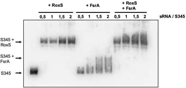
RoxS interacts directly with S345 in vitro.
358
To confirm the potential interaction between S345 and RoxS or FsrA, we performed an Electrophoretic
359
Mobility Shift Assay (EMSA; Figure 3). S345 was mixed with increasing concentrations of RoxS or FsrA
360
and loaded on a non-denaturing acrylamide gel. The results show that RoxS can bind very efficiently to
361
S345, producing a sharp band of higher molecular weight and a full-shift of S345 even at the lowest
362
molar ratio of RoxS to S345 tested (0.5). Complex formation between FsrA and S345 was less efficient
363
and the complex was less well defined, but nonetheless visible. A full shift of S345 was not apparent
364
even at a 2-fold excess of FsrA (Figure 3). When both RoxS and FsrA were incubated together with
365
S345, the interaction was clearly in favour of RoxS, with only trace quantities of the FsrA-S345 complex
366
visible. These results suggest that RoxS has a higher affinity for S345 than FsrA and are in agreement
367
with the longer predicted duplex between these two sRNAs.
368
369
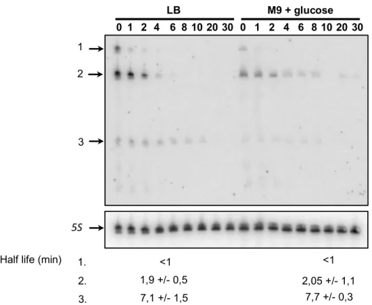
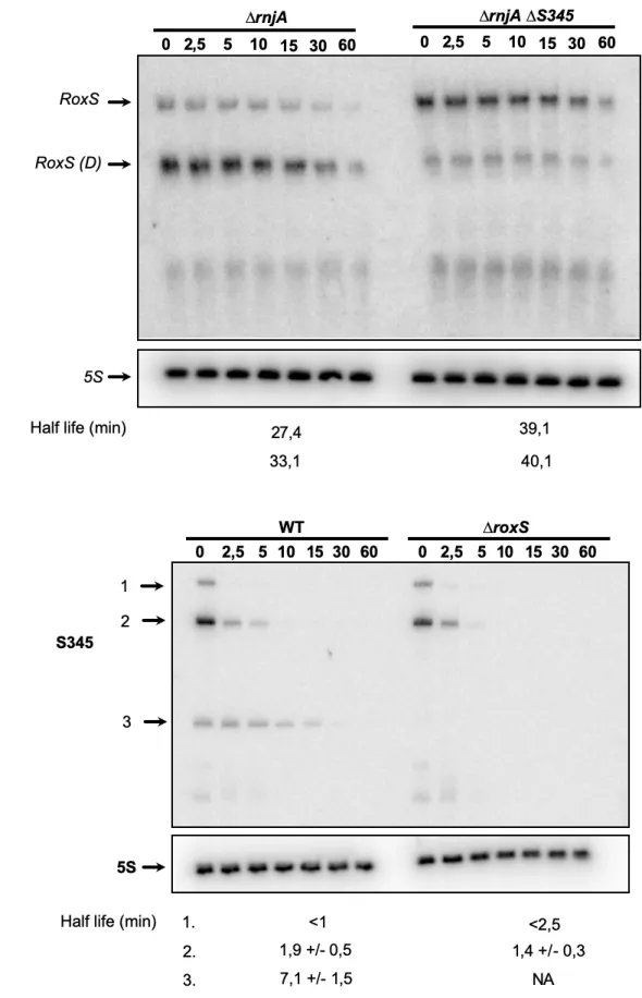
S345 is a highly processed sRNA
370
To begin to characterize S345, we first assayed its expression pattern and stability in the same
371
conditions as those used in the crosslinking experiment (LB and in M9 minimal medium + glucose).
372
Northern blot analysis of total RNA isolated at different times after the addition of rifampicin to block new
373
transcription showed that the level of the S345 RNA is higher in LB than in M9 at mid-exponential phase
374
(Figure 4). Moreover, three major forms of S345 were detected. The approximate sizes for species 1, 2
and 3 are 230 nts, 185 nts and 120 nts, respectively (Supplementary Figure 2A). The half-life of the
376
largest species (1) was less than 1 minute, while the dominant species (2) had a slightly greater stability,
377
with a half-life of 1.9 minutes in LB. The shortest species (3) had the longest half-life: 7.1 minutes in LB
378
(Figure 4). Since the half-lives of the three forms of S345 are similar in M9 + glucose, the lower levels
379
of S345 in this medium are most likely due to transcriptional regulation (see below).
380
381
The 5’-end of S345 was suggested from the sequencing product of the LIGR-seq data and was
382
confirmed by primer extension using an oligo close to the putative S345 transcriptional terminator
383
(supplementary Figure 2B). We were able to predict a putative sigma-A promoter that fits perfectly with
384
this mapped 5’ end (Figure 1). Moreover, the distance between the mapped 5’ end and the putative
385
transcriptional terminator is 229 nts, which corresponds well with the size of the largest band detected
386
by Northern blot and suggests that species 1 corresponds to the primary S345 transcript.
387
388
The Northern blot in Figure 4 was performed with an oligonucleotide probe starting 30 nts from the 5’
389
end of S345. A second probe starting only 10 nts from the 5’ end gave a similar pattern (data not shown).
390
We thus deduced that the three major forms of S345 have the same 5’ end and that species 2 and 3
391
are processed from the primary transcript at 3’ proximal sites. In agreement with this hypothesis, when
392
S345 was first identified by tiling array, an extended 3’ region was identified that was annotated as S346
393
12. The size of our proposed primary transcript corresponds to the sum of the annotated segments S345
394
+ S346. Our LIGR-seq data showed numerous truncations of S345 at its 3’ end and allowed us to
395
determine an approximate position for the cleavage site generating species 2 (Figure 2B). The
396
processing of the 3’ end of S345 was further confirmed as we were also able to map the 5’ end of a 3’
397
degradation product (*) stabilized in a ∆rnjA mutant strain by primer extension. This corresponds to an
398
endonucleolytic cleavage event occurring at the end of the duplex between RoxS and S345
399
(Supplementary Figure 2 and Figure 2B). The upstream cleavage product, protected from degradation
400
due to its hybridisation with RoxS corresponds to the smallest (120 nts) S345 species (species 3). These
401
observations suggest that S345 is quickly processed near its 3’ end to form species 2 and 3, in
agreement with the shorter half-life of the full length S345 RNA compared to its two derivatives (Figure
403
4).404
405
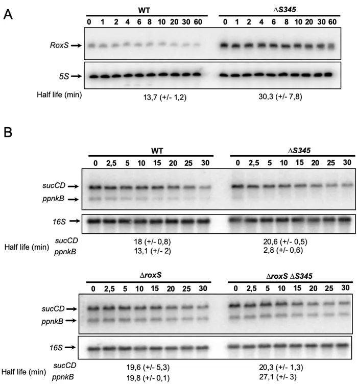
S345 destabilises RoxS406
To determine whether S345 had an effect on RoxS levels or stability in vivo, we measured the rate of
407
RoxS RNA degradation before and after the addition of rifampicin to WT and ∆S345 mutant strains. The
408
experiment was done in LB, since S345 is expressed at higher levels in this medium. Samples were
409
taken over a time course of 0 to 60 minutes and the RNA analysed by Northern blot. RoxS expression
410
was significantly higher in the absence of S345 (Figure 5A). The half-life of RoxS in the presence of
411
S345 was 13.2 minutes, whereas in the absence of S345 the half-life increased to 46.3 minutes. This
412
result shows that expression of S345 leads to destabilization of the RoxS sRNA. In contrast, deletion of
413
S345 has no impact on the stability of FsrA in LB media (Supplementary Figure 3).
414
415
We also calculated the relative amount of S345 and RoxS present in the cells grown in LB. In 5 µg of
416
total RNA, S345 and RoxS were present at approximately equimolar amounts (10 fmol each;
417
supplementary Figure 4). This result shows that there is sufficient S345 in the cell to completely titrate
418
all RoxS present in the cell under equilibrium conditions and suggests that it could act as an RNA sponge
419
to counteract RoxS activity by titrating it away from its targets.
420
421
Deletion of S345 leads to destabilisation of FsrA and RoxS targets
422
RoxS has been previously shown to negatively impact the stability of the ppnKB and sucCD mRNAs
423
encoding an NAD(H) kinase and succinate dehydrogenase, respectively 14. If S345 indeed modulates
424
the availability of RoxS to interact with its targets, we would predict that the half-life of these transcripts
425
would decrease in the ∆S345 strain due to the additional free RoxS in the cell (Figure 5A). In Northern
426
blot experiments performed on cells growing in LB medium, the half-life of the ppnKB mRNA was indeed
427
decreased 4.7-fold in ∆S345 cells compared to WT (Figure 5B), consistent with the increased amounts
428
of RoxS in the ∆S345 strain. To confirm that the effect on the half-life of the ppnkB mRNA in this strain
was due to the increase in RoxS levels, we constructed a strain lacking both sRNAs (S345 and RoxS).
430
As expected, the ppnKB mRNA became stable again in the ∆roxS ∆S345 double mutant with a half-life
431
similar to a strain lacking RoxS alone (Figure 5B). This result confirms that the destabilization of the
432
ppnKB mRNA in the ∆S345 strain is RoxS-dependent. We thus propose that S345 be renamed RosA,
433
for regulator of sRNA A.
434
435
Intriguingly, unlike ppnKB, the rate of degradation of the sucCD mRNA was not affected in ∆rosA cells,
436
with its half-life remaining at around 19 minutes in both the WT and the ∆rosA strain in LB medium
437
(Figure 5B). We previously showed that RoxS is processed by RNase Y to remove the first 20 nts of the
438
transcript producing a shorter, functional version of the sRNA called RoxS (D) 14. RoxS (D) is far more
439
efficient at competing with the ribosome for binding to the sucCD transcript than the full-length RoxS
440
sRNA in vitro 14. Removal of the first 20 nts of RoxS removes a significant portion of a 5’ stem loop,
441
freeing up nucleotides to base-pair with the sucCD Shine-Dalgarno (SD) region. We therefore asked
442
whether RosA had an effect on RNase Y processing of RoxS. RoxS (D) can be readily detected in a
443
strain lacking the 5’-3’ exoribonuclease RNase J1 (encoded by rnjA), since this RNase is involved in the
444
rapid degradation of the processed species. We therefore performed Northern blots on cells treated with
445
rifampicin to compare the relative amounts and half-lives of RoxS and RoxS (D) in ΔrnjA versus ΔrnjA
446
∆rosA cells. Figure 6 shows that RoxS is efficiently processed to produce RoxS (D) in the ΔrnjA strain
447
and is the most dominant form of RoxS in this strain. In contrast, in the double ΔrnjA ΔrosA mutant, full
448
length RoxS was the dominant version of RoxS. Thus, RosA increases the efficiency of processing of
449
RoxS to its truncated form. This is likely because base pairing with RosA is predicted to free up the
450
RNase Y cleavage site in RoxS that is normally hidden within the duplex structure of the 5’ stem-loop
451
(Figure 2A).
452
453
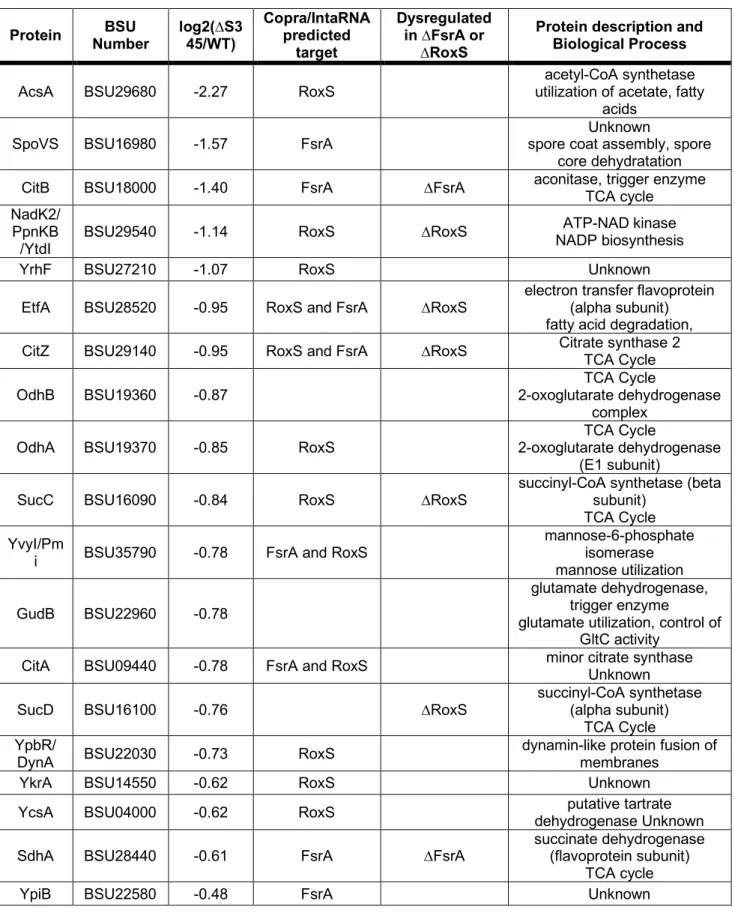
To determine the global effect of RosA on RoxS and FsrA targets, and potentially identify other roles for
454
this non-coding RNA, we performed a global proteomics analysis comparing the WT and ∆rosA deletion
455
strains grown to mid-exponential phase in LB. The proteomes were analysed by label free quantitative
proteomics. We detected 1463 proteins in the LC MS/MS analysis and identified 19 proteins that showed
457
statistically significant (P value <0.05) reduced levels in the ∆rosA strain compared to WT (Table 1).
458
Interestingly, seven of these proteins have already been assigned to the FsrA and RoxS regulons 14,15,21:
459
CitB and SdhA have been assigned to the FsrA regulon, and PpnKB, CitZ, EtfA, SucC and SucD are
460
members of the RoxS regulon. Most of the other proteins showing reduced levels in the ∆rosA mutant
461
are predicted by CopraRNA or IntaRNA 44,45 to be direct targets of FsrA and/or RoxS, and have been
462
shown to bind similar metal ions and other cofactors to the proteins encoded by other RoxS/FsrA mRNA
463
targets. This fits with the general agreement that members of the FsrA and RoxS regulons are involved
464
in regulating genes involved in iron homeostasis and oxidoreduction 14,21. The reduced levels of the FsrA
465
and RoxS targets in the ∆rosA strain supports the idea that RosA counteracts regulation by both RoxS
466
and FsrA and suggests that its primary role is as a sponge for these two sRNAs.
467
468
Production of the short form of RosA requires RoxS
469
We showed above that RosA is important for the processing of RoxS to RoxS (D); we therefore
470
wondered whether the converse was also true, i.e. whether RoxS had an effect on the processing of
471
RosA. In Figure 6B we analysed the expression pattern and degradation rates of the three RosA species
472
in the presence and absence of RoxS. In the presence of RoxS, all three forms of RosA were detected
473
as seen above (Figure 4). However, in the absence of RoxS, the RosA species 3 was completely absent.
474
We conclude that the interaction with RoxS plays a role in the processing of RosA to its smallest form.
475
The size of species 3 (~120 nts) is consistent with an RNA that extends from the mapped 5’ of all three
476
RosA species to the end of the duplex with RoxS around nt 116 (Figure 2B). The duplex would protect
477
the 3’ end of species 3 from 3’ exoribonucleases, consistent with its relatively long half-life compared to
478
species 1 and 2. No effect on RosA processing could be seen upon deletion of FsrA under these growth
479
conditions (Supplementary Figure 2).
480
481
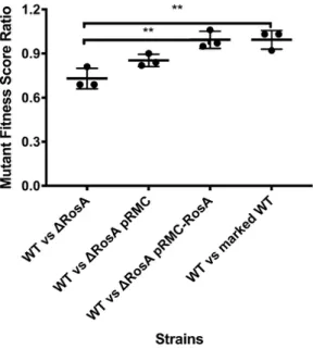
RosA provides a fitness benefit for B. subtilis under conditions of oxidative respiration
482
We asked whether RosA had an impact on cell doubling time by comparing the growth rate of the ∆rosA
483
strain to that of the WT. No major difference in growth rate was seen in either LB or in M9. To ask
484
whether there was a more subtle fitness cost to the cells lacking RosA, we performed competition assays
485
between WT and ∆rosA cells in LB medium. We mixed the WT strain marked with a spectinomycin
486
antibiotic resistance cassette and the phleomycin resistant ∆rosA mutant at a 1:1 ratio, which was
487
confirmed by colony counts carried out on the starting culture. We then counted the number of ∆rosA
488
and WT bacteria after 24 hours. The ∆rosA strain was recovered at significantly lower levels than the
489
WT suggesting that it is at a competitive disadvantage (Figure 7). In a control experiment, we also
490
competed a phleomycin resistant strain deleted for yqbR, a gene located on the Skin prophage region,
491
that was shown to be transcriptionally inactive in LB by Nicolas et al 12. This strain retained a 1:1 ratio
492
with the WT strain after 24 hours. We were also able to restore the fitness deficit of the ∆rosA strain with
493
ectopic expression of RosA at the amyE locus. We propose that the reduction in the levels of enzymes
494
of the TCA cycle, targeted by increased expression of FsrA and RoxS in the ∆rosA strain, gives these
495
bacteria a fitness disadvantage as they are unable to generate ATP as quickly the WT strain.
496
497
RosA is subject to carbon catabolite repression
498
To begin to understand under which physiological conditions RosA might act as a sponge of FsrA and
499
RoxS, we investigated how transcription of RosA is controlled. We used the DBTBS server to determine
500
which transcription factors are predicted to bind to the RosA promoter region and regulate its
501
transcription 46. DBTBS predicted a binding site for the transcriptional regulator CcpA between -1 to +
502
12 relative to the mapped 5’ end of RosA (Figure 1). CcpA mediates carbon catabolite repression in B.
503
subtilis, repressing catabolic genes and activating genes involved in excretion of excess carbon 47. The
504
prediction of a CcpA binding site in the promoter region of RosA was corroborated by Marciniak et al.
505
who identified the coordinates of the CcpA binding site in front of RosA 48. The expression profile of
506
RosA in the 104-condition tiling array data for B. subtilis was very similar to known members of the CcpA
507
regulon, such as MalA, AcoA and AbnA, consistent with the idea that RosA is a CcpA regulated sRNA
508
12.509
510
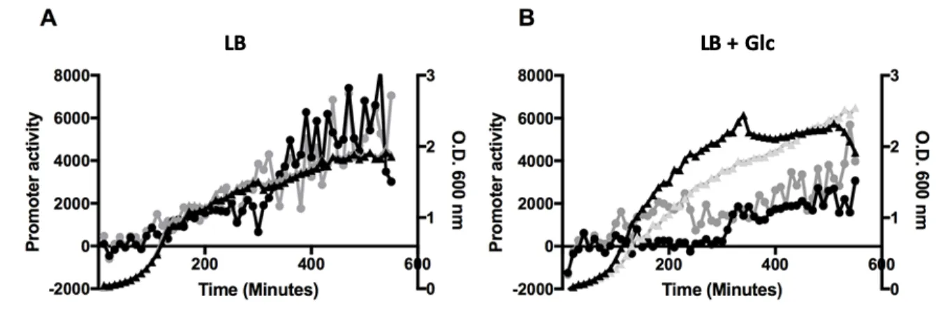
To confirm the regulation of rosA by CcpA we fused the promoter of rosA to GFP using the BaSysBioII
511
vector 32. We monitored expression of this fusion in WT B. subtilis and in an isogenic mutant lacking the
512
ccpA gene. No difference in ProsA-GFP expression could be seen between the WT and the ∆ccpA
513
strain in LB medium (Figure 8A). Addition of 0.3 % (w/v) glucose to the medium resulted in repression
514
of rosA promoter activity in the WT strain (Figure 8B), whereas in the absence of ccpA the rosA promoter
515
remained active, as predicted (Figure 8B).
516
517
We performed a similar experiment where we measured the levels of RosA RNA in a defined medium
518
with 1% malate or arabinose by Northern blot (Figure 8C). RosA levels were similar in WT and ∆ccpA
519
mutant strains grown in arabinose where CcpA is inactive on its targets. In contrast, as observed with
520
the promoter fusion, RosA expression was repressed in the WT strain and this repression was alleviated
521
in the ∆ccpA mutant strain grown in a medium supplemented with malate.
522
523
We also measured RosA expression during a switch in carbon source. B. subtilis WT and ∆ccpA strains
524
were first grown to late exponential phase in a defined medium with arabinose as the sole carbon source,
525
before adding 1% malate to promote carbon catabolite repression. Cells were harvested during
526
exponential phase and 30 and 60 min after addition of malate and RosA RNA levels were measured by
527
Northern blot (Figure 8D). Expression of RosA decreased after addition of malate in both the WT and
528
∆ccpA strain. However, the level of RosA was higher in ∆ccpA strain than in the WT demonstrating that
529
RosA is subject to the catabolite repression, and that this regulation is partially CcpA dependent. In
530
contrast, when the same membrane was re-probed for RoxS, we observed expression of RoxS was
531
induced upon addition of malate as previously observed 17. These experiments confirm that RosA is a
532
carbon catabolite responsive sRNA controlled by CcpA and that RoxS and RosA are important to
533
manage the reprogramming of gene expression during a switch in carbon sources.
534
535
536
DISCUSSION
537
538
In this study we report the use of in vivo RNA cross-linking using the psoralen AMT to globally identify
539
RNA-RNA interactions occurring in the Gram-positive model organism B. subtilis. Our results identified
540
hundreds of potential interactions, including previously well described sRNA-mRNA interactions. Two
541
of three known sRNAs containing C-rich regions in B. subtilis, FsrA and RoxS, have been shown to
542
target transcripts encoding essential components of central metabolism using their C-rich regions 14,15,20.
543
In addition to the identification of known and new mRNA targets for RoxS and FsrA, we also showed
544
that these two sRNAs interact with a new sRNA, S345, that we renamed RosA in this study.
545
Deletion of RosA from the genome of B. subtilis leads to a 3.5-fold increase of the half-life of RoxS
546
showing that RosA controls RoxS turnover. In parallel, a proteomic analyses in the ∆rosA strain show a
547
reduced levels of the known RoxS and FsrA targets like the TCA cycle enzymes, SucCD, OdhAB, CitZ,
548
SdhA and CitB 14,15,21. Many of the other proteins with reduced levels, identified by the proteomic
549
experiment, were also predicted to be targets of either RoxS or FsrA using CopraRNA 44,45. A predicted
550
target of RoxS is acsA, which encodes a key enzyme in central metabolism since it catalyses the
551
conversion of ATP, acetate and CoA to AMP, diphosphate and acetyl-CoA, thus acting as a balancing
552
point for the levels of CoA and acetyl-CoA in the cell 49. Furthermore, the SrtN protein is used by the cell
553
to deacetylate AcsA and this reaction depends on NAD+ 50. The goal of RoxS-mediated reduction in
554
AcsA levels may be to reduce non-essential NAD+ consumption. Moreover, we showed that the level of
555
RosA and RoxS is comparable in LB and that one-to-one mixtures of RosA and RoxS in vitro result in
556
full-duplex formation. Thus, these results show that RosA has the potential to be a highly efficient sponge
557
of RoxS and FsrA activity in B. subtilis cells. Our data show that RosA acts differently on the two target
558
RNAs. FsrA is sequestered in classic sponge RNA activity as the levels of the FrsA RNA remain
559
unchanged, but its proteins targets are reduced in the absence of RosA. Whereas for RoxS we have
560
shown that the levels, processing and its target efficacy are being affected.
561
562
In the field of eukaryotic RNA regulation, sponge RNAs are well-accepted as part of the regulatory
563
landscape 51 and this idea has recently been getting increased traction in bacteria. Indeed, several
564
sponge RNAs have been described in Gram-negative organisms and, intriguingly, many are derived
565
from other transcripts (reviewed by Figueroa-Bossi and Bossi 42 and Azam and Vanderpool 41). In
566
contrast, RosA is a stand-alone sRNA. Interestingly, another stand-alone sRNA in S. aureus, namely
567
RsaI (RsaOG), was also shown to be CcpA-regulated and to interact with the sRNAs RsaG, RsaD and
568
RsaE, the RoxS homologue in S. aureus, 52. RsaI, like RosA, contains two G-rich regions to bind to
569
RsaG, RsaD and RsaE. These results suggest that RsaI and RosA could fulfil the same functions in S.
570
aureus and B. subtilis and that similar sponge RNA-mediated regulatory pathways exist in Firmicutes to
571
balance the metabolic requirements of the cell. Indeed, RsaI is conserved in the genus Staphylococcus
572
but not in Bacilli, while RosA is conserved in some Bacilli but not in the Staphylococci. The role of RsaI
573
as a sponge RNA remains to be definitively proven since the impact of RsaI on RsaE, RsaD and RsaG
574
mRNA targets has not yet been investigated. In contrast to RosA, RsaI has also been shown to
575
additionally have a C-rich region used to bind mRNA targets. It could thus act as both a direct regulator
576
and as an sRNA sponge. The absence of equivalent C-rich regions in RosA may limit its function to that
577
of a sponge RNA. However, we plan to study whether RosA can directly regulate its own mRNA targets.
578
579
In this study, we identified three forms of RosA, with different half-lives. Full length RosA (229 nts) is
580
very short-lived and is quickly processed to the 185 nts form that appears to be the main functional form
581
of RosA. The shortest form of RosA (species 3) has a half-life of 7.1 minutes and its generation is RoxS
582
dependent. We believe that this form of RosA corresponds to a stable degradation product protected
583
from 3’ degradation by duplex formation with RoxS. None of the three most commonly used RNases in
584
B. subtilis, RNase J1, RNase III or RNase Y could account for the processing of RosA to its different
585
forms (data not shown). The role of RosA in facilitating the processing of RoxS and the possible
586
persistence of a RoxS-RosA duplex in cells (RosA species 3) raises the interesting question of whether
587
RoxS can be recycled from RosA to regulate mRNAs such as sucCD that prefer the shorter form of
588
RoxS? One could imagine that this duplex might be a reservoir of mostly processed RoxS, that could
switch to new partners for which it had a greater affinity. Further experiments are required to explore
590
this possibility.
591
592
Expression of RoxS is tightly controlled by two transcription factors, ResDE and Rex 14,17. Why then is
593
this additional level of post-transcriptional regulation of RoxS by RosA required? Our previous data
594
suggests that RoxS is involved in readjusting the transitory imbalances in NAD/NADH ratio that occur
595
upon encountering carbon sources such as malate. Through its role in reducing NADH levels, RoxS
596
eventually increases the DNA binding capacity of the transcriptional activator Rex, turning down its own
597
expression. However, RoxS is a relatively stable sRNA, with a half-life of 13 min in a WT strain that
598
increases to >45 min in the absence of RosA. The use of this non-coding sponge RNA is thus likely be
599
a way to dial down RoxS activity more efficiently than by simply turning off transcription, first by
600
neutralizing the C-rich regions involved in the regulation of all known targets so far and then by
601
stimulating its degradation. We propose that RosA accelerates the degradation of RoxS by stimulating
602
the opening of the 5’ stem loop of RoxS, where RNase Y is known to cleave to produce the truncated
603
form of RoxS, named RoxS (D), i.e. the processing pathway that leads to the functional form of RoxS
604
required for sucCD regulation is also the first step in RoxS turn-over. In agreement with this hypothesis,
605
the 45 min half-life of RoxS observed in the ∆rosA strain is similar to that measured previously in a strain
606
deleted for RNase Y 14.
607
608
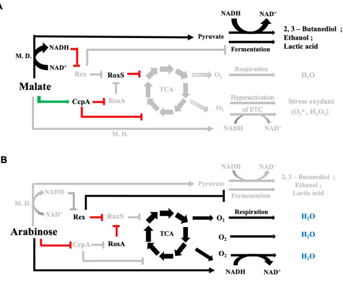
We determined that RosA is transcriptionally repressed by the main carbon catabolite repressor in B.
609
subtilis, CcpA, and that the expression of RoxS and RosA is anticorrelated during a switch of carbon
610
source. When B. subtilis is grown on one of its preferred carbon sources such as malate 53, a large
611
proportion of the carbon is metabolized only as far as pyruvate and acetyl CoA by malate
612
dehydrogenase (Figure 9). These enzymes use NAD as a co-factor, leading to an increase of NADH
613
concentration in the cell known to inhibit the DNA binding abilities of the transcriptional regulator Rex.
614
This inhibition allows the transcriptional derepression of RoxS and, instead of directing malate into the
615
TCA cycle, malate is converted to lactate and acetate via fermentation pathways normally repressed by
Rex. Fermentation allows the regeneration of NAD+ from NADH. Concomitantly, RosA is repressed by
617
CcpA allowing RoxS to bind its targets including mRNAs encoding enzymes of the TCA cycle which use
618
NAD as co-factor. CcpA enables B. subtilis to quickly adapt to the presence of these preferred carbon
619
sources. Indeed, CcpA represses genes involved in the metabolism of secondary carbon sources and
620
turns down expression many of the enzymes of the TCA cycle and transporters of TCA
cycle-621
intermediates, to ensure resources are not wasted 47,54 (Figure 9). CcpA also activates the transcription
622
of genes whose products are responsible for overflow metabolism when the bacteria are grown on a
623
preferred carbon source. The targeting of these metabolic pathways is strikingly similar to what was
624
observed previously by Durand et al. for RoxS, i.e. CcpA and RoxS have many overlapping targets 17
625
(Figure 10). In contrast, when B. subtilis is grown on a non-preferred carbon sources like arabinose, the
626
inactivation of the carbon catabolite protein CcpA will allow the transcriptional derepression of the RosA
627
sRNA and other CcpA regulated genes, including those encoding enzymes of the TCA cycle. RosA in
628
turn sponges RoxS and impairs the post-transcriptional repression of RoxS targets, also including
629
mRNAs implicated in the TCA cycle. Rex, for its part, represses the fermentation pathways (Figure 10).
630
631
The discovery here of the RosA RNA sponge under the control of the transcription factor CcpA, provides
632
the missing link between RoxS and CcpA. In other words, RoxS is connected to the CcpA regulon via
633
the RosA non-coding RNA, and RoxS ensures an additional, potentially more rapid control at the
post-634
transcriptional level for more than 30 % of genes that are regulated by CcpA. The effect of RosA on
635
RoxS also significantly expands CcpA regulon.
636
637
CONCLUSIONS
638
We have shown that in vivo AMT crosslinking of RNA is a suitable method to identify novel RNA-RNA
639
interactions including sRNA interactions. We have focused here on a novel interaction between the two
640
sRNAs FsrA and RoxS with the RNA sponge S345 that we have renamed RosA (Regulator of sRNA A)
641
and have highlighted its role in balancing the metabolic state of the cell. However, there remains many