HAL Id: tel-01682211
https://tel.archives-ouvertes.fr/tel-01682211
Submitted on 15 Jan 2018HAL is a multi-disciplinary open access
archive for the deposit and dissemination of sci-entific research documents, whether they are pub-lished or not. The documents may come from teaching and research institutions in France or abroad, or from public or private research centers.
L’archive ouverte pluridisciplinaire HAL, est destinée au dépôt et à la diffusion de documents scientifiques de niveau recherche, publiés ou non, émanant des établissements d’enseignement et de recherche français ou étrangers, des laboratoires publics ou privés.
The role of R-spondin3 in coronary artery formation and
novel roles for retinoic acid signaling in cardiac
development and repair
Fabio da Silva
To cite this version:
Fabio da Silva. The role of R-spondin3 in coronary artery formation and novel roles for retinoic acid signaling in cardiac development and repair. Agricultural sciences. Université Côte d’Azur, 2017. English. �NNT : 2017AZUR4082�. �tel-01682211�
École doctorale Sciences de la Vie et de la Santé (ED85)
Unité de recherche : INSERM U1091/CNRS UMR7277 / UCA
Thèse de doctorat
Présentée en vue de l’obtention du
grade de docteur en interactions moléculaires et cellulaires
de
L’UNIVERSITE COTE D’AZUR
par
Fabio Da Silva
Etude du rôle de R-spondin3 dans la formation des artères coronaires et
des nouvelles fonctions dans la signalisation de l'acide rétinoïque au
cours du développement et de la réparation cardiaques.
Dirigée par Andreas Schedl
Soutenue le 13 Octobre 2017
Devant le jury composé de :
Andreas
Schedl
Dr., iBV
Directeur de thèse
Kay
Wagner
Dr., iBV
Président du Jury
Pascal
Dollé
Dr., IGBMC
Examinateur
Ramón
Mu
ñoz Chápuli Dr., Universidad de Málaga Rapporteur
Robert
Kelly
Dr., IBDM Rapporteur
TABLE OF CONTENTS
RESUMÉ/ABSTRACT
... 3
ACKNOWLEDGEMENTS
……… 5
CHAPTER I: INTRODUCTION
-Part I: Heart morphology and functions
……… 7
-Part II: Cardiogenesis and cell lineages of the heart
………. 12
-Part III: Coronary vessel development
………. 20
-Part IV: Cardiac regeneration
………. 29
-Part V: Wnt signaling in cardiac development and repair
……….. 36
-Part VI: The R-spondin protein family
………... 45
-Part VII: Retinoic Acid signaling in the heart
……… 60
CHAPTER II: THE ROLE OF R-SPONDIN3 IN CORONARY ARTERY
FORMATION
-Part I: Project description
……… 87
-Part II: Schematic of project description
……….. 90
-Part III: Manuscript entitled:
“Coronary artery formation is driven…… 91
by localized expression of R-
spondin3”. Cell Reports 20, 1745-1754.
-Part IV: Future perspectives
……… 111
CHAPTER III: NOVEL FUNCTIONS FOR RETINOIC ACID SIGNALING IN
CARDIAC DEVELOPMENT AND REPAIR
-Part I: Project description
……… 115
-Part II: Schematic of project description
……….. 119
-Part III
: Manuscript entitled: “Retinoic acid signaling promotes ……….. 120
cardiomyocyte survival
in cardiac development and repair”. In preparation.
-Part IV: Future perspectives
……… 173
CHAPTER IV: CONCLUSION
-
General conc
lusion……….. 178
CHAPTER V: BIBLIOGRAPHY
RESUMÉ
Les maladies coronariennes sont l'une des principales causes de décès dans le monde. Comment les artères coronaires sont modelées et quelles sont les molécules de signalisation qui régissent ce processus, sont des mécanismes mal compris. Dans la première partie de ma thèse, j'ai identifié le modulateur de signalisation Wnt Rspo3 comme un régulateur crucial de la formation de l'artère coronaire dans le cœur en développement. Rspo3 est spécifiquement exprimé autour des branches coronaires à des moments critiques dans leur développement. L'ablation temporelle de Rspo3 à E11.5 conduit à une diminution de la signalisation de β-caténine et à une réduction de la prolifération spécifique des artères. En conséquence, les branches coronariennes sont défectueuses et l'arbre artériel ne se forme pas correctement. Ces résultats identifient un mécanisme par lequel l'expression localisée de RSPO3 induit la prolifération des artères coronaires à leurs branches permettant leur formation.
Le traitement des patients qui se remettent d'un infarctus du myocarde (IM) est difficile car les cardiomyocytes ont une capacité très limitée à proliférer et à régénérer le cœur endommagé. La voie de signalisation de l'acide rétinoïque (AR) est essentielle pour le
développement cardiaque et joue un rôle protecteur dans les cœurs endommagés. Les
mécanismes exacts et les types de cellules impliqués dans cette réponse protectrice ne sont pas clairs. Pour la deuxième partie de ma thèse, j'ai utilisé une nouvelle lignée rapportrice de l’AR inductible au TAM développée dans notre laboratoire et j'ai observé une réponse spécifique des cardiomyocytes pendant la fin de la gestation et après l'IM. L'ablation de la signalisation de l’AR par délétion génétique des enzymes Raldh1/2/3 entraîne une augmentation de l'apoptose myocytaire à la fin du développement tardif et après l'IM. Le séquençage des ARNs des cardiomyocytes primaires révèle que le traitement à l’AR réprime l'expression de Ace1, indiquant un nouveau lien entre la signalisation AR et le système Rénine Angiotensine dans le contexte de la réparation cardiaque.
ABSTRACT
Coronary artery disease is one of the leading causes of death worldwide. How coronary arteries are remodeled and the signaling molecules that govern this process are poorly understood. For the first part of my thesis, I have identified the Wnt-signaling modulator Rspo3 as a crucial regulator of coronary artery formation in the developing heart. Rspo3 is specifically expressed around the coronary stems at critical time-points in their development. Temporal ablation of Rspo3 at E11.5 leads to decreased β-catenin signaling and a reduction in arterial-specific proliferation. As a result, the coronary stems are defective and the arterial tree does not form properly. These results identify a mechanism through which localized expression of RSPO3 induces proliferation of the coronary arteries at their stems and permits their formation.
Treating patients recovering from myocardial infarction (MI) is difficult since cardiomyocytes have a very limited capacity to proliferate and regenerate the damaged heart. The Retinoic Acid (RA) signaling pathway is essential for cardiac development and plays a protective role in damaged hearts. The exact mechanisms and cell types involved in this protective response is unclear. For the second part of my thesis, I have utilized a novel inducible RA reporter line developed in our lab and I have observed a cardiomyocyte-specific response during mid-late gestation as well as after MI. Ablation of RA signaling through genetic deletion of the Raldh1/2/3 enzymes leads to increased myocyte apoptosis both during late development and after MI. RNA sequencing analysis of primary cardiomyocytes reveals atRA treatment represses
Ace1 expression, providing a novel link between RA signaling and the Renin Angiotensin System in the context of heart repair.
ACKNOWLEDGEMENTS
Before embarking on the long and difficult journey of defending a PhD, I had no idea how to think, act or perform like a researcher. I had no idea how to be a scientist. How to be an independent thinker capable of approaching a difficult question, formulating a hypothesis and carrying out the necessary experiments to either prove or disprove my initial hypothesis. But practice makes perfect. And with time I learned to work efficiently, to be innovative and to conduct my work with scientific rigour. However, this transformation into what I am today would not have been possible without the support of many people.
I would like to start off by thanking my supervisor, Andreas Schedl, for always believing in me and for encouraging me to pursue my research interests, wherever they took me. Being the only one working on the heart was undoubtedly difficult, but the experience molded me into a stronger, more capable researcher. Most other supervisors would not have supported this, but Andreas did, so, from the bottom of my heart, thank you! I would also like to thank Fariba Jian Motamedi for her tremendous dedication and enormous skill in performing the myocardial infarction surgeries. This VERY IMPORTANT part of the project would, without a shadow of a doubt, not have been possible without her. I would also like to thank Ana Sofia Rocha, my initial supervisor, who pretty much taught me everything I know. She showed me the ropes and encouraged me to always strive for the best. More importantly, she taught me to be critical not only of my own work, but of the scientific literature in general. Until today, I utilize the knowledge I gained from Ana, and I am sure I will continue to do so for the rest of my scientific career. It goes without saying that my PhD would have been a lot more difficult and less productive were it not for the continuous help of Kay Wagner. His technical expertise and willingness to teach were great assets to me, and I would have been lost without his guidance. Dr. Robert Kelly’s advice as an expert in the field was also invaluable, and I am grateful to him for taking the time
to help me. And of course, I would like to thank all of the members of my lab. The lab meetings were great, the friendships, even better!
Completing a PhD requires a lot of technical and theoretical knowledge, as well as many, many, many hours of work. It also requires emotional and mental fitness. Without the help of my family, I do not think I would have had the will and right mindset to accomplish all that I have during my PhD. So I would like to thank my mother, father, brother and grandmother for always being there for me during the past four years. You guys kept me sane and focused. Without your moral support I would have had a much harder time getting through the many tough moments presented to me during my PhD. And last, but DEFINITELY not least, I would like to thank my lovely wife. Thanks for waiting and thanks for always being there for me. You are, and always will be, my rock. This PhD is dedicated to you, meu amor.
CHAPTER I: INTRODUCTION
I.
Heart morphology, functions and disease
“The heart is, as it were, the hearthstone and source of the innate heat by which the animal is governed.”
The heart has played an important part in understanding the human body since antiquity. In his treatise, On the usefulness of the Parts of the Body, the Greek physician Aelius Galenus described the heart as the body’s innate source of heat and as the organ most closely related to the soul. Since then our understanding of the anatomy, physiology and function of the heart has evolved significantly. Yet cardiovascular-related diseases remain the number one cause of death worldwide. Hence, there is a need for a deeper understanding of the human heart and its associated pathologies.
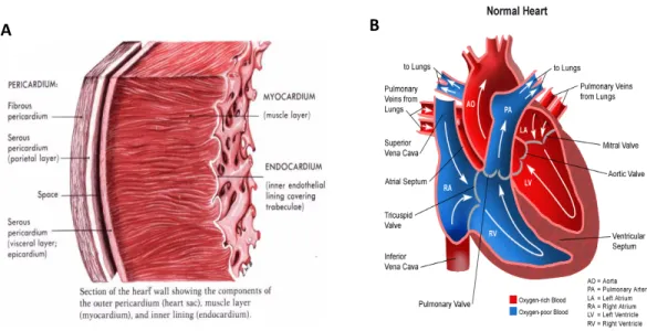
The human heart is a muscular organ located in the midline of the thoracic cavity. It is hollow and cone shaped and consists of four separate chambers. The wall of the heart is comprised of three layers: the outer pericardium, the middle myocardium, and the inner endocardium (Figure 1A). The pericardium is the outer most layer that is composed of connective tissue and serves to protect the heart by reducing friction. The myocardium is made up of cardiac muscle and is surrounded by blood vessels and specialized nerve fibers. The function of the myocardium is to contract in a coordinated fashion in response to nervous pulses transmitted by the cardiac conduction system. The endocardium is the inner most layer of the heart and is made up mainly of blood vessels. The endocardium acts as a barrier between the blood and the heart. (Clark, 2005).
The heart plays a central role in the cardiovascular system, acting as a mechanical pump that distributes blood throughout the entire body. The heart’s four chambers consist of two atria and two ventricles. Deoxygenated blood from the body enters the heart via the right atrium through
the superior and inferior vena cavae. This blood is pumped into the right ventricle and then through the pulmonary artery into the lungs, where it is oxygenated and delivered back to the left atrium via the pulmonary veins. The oxygenated blood is then pumped to the left ventricle and eventually through the aorta to the rest of the body (Figure 1B). Anatomically, the left ventricle contains a much thicker muscular layer than the right since it pumps blood to all body parts and has a much higher resistance to blood flow. The atria are separated by a solid wall known as the interatrial septum and the ventricles are separated by the interventricular septum. This separation is necessary to avoid mixing of oxygenated (left) and deoxygenated (right) blood. One way blood flow in the heart is ensured by a system of bi or tricuspid valves that are attached to the ventricular walls by strong fibers known as chordae tendinae. The atrioventricular valves, which consist of the bicuspid mitral valve (right) and the tricuspid atrioventricular valve (left), ensure one-way blood flow between the atria and ventricles. The pulmonary and aortic valves (tricuspid semilunar valves) ensure one way blood flow from the ventricles to the pulmonary artery and aorta respectively (Clark, 2005).
A
A B
Figure 1. Anatomy of the heart. A, Cross section of the heart wall showing the various layers of the heart. B, diagram showing the various compartments and components of the heart. White arrows delineate the flow of
Coordinated contraction of the atria and ventricles is achieved by a specialized subset of cardiac muscle cells that innervate the myocardium. These cells, collectively known as the cardiac conduction system, are responsible for conducting an electrical pulse that stimulates muscle contraction within the heart. The pulse begins at the sinoatrial node (SAN), which is located in the right atrium near the superior right vena cava and is composed of a specialized mass of cells capable of autonomously initiating pulses. The pulse then passes along fibers of the conduction system, promoting atrial contraction, until it reaches the atrioventricular (AV) node. The AV node delays the pulse slightly, allowing the atria to refill with blood before ventricular contraction. From the AV node the pulse passes into a large AV bundle (bundle of His) and then spreads into the Purkinje fibers, which extend into the ventricles and permit the pulse to stimulate contraction of the ventricular myocardium (Clark, 2005).
In order to ensure the heart’s nutritional and oxygen requirements are met, blood is supplied to the myocardium by a dedicated system of coronary arteries, veins and capillaries. Coronary circulation begins after left ventricular contraction, with the movement of blood from the aorta into two coronary orifices located just above the aortic valves. Connected to these orifices are the left and right coronary arteries, which extend into the ventricles and then branch off into a network of smaller arteries, arterioles and capillaries that supply the entire myocardium with oxygenated blood. After supplying the heart muscle with oxygen and nutrients the blood is then distributed to a system of venules and veins, which empty out into the right atrium via the coronary sinus (Reese et al, 2002).
Congenital heart disease (CHD) is a structural abnormality of the heart and/or great vessels that is present at birth. It is the most common birth defect, affecting nearly 1% of newly born infants. Moreover, it is estimated that around 30% of prenatal loss can be attributed to heart malformations (Brunneau, 2008). CHD is usually characterized based on a combination of anatomic and physiological phenotypes. These include conotruncal impairments such as ventricular and/or atrial septa defects, outflow tract malformations, defects resulting from abnormal left-right relationships,
valve abnormalities, and a broad range of other abnormalities (Zeidi and Brueckner, 2017). A special subset of CHDs involves defective development and/or anatomic variation of the coronary arteries. These diseases, known as coronary artery anomalies (CAA), can lead to serious clinical repercussions such as sudden cardiac death (Angelini, 2007). Overall, approximately one third of CHD patients have severe defects that require surgical intervention in the first year of life. Despite progress in medical and surgical treatments, CHD remains the leading cause of mortality from birth defects in the developed world and among the world’s poorest populations CHD has a greater contribution to cardiovascular disease than ischaemic heart disease or stroke (Zeidi and Brueckner, 2017). The majority of CHDs are genetic and caused by mutations in genes that are essential for proper heart formation during embryonic development (Brunneau, 2008). Hence, understanding these pathways is essential in order to improve the diagnosis and treatment of patients with CHDs.
Cardiovascular diseases resulting from stroke and acute myocardial infarction (MI) are among the leading causes of death worldwide killing up to 17.5 million people a year (Mozaffarian et al., 2015). Myocardial infarction (MI) results from occlusion of a coronary artery after atherosclerotic plaque rupture and thrombosis (Antman and Braunwald, 2001). The resulting lack of blood flow to the myocardium leads to rapid cellular death, which then triggers a massive inflammatory response (Frangogiannis, 2014). The inflammatory response gradually clears out the injury site leaving sparse tissue with enlarged capillaries. Eventually the gap fills with granulation tissue, which, after a certain period of time, begins to mesh into a dense non-functional scar. To compensate for the loss of functional tissue, the myocardium surrounding the infarct area undergoes a hypertrophic response and a series of remodeling steps, leading to ventricular dilation. Although the initial reaction to MI is protective and acts to limit the spread of the damage, the excessive scar formation and ventricular remodeling acts as a barrier to proper electromechanical coupling between healthy regions of the heart; thus, compromising efficient and synchronous contraction of the myocardium (Aisagbonhi et al, 2015). The issue is further
aggravated by the fact that cardiomyocytes, the principle cell-type comprising the contractile myocardium, have a very limited capacity to proliferate and regenerate the damaged area (Roij, 2016). Hence, the heart cannot recover and over time its function continues to decrease, eventually leading to heart failure and death. Interestingly, many of the molecular programs essential for proper heart formation during embryonic development are reactivated following myocardial infarction (Roij, 2016). These pathways influence several aspects of the inflammatory response, ventricular remodeling and cell death. Understanding the contributions of these pathways to myocardial infarction is, thus, an integral part of designing novel regenerative treatments for patients suffering from myocardial infarction.
The mouse serves as an excellent model to study the molecular cues involved in cardiac development and disease. It is a well characterized mammalian species and many of the signaling pathways active during cardiac development and repair in the mouse play a similar role in humans (Roij, 2016). Furthermore, myocardial infarction can be easily modeled in the mouse and the wide variety of transgenic lines available allow us to study and manipulate the various pathways activated during cardiac remodeling and repair (Aisagbhoni et al., 2015). Hence, by combining the knowledge gained from murine developmental studies and MI models, scientists can gain a deeper understanding of the molecular mechanisms activated in the post ischaemic heart in order to design novel regenerative treatments for patients suffering from cardiovascular diseases.
II.
Cardiogenesis and cell lineages of the heart
Cardiogenesis:
The embryonic heart is the first organ to function and it is essential for the distribution of oxygen and nutrients during embryogenesis (Vincent and Buckingham, 2010). In the mouse, heart development begins at Embryonic day 6.5 (E6.5) with specification of the cardiogenic mesoderm
in bilateral territories of the lateral mesoderm. At E7.5, cardiac progenitor cells extend towards the midline to form the cardiac crescent. The cardiac crescent is composed of two distinct populations of pre-cardiac cells, the so-called first and second heart fields. At E8.0, the cardiac crescent fuses at the midline and gives rise to the linear heart tube (Brade et al., 2016).
The linear heart tube is a primitive structure containing two tissues, the inner endocardium and the outer myocardium, which are separated by a thick extracellular matrix termed the cardiac jelly. Shortly after it is formed, the linear heart tube initiates a characteristic rightward looping that realigns and expands the heart, giving rise to a complex structure with four domains (MacGrogan et al., 2010). This looping and expansion of the heart tube occurs via two mechanisms: cell proliferation, and recruitment of additional cells (Brade et al., 2013). The additional cells are added to the arterial and venous poles of the heart and they originate primarily from the secondary heart field. SHF progenitors mainly contribute to the outflow tract (OFT), the right ventricle (RV) and a large portion of the inflow region (atria) (Buckingham et al., 2005; Kelly, 2012). The left ventricle (LV) is mainly derived from the first heart field. From E9.5-10.5 the atrioventricular canal, which separates the developing atria and ventricles, begins to form along with the valve primordia. Chamber development also progresses with trabeculae formation in the left and right ventricles (MacGrogan et al., 2010).
At E10.5 the developing heart is populated by two sources of extra cardiac cells: the proepicardium and the cardiac neural crest (Brade et al., 2010). The proepicardium gives rise to the epicardium, which consists of a single layer of cells that completely surrounds the heart. From E11.5-14.5, many epicardial cells undergo epithelial to mesenchymal transition and migrate into the heart, giving rise to various cell-types such as vascular smooth muscle cells, cardiac fibroblasts and a subset of endothelial cells (Brade et al., 2010). Additionally, the epicardium supports cardiomyocyte proliferation by secreting soluble growth factors to the underlying myocardium (Perez Pomares and de la Pompa, 2011). At E9.5 a subpopulation from the cranial neural crest, termed the cardiac neural crest cells (CNCC), begins to migrate towards the
pharyngeal arches. As they enter the heart, CNCCs differentiate into smooth muscle cells of the distal outflow tract and proximal coronary arteries (Brade et al., 2013; Arima et al., 2012). The cardiac neural crest also plays an essential role in signaling to the myocardium and promoting septation of the great arteries (pulmonary artery and aorta) as well as the atrial and ventricular chambers (Brade et al., 2013).
By E14.5 the heart is fully septated and the chambers and cell types are well-defined. The heart continues to proliferate, grow and mature throughout development and the post-natal period until 7 days post birth (P7), at which point the cardiomyocytes drastically slow down their proliferation rates. As the pup matures, the heart continues to grow but does so mainly via hypertrophy rather than hyperplasia (Xiao et al, 2017).
Figure 2. Different steps in heart development. A-B, Specification of cardiogenic mesoderm from primitive streak (PS) and
formation of the cardiac crescent (CC). C–E, Formation and looping of the heart tube with contributions of cardiac neural crest cells (cNCC), which migrate from the pharyngeal arches (PA) to the arterial pole (AP). The proepicardial organ (PEO) forms in the vicinity of the venous pole (VP). F, The looped heart tube, with the cardiac compartments—OFT, outflow tract; RA, right atrium; LA, left atrium; RV, right ventricle; LV, left ventricle. G, The mature heart which has undergone septation—IVS, interventricular septum; AA, aortic arch; Ao, aorta; PT, pulmonary trunk; PV, pulmonary vein; SVC, superior caval vein; IVC, inferior caval vein. The first heart field (FHF) and its myocardial contribution are shown in red, the SHF and its derivatives in dark green (myocardium) and pale green (vascular endothelial cells), cNCC in yellow, and PEO derivatives in blue (Vincent
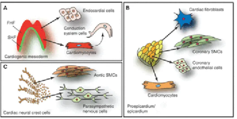
Overall the heart is a complex organ formed of various cell types. These include atrial and ventricular cardiomyocytes, vascular smooth muscle of the great arteries and coronary vessels, endothelial cells of the vasculature and endocardium, cardiac fibroblasts, epicardial cells and interstitial cells of the AV and semilunar valves. Cardiomyocytes make up the majority of the heart by volume but constitute only around 25-35% of the total number of cells. The rest is made up of other cell types such as cardiac fibroblasts and endothelial cells. Originally it was thought that fibroblasts were the principle non-myocyte cell type, constituting up to 50% of the cells in the heart. However, these estimates were probably overestimated due to the lack of specific markers for fibroblasts. Recent work with novel antibodies more specific to fibroblasts placed their number at around only 10% of all cells. In this study, endothelial cells were determined to be the principle non-myocyte cell type making up around 45% of the total number of cells (Pinto et al., 2016). Overall, the cell types of the heart arise at different times during cardiac formation and originate from different sources. Characterizing the origin and characteristics of these sources along with their precise contributions to the cell types of the heart has required a tremendous amount of work and has been the subject of much debate.
Cardiogenic mesoderm:
The cardiogenic mesoderm, which harbors the so called first and secondary heart fields, forms the major proportion of the ventricular, atrial, and outflow tract myocardium. Additionally, these progenitors contribute cells to the endocardium, the conduction system and the aortic and pulmonary cushions (Kelly et al., 2012). Mesoderm induction is regulated by numerous signaling pathways such as Nodal, bone morphogenetic protein (BMP) and WNT signals, as well as fibroblast growth factors (FGF) (Kimmelman et al., 2006; Noseda et al., 2011) . Expression of the T box transcription factor Brachybury/T (Bry), a direct target of Wnt/ -catenin signaling, marks
mesodermal cells ingressing through the primitive streak. Commitment of these cells towards a cardiac fate requires inhibition of canonical WNT/ -catenin signaling and activation of non-canonical WNT signaling (Gessert and Kuhl, 2011). After ingression through the PS, cardiac progenitor cells migrate to the anterior lateral position caudal to the head folds to form the cardiac crescent. At this time-point the first and second heart fields can be distinguished by their positions in the crescent (FHF progenitors more anterior and lateral in respect to SHF). Although the FHF cells already differentiate at this stage, the SHF cells remain in a proliferative progenitor state. It is only after entering the heart at a later time-point that they differentiate into mature cells (Kelly et al., 2012).
Although no genes are uniquely expressed in the early FHF progenitors, the SHF precursors are marked by expression of the LIM-homeobox transcription factor islet-1 (Kelly, 2012). Islet-1 expression is dependent on canonical WNT signaling (Cohen et al., 2008), and it is essential for the survival, proliferation and migration of SHF progenitors. As SHF progenitor cells migrate into the heart and differentiate, Islet-1 expression is extinguished (Cai et al., 2003). Several studies have shown that the molecular signature Isl1+/Nkx2.5+/Flk1+ marks a specific pool of primitive SHF progenitors that are multipotent and give rise to both myocytic and vascular cells. In particular, Isl1+/Nkx2.5+ descendants that have lost Flk expression form cardiomyocytes and smooth muscle cells (SMCs) and extensively contribute to the proepicardial organ (Zhou et al., 2008), whereas the Isl1+/Flk1+ subset differentiates to form endothelial cells and SMCs (Moretti et al., 2006). Interestingly, the SHF can be further subdivided into two different fields: the anterior heart field (AHF) and the posterior heart field (PHF). The AHF is marked by expression of genes such as Fgf10 and Mef2c, and it contributes to the outflow tract and right ventricle. The PHF expresses islet-1 but not any of the specific AHF heart field markers, and it contributes mainly to the atria (Zaffran et al.,2014). A critical transcription factor expressed in the PHF is Tbx5, which contributes to atrial septation and differentiation.
The fate of SHF progenitors is controlled by many different signaling pathways. FGF signaling promotes progenitor cell proliferation within the SHF and SHH-mediated signaling from the endoderm along with midline (neural tube) canonical WNT signaling maintain stemness and prevent differentiation of the SHF progenitors (Kelly 2012). BMPs, notch and non-canonical WNT signals promote differentiation of the SHF progenitors (Vincent and Buckingham, 2010). Epigenetic factors and microRNAs also play important roles in the progression of SHF progenitors to differentiated myocyte and non-myocyte cell types (Liu and Olson, 2010).
The Proepicardium:
The epicardium comprises the outer most layer of the heart and is essential for proper cardiac development. The epicardium, arises between E9.5 and E11.5 and is derived from a cluster of cells known as the proepicardium (PE). Apart from providing the heart with a protective outer layer, the epicardium is essential for coronary vessel development and formation of the compact myocardial layer. Defects in epicardial development generally lead to severe cardiac deformations and embryonic death around mid-late gestation (Brade et al., 2013).
The PE arises from the coelomic mesenchyme of the septum transversum in close proximity to the venous pole of the heart at E8.5 (Manner et al., 2001). PE induction, growth and maintenance depends upon opposing interactions between FGF signaling, which induces a proepicardial fate in the posterior splanchnic mesoderm, and BMP signaling, which drives myocardial differentiation of this population (Schlueter and Brand, 2012). The majority of PE progenitors are marked by expression of T-box18 (Tbx18) and Wilm’s tumour protein1 (Wt1) (Brade et al., 2012), with many of the Wt1 progenitors being derived from Nkx2.5+ and Isl1+ precursors (Zhou et al., 2008). An additional subpopulation of cells marked by the transcription factors Semaphorin3d (Sema3D) and Scleraxis (Scx) has been described, suggesting the PEO is a heterogeneous organ (Katz et al., 2012). Beginning at E9.5, free floating vesicles from the PE migrate towards the heart and upon contact flatten and spread out on the naked myocardium
(Brade et al., 2013). Cell adhesion molecules such as vascular adhesion molecule (VCAM) and b4-a1-integrin play a crucial role in this process, which is completed around E11.5 (Yang et al., 1995).
After the epicardium has been formed a complex array of signaling pathways work together to drive: (1) epicardial epithelial to mesenchymal transition and formation of epicardial-derived cells (EPDCs), (2) differentiation of EPDCs into different cell lineages, (3) compact zone proliferation, and (4) coronary vessel development. Wt1 and Tbx18 signaling are key factors for normal progression of epicardial EMT as well as subsequent EPDC migration and differentiation (Brade et al., 2013). FGFs, Notch and retinoic acid play prominent roles in promoting cardiomyocyte proliferation and formation of the compact zone (Sucov et al., 2009). Although there has been much debate about the exact contribution of EPDCs to different cell lineages, there is a general consensus that the majority of cardiac fibroblasts and vascular smooth muscle cells of the heart are derived from the epicardium (Brade, 2013). Whether or not cardiomyocytes are derived from the epicardium is not clear. Lineage tracing with a Tbx18-Cre suggests a small portion of cardiomyocytes derive from the epicardium, but these studies are muddled by the fact that cardiomyocyte-specific expression Tbx18 is detected between E10.5-16.5 (Brade et al., 2013). In terms of contribution to the vasculature of the heart, the majority of the endothelium seems to be derived from different sources (venous endothelium of the sinus venosus (Red Horse et al., 2010) and ventricular endocardium (Wu et al., 2012)). However, recent studies show that a small portion of endothelial cells are in fact derived from the Scx+/Sema3D+ population of the PEO (Katz et al., 2012).
The Cardiac Neural Crest:
CNCCs are a subpopulation of the cranial neural crest cells that delaminate from the neural tube and migrate on preset routes to the heart, reaching the pharyngeal arches 3,4, and 6 by E10.5 (Brade et al., 2013). The induction of CNCC progenitors is promoted by various molecular
cues such as BMP/TGF- , FGF, WNT/ -catenin, FGF and retinoic acid signaling (Vincent and Buckingham, 2010). Their migration towards the heart is promoted by ligands of the ephrin family, sempahorins, connexin-43 and many other chemical attractants (Kuriyama and Mayor, 2008). Once the CNCCs reach the pharyngeal arches, endothelin, TGF- and PDGF signaling promote their role in patterning the aortic arch arteries (Hutson and Kirby, 2007). In addition, to aortic arch patterning, CNCCs also play a role in promoting OFT development and septation. Defective CNCC induction and/or migration has been shown to lead to shortening and defective loop formation of the OFT. These phenotypes are generally caused by altered SHF progenitor addition to the developing OFT (Brade et al., 2013). The cellular contribution of CNCCs to the heart remains an ongoing debate, but it is generally thought that they differentiate into smooth muscle cells of the distal outflow tract and proximal coronary arteries, as well as a portion of insulating-glial cells of the cardiac conduction system (Brade et al., 2013).
Important transcription factors in cardiac development:
One of the first markers of cardiac progenitor cells is Mesp1, which is required for the delamination of these cells from the primitive streak (Vincent and Buckingham, 2010). A
Mesp1-Figure 3. Embryonic heart progenitor contributions to different cardiac compartments and cell types during heart morphogenesis in mouse development. A, Cardiogenic mesoderm, B, proepicardium/epicardium, and C, cardiac neural crest cell lineage diversification (Brade et al., 2013).
Cre line crossed to a Rosa26 conditional reporter marks all of the mesoderm-derived cardiac cells of the heart (Saga et al., 2009). It has been proposed that Mesp1 acts as a master regulator of cardiovascular cell fates by down regulating pluripotency and early mesodermal genes and up regulating key transcription factors such as Gata4 or Nkx2-5. Mesp1 has also been reported induce cardiogenesis by inhibiting canonical WNT signaling through promoting the expression of the WNT inhibitor Dkk1 (Vincent and Buckingham, 2010).
Other important transcription factors include Gata4 and Nkx2-5, which are expressed in the cardiac crescent where myocardial cell differentiation first takes place. T-box transcription factors such as Tbx5 and Hand1/2 (basic helix-loop-helix), as well as Mef2c (MADS-box) factors are also involved in cardiogenic differentiation. In the context of master regulators, Gata4 and Tbx5, along with the chromatin remodeling complex Baf60c/Smarcd3, can induce beating myocardial tissue when ectopically expressed in mesoderm. Gata4 and Baf60c induce Nkx2-5 expression which acts with Gata4 to initiate the cardiac program, while Tbx5 plays an important role in inducing full differentiation (Takeuchi and Bruneau, 2009). By manipulating the expression of these along with many other transcription factors many groups have been able to generate functional myocardial cells from precursor populations. In fact, recent work using viral vectors to induce the expression of the transcription factors Gata5, Mef2c and Tbx5 (GMT), showed that functional cardiomyocytes could be reprogrammed from adult fibroblasts (Qian et al., 2012). Hence, the clinical significance of not only delineating the cell lineages of the heart but understanding the transcriptional programs involved in their differentiation and maturation becomes clear and opens up numerous avenues towards novel regenerative treatments.
III.
Development of the Coronary vasculature
Overview of coronary vessel development
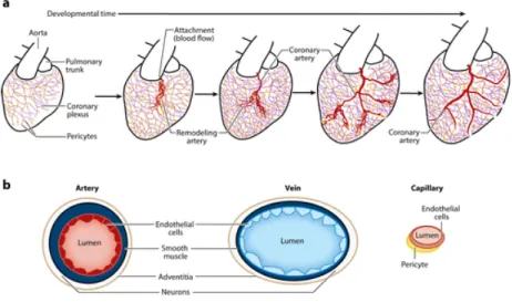
Early in embryogenesis, the heart contains a thin layer of myocardial muscle that is easily oxygenated by blood flowing through its lumen. As the heart grows and the compact myocardial layer is formed, blood vessels emerge in order to support cell proliferation. Coronary vessel development begins with the formation of an immature vascular plexus on the dorsal part of the atrioventricular groove. This vascular plexus then undergoes branching morphogenesis followed by massive expansion to cover the entire heart (Red Horse et al., 2010). The plexus eventually connects with the aorta at the coronary orifices and coronary circulation is initiated (Figure 4) (Sharma et al., 2017). Following the initiation of blood flow the vascular plexus is remodeled into mature arteries, capillaries and veins. The result is a mature circulation system that supports efficient oxygenation of the myocardium.
The mature coronary vascular system is composed of various cell types that originate from different sources. In total, they comprise up to 60% of the non-myocyte population of cells (Pinto et al., 2016). All coronary vessels are lined by a single layered endothelium comprised of endothelial cells with specialized functions. Surrounding the endothelial layer are mural cells, which include smooth muscle cells covering arteries, and pericytes around capillaries. Veins also contain a smooth muscle layer but at a lower density than arteries. In mice large arteries are located deep within the myocardium (intramyocardial) and veins are closer to the surface (subepicardial). Capillaries are located throughout the entire heart (Sharma et al., 2017).
Origins of the coronary endothelium:
The origin of the coronary vessels has been a topic of great interest for over a century. Initial observations of coronary connections lead scientists to believe coronary arteries and veins budded from the aortic orifices and sinus venosus respectively (Bennett, 1936; Goldsmith and Butler, 1937). However, through a series of transplantation studies in the chick, it was demonstrated that the coronary endothelium originated from an extra cardiac source, namely the proepicardial organ (Manner, 1999; Poelmann et al., 1993). For many years the PEO origin of coronary endothelial cells was accepted as a universal model for most complex organisms, including the mouse. However, with the advent of novel Cre recombinase lineage tracing systems, the PEO origin model for the mouse was challenged and gradually replaced by a more complex one (Sharma et al., 2017).
In order to gain a full appreciation for the intricate work on tracing the origins of the coronary endothelium, it is important to first introduce genetic lineage tracing techniques and to discuss their advantages and limitations. Lineage tracing in mammalian systems relies on Cre-loxP recombination and specific reporter lines. This method involves creating transgenic mice that
Figure 4. Structure and cellular components of the coronary vasculature. A, Schematic of the developmental
events leading to mature coronary arteries. First, a coronary plexus (purple) covered in pericytes (yellow) migrates over the surface of the heart and into the myocardium. Then, plexus vessels attach to the aorta to initiate blood flow, triggering arterial remodeling (red ) that ultimately leads to mature arteries. B, Depiction of the major cell types comprising the coronary vasculature. (Sharma et al., 2017)
express Cre recombinase under a specific enhancer/promoter which restricts Cre expression to particular cell lineages. Selection of an enhancer or promoter region is usually achieved by identifying an endogenous gene that is exclusively expressed in the cell type of interest. The Cre is generally inserted as a constitutive transgenic, or as a “Knock-In” within the gene of interest. More sophisticated techniques utilize a Cre-fused to two mutated estrogen receptors (ERT2). Under basal conditions the ERs prevent Cre translocation into the nucleus. However, when an exogenous source of tamoxifen (which binds the ER receptor) is provided, the complex can enter the nucleus and excise loxP-flanked genes. This technique is beneficial since it allows for temporal control of Cre expression in addition to spatial control. Cre lines are then crossed with reporters that contain a marker protein such as GFP or -galactosidase under the control of a broadly expressed promoter (eg. CAG). These constructs are generally knocked into an accessible genomic location such as the Rosa26 locus. In the absence of Cre recombinase, a loxP flanked stop cassette blocks reporter expression. Cre expression in the progenitor cell leads to excision of the stop cassette, allowing expression of the reporter protein. Because this labeling is permanent and heritable, the cell and all of its descendants are marked. However, the results obtained from genetic lineage tracing must be interpreted carefully. In order for the system to work, expression of the endogenous gene must be limited to the cell of interest. Even if low levels are detected in potential progeny/descendants of the cell of interest, reporter activation can occur. This would mean that the labelled progeny are not descended from the original cell of interest since they promoted Cre expression on their own (Tian et al., 2015).
In order to test whether the coronary endothelium was in fact derived from the PEO, epicardial-specific cre lines (WT1-Cre, Tbx18-Cre) were generated and tested in the mouse. However, very few endothelial cells were labelled by these lines (Cai et al., 2008; Zhou et al., 2008), suggesting an alternative origin for the murine coronary endothelium. Following these revelations several studies were conducted to address this mystery. Through this work it was determined that the endothelial cells of the coronary vessels originated for three different sources:
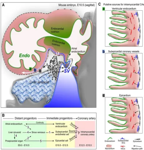
the sinus venosus, the ventricular endocardium and a specialized subset of cells in the proepicardium (Sharma et al., 2017).
The sinus venosus (SV) is a transient structure in cardiovascular development that receives venous blood from the embryo and shuttles it to the atrium. As the heart matures, the SV becomes integrated into the right atrium, forming the coronary sinus. Recent work from Red Horse et al., 2010, has demonstrated the importance of vascular sprouting from the SV in coronary vessel development. Through a combination of single-cell labeling and clonal analysis the authors were able to show a lineage relationship between SV endothelium and the coronary veins, capillaries and arteries. Because the SV is initially venous, the study proposed a mechanism through which sprouting of SV venous endothelial cells is accompanied by dedifferentiation into progenitor cells capable of adopting a venous or arterial fate. These cells then spread throughout the surface of the heart to form an immature vascular plexus located in the subepicardial space. Vessels destined to become arteries then migrate towards the inner myocardium and undergo arterial differentiation, while those that remain in place adopt a venous identity. To support the SV origin of the endothelial cells, Tian et al., 2013, developed an Apelin-CreERT2 line capable of tracing endothelial cells originating from the SV. Using this line they were able to show that the majority of the coronary vasculature was indeed derived from the SV as predicted by Red Horse et al.
In parallel to the experiments by Red Horse et al. and Tian et al., data began to emerge which proposed an alternative source of coronary endothelial cells: the ventricular endocardium. Through a series of clonal and histological observations it was demonstrated that endothelial lined structures filled with blood cells, termed “blood islands”, were capable of budding from the ventricular endocardium onto the surface of the heart (Tian et al., 2013). These so called “blood islands” were observed to arise at the surface of the dorsal and ventral midline, from where they would migrate deeper into the myocardium and form coronary vessels. Evidence suggested that the blood cells within these structures emerge because the endocardium is hemogenic and able
to differentiate into hematopoietic cells. Lineage tracing experiments using an endocardial-specific
Nfatc1-Cre line confirmed these observations. In these studies a large portion of coronary arteries and capillaries, but very few veins, were labelled by the Nfatc1-Cre line (Wu et al., 2012).
A third source of coronary endothelium in the mouse heart was revealed with Cre-based lineage-tracing studies on a subset of epicardial cells positive for the markers Scx or Sema3D. These cells, which were not labeled with the initial epicardial Cre lines, represent a separate population within the PEO that only partially overlap with Tbx18+ and Wt1+ cells. Lineage tracing using Scx-Cre or Sema3D-Cre lines revealed these cells could give rise to a portion of coronary endothelial cells. More specifically, the Sema3D-Cre line labeled endothelial cells of the sinus venosus, whereas the Scx-Cre gave rise to endocardium (Katz et al., 2012).
The above studies provided evidence that the coronary endothelium derived from three different sources. However, there was still much confusion over the levels of contribution from each source. A subsequent report aimed to address this issue by quantifying lineage-tracing data from all three sources using the Apelin-CreER (SV), Nfatc1-Cre (endocardial) and Sema3d-Cre (PEO) lines. The results revealed a striking compartmentalization of the areas populated by different progenitors. Sinus venosus cells gave rise to the majority of vessels on the dorsal and right lateral sides and almost half of the vessels on the left lateral side, with minimal contribution to the mid-ventral aspect and in the ventricular septum. Complementary to the sinus venosus traced cells, endocardium lineage tracing gave rise to vessels in the left mid-ventral aspect and ventricular septum. Sema3D-Cre and Scx-Cre derived cells were significantly lower when compared to the two aforementioned sources (<20%) and were distributed evenly among the outer circumference of the heart (Chen et al., 2014). However, more recent work using a Gata4-Cre to perform lineage tracing from the septum transversum demonstrated that the contribution of the PEO is larger than previously calculated, reaching over 20% of coronary endothelial cells in certain parts of the heart (Cano et al., 2015).
Although the simultaneous use of sinus venosus and endocardial lineage-tracing reagents clearly showed complementary contributions between these two progenitors, it was determined that there was a small percentage of cross labeling with the Cre lines. In particular, the Nfatc1-Cre line was shown to recombine in the sinus venosus and in parts of the coronary endothelium itself (Zhang et al., 2016). To address this issue a more specific endocardial Cre line (Nrp3-CreER) was developed to fully exclude sinus venosus labeling in endocardial lineage traces. It was observed that this line labelled very few coronary vessels; thus, demonstrating that the majority of coronary vessels believed to be derived from the endocardium, were actually derived from the sinus venosus.
Figure 5, Origin of coronary endothelial cells. A and B, The 3 major sources of coronary vessels: the proepicardium (PE), sinus
venosus (SV), and endocardium (Endo) are intimately associated with each other during heart development. The PE is a transient structure (gray color) that is wedged into the atrioventricular groove between liver sinusoids and SV, and eventually gives rise to the epicardium covering the heart. The SV (blue) is the venous inflow tract. Venous cells from SV sprout onto the heart and produce subepicardial coronary vessels. The endocardium (green) lines the heart lumen. Black-dashed arrows denote movement from one compartment to another, potentially complicating lineage-tracing experiments. Numbers in B correspond to those in A showing location of migration events. C, Three putative sources for intramyocardial coronary arteries (CAs) in the developing heart. Arrows indicate corresponding migration path. EC indicates endothelial cell; VEC, vascular endothelial cell. (Tian et al., 2015)
Coronary Artery Smooth Muscle
Another important cell type for coronary vessels are smooth muscle cells. During the remodeling phase of vascular plexus formation, smooth muscle cells are layered around the coronary arteries to provide mechanical support (Sharma et al., 2017). Coronary artery segments closer to the aorta that are exposed to higher blood pressure have multiple layers of smooth muscle while more distal ones have only a single layer. Lineage tracing with several epicardial-specific cre lines (Wt1-Cre, Tbx18-Cre, Gata5-Cre Tcf21-CreER) has revealed that the majority of vascular smooth muscle is derived from the epicardium (Cai et al., 2008; Zhu et al., 2008; Acyara et al., 2012). More specific lineage tracing experiments using Ng2-Cre and Notch3-Cre lines demonstrated the intermediate progenitors for smooth muscle to be cardiac pericytes (Volz et al, 2015). Much like the endothelial cells of the heart, the vascular smooth muscle of the coronary arteries also has multiple origins. Lineage tracing experiments with the Wnt1-Cre, which labels the CNCC, showed that the smooth muscle around the proximal coronary arteries is derived from the preotic neural crest (Jiang et al., 2000). Recently, a third source for coronary smooth muscle has been discovered. In this study it was determined that the endocardial-derived cardiac cushion mesenchyme can also provide a source of coronary mural cells (smooth muscle and pericytes). These cells were observed at a higher frequency in the ventricular septum when compared to the left and right lateral walls (Chen et al., 2016).
Molecular programs driving coronary vessel development
Coronaries are believed to derive from vascular sprouts originating in the SV. Mechanistically, it is not clear exactly how this happens but many signaling pathways have been shown to be involved in this process. For instance, Vegfr3, which is generally involved in angiogenic sprouting, is expressed at the site where SV cells enter the heart (Red Horse et al., 2010). Myocardial-specific Angiopoetin1 signaling to the SV promotes migration of venous cells
into the heart. Angiopoetin1 is also reported to be involved in venous differentiation as deletion of
Ang1 in the myocardium resulted in defective venous, but not arterial formation (Arita et al., 2014).
Vegfc is another essential signaling factor involved in SV sprouting. Ablation of Vegfc, which is normally expressed in the epicardium, leads to delayed vascular sprouting and reduced vessel density near the outflow tract. This was determined to be due to migration defects rather than a decrease in proliferation (Chen et al., 2014). Calcineurin-Nfat signaling is also essential for vascular plexus formation as deletion of Cnb1 delays vascular growth near the SV (Zeini et al., 2009). Mechanistically this is attributed to defective tube formation during the initial stages of plexus formation. Myocardial FOG2 expression has also been shown to promote plexus formation by promoting the expression of angiogenic factors and inhibiting anti-angiogenic ones (Ma et al., 2008). FGFs indirectly support coronary vessel development by promoting myocardial growth, which, through the expression of angiogenic growth factors (Vegf-a,b,c and Ang2), promotes vascular sprouting (Lavine et al., 2006). Sonic hedgehog (Shh) signaling plays an important role in arterial venous differentiation. Ablation of SHH receptors in either the epicardium or myocardium leads to less arteries and veins respectively (Lavine et al., 2008).
The derivation of coronary arteries from the so called “blood islands” of the endocardium is attributed to myocardial-derived VEGFA signaling to VEGFR2-expressing endocardial cells. Knockout of either molecule in their associated cell types diminishes the number of coronary arteries in the compact myocardium (Wu et al., 2012). Recent studies have begun to decipher the mechanisms involved in epicardial-endothelial transition. One study suggests that the Hippo pathway transcription factors Yap/Taz promote the proliferation, epithelial–mesenchymal transformation (EMT), and differentiation of Sema3d+ cells into coronary endothelial cells (Singh et al., 2016).
Another important aspect of coronary vessel formation involves proper connection of the coronary arteries to the aortic orifices. The proximal left and right coronary arteries are derived from the primitive vascular plexus in a well-defined “ïn growth model”. That is, rather than budding
from the aorta, as was initially thought many years ago, the proximal coronaries grow into the aortic orifices and establish a connection as early as E12.5 (Tian et al., 2013). The establishment of this connection is essential for plexus maturation as it initiates blood flow, which triggers the arterialization of undifferentiated plexus vessels (Sharma et al., 2017). Vegfc and the chemokine
Cxcr4 are highly expressed in the walls of the outflow tract and their receptors, Vegfr2 and Cxcl12 are expressed by endothelial cells of the plexus (Chen et al., 2014; Ivins et al., 2015). Deletion of either Vegfc or Cxcr4 leads to defective stem formation and severe plexus defects. In both cases the phenotype is attributed to migration deficiencies, although with Vegfc the defect in stem formation may be secondary to reduced vessel density around the outflow tract.
The differentiation of precursor cells from the epicardium and neural crest into smooth muscle is also a complex process regulated by many factors. To form vascular smooth muscle, epicardial cells must undergo EMT that allows them to leave the surface of the heart and migrate into deeper layers (Sharma et al., 2017). In terms of cellular differentiation, evidence indicates that the decision made by EPDCs to form smooth muscle rather than cardiac fibroblasts occurs right at the surface, where it is coupled to the EMT process. Canonical Wnt signaling has been shown to play a role in supporting epicardial EMT and deletion of -catenin with the Gata5-Cre leads to defective smooth muscle layer formation (Zamora et al., 2007). Deletion of Pdgfrβ and Myocardin-related transcription factors A and B also leads to defective smooth muscle differentiation (Trembley., 2015). Retinoic acid signaling (by controlling Tcf21 expression) delays smooth muscle long enough for the coronary endothelium to form functional tubes (Braitsch et al., 2012).
Edn/Ednra1 signaling promotes the differentiation of smooth muscle from the preoptic neural crest and defects in this pathways result in ectopic connections of the coronary arteries with the aorta (Jiang et al., 2000). Notch3 stimulates the induction of contractile proteins in pericytes surrounding the developing coronary arteries, which is in response to Jagged1 expression in the endothelium (Volz et al., 2015).
Overall, coronary vascularization is a precisely timed and fine-tuned process, and one that is regulated by many cell lineages and signaling pathways. Important cell types involved in coronary vascularization include venous cells of the SV, endocardial cells, epicardial cells, the cardiac neural crest and cardiac myocytes. Together, these cells coordinate coronary plexus formation and maturation via various autocrine and paracrine molecular cues. Disruption of these processes leads to defects in coronary circulation and serious consequences such as embryonic lethality.
IV.
Cardiac Regeneration
Acute myocardial infarction leads to rapid cell death and replacement of healthy tissue with a non-functional scar. Despite the availability of medical therapies, heart function continues to decline after MI, resulting in heart failure and eventual death (Frangogiannis, 2014). A central problem with treating heart disease is that adult mammals do not sufficiently regenerate cardiomyocytes to compensate for lost cardiomyocytes (Senyo et al, 2014). Recent work has elucidated some of the mysteries behind cardiomyocyte regeneration during homeostasis and repair. This knowledge has been used to generate novel strategies for treating heart disease based on stimulating endogenous repair mechanisms and reprogramming cardiomyocytes from other cell types.
Cardiomyocyte proliferation
Historically, the heart has been viewed as a post mitotic organ in which the primary parenchymal cells, cardiomyocytes, do not proliferate (Senyo et al., 2014). In a study performed in the 1950s, scientists measured the increase in cardiomyocyte cross sectional area in the left ventricular papillary and concluded that cardiomyocyte enlargement could fully account for myocardial growth between birth and adulthood (Linzbach, 1950). In mice, cardiomyocytes are
thought to proliferate vigorously until postnatal day 5, after which point they begin exiting the cell cycle. By postnatal day 7, hearts lose regenerative capacity and between P10 and P21, CMs become terminally differentiated and quiescent. This transition is accompanied by a downregulation of cell cycle factors such as Cyclin/cdk complexes and up regulation of cell cycle inhibitors such as P21/P27 (Naqvi et al., 2014). An interesting feature of mammalian cardiomyocytes is that many undergo bi-nucleation, a common marker of terminal differentiation. In mice, bi-nucleation occurs in 80-90% of cardiomyocytes between postnatal day 5 and 10. In humans, 30% of CMs are bi-nucleated in the newborn heart and although there is no rapid increase in bi-nucleation during the postnatal period, CM ploidy is active well into adulthood (Senyo et al., 2014)
Recent studies using novel technological advances have since challenged the notion that adult mammalian CMs are quiescent. One of the first indications that adult CMs could proliferate came from a study by Soonpaa et al. 1997, wherein mice were injected with tritiated thymidine and analyzed two hours later. From these experiments, the authors detected a 0.0005% labeling frequency of cardiomyocytes. In another series of studies, long-term bromo-deoxyuridine (BrdU) labeling demonstrated a basal CM proliferation rate of 1% per year in adult mice (Li et al., 1996; Malliaras et al., 2013). Perhaps the most stunning evidence for cardiomyocyte proliferation in humans was revealed by Carbon 14 (C14) dating studies. C14 birth dating makes use of the transient increase of C14 in the biosphere that occurred in the 1950s-1960s due to above ground nuclear testing. The C14, which was taken up by through diet and incorporated into genomic DNA, can be used as a time-stamp to calculate the mean birth date of a stable cell population. This can then be compared with the age of the source individual to calculate the generation rate of new cells (Senyo et al., 2014). By using this technique, the Frisen group was able to demonstrate that new cardiomyocytes formed at a rate of approximately 1.5% per year at age 25 years; however, this rate tended to decrease substantially in the latter half of life (Bergmann et al., 2009). This data was confirmed through the use of imaged-based assays in tissue samples procured from donor
hearts prior to heart transplantation. From this study the authors calculated a CM proliferation rate of about 1.9% at 20 years of age. Furthermore, through direct stereological quantification of cardiomyocytes in humans, the authors also demonstrated that CM number increases from 1.1 to 3.7 billion during a 20 year time period after birth (Mollova et al., 2013). A similar change is reported in rodents (Li et al., 1996) although a new study indicates that a large proportion of this increase happens during the pre-adolescent period (P15), during which time mice experience a 40% increase in CM number (Naqvi et al., 2014).
To assay the role of a potential progenitor cell contribution to CM proliferation, multiple groups have used the α-Myosin Heavy Chain (MHC) CreERT2 crossed with a GFP reporter line. In a study by Hsieh et al., 2007, it was demonstrated that during normal aging in mice, the percentage of preexisting cardiomyocytes remained unchanged. Influx of cardiomyocytes generated from undifferentiated progenitor cells should have resulted in a decrease in the percentage of GFP-positive CMs but this “dilution” was not observed, indicating that the majority of new CMs generated after birth in the mammalian heart arose from endogenous cell proliferation. Regarding one specific progenitor cell type marked by c-Kit expression, conflicting results have emerged.Ellison et al. 2013, reported a 0.15% generation rate for c-kit derived cardiomyocytes in a 4-week period of normal aging. This, however, contrasted with results from Zaruba et al. 2010, who, by using a more direct lineage tracing approach with a c-Kit knock in Cre, determined that C-KIT positive cells did not generate novel cardiomyocytes to a significant degree.
In adult mouse models of myocardial damage, the story becomes more complex. After experimental myocardial infarction, border zone CMs exhibit a 10-fold increase in cell cycle activity (Senyo et al., 2014). Using the MHC transgenic model, Malliaras et al. demonstrated a dilution of GFP positive cells after damage, indicating a larger role for progenitor cells than endogenous proliferation. However, by combining the Cre model with stable isotope mapping, Senyo et al. 2013 observed cell cycle activity in preexisting cardiomyocytes at the injury border. These contrasting observations may be attributed to varied long-term viability of GFP- and GFP+
populations, although an evaluation of short term apoptosis or proliferation rates did not show any differences. In addition, there is growing evidence that activation of the surrounding epicardium may contribute to myocardial repair after injury (Senyo et al., 2012). Epicardial cells that demonstrate an epithelial to mesenchymal transition can lead to myocardial vascularization and, possibly, to cardiomyocyte formation (Huang et al., 2012). However, there is much controversy regarding the latter and treatments aimed at promoting epicardial-cardiomyocyte differentiation have had conflicting results (Smart et al., 2011; Zhou et al., 2012).
In an attempt to explain the limited proliferative capacity of adult CMs, two possibilities have been proposed: (1) the presence of the differentiated sarcomeric cytoskeleton prohibits cell division and; (2) the binucleated and polyploid phenotype prevents cell cycle re-entry (Senyo et al., 2014). To address the first point, cardiomyocyte mitotic figures have been reported in adult zebrafish and neonatal mice during cardiac regeneration following MI (Jopling et al., 2010; Porello et al., 2011). Both neonatal and adult zebrafish cardiomyocytes are capable of disassembling their sarcomeres in order to reenter the cell cycle, indicating that presence of sarcomeres may not be prohibitive to cardiomyocyte division. Hence, this theory has its limitations. In support of the binucleation theory, it has been demonstrated that exogenously induced proliferation of differentiated adult mouse cardiomyocytes in vitro occurs primarily in the mononucleated portion (Bersell et al., 2009). Furthermore, most adult zebrafish are mononucleated which is consistent with their proliferative activity during regeneration (Wills et al., 2008). A similar feature is observed in Newt cardiomyocytes (Bettencourt-Dias et al., 2003), indicating that binucleation is indeed correlated with the inability of CMs to proliferate.
Novel regenerative approaches for patients suffering from heart damage
The majority of cardiac regenerative approaches have involved stimulating endogenous repair mechanisms and transplantation of cells with potential progenitor features into infarcted myocardium. Types of stem cells used include bone marrow derived stem cells, human embryonic
stem cells and induced pluripotent stem cells. Reprogramming of adult progenitor cells in vivo is an additional technique that has shown much promise.
The observation that neonatal mice exhibit regenerative capacity in response to surgical procedure has sparked interest in modulating adolescent-adult cardiomyocyte proliferation (Senyo et al., 2014). Forced expression of CYCLINB1/B2 and knockdown of p21/p27 have been shown to increase CM proliferation in vitro (Bicknell et al., 2004; Di Stefano et al., 2011). In vivo, CYCLIN D2 overexpression resulted in increased cardiomyocyte DNA synthesis and reduced scarring after MI in mice (Pasumarthi et al., 2005). Similarly, transgenic expression of CYCLIN A2 increased cardiomyocyte cycling and myocardial regeneration (Chaudry et al., 2004). Delivery of neuregulin (NRG10), fibroblast growth factor (FGF1) with pharmacologic p38 MAP kinase blockade, and periostin peptide have all been demonstrated to promote myocardial repair as well (Bersell et al., 2009; Engel et al., 2006; Kuhn et al., 2007). Thus, the modulation of endogenous repair pathways provides a novel therapeutic avenue for regenerative treatments, although it is no quite clear how these would fare in clinical trials.
Bone marrow-derived stem cells (BMDSCs) are able to differentiate into a wide variety of cells including cardiomyocytes and can be used for autologous transplantation in patients suffering from heart disease (Senyo et al., 2014). Many trials, such as the REPAIR-AMI, have shown improved outcomes in heart function after BMDSC transplantation (Assmus et al., 2010). However, two recent clinical trials have been somewhat discouraging. Neither the TIME trial nor the POSEIDON trial showed any significant improvement in ventricular function after transplantation (Traverse et al., 2012; Hare et al., 2012). A large multinational trial (BAMI) is being conducted in Europe to further address this issue.
Embryonic stem cells (ESCs) provide the possibility of generating an unlimited amount of cardiomyocytes in vitro. By treating ESCs with ACTIVIN-A and BMP4 one can generate a highly purified population of ESC-derived CMs, that when transplanted in vivo, demonstrate enhanced survival rates (Laflamme et al., 2007). Human ESC-derived CMS can also electromechanically