HAL Id: hal-02266283
https://hal.archives-ouvertes.fr/hal-02266283
Submitted on 14 Aug 2019
HAL is a multi-disciplinary open access
archive for the deposit and dissemination of
sci-entific research documents, whether they are
pub-lished or not. The documents may come from
teaching and research institutions in France or
abroad, or from public or private research centers.
L’archive ouverte pluridisciplinaire HAL, est
destinée au dépôt et à la diffusion de documents
scientifiques de niveau recherche, publiés ou non,
émanant des établissements d’enseignement et de
recherche français ou étrangers, des laboratoires
publics ou privés.
Inger Lauritzen, Nicolas Blondeau, Catherine Heurteaux, Catherine
Widmann, Georges Romey, Michel Lazdunski
To cite this version:
Inger Lauritzen, Nicolas Blondeau, Catherine Heurteaux, Catherine Widmann, Georges Romey, et al..
Polyunsaturated fatty acids are potent neuroprotectors. EMBO Journal, EMBO Press, 2000, 19 (8),
pp.1784-1793. �10.1093/emboj/19.8.1784�. �hal-02266283�
Inger Lauritzen, Nicolas Blondeau,
Catherine Heurteaux, Catherine Widmann,
Georges Romey and Michel Lazdunski
1Institut de Pharmacologie MoleÂculaire et Cellulaire, CNRS UPR 411, 660 route des Lucioles, Sophia Antipolis, 06560 Valbonne, France
1Corresponding author
e-mail: [email protected]
Results reported in this work suggest a potential
therapeutic value of polyunsaturated fatty acids for
cerebral pathologies as previously proposed by others
for cardiac diseases. We show that the
polyunsatur-ated fatty acid linolenic acid prevents neuronal death
in an animal model of transient global ischemia even
when administered after the insult. Linolenic acid also
protects animals treated with kainate against seizures
and hippocampal lesions. The same effects have been
observed in an in vitro model of seizure-like activity
using glutamatergic neurons and they have been
shown to be associated with blockade of glutamatergic
transmission by low concentrations of distinct
poly-unsaturated fatty acids. Our data suggest that the
opening of background K
+channels, like TREK-1 and
TRAAK, which are activated by arachidonic acid and
other polyunsaturated fatty acids such as
docosahexa-enoic acid and linolenic acid, is a signi®cant factor in
this neuroprotective effect. These channels are
abun-dant in the brain where they are located both pre- and
post-synaptically, and are insensitive to saturated
fatty acids, which offer no neuroprotection.
Keywords: background K
+channels/epilepsy/
excitotoxicity/ischemia/polyunsaturated fatty acids
Introduction
There is now a considerable literature describing the
bene®cial effects of polyunsaturated fatty acids (PUFAs)
in the prevention of coronary heart disease (Nordoy,
1999), in decreasing the risk of sudden cardiac death (Leaf
and Kang, 1996; Albert et al., 1998) and particularly in
preventing fatal ventricular arrhythmias (Nair et al., 1997;
Leaf et al., 1999). There are also some indications that
PUFAs might have a bene®cial effect on various brain
functions such as epileptic seizures (Vreugdenhil et al.,
1996), depression (Hibbeln, 1998) or bipolar and other
behavioral diseases (Stoll et al., 1999). PUFAs seem to
exert their bene®cial effect by decreasing cardiac and
neuronal excitability. Obvious candidates for this effect
are the neurotransmitter receptors and ion channels that
underly this excitability (Leaf et al., 1999).
The purpose of this work is two-fold. First, it is
important to demonstrate that PUFAs are indeed
neuro-protectors as well as anti-epileptic molecules in vivo using
animal models of transient global ischemia or kainate
(KA)-induced seizures. This demonstration having been
made, we analyze using an in vitro system how PUFAs
exert their protective action. Hypoxic±ischemic and
epileptic neuronal injuries have been associated with an
excessive activation of post-synaptic glutamate receptors,
and glutamate toxicity itself has been linked to lethal
in¯ux of Ca
2+mainly through cell membrane channels of
the N-methyl-
D-aspartate (NMDA) subtype of glutamate
receptors (SiesjoÈ and Wieloch, 1985; Choi, 1994; Barnard,
1997). We show that PUFAs block neuronal death by
inhibiting glutamatergic transmission. How do they exert
this effect? We worked out that among the different
neurotransmitter receptors and transporters and ionic
channels that are modulated by PUFAs, a new class of
K
+channels with two pore-forming domains (2-P
domains) having the properties of background K
+channels
could be particularly interesting candidates. Some of these
channels, which have been cloned recently (Lesage and
Lazdunski, 1999), are activated by arachidonic acid (AA)
and other PUFAs (Kim et al., 1995; Fink et al., 1996,
1998; Patel et al., 1998). These channels are expressed in
the heart (Kim and Clapham, 1989) and are abundant in
the brain (Kim et al., 1995). Their activation in the neurons
would be expected to hyperpolarize synaptic terminals,
decreasing glutamate release and/or producing a
post-synaptic hyperpolarization, which would favor the
block-ade of the NMDAreceptor-associated channel by Mg
2+and also counterbalance depolarization produced by the
glutamate action on other types of ionotropic glutamate
receptors. We indeed observed that K
+channels activated
by PUFAs are present both pre- and post-synaptically and,
consequently, they might be important players in the
blockade of glutamatergic transmission by PUFAs, which
results in a potent neuroprotective effect.
Results
Linolenic acid, but not palmitic acid, protects
against ischemia-induced neuronal death
Neuroprotection by the PUFAlinolenic acid (LIN) was
evaluated in an in vivo model of cerebral global ischemia
(Pulsinelli and Brierley, 1979; Schmidt-Kastner and
Freund, 1991). Complete forebrain ischemia for 10±
30 min induced neuronal cell death of CA1 pyramidal
cells in the hippocampus (Figure 1i). Seven days after a
20 min global ischemia, the number of surviving CA1
neurons, as measured by Nissl's staining, was decreased to
14 6 5% (Figure 1b and i) as compared with sham control
(Figure 1a and i). Injection of LIN [10 mM (5 ml),
intracerebroventricularly (i.c.v.)], a PUFAthat is found in
high quantities in vegetable oils, 30 min before induction
of ischemia almost completely inhibited neuronal loss
(81 6 6% of cell survival; Figure 1c and i). These results
Polyunsaturated fatty acids are potent
neuroprotectors
The EMBO Journal Vol. 19 No. 8 pp. 1784±1793, 2000
were con®rmed by the lack of TUNEL staining of cells in
the CA1 pyramidal cell layer (Figure 1f). The potent
neuroprotective effect of LIN was observed for the three
different times of ischemia (10, 20 and 30 min) (Figure 1i).
In contrast to LIN, administration of the saturated fatty
acid palmitic acid (PAL) failed to protect the brain and
considerable neuronal loss of CA1 pyramidal neurons was
observed after this treatment (Figure 1i). LIN is also
protective when administered intravenously (i.v.). LIN
(100 nmol/kg, i.v.), injected 30 min before (Figure 1g) or
30 min after (Figure 1h) the toxic insult, fully prevented
neuronal loss of CA1 neurons. We also analyzed the
effects of another PUFA: AA. Although this fatty acid also
had bene®cial effects, they were less pronounced and less
Fig. 1. Neuroprotection by PUFAs against global ischemia. Photomicrographs of hippocampal CA1 regions of rats 7 days (Nissl's stain) or 3 days (TUNEL labeling) after: sham operation (a and d), 20 min global ischemia (b and e) or injection of LIN [10 mM (5 ml), i.c.v.] 30 min before 20 min ischemia (c and f). (g and h) Nissl's stained CA1 regions 7 days following an i.v. injection of LIN (100 nmol/kg) 30 min before (g) or 30 min after (h) 10 min ischemia. In each case, the photomicrographs illustrate one representative example of six rats per experimental group. The scale bar in (f) corresponds to 50 mm in (a±c) and (g±h) and 150 mm in (d±f). (i) Histograms representing the percentage of cell survival assessed in Nissl's stained sections per 1 mm linear length of hippocampal CA1 pyramidal layer in the different experimental groups. Values are plotted as a percentage (6 SEM) of control neuronal density (sham-operated rats). Data represent the means (6 SEM) of counting 10 brain sections per rat (n = 6 per group). Statistical analysis was performed on normalized percentage neuronal densities by analysis of variance followed by Tukey's w test for multiple comparisons. Differences compared with the respective ischemic controls that were signi®cant at p <0.001 are indicated by a star.
reproducible than those for LIN; we therefore decided to
use LIN throughout all the in vivo experiments.
Linolenic acid, but not palmitic acid, prevents
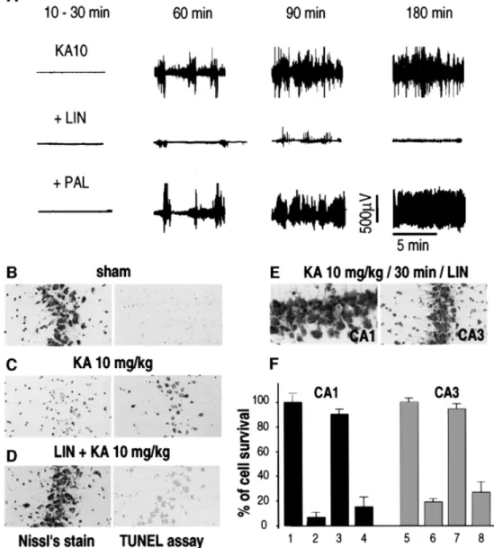
kainate-induced seizures and neuronal death
The glutamate analog KAcauses a well characterized
seizure syndrome resembling human temporal lobe
epi-lepsy and is associated with excitotoxic neurodegeneration
in CA1 and CA3 sub®elds (Nadler et al., 1978; Lothman
and Collins, 1981). Administration of KA [10 mg/kg,
intraperitoneally (i.p.)] into rats induced behavioral stage
4±5 seizures and electroencephalogram (EEG) seizure
activity (Figure 2A). Injection of LIN [10 mM (5 ml), i.c.v.]
before KAtreatment blocked the epileptiform activity
(Figure 2A). In contrast, PAL failed to prevent the
epileptiform activity (Figure 2A). Nissl's staining and
TUNEL labeling con®rmed the neuroprotection produced
by PUFAs against KA-induced excitotoxicity in the
hippocampus (Figure 2B±D). LIN [10 mM (5 ml), i.c.v.]
administered to rats before KAproduced a considerable
inhibition of neuronal loss (Figure 2C and F). In this case,
90 6 5% of CA1 neurons and 95 6 3% of CA3 neurons
survived the KAtreatment (Figure 2F). Palmitic acid did
not prevent the neuronal damage induced by KAin the
hippocampus (Figure 2F). Again, LIN was also found to be
neuroprotective when it was administered i.v. either 30 min
before (100 nmol/kg) (not shown) or 30 min after
(500 nmol/kg) (Figure 2E) the injection of KA.
Fig. 2. Neuroprotection by LIN against kainic acid-induced excitotoxicity. (A) EEG recordings illustrating the effects of LIN and PAL on seizure responses to systemic injection of KA (10 mg/kg) at distinct time points following the injection of KA. LIN and PAL were administered [10 mM (5 ml), i.c.v.] 30 min before KAtreatment (n = 6). (B±D) Photographs of CA3 substructures highlighting the effects of LIN and PAL [10 mM (5 ml), i.c.v.] on neuronal damage following KAtreatment. Rats were collected 7 or 3 days after KAtreatment for Nissl's staining (left panels) or TUNEL labeling (right panels), respectively. (E) Photographs of CA1 (left) and CA3 (right) substructures of Nissl's stained sections after i.v. injection of LIN (500 nmol/kg) 30 min after the KAinjection. (F) Histograms represent the percentage of cell survival assessed in Nissl's stained sections per 1 mm linear length of CA1 and CA3 pyramidal layers 7 days following KA injection. Values are plotted as a percentage (6 SEM) of control neuronal density (saline-injected rats). 1 and 5, sham; 2 and 6, KA10 mg/kg; 3 and 7, LIN [10 mM (5 ml), i.c.v.] + KA10 mg/kg; 4 and 8, PAL [10 mM (5 ml), i.c.v.] + KA10 mg/kg. Data represent the counting of 10 brain sections per rat (n = 6 per group). Statistical analysis was performed as described in Figure 1. I.Lauritzen et al.
PUFAs, but not saturated fatty acids, inhibit
abnormal synaptic transmission and associated
neuronal death in neuronal cultures
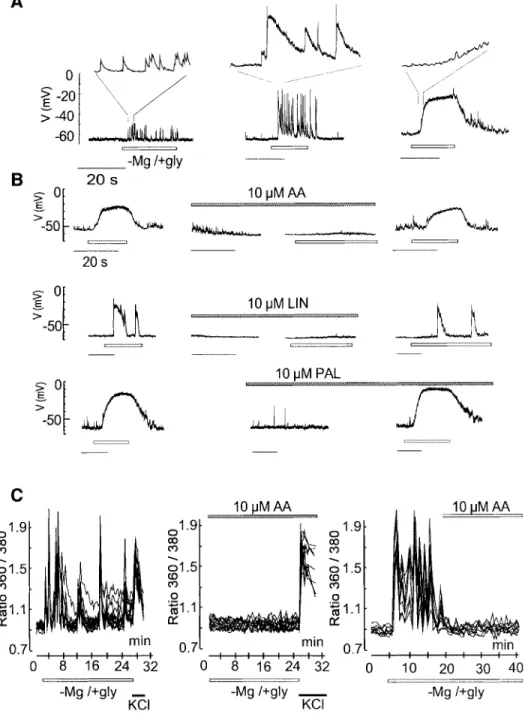
The effects of AA, LIN and other PUFAs were studied in
an in vitro model of excitotoxicity (Abele and Miller,
1990; Abele et al., 1990; Rose et al., 1990) in which mouse
cerebellar granular neurons were exposed to a Mg
2+-depleted glycine-supplemented medium (±Mg/+gly). The
removal of Mg
2+acts by enhancing the spontaneous
release of the neurotransmitter glutamate and by relieving
the Mg
2+block of NMDAreceptors, whereas glycine acts
as a positive allosteric regulator of the NMDAreceptor
(Abele et al., 1990). When granule cells are in culture, the
±Mg/+gly medium induces a large increase in synaptic
activity, observed as EPSPs (excitatory post-synaptic
potentials) (Figure 3Aand B) producing large and
immediate ¯uctuations in intracellular Ca
2+levels
(Figure 3C). These events are due to enhanced glutamate
release at presynaptic terminals. They can be completely
blocked by both tetrodotoxin (TTX, 1 mM), which
eliminates synaptic transmitter release, and by
D(±)-2-amino-5-phosphonopentanoic acid (D-AP5; 20 mM), an
antagonist of the NMDAreceptor at the post-synaptic level
(not shown). Exaggerated [Ca
2+]
i
levels in granule neurons
exposed to ±Mg/+gly for 30±45 min were followed by
neuronal death over the next 6±10 h (Figure 4).
AA (10 mM) completely inhibited both electrical
discharges (Figure 3B), [Ca
2+]
i
¯uctuations (Figure 3C)
and neuronal death (Figure 4, p <0.001). Even when
applied 10 min after the induction of [Ca
2+]
i
¯uctuations
by the ±Mg/+gly medium, AA effectively reduced the
[Ca
2+]
i
¯uctuations (Figure 3C, right panel). Not only AA,
but other PUFAs such as LIN (10 mM) (Figure 3B) and
docosahexaenoic acid (DO) (10 mM) (not shown), also
completely abolished the induction of EPSPs discharges,
[Ca
2+]
i
¯uctuations (not shown) and neuronal death
determined by the lactate dehydrogenase (LDH)-ef¯ux
assay (Figure 4B) or TUNEL labeling (not shown). No
protection was obtained by the methylester of DO or the
saturated fatty acids PAL and stearic acid at similar
concentrations (Figure 4B). The effect of AA is
concen-tration dependent (Figure 4C); full protection was
observed at 10 mM A A .
The neuroprotective effects of PUFAs may be
linked to K
+channel opening
Several lines of evidence suggest that the potent
neuroprotective effects of PUFAs may be linked at least
in part to K
+channel opening. First, the protective effects
of PUFAs are strongly dependent on external K
+concen-trations. Protection was observed only as cerebellar
granular neurons were exposed to physiological [K
+]
e
(5 mM), whereas it was not seen in a medium containing
25 mM external K
+(Figure 4D). Viability of cells exposed
to ±Mg/+gly alone for 45 min and estimated 8 h later was
the same at low and high K
+concentrations. Moreover,
AA was unable to prevent a rise in [Ca
2+]
i
when cells were
exposed to high external K
+following the ±Mg/+gly
exposure (Figure 3C). The calcium rise provoked by high
external K
+was higher and more long-lasting in the
presence of PUFAs. If driving the membrane potential
close to the K
+equilibrium potential (E
K
) by activating
background K
+channels is the mechanism by which
PUFAs exert neuroprotection, then protection can only
occur in low [K
+]
e
, as observed in this work.
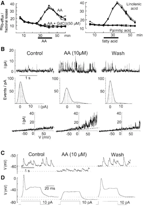
86
Rb
+ef¯ux analysis is an effective way to analyze the
properties of cloned PUFA-activated K
+channels
(Maingret et al., 1999). It has been used here to provide
information about both synaptic and non-synaptic
chan-nels activated by fatty acids in the cerebellar granule cell
cultures. AA, LIN (Figure 5A) and DO (not shown) all
stimulate a large
86Rb
+ef¯ux in these cultures with
properties that are the same as those previously observed
with COS cells transfected with the TRAAK channel, a
2-P domain K
+channel with speci®c expression in neurons
(Maingret et al., 1999). Neither the methyl derivative of
DO, nor saturated fatty acids, were capable of inducing
any
86Rb
+ef¯ux. Gadolinium completely blocked the
86Rb
+ef¯ux (Figure 5A, left panel). The classical blockers
of K
+channels, TEA, 4-AP or charybdotoxin, had no
effects, whereas Ba
2+at high concentrations (up to 2 mM)
partially reduced the
86Rb
+ef¯ux (not shown).
Outside-out patch analysis also independently revealed the
pres-ence of non-inactivating K
+channels, which are reversibly
activated by 10 mM AA. These channels have
electro-physiological properties close to those of the recently
cloned AA- and PUFA-activated TRAAK and TREK-1
(another 2-P domain K
+channel) channels (Fink et al.,
1996, 1998) and to the native AA-activated channels
previously recorded in cultured mesencephalic and
hypothalamic neurons (Kim et al., 1995) (Figure 5B).
Whole-cell recordings showed an inhibitory effect of
PUFAs on neuronal excitability (Figure 5C and D). In
most neurons, addition of AA resulted in a decrease of the
excitability due to a hyperpolarization of the cell
mem-brane by 5±10 mV (Figure 5C and D). Other authors have
similarly described a potent inhibitory effect of PUFAs on
the frequency of action potentials in CA1 and CA3
neurons together with a hyperpolarization of the resting
membrane potential (Xiao and Li, 1999).
The RT±PCR analysis (not shown) and
immunocyto-chemistry, using polyclonal antibodies directed against the
channel proteins (Figure 6A), con®rmed the presence of
both TREK-1 or TRAAK channels in cultured granule
neurons. Both channel proteins are clustered throughout
cell body membranes and ®ber bundles. Both TRAAK
(Figure 6A) and TREK-1 (not shown) are present at both
non-synaptic and synaptic sites identi®ed by the synaptic
marker synapsin I. TREK-1 and TRAAK proteins are
highly expressed in the hippocampus (Figure 6B±D), a
particularly vulnerable structure in global ischemia and
KA-induced seizures. In this region, TRAAK is located to
cell bodies (Figure 6D), whereas TREK-1 is absent from
cell bodies but abundant in neuropile, notably in dendrites
(Figure 6C and D). AA activation of sustained dendritic K
+channels has recently been identi®ed in CA1 pyramidal
neurons (Colbert and Pan, 1999).
Discussion
The present study shows a major neuroprotective effect of
the n-3 PUFALIN in in vivo models of both global
ischemia and KA-induced epilepsy. LIN, which is present
in vegetable oils, is neuroprotective when administered
preventively in one single i.v. dose, 30 min before the
ischemic or epileptic insult. It also provides potent
neuroprotection when administered as long as 30 min after
the neurotoxic insult. Therefore, the potential therapeutic
value of this PUFAand other related compounds in
neurological diseases associated with hyperexcitability,
excessive glutamate release and neuronal death is worth
exploring in the same way as is being done for
ischemic-induced cardiac sudden death (Leaf et al., 1999).
The neuroprotective effects of LIN and other PUFAs
have also been observed in an in vitro model of
excitotoxicity with mouse cerebellar granular neurons in
Fig. 3. PUFAs abolish the ±Mg/+gly-induced excitatory discharges and calcium ¯uctuations in granule cells. Whole-cell recordings were performed from individual cells in distinct culture dishes. (A) Lower traces: typical patterns of excitatory synaptic activities in response to ±Mg/+gly. Upper traces: expanded portions (2 s) from each pattern. Left: increase in the frequency of EPSP. Middle: the synaptic potentials were larger and lasted longer. Right: numerous EPSP of small amplitude led to a sustained depolarization to about ±20 mV. In all cases, TTX (1 mM) completely prevented the EPSPs and the effects of ±Mg/+gly were reversed upon wash-out. (B) Left: synaptic activities elicited in response to ±Mg/+gly. Middle: after a complete recovery upon wash-out, the granule cells were pretreated for 1 min with the different fatty acids (10 mM) prior to the application of ±Mg/+gly in the presence of fatty acids. The unsaturated fatty acids (AA, LIN) but not the saturated fatty acid (PAL) prevent the ±Mg/+gly-induced intense synaptic activities. Right: removal of the PUFAs led to the restoration of the ±Mg/+gly-induced synaptic responses. (C) Internal calcium ¯uctuations measured by Fura-2-AM. Basal synaptic activity and [Ca2+]
ilevels were in each case determined by perfusing the
cells with a standard salt solution at the beginning of the experiment, and all washes were performed with a similar solution. Data represent [Ca2+] i
in eight individual neurons out of a total of ~30 neurons from the same culture dishes. The Ca2+peaks in the eight neurons, chosen for this typical
illustration, coincide, indicating that these neurons are all interconnected. Experiments were repeated at least three times. AA (10 mM) was present during the whole exposure time (middle panel) or added 10 min after the induction of calcium ¯uctuations (right panel). In the left and middle panels, cells were exposed to 25 mM KCl upon 25 min ±Mg/+gly treatment.
I.Lauritzen et al.
culture. PUFAs potently eliminated the build-up of
neurotoxic exaggerated [Ca
2+]
i
levels in granule cells
and the associated neuronal death. Their protective effect
is probably largely due to the fact that PUFAs abolish
EPSP discharges by inhibiting glutamatergic synaptic
transmission; none of the effects observed with PUFAs in
vivo and in vitro are observed with saturated fatty acids.
The most important message in this work is the very
potent neuroprotective effect of PUFAs associated with
PUFA-induced blockade of glutamatergic transmission.
However, it seemed to us that it would also be useful to try
to identify the molecular target(s) involved in the
bene-®cial action of these PUFAs. AA and other PUFAs have
multiple effects on glutamate receptors, glutamate
trans-porters and ion channels. Some of these effects would be
clearly expected to favor hyperexcitability and/or
neuro-toxicity instead of providing neuroprotection. They
include PUFAinhibition of the activity of several
voltage-sensitive K
+channels (Honore et al., 1994;
Meves, 1994; Keros and McBain, 1997), the inhibition
of glutamate transporters (Gegelashvili and Schousboe,
1997) and the activation of NMDAreceptors (Miller et al.,
1992; Nishikawa et al., 1994) [other types of glutamate
receptors seem to be only modestly changed in their
function (Miller et al., 1992; Nishikawa et al., 1994)].
Other previously described effects of PUFAs will certainly
contribute to a decrease of synaptic glutamate
transmis-sion and an increase of neuroprotection. They include the
partial inhibition of voltage-sensitive Na
+channels (Fraser
et al., 1993; Vreugdenhil et al., 1996) and of
voltage-sensitive Ca
2+channels (Vreugdenhil et al., 1996).
Another important class of candidates for the
neuropro-tective action of PUFAs are the recently cloned TREK-1
and TRAAK channels (Fink et al., 1996, 1998), which
Fig. 4. PUFAs, but not saturated fatty acids, protect against ±Mg/+gly-induced excitotoxicity in cultured cerebellar granular neurons. Cultures were treated with ±Mg/+gly during 45 min and cell viability estimated at 8 h post-treatment. (A) Phase-contrast photomicrographs. (B±D) Estimation of viability by the LDH-ef¯ux assay. In (B±D), columns represent the sum of the LDH activities released into the culture media during the 8 h post-treatment. Data are in each case shown as averages 6 SEM obtained from nine dishes in three independent experiments. *p <0.05 and ***p <0.001 as compared with ±Mg/+gly. In (D), cells were exposed to ±Mg/+gly in the presence of AA (10 mM) and then shifted to culture medium containing either 5 mM KCl or 25 mM KCl. Viability in control cells, exposed to 5 mM or 25 mM KCl, was identical.
seem to correspond closely to a class of background
channels identi®ed in the heart (Kim and Clapham, 1989)
and in the brain (Kim et al., 1995) and now in the
cerebellum (this work), and which are potently activated
by AA and other PUFAs but are unaltered in their function
by saturated fatty acids.
Although K
+ef¯ux has been proposed to contribute to
neuronal apoptosis (via the NMDAreceptor) in
non-physiological concentrations of external Na
+and Ca
2+(Yu
et al., 1999), we and others have shown that K
+channel
openers such as those that activate ATP-sensitive
K
+channels (K
ATP
) (Quast, 1993) are potent
neuropro-tectors (Abele and Miller, 1990; Heurteaux et al., 1993;
Lauritzen et al., 1997) and potent inhibitors of
hyperexcit-ability (Abele and Miller, 1990). However, these
com-pounds, such as cromakalim or pinacidil, protect only
when administered before the ischemic insult (Heurteaux
et al., 1993) and they have side-effects, since they also
produce hypotension by activating vascular K
ATPchannels
(Quast, 1992).
Activators of background K
+channels will in general be
more potent than inhibitors of other types of ionic
Fig. 5. AA contributes to synaptic inhibition in granule cells by activating background K+channels. (A) Kinetics of86Rb+ef¯ux evoked by 50 mM
AA (left) or 50 mM LIN (right) in cerebellar granular cultures. Four independent kinetics are shown per experimental condition. Fatty acids were added as indicated by a horizontal bar. (B) Activation of background K+channels by AA. Upper traces: single channel K+currents recorded in an
out patch held at +60 mV. Left, control; middle, after 2 min exposure to 10 mM AA; right, after a 5 min wash. Middle traces: same outside-out patch. Density histograms corresponding to 10 s recordings. Lower traces: I±V curves from another outside-outside-outside-out patch constructed with a voltage ramp of 500 ms duration from ±100 to +100 mV. (C and D) Whole-cell recordings of the effects of AA (10 mM) on neuronal excitability. (C) Left, spontaneous EPSP. Middle, 10 mM AA induced a membrane hyperpolarization and a reduction of both the mean EPSP amplitude and frequency. Right, after a 10 min wash period. (D) Left, control action potential evoked by a current step of 10 pA. Middle, AA caused membrane
hyperpolarization associated with a decreased input resistance and 10 pAwas unable to trigger an action potential. Right, after a 10 min wash. I.Lauritzen et al.
channels. Indeed, an activation of only a small fraction of
these channels, and particularly of TREK-1 and TRAAK
channels, which are not very active in the absence of
PUFAstimulation, might produce substantial changes in
the resting potential as well as in the ®ring properties of
neurons. The dependence on external K
+of the protection
by AA and other PUFAs would be consistent with a
dominant effect of the activation of background K
+channels. However, the ®nal demonstration would require
the use of speci®c blockers of TREK-1 and TRAAK
channels, as has been performed previously for K
ATPchannels using the blocker glibenclamide (Heurteaux et al.,
1993). Speci®c TREK-1 and TRAAK blockers have not
yet been discovered, and unfortunately we cannot use
Gd
3+, which has multiple effects and inhibits many
different types of ion transport systems. Therefore, at the
present time, it is probably safe to conclude that if
PUFA-activated K
+channels have a signi®cant role in
neuropro-tection, the bene®cial effects of PUFAs might well be due
to a combination of actions on TREK-1 and TRAAK-like
channels and on voltage-dependent Na
+and Ca
2+chan-nels. Interestingly, other activators of PUFA-activated 2-P
K
+channels, such as riluzole (Duprat et al., 2000) or
volatile anesthetics (Patel et al., 1999), also inhibit
neuronal excitability and are also neuroprotective
(Mizoule et al., 1985; Ettaiche et al., 1999;
Lang-Lazdunski et al., 1999; Walker et al., 1999).
There is no ef®cacious treatment of irreversible brain
damage following cerebral ischemia and, whatever the
molecular mechanism, the observation that PUFAs such as
LIN that are present in our diets are neuroprotective, even
after the neurotoxic insult has taken place, seems
prom-ising and worth further clinical exploration. Our work
shows that neuroprotection is not restricted to n-3 PUFAs,
such as LIN and DO (a major component of ®sh oil), since
n-6 PUFAs such as AA also have comparable effects on
excitability. However, the bene®cial effects of AA in the
in vivo models were less pronounced. These ®ndings are
likely to be explained by the fact that AA can be
transformed into various other compounds by the
cyclo-oxygenase and lipcyclo-oxygenase pathways and has many toxic
oxidized metabolites (Farooqui and Horrocks, 1991;
Lafon-Cazal et al., 1993). Therefore, further therapeutic
assays with PUFAs should probably be restricted to n-3
PUFAs.
Materials and methods
Cell cultures and toxicity experiments
Primary cultures of cerebellar granule cells were prepared from 5- to 6-day-old Balb-C mice (Charles River, France) as described (Schousboe et al., 1989). Cells were plated at a density of 3 3 106 cells/35 mm
culture dish. For toxicity experiments, neurons were washed once and incubated for 45 min in physiological salt solution of the following composition (in mM): 118.2 NaCl, 4.8 KCl, 1.2 NaH2PO4, 2.5 CaCl2,
10 HEPES and 33 glucose, with or without 3 MgCl2. The Mg2+-free
buffer contained 100 nM glycine. Following treatments, the cells were returned to culture medium containing either 5 or 25 mM KCl as indicated. All drugs were added at the beginning of the toxic exposure and removed at the end.
Cell survival was evaluated 8 h after the treatment using the LDH assay (Koh and Choi, 1987). At this time point, no cell death had occurred in Fig. 6. (A±D) Immunostaining with a-TREK1 and a-TRAAK antibodies on cerebellar granule cultures (A), or brain sections at the level of the hippocampus (B±D). (A) Clustering of TRAAK proteins (left) and synapsin I (middle) in ®ber bundles of granule neurons with some overlap of the two proteins (right). (B) Low-power photomicrographs of the hippocampus. CA1±CA3, Ammon's horn; DG, dentate gyrus; g, granular cells; h, hilus; p, pyramidal neurons; sl, stratum lucidum; slm, stratum lacunosum moleculare; so, stratum oriens; sr, stratum radiatum. (C) Confocal microscopic images of TREK-1 (left) and synapsin I (middle) labeling in stratum lucidum of the CA3. Right, signi®cant although not complete overlap of the two proteins. (D) High-power photomicrograph of the CA1 region following a-TREK-1 or a-TRAAK labeling, respectively. The scale bar corresponds to 10 mm in (A), (C) and (D) and to 50 mm in (B).
control cells, as veri®ed by both LDH ef¯ux and TUNEL labeling, performed as described in Lauritzen et al. (1997). LDH activities, measured as OD340, were expressed as activity present in the total
medium volume/total LDH activity (media + cells), and presented in the ®gures as release of LDH as a percentage of that in control cells. The percentage LDH activities were used for statistical analysis by ANOVA followed by a Fischer PLSD for multiple comparisons (*p <0.05, **p <0.01 and ***p <0.001).
[Ca2+]
imeasurements
Intracellular calcium levels were measured in single neurons using Fura-2 microspectro¯uorimetry, as described (Grynkiewicz et al., 1985; Lauritzen et al., 1997).
86Rb+ef¯ux studies
All the86Rb+ef¯ux studies were conducted at room temperature (RT) on
5- to 6-day-old cultures (grown in Falcon 12 well dishes at 2 3 106cells/
well) as described in Maingret et al. (1999).86Rb+release was expressed
as fractional release using a computer program adapted for the calculations. In each experiment, background ef¯ux was determined by incubating sister cultures in vehicle without drugs.
Electrophysiology
The whole-cell and outside-out con®gurations of the patch±clamp technique were used (Patel et al., 1998). The external solution at pH 7.4 contained (in mM): 140 NaCl, 5 KCl, 1 CaCl2, 2 MgCl2, 10 HEPES±
NaOH. Pipette solutions contained either the external medium (cell attached) or an internal solution at pH 7.3 with (in mM): 140 KCl, 2 MgCl2, 10 HEPES±KOH, 5 EGTA. Cells were continuously superfused
with a microperfusion system during the time course of the experiments (0.1 ml/min) at RT.
Global ischemia and kainate administration
Transient complete forebrain ischemia (Pulsinelli and Brierley, 1979) was produced in male Wistar rats (250 g) as described previously (Heurteaux et al., 1993).
Kainate was administered i.p. at a dose of 10 mg/kg body weight. The EEG was recorded using a twisted stainless steel bipolar electrode implanted under stereotaxic guidance in CA1 ®eld (A3.8, L2, H2.7) and a ground electrode screwed in front of the olfactory bulb in the middle plane. EEG recordings were monitored for four consecutive hours.
LIN and PAL were injected either i.c.v. at a dose of 10 mM (5 ml) 30 min before the induction of ischemia or KAor i.v. 30 min (100 nmol/kg body weight) before or 30 min (100 or 500 nmol/kg body weight) after the neurotoxic insult. Sham-operated animals and rats injected with vehicle were used as controls.
Analysis of ischemia- or kainic acid-induced hippocampal damage
Three or 7 days (TUNEL assay or Nissl's stain, respectively) following ischemia or KAtreatment, the animals were killed and their brains were removed. Frozen coronal sections (12 mm) were taken of the hippocampal area, stained with cresyl violet (Nissl's stain) and assessed for neuronal death. The neuronal density of CA1 and CA3 sectors, i.e. the number of intact pyramidal cells per 1 mm linear length of the CA1/CA3 stratum pyramidale observed in each section, was quanti®ed according to the method of Kirino et al. (1986). The TUNEL assay (Gavrieli et al., 1992) was performed using the terminal deoxynucleotidyl transferase-mediated dUTP-biotin nick end labeling kit from Boehringer Mannheim. TUNEL labeling was visualized with 3¢-diaminobenzidine and nickel chloride. Immunostaining
a-TREK-1 and a-TRAAK antibodies were raised against a glutathione S-transferase fusion protein containing the C-terminus (Pro296±Val398) of TRAAK and the N-terminus (Met1±Trp44) of TREK-1, as described (Lesage et al., 1996). Neuronal cultures were ®xed, blocked with goat serum and incubated with a-TREK-1 (1:600) or a-TRAAK (1:600) and mouse monoclonal antibody to synapsin I (1:1000; Bioproducts) for 2 h at RT. Appropriate secondary antibodies (goat anti-rabbit tetramethyl-rhodamine isothiocyanate and donkey anti-mouse±¯uorescein isothio-cyanate; Jackson Laboratories) were used to visualize the proteins. For immunohistochemistry on mice brain sections, adult BalbC mice were killed after transcardial perfusion with phosphate-buffered 4% para-formaldehyde. Floating brain sections (50 mm) were cut on a vibratome (Leica). Immunostaining with a-TREK-1 and a-TRAAK antibodies was performed using the Vector Elite ABC kit. Sections were incubated overnight at 4°C with TREK-1 or TRAAK antibodies, respectively (1:800
dilution). The antigen±antibody complexes were visualized by reaction with 3¢-diaminobenzidine and H2O2.
Acknowledgements
We are grateful to Drs F.Lesage and R.Reyes for the preparation of antibodies and to Drs W.Ferlin and A.Patel for careful reading of the manuscript. Thanks are due to F.Aguila, D.Doume and V.Lopez for technical assistance. This work was supported by the Centre National de la Recherche Scienti®que (CNRS), the Association pour la Recherche sur le Cancer (ARC), the Conseil Regional (PACA) and the Association FrancËaise contre les Myopathies (AFM).
References
Abele,A.E. and Miller,R.J. (1990) Potassium channel activators abolish excitotoxicity in cultured hippocampal pyramidal neurons. Neurosci. Lett., 115, 195±200.
Abele,A.E., Scholz,K.P., Scholz,W.K. and Miller,R.J. (1990) Excitotoxicity induced by enhanced excitatory neurotransmission in cultured hippocampal pyramidal neurons. Neuron, 4, 413±419. Albert,C.M., Hennekens,C.H., O'Donnell,C.J., Ajani,U.A., Carey,V.J.,
Willett,W.C., Ruskin,J.N. and Manson,J.E. (1998) Fish consumption and risk of sudden cardiac death. J. Am. Med. Assoc., 279, 23±28. Barnard,E.A. (1997) Ionotropic glutamate receptors: new types and new
concepts. Trends Pharmacol. Sci., 18, 141±148.
Choi,D.W. (1994) Glutamate receptors and the induction of excitotoxic neuronal death. Prog. Brain Res., 100, 47±51.
Colbert,C.M. and Pan,E. (1999) Arachidonic acid reciprocally alters the availability of transient and sustained dendritic K+ channels in
hippocampal CA1 pyramidal neurons. J. Neurosci., 19, 8163±8171. Duprat,F., Lesage,F., Patel,A.J., Fink,M., Romey,G. and Lazdunski,M.
(2000) The neuroprotective agent riluzole activates the two P-domain K+channels TREK-1 and TRAAK. Mol. Pharmacol., in press.
Ettaiche,M., Fillacier,K., Widmann,C., Heurteaux,C. and Lazdunski,M. (1999) Riluzole improves functional recovery following ischemia in the rat retina. Invest. Ophthalmol. Vis. Sci., 40, 729±736.
Farooqui,A.A. and Horrocks,L.A. (1991) Excitatory amino acid receptors, neural membrane phospholipid metabolism and neurological disorders. Brain Res. Rev., 16, 171±191.
Fink,M., Duprat,F., Lesage,F., Reyes,R., Romey,G., Heurteaux,C. and Lazdunski,M. (1996) Cloning, functional expression and brain localization of a novel unconventional outward recti®er K+channel.
EMBO J., 15, 6854±6862.
Fink,M., Lesage,F., Duprat,F., Heurteaux,C., Reyes,R., Fosset,M. and Lazdunski,M. (1998) Aneuronal two P domain K+channel stimulated
by arachidonic acid and polyunsaturated fatty acid. EMBO J., 17, 3297±3308.
Fraser,D.D., Hoehn,K., Weiss,S. and MacViar,B.A. (1993) Arachidonic acid inhibits sodium currents and synaptic transmission in cultured striatal neurons. Neuron, 11, 633±644.
Gavrieli,Y., Sherman,Y. and Ben-Sason,S.A. (1992) Identi®cation of programmed cell death in situ via speci®c labeling of nuclear DNA fragmentation. J. Cell Biol., 119, 493±501.
Gegelashvili,G. and Schousboe,A. (1997) High af®nity glutamate transporters: regulation of expression and activity. Mol. Pharmacol., 52, 6±15.
Grynkiewicz,G., Poenie,M. and Tsien,R.Y. (1985) Anew generation of Ca2+indicators with greatly improved ¯uorescence properties. J. Biol.
Chem., 260, 3440±3450.
Heurteaux,C., Bertaina,V., Widmann,C. and Lazdunski,M. (1993) K+channel openers prevent global ischemia-induced expression of
c-fos, c-jun, heat shock protein and amyloid b-protein precursor genes and neuronal death in rat hippocampus. Proc. Natl Acad. Sci. USA, 90, 9431±9435.
Hibbeln,J.R. (1998) Fish consumption and major depression. Lancet, 351, 1210±1213.
HonoreÂ,E., Barhanin,J., Attali,B., Lesage,F. and Lazdunski,M. (1994) External blockade of the major cardiac delayed-recti®er K+channel
(Kv1.5) by polyunsaturated fatty acids. Proc. Natl Acad. Sci. USA, 91, 1937±1944.
Keros,S. and McBain,C.J. (1997) Arachidonic acid inhibits transient potassium currents and broadens action potentials during electrographic seizures in hippocampal pyramidal and inhibitory interneurons. J. Neurosci., 17, 3476±3487.
I.Lauritzen et al.
Kim,D. and Clapham,D.E. (1989) Potassium channels in cardiac cells activated by arachidonic acid and phospholipids. Science, 244, 1174± 1179.
Kim,D., Sladek,C.D., Aguado-Velasco,C. and Mathiasen,J.R. (1995) Arachidonic acid activation of a new family of K+channels in cultured
rat neuronal cells. J. Physiol. (Lond.), 484, 643±660.
Kirino,T., Tamura,A. and Sano,K. (1986) A reversible type of neuronal injury following ischemia in the gerbil hippocampus. Stroke, 17, 455± 459.
Koh,J.Y. and Choi,D.W. (1987) Quantitative determination of glutamate mediated cortical neuronal injury in cell culture by lactate dehydrogenase ef¯ux assay. J. Neurosci. Methods, 20, 83±90. Lafon-Cazal,M., Pietri,S., Culcasi,M. and Bockaert,J. (1993)
NMDA-dependent superoxide production and neurotoxicity. Nature, 364, 535±537.
Lang-Lazdunski,L., Vaillant,N., Widmann,C., Heurteaux,C. and Lazdunski,M. (1999) Riluzole improves neurological function following severe spinal cord ischemia. J. Thorac. Cardiovasc. Surg., 117, 881±889.
Lauritzen,I., De Weille,J.R. and Lazdunski,M. (1997) The potassium channel opener (±)-cromakalim prevents glutamate-induced cell death in hippocampal neurons. J. Neurochem., 69, 1570±1579.
Leaf,A. and Kang,J.X. (1996) Prevention of cardiac sudden death by n-3 fatty acids: a review of the evidence. J. Intern. Med., 240, 5±12. Leaf,A., Kang,J.X., Xiao,Y.-F., Billman,G.E. and Voskuyl,R.A. (1999)
The antiarrhythmic and anticonvulsant effects of dietary n-3 fatty acids. J. Membr. Biol., 172, 1±11.
Lesage,F. and Lazdunski,M. (1999) Potassium channels with two P-domains. In Kurachi,Y., Jan,L.Y. and Lazdunski,M. (eds), Potassium Ion Channels. Molecular Structure, Function and DiseasesÐCurrent Topics in Membranes. Academic Press, San Diego, CA, pp. 199±222.
Lesage,F., Guillemare,E., Fink,M., Duprat,F., Lazdunski,M., Romey,G. and Barhanin,J. (1996) TWIK-1, a ubiquitous human weakly inward rectifying K+channel with a novel structure. EMBO J., 15, 1004±
1011.
Lothman,E.W. and Collins,R.C. (1981) Kainic acid induced limbic seizures: metabolic, behavioral, electroencephalographic and neuropathological correlates. Brain Res., 218, 299±318.
Maingret,F., Fosset,M., Lesage,F., Lazdunski,M. and HonoreÂ,E. (1999) TRAAK is a mammalian neuronal mechano-gated K+channel. J. Biol.
Chem., 274, 1381±1387.
Meves,H. (1994) Modulation of ion channels by arachidonic acid. Prog. Neurobiol., 43, 175±186.
Miller,B., Sarantis,M., Traynelis,S.F. and Attwell,D. (1992) Potentiation of NMDAreceptor currents by arachidonic acid. Nature, 355, 722± 725.
Mizoule,J., Meldrum,B., Mazadier,M., Croucher,M., Ollat,C., Uzan,A., Legrand,J.J., Gueremy,C. and Fur,G.L. (1985) 2-amino-6-tri¯uoromethoxy benzothiazole, a possible antagonist of excitatory amino acid neurotransmission. I. Anticonvulsant properties. Neuropharmacology, 24, 767±773.
Nadler,J.V., Perry,B.W. and Cotman,C.W. (1978) Intraventricular kainic acid preferentially destroys hippocampal pyramidal cells. Nature, 271, 676±677.
Nair,S.S., Leitch,J.W., Falconer,J. and Garg,M.L. (1997) Prevention of cardiac arrhythmia by dietary (n-3) polyunsaturated fatty acids and their mechanism of action. J. Nutr., 127, 383±393.
Nishikawa,M., Kimura,S. and Akaike,N. (1994) Facilitatory effect of docosahexaenoic acid on N-methyl-D-aspartate response in pyramidal neurones of rat cerebral cortex. J. Physiol., 475, 83±93.
Nordoy,A. (1999) Dietary fatty acids and coronary heart disease. Lipids, 34, S19±S22.
Patel,A.J., HonoreÂ,E., Maingret,F., Lesage,F., Fink,M., Duprat,F. and Lazdunski,M. (1998) Amammalian two pore domain mechano-gated S-type K+channel. EMBO J., 17, 4283±4290.
Patel,A.J., HonoreÂ,E., Lesage,F., Fink,M., Romey,G. and Lazdunski,M. (1999) Inhalational anesthetics activate two-pore-domain background K+channels. Nature Neurosci., 2, 422±426.
Pulsinelli,W.A. and Brierley,J.B. (1979) A new model of bilateral hemispheric ischemia in the unanesthetized rat. Stroke, 10, 267±272. Quast,U. (1992) Potassium channel openers: pharmacological and
clinical aspects. Fundam. Clin. Pharmacol., 6, 279±293.
Quast,U. (1993) Do the K+channel openers relax smooth muscle by
opening K+channels? Trends Pharmacol. Sci., 14, 332±337.
Rose,K., Christine,C.W. and Choi,D.W. (1990) Magnesium removal induces paroxysmal neuronal ®ring and NMDAreceptor-mediated
neuronal degeneration in cortical cultures. Neurosci. Lett., 115, 313± 317.
Schmidt-Kastner,R. and Freund,T.F. (1991) Selective vulnerability of the hippocampus in brain ischemia. Neuroscience, 40, 599±636. Schousboe,A., Meier,E., Drejer,J. and Hertz,L. (1989) Preparation of
primary cultures of mouse (rat) cerebellar granule neurons. In Shahar,A., de Vellis,J., Vernadakis,A. and Haber,B. (eds), A Dissection and Tissue Culture Manual of the Nervous System. Alan R.Liss, New York, NY, pp. 203±206.
SiesjoÈ,B.K. and Wieloch,T. (1985) Brain injury: neurochemical aspects. In Becker,D.P. and Povlishock,J.T. (eds), System Trauma. William Byrd Press, Richmond, VA, pp. 513±532.
Stoll,A.L., Severus,E., Freeman,M.P., Rueter,S., Zboyan,H.A., Diamond,E., Gress,K.K. and Marangell,L.B. (1999) Omega 3 fatty acids in bipolar disorder: a preliminary double-blind, placebo-controlled trial. Arch. Gen. Psychiatry, 56, 413±416.
Vreugdenhil,M., Bruehl,C., Voskuyl,R.A., Kang,J.X., Leaf,A. and Wadman,W.J. (1996) Polyunsaturated fatty acids modulate sodium and calcium currents in CA1 neurons. Proc. Natl Acad. Sci. USA, 93, 12559±12563.
Walker,M.C., Perry,H., Scaravilli,F., Patsalos,P.N., Shorvon,S.D. and Jefferys,J.G. (1999) Halothane as a neuroprotectant during constant stimulation of the perforant path. Epilepsia, 40, 359±364.
Xiao,Y.-F. and Li,X. (1999) Polyunsaturated fatty acids modify mouse hippocampal neuronal excitability during excitotoxic or convulsant stimulation. Brain Res., 846, 112±121.
Yu,S.P., Yeh,C.-H., Strasser,U., Tian,M. and Choi,D.W. (1999) NMDA receptor-mediated K+ ef¯ux and neuronal apoptosis. Science, 284,
336±339.
Received January 24, 2000; revised February 22, 2000; accepted March 3, 2000