HAL Id: hal-01861372
https://hal.archives-ouvertes.fr/hal-01861372
Submitted on 24 Aug 2018
HAL is a multi-disciplinary open access
archive for the deposit and dissemination of
sci-entific research documents, whether they are
pub-lished or not. The documents may come from
teaching and research institutions in France or
abroad, or from public or private research centers.
L’archive ouverte pluridisciplinaire HAL, est
destinée au dépôt et à la diffusion de documents
scientifiques de niveau recherche, publiés ou non,
émanant des établissements d’enseignement et de
recherche français ou étrangers, des laboratoires
publics ou privés.
amidotransferase MurT/GatD complex from
Streptococcus pneumoniae
Cécile Morlot, Daniel Straume, Katharina Peters, Olav Hegnar, Nolwenn
Simon, Anne-Marie Villard, Carlos Contreras-Martel, Francisco Leisico,
Eefjan Breukink, Christine Gravier-Pelletier, et al.
To cite this version:
Cécile Morlot, Daniel Straume, Katharina Peters, Olav Hegnar, Nolwenn Simon, et al.. Structure of
the essential peptidoglycan amidotransferase MurT/GatD complex from Streptococcus pneumoniae.
Nature Communications, Nature Publishing Group, 2018, 9, pp.3180. �10.1038/s41467-018-05602-w�.
�hal-01861372�
Structure of the essential peptidoglycan
amidotransferase MurT/GatD complex
from
Streptococcus pneumoniae
Cécile Morlot
1
, Daniel Straume
2
, Katharina Peters
3
, Olav A. Hegnar
2
, Nolwenn Simon
1
, Anne-Marie Villard
1
,
Carlos Contreras-Martel
1
, Francisco Leisico
4
, Eefjan Breukink
5
, Christine Gravier-Pelletier
6
,
Laurent Le Corre
6
, Waldemar Vollmer
3
, Nicolas Pietrancosta
6
, Leiv Sigve Håvarstein
2
& André Zapun
1
The universality of peptidoglycan in bacteria underlies the broad spectrum of many
suc-cessful antibiotics. However, in our times of widespread resistance, the diversity of
pepti-doglycan modi
fications offers a variety of new antibacterials targets. In some Gram-positive
species such as
Streptococcus pneumoniae, Staphylococcus aureus, or Mycobacterium
tubercu-losis, the second residue of the peptidoglycan precursor, D-glutamate, is amidated into
iso-D-glutamine by the essential amidotransferase MurT/GatD complex. Here, we present the
structure of this complex at 3.0 Å resolution. MurT has central and C-terminal domains
similar to Mur ligases with a cysteine-rich insertion, which probably binds zinc, contributing
to the interface with GatD. The mechanism of amidation by MurT is likely similar to the
condensation catalyzed by Mur ligases. GatD is a glutaminase providing ammonia that is
likely channeled to the MurT active site through a cavity network. The structure and assay
presented here constitute a knowledge base for future drug development studies.
DOI: 10.1038/s41467-018-05602-w
OPEN
1Université Grenoble Alpes, CNRS, CEA, IBS UMR 5075, 38044 Grenoble, France.2Faculty of Chemistry, Biotechnology and Food Science, Norwegian
University of Life Sciences, Ås 1432, Norway.3Centre for Bacterial Cell Biology, Institute for Cell and Molecular Bioscience, Newcastle University, Newcastle Upon Tyne NE2 4AX, United Kingdom.4Departamento de Química, Universidade Nova de Lisboa, Caparica 2829-516, Portugal.5Membrane Biochemistry
and Biophysics, Department of Chemistry, Faculty of Science, Utrecht University, Utrecht 3584, The Netherlands.6Université Paris Descartes, Laboratoire de
Chimie et Biochimie Pharmacologiques et Toxicologiques UMR 8601 CNRS, Sorbonne Paris Cité (USPC), Paris 75006, France. Correspondence and requests for materials should be addressed to A.Z. (email:andre.zapun@ibs.fr)
123456789
P
eptidoglycan is a defining trait of bacteria and its
metabo-lism is targeted by major antibiotics. A mesh-polymer that
surrounds the cell, defining its shape and providing
resis-tance to osmotic pressure, peptidoglycan is extraordinarily
con-served, consisting of chains of alternating N-acetylglucosamine
(GlcNAc) and N-acetylmuramic acid (MurNAc) that are
cross-linked by peptide bridges attached to the MurNAc
1.
On the other hand, peptidoglycan is also very diverse.
Pepti-doglycan is thin and mono-layered, or thick and multi-layered, in
Gram-negative or Gram-positive bacteria. Chain length ranges
from a few units (Staphylococcus aureus) to several hundred
(Bacilli), with reducing ends or terminating with
1,6-anhy-droMurNAc. Glycan chains are modified by various degrees of
N-deacetylation, and O-acetylation and N-glycolylation
1.
The peptide component shows even greater variety arising
from the diverse residues assembled into the precursor
penta-peptide by the Mur ligases C, D, E, and F
1. The
first residue is
generally an L-Ala, but can be Gly or L-Ser. The second amino
acid is D-Glu. The third residue attached to the C5 of the D-Glu
can be L-Lys, as in Streptococcus pneumoniae, or a
meso-diami-nopimelate, as in many Gram-negative species, or a variety of
other residues. The fourth and
fifth residues, added as dipeptide,
is generally Ala, but can be Ser or
D-Ala-D-lactate.
Peptide cross-linking and maturation at the cell surface can
also differ. The D,D-transpeptidase activity of the
penicillin-binding proteins (PBPs) forms the most common 3–4 linkage
with or without intervening branches. PBPs are also responsible
in some Corynebacteria for the 2–4 linkage with an intermediate
diamino acid. L,D-transpeptidases can form 3–3 linkages in many
species, and account for most of the cross-linking in
Myco-bacteria, Clostridia, and in some
β-lactam-resistant bacteria. The
fifth and fourth residues can be trimmed by carboxypeptidases.
The degree of cross-linking and trimming is variable
1.
The modification of the precursor peptide in the cytoplasm
provides additional diversity. Amino acids can be added on the
second or third residue to form branches, such as the
pentagly-cine in S. aureus, and the L-Ala-L-Ala or L-Ser-L-Ala in
S. pneumoniae. These branches are generally part of the
cross-links. The meso-diaminopimelate is amidated in some species
such as Bacillus subtilis. At the second position, the D-glutamate
can be amidated into D-iso-glutamine in many Gram-positive
organisms by the MurT/GatD amidotransferase complex that is
the subject of our study.
MurT/GatD amidates the C1 carboxylate of the D-Glu to form
a D-iso-Gln at the second position of the membrane-bound
peptidoglycan precursor lipid II (Fig.
1
). This activity was long
known in S. aureus extract
2, but the responsible operon
(murT-gatD) was identified only recently
3,4. MurT is homologous to the
Mur ligases that elongate the peptide of the UDP-linked soluble
precursors
5–8. GatD belongs to the class I glutaminase with a
characteristic catalytic Cys-His-Asp/Glu triad, and is homologous
to the glutaminase domain of a variety of amidotransferases such
as cobyric acid synthase, CTP synthetase, or imidazole glycerol
phosphate (IGP) synthase
9. GatD is thought to hydrolyze
L-glutamine to generate ammonia, which is used by MurT to
amidate lipid II. By analogy with reactions catalyzed by Mur
ligases, the acceptor carboxyl of the D-Glu is thought to be
activated by phosphorylation
10.
In vitro, MurT/GatD from S. aureus was shown to amidate
both lipid II and its immediate precursor lipid I, which lacks
GlcNAc, but none of the soluble UDP-linked precursors
4. L-Gln
is the nitrogen donor in neutral and basic conditions (pH 7.5 and
8.5). Ammonia could also serve as direct nitrogen source at pH
8.5, which is consistent with the mechanism of glutamine
ami-dotransferases, where ammonia is transferred to the synthase
subunit
9. When ammonia was provided directly, amidation still
required presence of GatD
4. Amidated lipid II was shown to be
the preferred substrate for transpeptidation by PBPs from S.
pneumoniae
11.
In the quest for novel antibacterial target, MurT/GatD is
par-ticularly important due to its presence in mycobacteria,
staphy-lococci, enterococci, and streptococci. The essentiality of
murTgatD has been demonstrated in important pathogens such
as Mycobacterium tuberculosis
12, S. pneumoniae
11,13,14, and S.
aureus
4,15,16. MurT/GatD depletion in S. aureus caused increased
susceptibility to
β-lactams and lysozyme, including in
methicillin-resistant strains
3,17.
β-Lactams antibiotics have been so successful due to their large
spectrum as they inhibit the D,D-transpeptidases that cross-link
peptidoglycan, a nearly universal feature. However, drugs with
narrower spectra are highly desirable. The diversity of the
pep-tidoglycan offers such opportunities. In this context, we present
the structure of a MurT/GatD complex, from the human
pathogen S. pneumoniae. The structure show that MurT is similar
to Mur ligases with a cysteine-rich insertion contributing to the
interface with GatD. Amidation probably proceeds like the
con-densation catalyzed by Mur ligases. GatD is a glutaminase
pro-viding ammonia that is likely channeled to the acceptor site
through a cavity network. This work, which reveals atomic details
of the active sites and interface, provides essential knowledge for
future efforts to design inhibitory molecules.
OH O O HN O O OH O O HN OH OH O P O P O OH HO O O NH O O HN O OH O N H HN O H2N NH O OH O 7 2 NH O HN O O O N H HN O NH O OH O P O OH OH NH O HN O O N H HN O NH O OH O ATP ADP MurT
Lipid II(Glu) Lipid II(iGln)
Pi MurT L-Gln L-Glu NH3 GatD H2O H2N H2N NH2
Fig. 1 Proposed reaction scheme of the lipid II amidation by MurT/GatD. The second residue of the pentapeptide is highlighted in pink Glu) or blue (D-iso-Gln). The putative phosphorylated intermediate is also shown
Results
Distribution of the murTgatD operon. We searched available
sequences to evaluate the distribution of murTgatD among
bac-teria. The operon is present in many Gram-positive bacteria of
the Terrabacteria superphylum, it is universally present in the
Actinobacteria and Chloroflexi, including in genera
Cor-ynabacterium, Mycobacterium, and Streptomyces. Among
Firmi-cutes, murTgatD is present in the whole Lactobacillales order that
includes genera: Streptococcus, Enterococcus, Lactococcus,
Lacto-bacillus; it is widespread in the Clostridiales, including in the
Clostridia. In Bacillales, murTgatD is ubiquitous in
Staphylo-coccaceae, but is nearly absent from Bacillaceae or Listeriaceae.
The operon is also ubiquitous in some classes of Cyanobacteria. A
few strains from Gram-negative species also contain the operon
suggesting gene transfer events.
Depletion of MurT/GatD. To investigate the function of
murTgatD in S. pneumoniae R6, an ectopic copy under the
control of the inducible promoter PcomX
was introduced and the
endogenous copy was deleted. Growth of the resulting strain
required the presence of the ComS inducer, confirming the
essentiality of MurT/GatD. Depletion of MurT/GatD with
reduced concentrations of inducer caused aberrant morphologies
(Supplementary Fig. 1). Mild depletion (2.5 nM ComS) resulted
in some elongated cells with aborted septa, whereas severe
depletion (1.5 nM ComS) produced a high proportion of aberrant
cells, elongated or bulging, showing that proper cell wall assembly
requires a functioning murTgatD operon, as observed previously
with strain D3913.
The effect of MurT/GatD depletion on peptidoglycan
compo-sition was examined after the growth was stunted by the lack of
inducer, which occurred at OD550
≈ 0.4 (Supplementary Table 1).
As expected, the proportion of (Glu)-muropeptides increased
from 18 to 42% upon depletion of MurT/GatD, indicating that a
minimal amount of around 58% amidated muropeptide is
required for growth. The proportion of cross-linked
muropep-tides decreased from 47 to 36%, which was also expected as
efficient cross-linking depends on the presence of iso-Gln
11.
Surprisingly, there was an increase in the proportion of branched
muropeptides from 16 to 57%. As branching depends on the
MurM enzyme, a strain was constructed by deleting murM in the
MurT/GatD depletion strain. In this strain without branched
muropeptide, depletion of MurT/GatD caused a similar increase
of (Glu)-muropeptides and decrease of cross-linking.
In vitro enzymatic activity. A continuous assay was set up for the
future evaluation of potential inhibitors. MurT/GatD utilizes
three reactants: L-Gln, ATP, and (Glu)-lipid II; and generates
four products: L-Glu, ADP, phosphate, and (iGln)-lipid II
(Fig.
1
). An ATPase assay was chosen, monitoring the generation
of ADP with coupled reactions that consumes NADH, which can
be quantified by its absorbance
18. Comparable kinetics were
observed when aliquots withdrawn after various time intervals
were analyzed by thin layer chromatography (Supplementary
Fig. 2). The identity of the two lipid II species was checked by
mass spectrometry (Supplementary Fig. 2). Although all enzymes
and reactants are soluble in water, including lipid II
(Supple-mentary Fig. 2), no activity was detected without detergent. It is
presumed that lipid II forms micelles (Supplementary Fig. 2), and
that tight packing of the muropeptide heads hinders access to the
enzyme. With 25
μM lipid II, the enzymatic activity was optimal
with n-dodecyl-β-D-maltoside (DDM) at concentrations between
5 and 30 mM. A DDM concentration of 20 mM (150-fold the
critical micelle concentration) was chosen to ensure a micelle
concentration superior to that of lipid II.
Apparent KM
was obtained for the three substrates, and kcat
was measured with lipid II (Table
1
, Fig.
2
). The apparent KM
for
lipid II is in the range of values reported for Mur ligases and their
UDP-linked substrates (e.g., 5 and 90
μM for MurD and C
6,19).
Turnover is similar to values measured with Mur ligases (e.g., 1 to
15 s
−1for MurD
20,21). Due to the limited availability of lipid II,
we used a concentration below its apparent KM
to measure the
dependence of kinetics on the concentrations of L-Gln or ATP.
Assuming that MurT has a mechanism similar to other Mur
ligases with an ordered binding of the substrates prior to products
release
22,23, these measurements allow the estimation of the
apparent KM
for ATP and L-Gln. An additional assumption is
that GatD is not limiting. The KM
for ATP is in the lower range of
values for Mur ligases (e.g., 17 and 100
μM for MurE and C
6,24).
The KM
for L-Gln is one order of magnitude lower than reported
for other amidotransferases such as the IGP synthase
25.
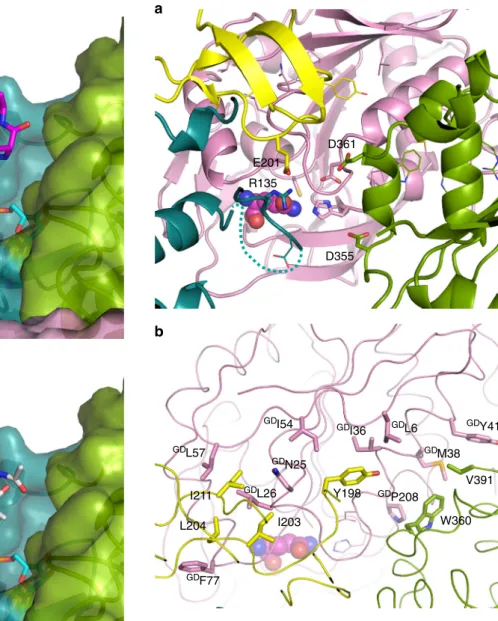
Overall structures of MurT and GatD. Despite the limited
resolution (3.0 Å, statistics in Supplementary Table 2), the
elec-tron density map allowed us to trace the main chains of all four
complexes in the asymmetric unit, revealing a triangular complex
with a large interface area adjacent to the enzymatic cavities of
MurT and GatD (Fig.
3
a). The N-terminal region (1–42) of MurT
was not visible although its presence was confirmed by mass
spectrometry, indicating that this short segment is mobile. The
central (43–302) and C-terminal (303–439) domains are
char-acteristic of the Mur ligases
26(Fig.
3
b, c and Supplementary
Fig. 3). The enzymatic cavity is nestled between the two domains,
with a large solvent-accessible area and one side contacting GatD
(Fig.
3
a).
The central domain of MurT superimposes to those of Mur
ligases with r.m.s.ds (root-mean-square deviations) of 2.9–3.1 Å
over 116–119 residues (shown with Mur C in Supplementary
Fig. 3). It consists of a six-stranded parallel
β-sheet (β2β3β1β4β5β6)
sandwiched between
α-helices α2,
α3,
α4
on one side and a
three-helix bundle (α1α5α6) on the other side,
flanked by a
three-stranded antiparallel
β-sheet (β11β13β12) (Fig.
3
b). In Mur ligases,
α-helices α2,
α3,
α4
(α5, 3/10-helix
η3
and
α7in MurC) are
flanked
by a fourth helix (α8, Supplementary Fig. 3) that is not formed in
MurT, as the corresponding segment (169–177) contains two
prolines. The central domain forms a mononucleotide-binding
domain including a typical P-loop (Walker A motif GxxGK)
27.
Residues 137–141 are not resolved in crystal structure. This loop
may form a mobile bridge extending towards the C-terminal
domain over the GatD active site. The most striking difference
between the central domain of MurT and those of Mur ligases is
the insertion of a cysteine-rich region (187–236) between β6
and
β11
(Fig.
3
b). This region will be further presented below.
The C-terminal domain of MurT consists of a six-stranded
β-sheet (β14β15β19β16β17β18) surrounded by four helices on one side
(α7,
η7,
η8, and
α8) and two on the other side (α9
and
α10)
Table 1 MurT/GatD enzymatic parameters
Initial velocities method Progress curves globalfit ApparentKM (μM) kcat (s−1)a ApparentKD (μM) kcat (s−1) Lipid II 220 ± 60 4 ± 1 193 ± 3 5.0 ± 0.5 ATP 16 ± 2 L-Gln 78 ± 8
ATPase activity was determined by measuring the generation of ADP with a coupled enzymatic assay at 30 °C and pH 7.5 with 1% DDM (w/v). Standard error is given
ak
(Fig.
3
c). Helices
α8
and
η7
interact with GatD. This domain
forms a Rossmann fold
28that superimposes onto those of Mur
ligases with r.m.s.ds of 2.9–3.4 Å over 129–159 residues (shown
with MurC in Supplementary Fig. 3).
The structure of GatD from pneumococcus is similar to that
from S. aureus (PDB# 5N9M; r.m.s.d. of 1.7 Å over 213 residues)
and to other class I glutaminases, such as the HisH subunit of IGP
synthase from Thermotoga maritima (PDB# 1GPW; r.m.s.d. of
2.9 Å over 190 residues) (Supplementary Fig. 4). It consists of a
seven-stranded
β-sheet (β1β3β2β4β5β15β14) surrounded by three
α-helices on one side (α2,
α3,
α4) and four on the other side (α1,
α5,
α6,
α7), and
flanked by two four-stranded β-sheets (β7β6β8β11
and
β10β9β12β13) (Fig.
4
a). A notable difference between
pneumococcal and staphylococcal proteins is the presence of a
long C-terminal
α7
in S. aureus, whereas this region mainly forms
a contorted loop in the S. pneumoniae complex (Supplementary
Fig. 4). The GatD active site faces MurT with solvent and
substrate access in a cleft between both subunits (Fig.
3
a).
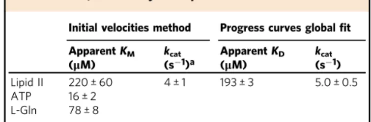
The MurT cysteine-rich insertion. The central domain of MurT
compared to that of Mur ligases, comprises a 50 residue-long
cysteine-rich insertion (187–236) (Fig.
3
b), with
five cysteines in
S. pneumoniae, in positions 205, 208, 227, 230, and 232, of which
the
first four are largely conserved. This cysteine tetrad is present
in Firmicutes with the exception of the Oenococcus, Weissella, and
Leuconostoc genera, where they are replaced by pairs of serine and
aspartate. The tetrad is also present in Cyanobacteria, Chloroflexi,
and some Actinobacteria.
The electron density map showed a great proximity of the
205, 208, 227, and 230 cysteine side chains. The best models
include two disulfide bonds, most likely between pairs 205–230
and 208–227 (Supplementary Fig. 5), although alternative
pairings cannot be ruled out. We found no evidence of a
coordinated metal ion.
The presence of these disulfide bonds is surprising, as MurT/
GatD functions in the cytoplasm where cysteines are reduced
29.
We suspected that these bonds had formed during crystallization,
following the gradual depletion of the reducing agent. To
determine if disulfide bonds were present immediately after the
purification in reducing conditions, accessible thiols in MurT/
GatD or in a mutant (
MT4C4S) with the four cysteines of the
disulfide bonds replaced by serines were alkylated by reaction
with iodoacetamide. The number of alkylations was determined
by mass spectrometry (Supplementary Fig. 6). The major species
exhibited six alkylations for wild-type MurT/GatD and two for
the
MT4C4S variant, implying that the four thiols of the cysteine
tetrad were reduced and accessible after purification. As the thiols
of
MTCys120 and
MTCys325 are buried, the two other accessible
thiols likely belong to
MTCys232 and
MTCys417. Note that the
active site thiol of GatD was also alkylated.
When the PDB databank was searched for domains similar to the
inserted region, the best match was the N-terminal zinc-ribbon
domain of subunit RBN9 of the yeast RNA polymerase II (PDB#
3QT1 chain I
30, Supplementary Fig. 5). Zinc-ribbons consist of a
three-stranded antiparallel
β-sheet supporting two “knuckles”, each
carrying two cysteines that coordinate a Zn
2+cation
31. The likeness
(main chain r.m.s.d. 1.22 Å over 29 residues) between the RNA
polymerase zinc-ribbon and the cysteine-rich insertion suggests that
the latter also binds zinc in vivo, raising the question of why zinc
was not found in our samples, despite the absence of chelators such
as EDTA during purification. It is possible that zinc has been
stripped by the high concentration of DTT as it chelates Zn
2+(KD
≈ 100 nM
32).
That disulfide bonds are not required for catalysis was
confirmed by the residual activity exhibited in vitro by the
MT
4C4S mutant (Table
2
). However, the association with GatD
was impacted by the quadruple mutation, as the ratio of the
Coomassie-stained MurT/GatD bands after SDS-PAGE was 1.40,
instead of 1.78 for the wild type (Supplementary Fig. 7). Also, the
MT
4C4S variant was degraded during storage at 4 °C. In vivo, the
quadruple mutation was not tolerated (Table
2
), although another
mutant with lower in vitro activity was viable (
GDY30A). It is
therefore likely that the protein is destabilized and its abundance
reduced in vivo.
MurT active site. Alignments of MurT and Mur ligases sequences
revealed residues conserved in the whole Mur family or in MurT
(Supplementary Fig. 8). To probe their importance, some residues
were substituted and the activity was evaluated in vitro. In most
cases, the conformation was likely unaffected as GatD was
co-purified with MurT in comparable amounts with a ratio of MurT/
GatD bands of 1.78 ± 0.15 (standard deviation). Partial digestion
with trypsin at 30 °C after the metal-affinity chromatography
resulted in similar fragment patterns for all active site variants,
indicating that conformation and stability were little affected
(Supplementary Fig. 9). GatD was resistant to digestion. The
35 kDa trypsin-resistant fragment of MurT could correspond to
the C-terminal part of the protein digested in the loop 135–143.
Interestingly, this part of the protein interacts with GatD, and
MurT in the absence GatD was fully degraded by trypsin. The
0 0.01 0.02 0.03 0.04 0 100 200 300 400 [Gln] (μM) 0 0.01 0.02 0.03 0 200 400 600 [ATP] (μM) Vi ( μ M s –1) Vi ( μ M s –1) Vi ( μ M s –1 ) 0 0.05 0.1 0.15 0.2 0.25 0.3 0 20 40 60 80 [Lipid II] (μM) 0.4 0.6 0.8 1 0 200 400 600 800 Time (s) Abs 340 nm
a
b
c
d
Fig. 2 Enzyme kinetics of MurT/GatD. Reactions were performed at pH 7.5 and 30 °C in 50 mM HEPES, 150 mM NaCl, 10 mM MgCl2, 2 mM TCEP and 1% DDM. The generation of ADP was followed by its phosphorylation by a pyruvate kinase using phosphoenolpyruvate as phosphate donor. The generated pyruvate was reduced into L-lactate by a lactate dehydrogenase consuming NADH, which was followed by its absorbance at 340 nm. The concentration of MurT/GatD, lipid II, ATP and L-Gln were 190 nM, 10μM, 2 and 10 mM, respectively, unless they were the varying component.a–c Initial velocities for the reaction with varying concentrations of L-Gln, ATP and lipid II, respectively. The solid curves represent the bestfits to the data of the Michelis-Menten equation (parameters apparentKMandVmaxare given in Table1).d Reaction progress curves with concentrations of lipid II varying between 0 and 80μM. The blue and red points are two independent data sets (3% of the data points are shown). Solid curves represent the best globalfits to the two data sets of a Michaelis-Menten model of the reaction (parameters apparentKDandkcatare given in Table1)
ability of some mutant MurT/GatD proteins to sustain growth of
S. pneumoniae was also tested (Table
2
).
To analyze the active site of MurT, we superimposed its
structure with those of Mur ligases in complex with ligands
(MurC PDB# 1P3D, r.m.s.d. of 3.6 Å over 303 residues; MurD
PDB# 2UAG, r.m.s.d. of 4.4 Å over 290 residues; MurE PDB#
1E8C, r.m.s.d. of 3.7 Å over 292 residues; MurF PDB# 4CVM and
4QF5, r.m.s.ds of 3.7 and 4.7 Å over 316 and 309 residues,
respectively, the latter superimposition is shown in Fig.
5
).
Models were also produced by computationally docking lipid II
together with ATP in the active site (Fig.
6
). The list of residues
making contacts with the modeled substrates is given in
Supplementary Table 3.
The Mur ligase structures show ATP sandwiched between the
central
and
C-terminal
domains
and
bound
to
the
mononucleotide-binding motif. In MurT, the deep cleft to
accommodate ATP is well defined and in the docking models
the nucleotide
fills a position very similar to that in Mur ligases.
The substitution of
MTLys59 of the P-loop (residues 52–60) by
alanine abolished the enzymatic activity, as observed with MurC
6.
The primary amine of this lysine in MurC
33and MurF
34is
coordinated by the two distal phosphates of ATP (Fig.
5
a), and
presumably plays a role in the transfer of the
γ-phosphate to the
acceptor peptide. Since the crucial role of
MTLys59 was
confirmed, the role of other conserved residues in ATP binding
can reasonably be inferred from the structures of MurC and
MurF, since the r.m.s.d. for residues of MurT and those of MurC
or MurF within 4.0 Å of the bound nucleotide is only 1.05 Å (89
atoms) and 0.98 Å (81 atoms), respectively.
MT
Gly58,
MTThr60, and
MTArg304 would interact with the
α-phosphate group (Fig.
5
a). However, in apo-MurT,
MTArg304
points away from the active site and the loop carrying this residue
would have to
flip by nearly 180° to interact with ATP. As
conserved
MTGly303 and
MTArg304 form the hinge between the
central and C-terminal domains, it is conceivable that
reorienta-tion of the two domains upon substrate binding might reposireorienta-tion
MT
Arg304.
MTGlu110 likely participates to ATP binding through
coordination of one Mg
2+ion, while asparagines 132, 270, and
273 probably interact with the adenine (Fig.
5
). No electron
density was found in the ATP-binding pocket despite
crystal-lization with ATP, or soaking in cryo-protectant solutions
containing ATP.
Predictions of the binding mode of lipid II is more uncertain as
substrate binding to Mur ligases offers little guidance. Lipid II
lacks the uridine bound by the N-terminal domain of Mur ligases
that also makes contacts with the pyrophosphate. Instead, the
muropeptide-pyrophosphate of lipid II is anchored to the
membrane by an undecaprenyl chain. The N-terminal region of
MurT is mobile and not visible, precluding prediction of
interactions with the pyrophosphate of lipid II.
Docking calculations suggest that the acceptor peptide is held
against the loop connecting
β2 and α2, which harbors the
conserved
MTGly83-Ala84-Asn85 motif (Fig.
7
).
MTAsn85 is fully
conserved in MurT, but its position is poorly defined by the
density map (Fig.
5
b). A fully conserved asparagine is also found
in MurD and MurF at the same position and the structures show
an interaction with the acceptor carboxylate of the substrate
peptide. Replacement of
MTAsn85 by an alanine extinguished the
activity of MurT/GatD (Table
2
).
MT
Asp112 and
MTGlu113 are conserved in MurT and MurC,
whereas they are replaced by a pair of serine residues in MurD
and MurE, or a glycine and a hydrophobic residue in MurF. In
MurC the two conserved acidic residues coordinate a Mg
2+ion
between ATP and the acceptor carboxylate
33. This arrangement
was also found in the docking models. When the acidic residues
in positions 112 and 113 were replaced by serines, the activity was
lost, indicating that they could play an important role akin to that
in MurC. In all Mur ligases, the Mg
2+ion coordinated by
residues 112 and 113 and the acceptor carboxylate is also
coordinated by a conserved histidine in an active site loop. The
corresponding loop in MurT (135–143) that is not fully resolved
in the crystal structure contains six conserved residues but lacks a
histidine. Models gave no indication that residues of this loop
could coordinate a Mg
2+ion as the histidine of Mur ligases.
In the high score docking models, localization of the acceptor
peptide is consistent with its position in Mur ligases, but its
orientation is unclear (Fig.
6
). While the acceptor carboxylate is
positioned close to the distal phosphate of ATP and a Mg
2+ion,
the peptide chain runs in various directions. The pose presented
in Fig.
6
a is consistent with the substrate orientation in Mur
ligases, whereas the pose presented in Fig.
6
b presents an opposite
orientation. The disaccharide has little or no interaction with the
protein, as observed for the MurNAc in Mur ligases. In the
computed models of the bound muropeptide, its lysine side chain
forms a salt bridge with the pyrophosphate. This is in contrast to
the situation in MurE where the ligand lysine is bridged to two
conserved acidic residues of the C-terminal domain
35. In MurT,
the corresponding side of the catalytic cleft is devoid of acidic side
chain, precluding a similar binding of the substrate lysine.
ATP and lipid II docked on the central-domain of the crystal
structure do not interact with the C-terminal domain. Both
domains are known to move relative to each other in Mur
ligases
26,36, and an acidic residue on a loop of the C-terminal
α8 α9 α10 α7 η7 η8 β14 β15 β16 β17 β18 β19 C N α1 α2 α3 α4 α5 α6 β1 β2 β3 β4 β5 β6 β7 β8 β9 β10 β11 β12 β13 C N C C N N MurT Cys-rich region GatD MurT Central MurT C-terminal
a
b
c
Fig. 3 Domains of MurT/GatD. Ribbon representations of a the MurT/ GatD amidotransferase complex,b the central and c the C-terminal domains of MurT. The glutamine substrate is shown as atom-colored spheres. N- and C-termini, as well as numbering ofα-helices and β-strands are indicated. Domains are labeled and colored in dark cyan (central domain), green (C-terminal domains), yellow (cysteine-rich zinc-ribbon-like insertion) or pink (GatD)
domain is hydrogen-bonded to the ATP ribose in the closed
conformation. The corresponding loop of MurT harbors three
consecutive conserved residues Lys321-Asn322-Pro323, and
MT
Asn322 may contact the ATP ribose if the domains are
brought in proximity. Reorientation of the domains may occur
when the hinge
MTArg304 interacts with ATP
36.
GatD active site. The conserved residues
GDCys107,
GDHis206,
and
GDAsp32 are in positions close to the Cys83-His193-Glu195
catalytic triad of the glutaminase of yeast IGP synthase
37(Fig.
4
b),
identifying them as the probable catalytic residues. The
corre-sponding residues in S. aureus GatD are Cys94-His189-Glu19.
Glycine substitution of Cys94 in S. aureus GatD abolished
amida-tion of lipid II
4, and substitution of either Cys94 or His189 by
alanine suppressed a residual glutaminase activity in the absence of
MurT
38. The glutamate of the triad in canonical class I glutaminases
is highly conserved, two residues downstream of the catalytic
his-tidine in sequence, but its catalytic role is uncertain as its
replace-ment with a non-acidic residue had minor effect on the glutaminase
activity of the human
γ-glutamyl hydrolase or the Escherichia coli
carbamoyl phosphate synthase
39,40. Whereas
GDAsp32 in S.
pneu-moniae GatD occupies a spatial position similar to the glutamate of
the glutaminase triad, Asp19 in S. aureus GatD has a different
orientation (Fig.
4
c). This aspartate is at the junction between helix
α1
and a loop joining strand
β2. In S. pneumoniae GatD, this loop
interacts with helix
η8
of MurT. As S. aureus GatD was crystallized
without MurT, the loop and Asp19 may have a non-native
con-formation. In T. maritima IGP synthase, Asp98 of the synthase
subunit contributes to the glutaminase activity
25. In MurT/GatD,
MT
Asp355 points towards GatD active site in an position analogous
to Asp98 in the IGP synthase. However, unlike in the latter
enzyme
25, when
MTAsp355 was replaced by an alanine, the activity
was reduced but not suppressed (Table
2
). The conserved
GDGly72-Gly73-Gly74 likely provide the oxyanion hole, which is common to
many hydrolases
41.
After complete building of GatD in the electron density map,
an elongated residual density was present in the vicinity of
GD
Cys107 and occupied a volume that matched the position of
the glutamine substrate in T. maritima IGP synthase
25. Although
no glutamine was added for crystallization, this compound could
have been trapped while the protein was produced in E. coli;
glutamine was thus tentatively placed in the residual density
observed in GatD active site (Fig.
4
).
Conserved
GDArg142 at the extremity of the catalytic cleft
opposite
GDCys107 appears to interact with the glutamine substrate.
The corresponding Arg128 in S. aureus GatD was found to interact
with a glutamine outside of the active site, and its replacement by an
alanine abrogated glutaminase activity
38. The two structures
support a model in which the side chain of
GDArg142 helps to
“capture” the substrate from the solution (Fig.
4
c).
MurT/GatD interface. The MurT/GatD complex buries 1231
Å2 of solvent-accessible area, which is typical of enzymatic
complexes
42. The MurT loop 135–143, including the
unre-solved stretch 137–141, is at the interface with GatD in a
position where it could connect the central and C-terminal
domains and form a channel to conduct ammonia to the
acceptor peptide. Individual replacement by alanine of four
conserved charged residues of the loop had contrasting
con-sequences. Whereas side chain ablation of
MTAsp136 and
MTArg140 reduced the enzymatic activity, that of
MTAsp139
and
MTGlu143 had minor effects. Although none of these
b
c
GatDSpn HisHSce D32 H206 D32 GatDSpn GatDSau G71 G73 G72 H206 C107 Gln C107 Gln Arg142 α1 α2 α3 α4 α5 α6 α7 β1 β2 β3 β4 β5 β6 β7 β8 β11 β9 β10 β12 β13 β14 β15 β16 N Ca
Fig. 4 Structure of GatD. a Ribbon representation of GatD fromS. pneumoniae with numbering of α-helices and β-strands. The putative glutamine molecule is shown as atom-colored spheres. N- and C-termini are indicated.b Superimposition of the active sites of GatD fromS. pneumoniae (GatDSpn, in pink) and of the glutaminase HisH fromS. cerevisiae (HisHSce, in light grey, PDB#1JVN). The glutamine and the active site residues from GatDSpnare labeled and shown as atom-colored sticks.c Superimposition of the active sites ofS. pneumoniae GatD (in pink) and of GatD from S. aureus (GatDSau, in dark grey, PDB# 5N9M). The glutamine and active site residues are shown as sticks, the numbering is that ofS. pneumoniae
substitutions changed the susceptibility to trypsin
(Supple-mentary Fig. 9), the
MTD136A and
MTD139A substitutions
impacted the interaction with GatD, as indicated by the
mod-ified ratio of purmod-ified MurT/GatD (Supplementary Fig. 7),
complicating the interpretation of the enzymatic data.
Facing GatD, an opening in MurT is surrounded by negative
charges provided by
MTGlu201,
MTAsp355, and
MTAsp361.
Flanking this negative gate is the positive charge of conserved
MT
Arg135, which forms a bridge with
MTGlu201 (Fig.
7
a and
Supplementary Figs 10 and 11). On the same side of the opening, a
hydrophobic patch is formed by residues of the cysteine-rich
insertion:
MTTyr198,
MTIle203,
MTLeu204,
MTIle211 (Fig.
7
b). This
hydrophobic surface is matched on GatD by a patch formed by
GD
Leu6, Cα and Cβ of
GDAsn25,
GDLeu26,
GDIle36,
GDIle54,
GDLeu57, and
GDPhe77. Another hydrophobic patch of MurT
formed by
MTTrp360 and
MTVal391 on the other side of the
negative gate is facing a corresponding patch on GatD formed by
GD
Gly34,
GDMet38,
GDTyr41, and
GDPro208 (Fig.
7
b and
Supplementary Figs 10 and 11). Lining this neutral patch and
completing the interface,
MTArg387 and
MTArg390 make
coulom-bic interaction with
GDGlu245.
Residues of the hydrophobic interacting surfaces are not
conserved with the exception of the pair
MTTrp360 and
GDGly34,
which are in direct contact. Ablation of the indole rings by the
MT
W360A substitution abolished the interaction as GatD could
not be co-purified with MurT (Supplementary Fig. 12). The
resulting lone MurT protein was more susceptible to tryptic
digestion than in the complex (Supplementary Fig. 9). This
destabilization may result from the loss of interaction with GatD
or directly from the absence of the indole ring, or from a
combination of these intramolecular and intermolecular effects.
The GatD active site does not communicate directly with the
opening in MurT. The passage is blocked by residues
GDAsn28-Thr29-Tyr30 (Supplementary Fig. 10), with
GDTyr30 in a
position analogous to that of Tyr136 in T. maritima IGP
synthase, where it plugs the entrance of the ammonia channel
25.
Substitution of Tyr136 by an alanine in IGP synthase uncoupled
the glutaminase and synthase but preserved the overall activity
25.
In contrast, the
GDY30A substitution severly reduced the MurT/
GatD activity in vitro although it was tolerated in vivo (Table
2
).
Viability of MurT/GatD point mutants. Different mutTgatD
alleles were introduced at the native locus in a strain containing
an ectopic copy of the operon. Deletion of the ectopic copy was
then attempted. Only the wild type and
GDY30A were tolerated,
despite the fact that the
GDY30A variant had a lower in vitro
activity than other variants (Table
2
). If less than 10% MurT/
GatD activity is sufficient for growth, as implied by the viability of
the
GDY30A substitution, the lethality of the
MTD355A and
MT
4C4S variants, which exhibit higher in vitro activity, may result
from a lower stability in vivo or from disruption of other cellular
interactions.
Table 2 Comparison of the activity and ability to sustain
growth of MurT/GatD variants
Variant Relative activity (%) Viability
WT 100 + MTK59A 2 ± 1 -MTN85A 1 ± 1 -MTD112S/E113S 3 ± 2 -MTD136A 10 ± 4 n.d. MTD139A 71 ± 11 n.d MTR140A 13 ± 5 n.d MTE143A 102 ± 30 n.d MT4C4S 23 ± 10 -MTD355A 28 ± 10 -GDY30A 7 ± 3 +
Relative activities are given as percentage of that of WT. Unless specified, substitutions were in MurT. The mean of three independent experiments is given with the standard deviation.
R304 N270 N273 T60 E110 K59 D112 E113 N132
a
R304 N273 N270 T60 E110 N85 D112 E113 K59 N132b
Fig. 5 The active site of MurT. a Superimposition of the active sites of MurT fromS. pneumoniae and MurF from Acinetobacter baumannii (PDB# 4QF5, ATP-bound form of MurF). The central domains of MurT and MurF are colored in dark cyan and light grey, respectively. The C-terminal domains of MurT and MurF are colored in green and dark grey, respectively. The bound ATP in MurF active site is shown as atom-colored sticks and Mg2+ions as green spheres.b Ribbon representation of MurT active site with putative positioning of ATP, Mg2+, and MurNac tripeptide inferred from substrate-bound forms of MurF (PDB# 4QF5 and 4CVK). The ATP, Mg2+, and the MurNac tripeptide are shown as orange, green and grey spheres, respectively. MurT residues predicted to interact with the substrates are labeled and shown as atom-colored sticks
Discussion
The essentiality of MurT/GatD is thought to result from the
optimal transpeptidase activity of PBPs with amidated
stem-peptides
11. As predicted, depletion of MurT/GatD reduced the
amount of cross-linked muropeptides (Supplementary Table 1).
Reduced cross-linking could lead to reduced O-acetylation
43,
which could in turn increase susceptibility to lysozyme
44.
The increase in branched peptides following depletion of MurT/
GatD was unexpected. Non-amidated lipid II could be a better
substrate for the MurM enzyme, thus inducing a balance between
amidation and branching. However, as depletion of PBP2b also
causes a surge in branched muropeptides
45, we favor a more
general explanation where branching is a phenotypic adaptation
that compensate for a weaker cell wall. This adaptation could
result from a greater availability of lipid II for MurM if its
flow to
the cell surface is reduced by the MurT or PBP2b depletion. This
adaptation, however, is insufficient to rescue cells completely
devoid of MurT/GatD, which remains an attractive drug target.
The limited resolution and absence of ligand leave a number of
interrogation open for speculation. Of particular interest is the
conformation of the loop 135–141. Two modeling resulted in
markedly different conformation. In a model generated by
SWISS-MODEL
46followed by energy minimization with
YASARA
47, the structure remained very close to the input crystal
a
b
Fig. 6 Models of the substrates ATP and lipid II docked in MurT active site. Representative poses of the two best score dockings are shown (a, b). The color scheme of MurT/GatD is as in Fig.3. ATP is in cyan with the phosphates in orange. For docking, a lipid II variant was used with the undecaprenol replaced by ethanol to avoid non-specific hydrophobic interaction. The disaccharide and ethanol are in white, the pentapeptide is in magenta except the D-glutamate in yellow. Mg2+ions are shown as green spheres GDL6 GDY41 GDM38 GDI54 GDN25 GDL26 GDL57 GDF77 GDI36 W360 V391 GDP208 Y198 I203 I211 L204
b
a
E201 R135 D355 D361Fig. 7 Interface of the MurT/GatD complex. a View of the negative gate of MurT facing the GatD active site seen from the MurT active site. MurT residues bordering the putative channel for the transfer of ammonia are labeled and shown as atom-colored sticks. TheGDCys107-GD His206-GDAsp32 residues of the GatD active site are shown as sticks without
labels. Other residues involved in the MurT/GatD interface are shown as atom-colored lines. The dashed line indicate the 137–143 loop missing from the structure of MurT. L-glutamine is shown as spheres.b View of the MurT/GatD interface with the residues involved in hydrophobic
interactions labeled and shown as atom-colored sticks. The main chains of the proteins are shown as loops and the residues involved in electrostatic interactions are shown in atom-colored lines
structure. In this model, the loop forms one side of the negative
gate of MurT (Supplementary Fig. 10).
MTAsp136 and
MTAsp139
interact with
MTArg140. In another model generated with
Dis-covery Studio, the structure was more affected with many
rear-ranged side chains. In this model, MurT shows no tunnel
entrance (Supplementary Fig. 10), but
MTArg140 protrudes inside
GatD active site and
MTArg135 closes the gate, while
MTAsp136
forms a salt bridge with
MTLys321. These models could represent
different conformations accessible to the
flexible loop.
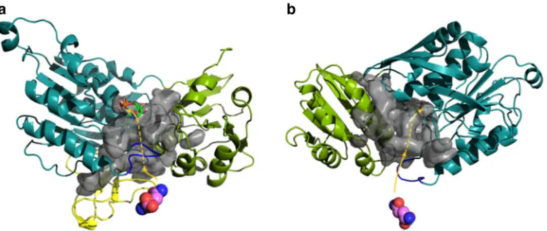
The opening in MurT facing the active site of GatD
commu-nicates with a network of cavities inside MurT (Fig.
8
). One branch
is formed within the central domain between the Zinc-ribbon-like
insertion and helix
α2, another branch is extended between helix α7
of the C-terminal domain and the Zinc-ribbon-like region. These
two branches may be obstructed by the reorientation of the
domains. A third channel reaches into the MurT active site and
likely serves to transfer ammonia generated by GatD. Escape of
ammonia could be prevented by narrowing of the active site and the
C-terminal domain contacting the substrates.
The hypotheses about substrate binding, loop conformation
and domain movement await the determination of structures
with bound ligands. The stabilizing role of zinc in the
cysteine-rich region also require further investigation. The structure
pre-sented here will be invaluable in these endeavors, while substrate
analogues and known inhibitors of Mur ligases will be tested to
initiate a search for antibacterials aimed at the major pathogens S.
pneumoniae, S. aureus, and M. tuberculosis.
Methods
Construction of a MurT/GatD depletion strain. Pneumococcal strains used and produced are listed in Supplementary Table 4. Depletion of MurT/GatD was performed by using the ComRS gene depletion system48. The strain SPH131
contains a replaceable Janus cassette behind the ComS-inducible PcomXpromoter of the ComRS system. This Janus cassette was substituted with the murTgatD genes by transforming SPH131 with an amplicon comprising the operon fused with the 1000 bp regionsflanking the Janus, giving rise to strain SPH476. This amplicon was constructed by amplifying the murTgatD operon from strain RH1 using the pri-mers ds188 and ds189 (Supplementary Table 5). The Janus upstream and down-stream regions were amplified using pairs khb31/khb36 and khb33/khb34. The three fragments were joined by overlap extension PCR49. Next, the native
murT-gatD operon in strain SPH476 was deleted by replacement with a Janus cassette. DNA fragments corresponding to the 1000 bp regions upstream and downstream of the native operon were amplified using pairs ds190/ds191 and ds192/ds193, respectively. Janus was amplified from strain SPH131 with the primer pair Kan484F/RpsL41R. The three PCR-products were fused by overlap extension PCR. The resulting amplicon was transformed into strain SPH476 generating strain SPH477, which required 0.2 µM ComS in the growth medium to drive ectopic expression of murTgatD. The Janus in the SPH477 genome was removed by transforming this strain with a DNA amplicon corresponding to the 1000 bp
regions upstream and downstream of the native murTgatD. These fragments were amplified from RH1 using the pairs ds190/ds195 and ds193/ds194, and joined by overlap extension PCR. The resulting strain, SPH478, required 0.2 µM ComS in the growth medium.
Depletion of MurT/GatD and cell wall isolation. For isolation of cell wall material from MurT/GatD-depleted cells, strain SPH478 wasfirst grown in 5 mL C medium to an OD550= 0.35 in the presence of 0.2 μM ComS. Then, the cells were washed once with 5 mL ComS-free C medium before they were transferred to a bottle containing 1 L of ComS-free C medium. Cells were incubated at 37 °C, and growth was monitored at OD550every 30 min. When growth levelled out because of MurT/GatD depletion (OD550≈ 0.4), cells were collected at 5000 g for 10 min. Cell wall material was isolated and analyzed as follows50. The cell pellet from 2 L of
culture was resuspended in 40 mL of ice-cold 50 mM TrisHCl (pH 7.0) and added dropwise to 150 mL of boiling 5% sodium dodecyl sulfate (SDS). After boiling for another 30 min, the suspension was centrifuged 45 min at 130,000 g and 25 °C. Following two washes with 30 mL of 1 M NaCl and repeated washes with water until free of SDS, the pellet was resuspended in 2–4 mL of water, one-third volume of acid-washed glass beads (0.17–0.18 mm diameter, Sigma) was added to disrupt cells in a FastPrep machine (FP120, Thermo Scientific). After filtration to remove glass beads, and clarification with a 5 min centrifugation at 10,000 g, the super-natant was centrifuged for 45 min at 130,000 g and 25 °C. The pellet was resus-pended and stirred for 2 h at 37 °C in 20 ml of 100 mM TrisHCl (pH 7.5), 20 mM MgSO4, 10μg mL−1DNase A and 50μg mL−1RNase I, prior to addition of 10 mM
CaCl2and 100μg mL−1trypsin and further incubation overnight at 37 °C, and enzymes were inactivated 15 min at 80 °C with 1% SDS. Cell wall was centrifuged for 45 min at 130,000 g at 25 °C, resuspended in 20 mL of 8 M LiCl, and incubated for 15 min at 37 °C. The procedure was repeated with 10 mM ethylenediaminete-traacetic acid (EDTA, pH 7.0). Cell wall was washed with water, acetone, and water before resuspension in 2–4 mL of water and lyophilization. To remove teichoic acids, 5 mg of cell wall was stirred with 48% hydrofluoric acid (HF) for two days at 4 °C. Peptidoglycan was centrifuged (45 min, 130,000 g, 4 °C), washed with water, 100 mM TrisHCl (pH 7.0), and twice with water. Peptidoglycan (0.5 mg in 200μL) was digested with 10μg of cellosyl in 20 mM sodium phosphate (pH 4.8) for 24 h at 37 °C. The reaction was stopped by heating at 100 °C for 10 min prior to cen-trifugation for 10 min at 13,000 g to clarify the solution. An equivalent volume of 500 mM sodium borate (pH 9.0) and a few crystals of NaBH4were added and incubated 30 min at 20 °C to reduce saccharrides, and the reaction was stopped lowering the pH to 4.0 with H3PO4. Muropeptides were analyzed by reversed-phase HPLC using a Prontosil 120–3-C18-AQ (250 4.6 mm, 3 μM) column on an Agilent 1100 system. The separation was obtained with a linear 135-min gradient from 0 to 30% methanol in 10 mM sodium phosphate (pH 6.0) and the column temperature was 55 °C. The muropeptides were quantified by their peak area.
Electron microscopy. To prepare MurT/GatD-depleted cells for morphological examination, strain SPH478 was grown in 5 mL C-medium containing 0.2 µM ComS to an OD550= 0.2. Then the cells were washed once with 5 mL ComS-free C-medium and diluted to OD550= 0.01 in 5 mL C-medium containing 0.2 µM, 2.5 nM or 1.5 nM ComS. When the cell cultures reached OD550= 0.3, the cells were harvested at 4000 g and diluted to OD550= 0.01 in 10 mL fresh C-medium con-taining the same concentrations of ComS as described above. Cultures reaching OD550= 0.3 were harvested at 4000 g for 5 min. Cells were washed once with ice cold PBS before they werefixed and prepared for scanning and transmission electron microscopy (SEM and TEM) as follows51. For SEM, cells were dehydrated
with 70 and 90% ethanol for 10 min each and then with 100% ethanol overnight,
a
b
Fig. 8 Putative channels allowing the transfer of ammonia inside MurT. Front (a) and back (b) view with the zinc-ribbon-like region removed to allow better view. The color scheme of the domains is as in Fig.3. The modeled loop 137–141 is shown in blue. The glutamine in the GatD active site is shown as atom-colored spheres. The cavity surface is in transparent gray with a possible path of the ammonia in pink
and subjected to critical point drying with liquid CO2. Samples were coated with Au-Pd and examined with a Zeiss EVO 50 EP scanning electron microscope. For TEM,fixed cells were post-fixed for 1 h at room temperature using 1% OsO4(w/v) and 1.5% K3[Fe(CN)6] (w/v) dissolved in water. Following three washing steps in water, cells were pre-stained for 30 min with 1% uranyl acetate. Next, cells were washed for three times in water and dehydrated with sequential 10-min incuba-tions in 70, 90, and 100% ethanol. Finally, cells were stepwise infiltrated in LR White resin as follows: LR White resin: EtOH in ratios 1:3 for 30 min, 1:1 overnight and 3:1 for 4 h, andfinally 100% LR White resin overnight followed by embedding in 100% LR White resin at 60 °C overnight. Thin sections were cut with a diamond knife mounted on an ultra-microtome (Leica, EM UC 6). The sections were counterstained with 1% KMnO4for 10 min. After staining, grids were washed thoroughly in water. The sections were examined under a FEI MORGAGNI 268 electron microscope.
Site-directed mutagenesis. Site-directed mutagenesis for the recombinant pro-duction of point mutants of MurT/GatD were performed on double-stranded expression plasmids using primers with the desired mutations (Supplementary Table 5)52. Site-directed mutagenesis for expression of MurT/GatD in S.
pneu-moniae was performed by using overlap extension PCR with primers containing the desired mutations (Supplementary Table 5). The complementary forward and reverse primers annealing internally in the murT or gatD genes were used in combination with the primers ds190 and ds193, which bind 1000 bp upstream and downstream of murTgatD. Point mutated versions of the murTgatD operon were introduced into the S. pneumoniae genome by replacement of the Janus cassette in strain SPH477. The ectopic copy of murTgatD behind PcomXwas then deleted by replacement with a Janus cassette amplified from strain SPH131 using the primer pair khb31/khb34. If the point mutation in murTgatD was not tolerated, the ectopic murTgatD could not be deleted.
Production of MurT/GatD. The complex was expressed from a modified pET-30 plasmid to allow co-expression in E. coli BL21 Star™ (DE3) of MurT with a N-terminal poly-histidine-tag and GatD without tag. After growth in Luria broth to saturation, expression was induced overnight at 20 °C by the addition of 0.5 mM IPTG. Cells were resuspended in 50 mM HEPES, pH 7.5, 300 mM NaCl, 10 mM MgCl2, 25 mM imidazole, 2 mM TCEP, and Complete™ protease inhibitors. After cell breakage with a Microfluidizer M−110P (Microfluidics) and removal of cellular debris and membranes by ultracentrifugation, the lysate was loaded onto a 1 mL HisTrap FF column (GE Healthcare) and proteins were eluted with imidazole concentration steps at 50, 100 and 200 mM. Fractions of the 100 mM imidazole step were pooled and diluted 10-fold in the same buffer without NaCl, imidazole or TCEP but with 5 mM DTT, and loaded onto a 8 mL Q Sepharose column (GE Healthcare). Proteins were eluted with a 150–450 mM NaCl gradient over 70 mL. After concentration using Amicon® Ultra-4 cells with a 30k cutoff, proteins were further purified by size-exclusion chromatography on an ENrich™ SEC650 column (BioRad) equilibrated with 50 mM HEPES, pH 7.5, 150 mM NaCl and 10 mM MgCl2, 2 mM DTT. For crystallization, the complex was concentrated again to more than 50 mg mL−1. Concentration was determined by measuring the absorbance at 280 nm with the extinction coefficient ε280= 63,860 M−1cm−1(0.788 mg-1mL
cm−1). For activity measurements of the wild type and mutant proteins, the anion exchange chromatography was omitted and thefinal buffer contained 2 mM TCEP instead of DTT.
Structure determination. Crystallization trials on the native MurT/GatD complex were performed in 96-well Greiner Crystal Quick plates with 100 nL protein (6 mg mL−1) and 100 nL reservoir solution. Native MurT/GatD crystallized in 0.2 M tri-ammonium or tri-sodium citrate buffers, pH 6.0–8.2, 20% PEG 3350 at 20 °C in 24–48 h. Crystallization conditions were manually refined and large native crystals werefinally obtained in 0.2 M tri-sodium citrate pH 6.1, 14% PEG 3350, 4 mM NiSO4. Native MurT/GatD crystals were cryoprotected by transfer into 0.2 M Tri-sodium citrate pH 6.1, 16% PEG 3350, 4 mM NiSO4, 20% glycerol, and then flash-frozen in liquid nitrogen. Seleno-methionine (SeMet) derivatives of MurT/GatD crystals were obtained in 0.2 M Tri-sodium citrate pH 6.2, 15% PEG 3350 and were cryoprotected by exchanging the mother liquor with crystallization solution con-taining 20% glycerol, andflash-frozen in liquid nitrogen. Native and SeMet MurT/ GatD crystals crystallized in the space group R3 with isomorphous unit cell parameters (Supplementary Table 2). Native diffraction data were collected on the automated ID30a-1 beamline of the European Synchrotron Radiation Facility (ESRF, Grenoble, France). Single-wavelength anomalous diffraction (SAD) data were collected on the BM30A-FIP beamline at ESRF. The diffraction data were reduced by using XDS53and phase determination was achieved using the SIRAS
(Single Isomorphous Replacement with Anomalous Scattering) method, including a heavy-atom search with SHELXD54, followed by phasing with SHARP55and
density modification with SOLOMON56. All calculations were performed
auto-matically using the autoSHARP package57. After automated model building using
PHENIX58, the crystallographic model was refined by using REFMAC559in the
CCP4 package, iterated with manual building. Water, ion and glutamine molecules were added manually in COOT60. Model quality was determined by using
PRO-CHECK61, with all residues within most favorable or allowed regions of the
Ramachandran plot. The data collection and refinement statistics are summarized in Supplementary Table 2).
Enzymatic assay. The activity of MurT/GatD was monitored through its hydrolysis of ATP. A classical ATPase assay was used where ADP is phosphorylated by pyr-uvate kinase from phosphoenolpyrpyr-uvate, pyrpyr-uvate being reduced to L-lactate by lactate dehydrogenase consuming NADH, which is followed by its absorbance at 340 nm62. For the determination of the enzymatic parameters, 10μL of the substrate
under investigation (lipid II, ATP or L-Gln) at 10-fold thefinal concentration in 50 mM HEPES, pH 7.5, 150 mM NaCl and 10 mM MgCl2were placed in a 1 cm quartz cuvette prior to the addition at time t= 0 of 90 μL of the reaction mix (minus the substrate being studied) in the same buffer to reach the followingfinal con-centrations: 250μM NADH, 1 mM potassium phosphoenolpyruvate, 25 μg mL−1 L-lactate dehydrogenase (porcine, Roche Diagnostics), 50μg.mL-1pyruvate kinase
(rabbit, Roche Diagnostics), 2 mM TCEP, 1% n-dodecyl-β-D-maltoside (DDM, Anatrace), 190 nM MurT/GatD, 10μM lipid II, 2 mM ATP and 10 mM L-Gln. The absorbance at 340 nm was recorded at 30 °C with a thermostated Uvikon 943 double beam spectrophotometer (Kontron Instruments). When investigating the activity of MurT/GatD mutants, 10μL of lipid II and 40 μL of MurT/GatD variant were placed in the cuvette prior to the addition of 50μL of reaction mix to reach the samefinal concentrations stated above, except lipid II at 20 μM. The amounts of NADH, potassium phosphoenolpyruvate, L-lactate dehydrogenase and pyruvate kinase were checked to be non-limiting.
Initial velocities (Vi) were measured by linearfitting of the reaction progress curves at early time points using the extinction coefficient of NADH at 340 nm ε = 6300 M−1cm−1. The apparent KMand Vmaxwere extracted from non-linearfitting of the Viat different substrate concentrations [S] to the Michaelis-Menten equation Vi= [S]Vmax/(KM+ [S]). Fittings were performed using the Kaleidagraph (Synergy) software. Alternatively, the complete progress curves were collectively fitted to a Michaelis-Menten model of the reaction to extract the apparent KDand kcatusing the Dynafit software (Biokin)63.
Thiol counting. The number of accessible free thiols was determined by alkylation under native condition of wild-type and 4C4S MurT/GatD and mass spectrometry analysis. One fourth-volume of 0.5 M iodoacetamide in 1.5 M Tris HCl pH 8.8 were added to the purified proteins for 5 min at room temperature prior to desalting on G-25 Sephadex resin (NAP-5 column, GE Healthcare) in 50 mM ammonium acetate pH 7. Samples were analyzed by electrospray mass spectrometry.
Docking. Calculations were performed in BIOVIA Discovery Studio 4.5 (Accelrys). The structure of MurT/GatD was cleaned, side-chain conformations were opti-mized for residues with inserted atoms, missing loops were modeled based on SEQRES information, and the protonation state was predicted at pH 7.5 (Prepare Protein protocol of DS 4.5). CDOCKER64was used to dockflexible ligands onto
the rigid protein.
To propare ligands, canonical tautomers were generated, keeping the largest fragments, standard charges of common functional groups were used, kekule structures were generated, ionization states at a given pH range and tautomers were enumerated (Prepared Ligand protocol of DS 4.5). Reasonable 3D conformations were created using Catalyst. High-temperature molecular dynamics were used to generate random ligand conformations from the initial ligand structure. Due to the highflexibility of lipid II, several conformations generated with the BEST algorithm65were docked to cover the full range of conformers. The lowest energy
poses were retained and clustered according to their binding mode and the interaction with key residues. Snapshots of the docked ligands were generated using BIOVIA DS Visualizer.
Data availability. Coordinates and structure factors have been deposited in the Protein Data Bank, with Accession No. 6FQB [https://doi.org/10.2210/pdb6FQB/ pdb]. The data that support thefindings of this study are available from the corresponding author upon request.
Received: 5 March 2018 Accepted: 17 July 2018
References
1. Vollmer, W., Blanot, D. & de Pedro, M. A. Peptidoglycan structure and architecture. FEMS Microbiol. Rev. 32, 149–167 (2008).
2. Siewert, G. & Strominger, J. L. Biosynthesis of the peptidoglycan of bacterial cell walls. XI. Formation of the isoglutamine amide group in the cell walls of Staphylococcus aureus. J. Biol. Chem. 243, 783–790 (1968).
3. Figueiredo, T. A. et al. Identification of genetic determinants and enzymes involved with the amidation of glutamic acid residues in the peptidoglycan of Staphylococcus aureus. PLoS. Pathog. 8, e1002508 (2012).