HAL Id: hal-01152947
https://hal.archives-ouvertes.fr/hal-01152947
Submitted on 20 May 2015
HAL is a multi-disciplinary open access
archive for the deposit and dissemination of
sci-entific research documents, whether they are
pub-lished or not. The documents may come from
teaching and research institutions in France or
abroad, or from public or private research centers.
L’archive ouverte pluridisciplinaire HAL, est
destinée au dépôt et à la diffusion de documents
scientifiques de niveau recherche, publiés ou non,
émanant des établissements d’enseignement et de
recherche français ou étrangers, des laboratoires
publics ou privés.
Femtosecond spin-state photo-switching dynamics in an
FeIII spin crossover solid accompanied by coherent
structural vibrations
Roman Bertoni, Maciej Lorenc, Jerome Laisney, Antoine Tissot, Alain
Moréac, Samir F. Matar, Marie-Laure Boillot, Eric Collet
To cite this version:
Roman Bertoni, Maciej Lorenc, Jerome Laisney, Antoine Tissot, Alain Moréac, et al..
Femtosec-ond spin-state photo-switching dynamics in an FeIII spin crossover solid accompanied by coherent
structural vibrations. Journal of Materials Chemistry C, Royal Society of Chemistry, 2015, 3 (30),
pp.7792-7801. �10.1039/C5TC00854A�. �hal-01152947�
a.Institut de Physique de Rennes, Université de Rennes1, UMR UR1-CNRS 6251,
F-35000 Rennes, France.
b.Institut de Chimie Moléculaire et des Matériaux d’Orsay, Université Paris-Sud,
UMR-CNRS 8182, Orsay, France.
c.
CNRS, Université de Bordeaux, ICMCB, 87 avenue du Dr. A. Schweitzer, Pessac, 33608 France.
∗ Corresponding author Email address: [email protected]
† Present address: Institut Lavoisier de Versailles, UMR CNRS 8180, Université de
Versailles Saint-Quentin, 78035 Versailles cedex, France.
Electronic Supplementary Information (ESI) available: Videos of the molecular vibrational modes calculated at 56 cm-1, 33cm-1 and 88 cm-1.
Femtosecond spin-state photo-switching dynamics in an Fe
IIIspin
crossover solid accompanied by coherent structural vibrations
R. Bertoni,a M. Lorenc,a J. Laisneyb, A. Tissotb,†, A. Moréaca, S. F. Matarc, M.-L. Boillotb and E. Colleta,*
We investigate light-induced excited spin-state trapping (LIESST) dynamics of an FeIII spin-crossover material from low (S=1/2) to high (S=5/2) spin states. Our results show that this process occurs only at the molecular level as evidenced by the linear dependence of the fraction of photo-switched molecules with the excitation density as well as with the initial fraction of low spin molecules. The inter-system crossing from photoexcited LS (S=1/2) to HS (S=5/2) occurs within 200 fs and is accompanied by coherent non-equilibrium vibrational relaxation in the photo-induced HS state. These results reveal similar dynamical features to those already reported for LIESST in FeII systems. The activation of coherent molecular vibrations is essential for reaching rapidly the HS potential on the timescale of molecular motions, whereas their fast damping allows an efficient trapping in the HS potential. The observed coherent oscillations are attributed to photoinduced molecules in the HS states, as supported by Raman spectroscopy at thermal equilibrium, and DFT analyses of molecular vibrations and TD-DFT calculations of optical absorption.
Introduction
In molecular science, light is known to be an efficient trigger for driving chemical reactions, breaking bonds or changing
electronic molecular states1. Reaction coordinates connecting
initial, photoexcited, and final photoinduced states are not simple to describe. Furthermore, the pathway followed on the potential energy surface is complex and numerous studies investigated how fast such processes are and how interconversion of molecular states proceeds. In the solid state too, light can switch molecules between different electronic states. Charge-transfer processes induced by light can lead to
photo-induced phase transition from insulating to metallic2 or
from neutral to ionic3 phases for example. Spin-crossover
(SCO) materials are bistable molecular systems able to switch between low spin (LS) and high spin (HS) states under the effect of different control parameters such as temperature or
pressure4. They are also prototype photoactive systems as
they can be interconverted through the light-induced excited spin-state trapping (LIESST) process from LS to HS state or conversely from HS to LS state (reverse-LIESST). LIESST in
crystals was discovered more than thirty years ago5: by
irradiating SCO solids with cw light at low temperature complete conversion of molecular states can be generated at the macroscopic scale of the material and the photo-induced state can be metastable up to a critical temperature referred
to as T(LIESST)6. In crystals, molecules may respond
cooperatively to light excitation2,3. In addition, light can control order between the individual molecules of the materials and generate new phases which cannot be reached at thermal
equilibrium7. This underlines the ability of molecular-based
materials to show response to light different from the response of the individual constituting molecules. There are also other examples in the literature of molecular materials showing cooperative response to light excitation, for which a single photon transforms several molecules and the response of the molecular material is more than the sum of individual
molecular events2,3. Spin-crossover has become a topical
research area over the last three decades, with many research
groups contributing to its advancement4. The very high
efficiency and speed of the photo-induced spin state switching between two states that completely differ in physical properties (color, volume, magnetic susceptibility) represent a conceptual challenge8. For Fe
II
systems the role of ligand-field
states was deeply investigated9. The study of reverse-LIESST
revealed a mechanism involving two main inter-system crossing (ISC) and therefore true intermediate electronic states (such as S=1) between the photoexcited HS (S=2) and the photoinduced LS (S=0) state, which is reached within 40 ps. But for LIESST, the irradiation into metal–ligand charge transfer (MLCT) or d-d bands of S=0 species promotes the system to the S=2 state within 160 fs. This timescale was determined with ultrafast optical spectroscopy, a powerful technique for investigating the photo-physical dynamics of
SCO compounds10. Complementary ultrafast X-ray absorption
spectroscopy studies revealed the structural dynamics related to ligand-cage dilation, which leads to the structural trapping
of the newly formed HS electronic state11. On the pathway
from MLCT to HS states, other not structurally relaxed electronic states serve as intermediates. Indeed, these states are populated on a timescale shorter than an oscillation period in the potential12.
For better understanding this process, it seems necessary to track the electronic and structural pathway followed by the system with the time resolution of elementary electronic and molecular events. Recent theoretical works suggested that such speed and efficiency of LIESST results from the instantaneous activation of molecular phonons after photo-excitation13. This is due to the important displacive nature of the inter-system crossing, which moves the system rapidly into the HS potential with a 0.2 Å elongation of Fe-N bonds. The experimental proof of such a conjecture was reported recently for an FeII system14. This study revealed that the structural
molecular reorganization, trapping the photoinduced
electronic state, occurs in a two-step sequence: the molecule elongates first (within 170 femtosecond) and bends afterwards, via a coherent vibrational energy transfer between these two main structural modes.
There are very few FeIII materials showing efficient or
long-lived LIESST15 but we evidenced in several SCO archetypes16,17
by time-resolved spectroscopy that transient FeIII HS state can
be generated. This is also the case of the FeIII SCO
[Fe(3-MeO-SalEen)]PF6 material studied here in the form of nanocrystals
(average size, 900(230)285(65)64(18) nm3)8a. Here we show
that the spin-state photo-switching dynamics in this FeIII
material occurs locally and that the structural trapping during LIESST is accompanied by coherent molecular dynamics.
Results and discussion
Thermal spin conversion.
The FeIII complex [Fe(3-MeO-SalEen)2]PF6 was originally
synthesized by Hendrickson18. H-3-MeO-SalEen is the
condensation product of 3-methoxy-substituted
salicylaldehyde and N-ethyl-ethylenediamine. This FeIII system
can switch under the effect of external parameter, such as temperature, between LS and HS states. Since the HOMO and LUMO orbitals have a strong d character, the two electronic states correspond to:
- a low spin one (LS) with S= 1/2 with an electronic configuration t2g5 eg0 in the octahedral-like description. More
rigorously, in the symmetry of the complex the electronic configuration is dπ5 dσ0.
- a high spin one (HS) with S= 5/2 corresponding to the electronic configuration t2g3 eg2 (or dπ3 dσ2).
In the form of single crystals, this compound shows a first-order phase transition from LS to HS phases at thermal equilibrium18,19. Nano-crystals of this material dispersed in
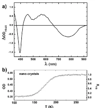
Fig.1 Difference of optical density in the (350-925 nm) spectral window between HS state (296 K) and LS state (16 K) measured on the nano-crystals sample (a). Photo-excitation is performed at 850 nm in the tail of the LS absorption band. OD at 550 nm (left axis) of the nano-crystal assembly during the crossover scaled to XHS (right axis) (b).
polymeric matrix lose the 1st order transition and show a more
gradual spin conversion16, as a result of both the pressure effects and anchoring processes (physisorption, capillarity) due
to the polymers20. It was recently demonstrated by
time-resolved studies that these FeIII nanocrystals exhibit photo-switching toward a transient HS state by photo-excitation of
the LS state8a. Here we present new results and detailed
analysis of the spin state photo-switching dynamics by means of femtosecond pump-probe optical spectroscopy. It is easier to investigate an assembly of nano-crystals dispersed in a PVP polymer film than single crystals: the low optical density of the nano-crystals allows to perform a detailed study over a broad spectral range from UV to IR. Single crystals restrict the range of wavelengths for spectroscopic probing due to the higher optical density (OD)8a,16a.
Fig. 1 shows the variation of optical density OD measured
on the films of nano-crystals, between pure LS state (16 K) and pure HS state (296 K). It reveals phenolate-to-iron charge-transfer bands (CT) in the near-infrared (NIR) - visible (VIS) with distinct features for LS and HS states. With respect to that of LS state, the OD of the HS state is higher in VIS and lower in NIR parts of the spectrum, with a well-defined isosbestic point
around 650 nm. The thermal fraction of HS molecules XHS(T) is
obtained by scaling the change of optical density OD(T) at a given temperature T to the change of optical density between LS (ODLS) and HS (ODHS) states:
The thermal behavior of XHS(T) obtained in this way is
displayed in Fig. 3(b) and indicates a gradual conversion spanning from 140 K to 220 K, in agreement with magnetic
Fig. 2. LS schematic energy diagram and frontier molecular orbitals obtained from DFT calculations. Red: d and d metal-like orbitals, blue : ligand-like
orbitals. The HOMO-LUMO gap (1.6 eV) is larger than the pump excitation at 1.45 eV (850 nm). With respect to the HOMO (strong weight on a Fe d orbital), a few doubly occupied molecular orbitals (centered on phenol groups) falls within the energy range corresponding to the photo-excitation.
In the following, we exploit these spectroscopic fingerprints, to study the ultrafast spin-state photo-switching and the resulting energy dissipation into the material.
Inter-System Crossing and vibrational cooling
The spin-state photo-switching is known to proceed via transient intermediate states, as the direct low energy excitation from LS to HS state is forbidden by spin parity. In the
case of FeII systems, the photo-switching at the molecular level
results from the promotion by light of an electron from the metal center to the ligand orbitals (MLCT excited state). For this FeIII [Fe(3-MeO-SalEen)2]PF6 system, we performed DFT calculations with Gaussian 09 package to establish the molecular orbital energy diagram. Fig. 2 shows that in the LS state the highest occupied molecular orbital (HOMO) and lowest unoccupied molecular orbitals (LUMO), both of Fe d predominant character, are separated by a 1.6 eV energy gap. We found that the low energy excitation at 850 nm (1.45 eV) therefore may promote an electron from a few symmetry-adapted and doubly-occupied molecular orbitals (centered on the phenol group, noted L) to the HOMO with a strong weight on the d orbital of the metal center (M). This LMCT state corresponds then to a L1dπ6dσ0 electronic configurations.
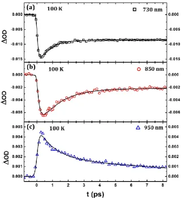
Fig. 3. Change of optical density at different probe wavelengths after fs excitation at 850 nm measured on [FeIII(3-MeO-SalEen)2]PF6 nano-crystals. Measurements are performed at 100K in pure LS state.
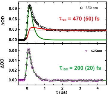
Fig. 4. Deconvolution of raw data into the relaxation kinetic of the INT electronic states (isc,, green line) and the population kinetic of the HS state taking into account vibrational cooling (vc, red line). Only 1/8 of the experimental data are shown for clarity.
By using femtosecond optical spectroscopy (see experimental section), it is possible to study the photo-switching dynamics since the different molecular states have peculiar optical spectra (Fig. 1). Selected time traces of transient OD measurements performed at different probing wavelengths are plotted in Fig. 3-5. The OD increase in the VIS part and bleaching in the NIR part (at the ps timescale), are characteristic of the formation of the HS state, as observed during the thermal LS to HS conversion (Fig. 1) and previously reported on similar samples8a,16.
Around time zero, the OD spectrum differs significantly from those of the LS ground state and of the HS photoinduced state, in terms of intensities or spectral range, where OD increases or decreases. This is the optical fingerprint of the LMCT state. The kinetic evolution of the optical density around the isosbestic point at 625 nm, where the OD of LS and photoinduced HS
states are the same8a, allows to selectively follow the dynamics
Fig. 5. Fit of the OD traces at 730, 850 & 950 nm with bi-exponential functions describing ISC and vibrational cooling.
The OD kinetic trace at 625 nm shows at first a rapid rise (limited by our time resolution) corresponding to the generation of the LMCT state, which then decays towards the final HS state. Rigorously speaking, the dynamics may involve a sequence of transient states between LS and HS, as already reported in the case of FeII systems. But since we do not observe clear fingerprints of different intermediate states, we consider only a lump intermediate state (INT) including the LS LMCT state (L1dπ6dσ0 electronic configuration) and other
possible intermediates, such as those resulting from L1dπ5dσ1
or L1dπ4dσ2 electronic configurations.
The inter-system crossing (ISC) sequence from LMCT to HS is well described at 625 nm by a single exponential decay. The fit
shown in Fig. 4 gives ISC = 200 ± 20 fs. For other
measurements with probe wavelengths away from the isosbestic point, the single exponential model fails to reproduce the observed dynamics and slower components are observed on the ps time-scale, as shown at 550 nm for example. This is especially true in the IR region (950 nm, Fig. 5), more sensitive to vibrational cooling since lower energetic levels are probed. Thus we used a two-exponential decay model for fitting both dynamics, the first time constant
describing the ISC being fixed to ISC = 200 fs. Depending on the
probe wavelength the vibrational cooling time constant vc
varies from 470 (50) fs at 550 nm and up to 1.6 (0.2) ps at 950 nm. At 730 and 850 nm the probe is sensitive both to vibrational cooling but also to LS state bleaching, thus making the interpretation more difficult. These measurements in the visible and infrared region reveal therefore that even if the
system has reached the HS potential within 200 (20) fs, more
than one picosecond is needed for the HS molecule to thermalize with its environment.
Fig. 6. (a-c), Oscillating component of OD at 550, 625, and 700 nm. (d-f) corresponding FFT of the experimental data, showing the activation of different modes around 85, 56 et 35 cm-1.
The vc1.6 ps observed here corresponds to the transfer of
the excess energy deposited on the molecule by the laser to
the lattice. This timescale is shorter than that reported for FeII
molecules in solution (up to 10 ps) with infrared experiments10,11. This may be due to the fact that in crystals the excited molecules interact efficiently with the lattice system via phonon-phonon coupling.
Coherent structural oscillations
A closer inspection of the OD time traces in Fig. 3-4 allows the observation of weak oscillating components. Fig. 6 (a-c) shows the deviation of the OD data from the exponential fits for different probe wavelengths. The corresponding Fourier transforms of the residual parts corresponding to positive delay are presented in Fig. 6 (d-f). At 550 nm, a prevailing
mode is observed around 85 cm-1 (similar oscillations are
observed at 525 and 475 nm in Fig. 3). At the wavelength on the isobestic point (625 nm) another mode is observed around
56 cm-1. These modes are equally observed at 700 nm, where a
third mode around 35 cm-1 also appears.
A similar vibrational coherence in the photoinduced high-spin state was reported for FeII SCO molecules in solution21 and more recently in [Fe(phen)2(NCS)2] crystals14. The mechanism
behind the high quantum efficiency of LIESST in FeII SCO
compounds was theoretically proposed to result from the instantaneous coupling of the excited (MLCT) state with the
phonons of the final (HS) state13. The first experimental proof
of this process was provided by femtosecond optical
pump-probe spectroscopy studies of LIESST in [Fe(phen)2(NCS)2]
crystals, where the breathing mode (113 cm-1) associated with
the elongation of the Fe-N bonds was clearly identified14: the instantaneous activation of this molecular breathing phonon drives the system from the MLCT to the HS potential within 170 fs, i.e. on the timescale of a half period of such molecular vibration. The fast damping of the key molecular phonon modes leads to the efficient trapping the HS state. It may also result from intermolecular contacts.
Fig. 7. Raman spectra obtained on single crystals for different polarization (1), (2) and (3) shown in the photographs on the top.
The present observation of the activation of such coherent structural vibrations during LIESST in FeIII SCO crystals may therefore be also attributed to such mechanism. However, there are very few vibrational spectroscopy studies of FeIII compounds in literature, even though these low frequencies vibrations are often attributed to ligand breathing or torsion
modes22. We thus performed Raman measurements at room
temperature, as well as calculations of molecular vibrations on
the [Fe(3-MeO-SalEen)]PF6 compound for gaining insights on
the frequencies and nature of such modes. Raman and DFT studies of the HS phase
We used a single crystal for finding the crystal axis orientation with x-ray diffraction and checking polarization effect on Raman signal. Fig. 7 shows the single crystal Raman spectra obtained at room temperature. Since the crystalline space group is and the molecules are in general positions in the unit cell16a, the modes split in two categories: the totally
symmetric ones (Ag and Raman active) where molecules
oscillate in phase with respect to inversion symmetry and the
antisymmetric ones (Au and IR active). The Raman spectra
reveal several low frequency modes, especially around 65, 85,
100, 145, 172, 181, 240 and 260 cm-1. In this frequency range,
the modes may be the skeletal modes of the [FeN4O2] core, but may also correspond to ligand distortions. Because of the Ci point symmetry of the crystal, there is no polarization selection: only the Raman intensity may depend on the sample orientation with respect to the laser polarization.
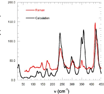
Fig. 8.Comparison of the calculated and experimental (averaged of the different polarizations) Raman spectra.
According to Fig. 7 the relative intensity of the different modes changes with sample orientation. The Raman mode around 85
cm-1 corresponds well to the coherent vibration observed
during LIESST in the present FeIII compound. The modes at 35
and 56 cm-1 could not be measured with our Raman set-up
because of the dielectric edge filter cut-off.
For understanding better the nature of the molecular vibrations involved during LIESST, we carried out molecular vibration frequencies calculations with DFT methods for an
isolated [Fe(3-MeO-SalEen)2]PF6 complex, after geometry
optimization, by using hybrid B3LYP functional with triple-ζ 6-311g(d,p) basis set within Gaussian09 code23,24. In Fig. 8 we compare the calculated Raman spectrum with the experimental results from Fig. 7, averaged over the different orientations measured on a single crystal for better comparison with calculations. The overall calculations reproduce quite well the observed Raman spectrum. A complete analysis of the modes and the nature of the vibrations will be published elsewhere and in this paper we
focus on the modes observed with time-resolved
spectroscopy. In the low frequency region, the calculated modes at 88, 56 and 33 cm-1 are close to those observed by Raman and/or time-resolved spectroscopy. The mode at 56
cm-1 (shown in video S1) is mainly associated with Fe-N
breathing of the FeN4O2 coordination sphere. The modes at 33cm-1 (video S2) and 88 cm-1 (video S3) are mainly ligand torsion modes. Since during the LS to HS conversion the structural reorganization involves both the molecular breathing (as Fe-L bonds elongate) and the ligand torsion, such modes should play an important role during the self-trapping process.
For understanding why different vibration modes are observed during LIESST depending on the probe wavelength, we need to analyze optical absorption of HS species through an accurate description of excited states and a time dependent DFT
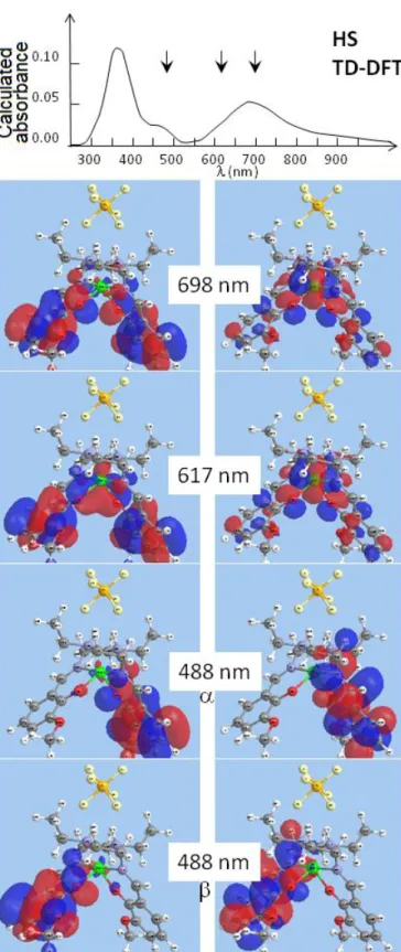
Fig. 9. Natural transition orbitals of hole (left) and particle (right) calculated in the case of HS species at 698, 617 and 488 nm, i.e. close to the experimental probes at 700, 625 and 550-475 nm. α and β designate spin ↑ and spin ↓. The calculated absorbance spectrum (top) underline the strong weight of these transitions.
The obtained natural transition orbitals (NTO) taking into account the hole–particle pairs resulting from light absorption can then be used to interpret our data.
Fig. 10. Schematic representation of the photoswitching trajectory along a simple reaction coordinate.
Fig. 9 shows the NTO corresponding to the hole–particle transition state around 500, 617 and 700 nm. The strong weight of the particle orbital on the Fe-N (and Fe-O) bonds for the transition at 617 nm explains the sensitivity to the breathing mode of the pump-probe data at 625 nm. The transition state around 700 or 500 nm are characterized by a strong weight of the particle orbital mainly located on one ligand and explain the sensitivity to ligand torsion.
Ultrafast LIESST dynamics in FeIII systems
Our investigation gives important insights on the ultrafast
mechanism of LIESST in a FeIII system, schematically
represented in Fig. 10. Molecular orbital calculations (Fig. 2) indicate that the photo-excitation of the LS (L2dπ5dσ0)
generates a LMCT state (L1dπ6dσ0). Time-resolved spectroscopy
data show that the LMCT state decays to the HS state (L2dπ3dσ2), possibly through other intermediate electronic
states such as L1dπ5dσ1 or L1dπ4dσ2. However, since the HS
potential is reached within 200 fs, these electronic states only
serve as mediators and are dynamically mixed. The different photoexcited molecules arrive "simultaneously" on the HS potential, where they oscillate. The global coherent oscillations experimentally observed underline that the phase is mainly kept between the different photo-excited molecules. Indeed, the observation of vibrational coherence in the
photoinduced HS state with oscillation around 85 cm-1 (392 fs
period), requires that the photoexcited molecules reach the HS potential on a timescale shorter than a half oscillation period
to avoid dephasing. This is in agreement with ISC = 200 ± 20 fs
observed by time-resolved spectroscopy (Fig. 4). This very
short time-scale is similar to the one reported for LIESST in FeII
SCO systems10,14 and also indicates that intermediates other
The instantaneous activation of several phonons of the HS state results from the important displacive nature of the mechanism. However, the structural change between LS and HS structures involves several degrees of freedom such as molecular breathing, torsion..., which may be activated with different timescales. Therefore, the transformation pathway connecting the initial photo-excited state to the photoinduced HS state is only roughly described by the classical single coordinate picture employed here in Fig. 10. A more complex multi-dimensional energy surface description has to be used for a more detailed analysis. However, these results indicate that on such short time-scale the wave functions describing the electronic and structural degrees of freedom are strongly coupled during the process. This LS-to-HS photoswitching dynamics is therefore very different in nature from the reverse
HS-to-LS one observed recently in a FeII SCO material, where a
true (vibrationally cooled) triplet state is populated and
depopulated within 10's ps9. Indeed, such slow reverse-LIESST
dynamics obey the Born-Oppenheimer approximation. Local nature of ultrafast LIESST
Another important question associated with LIESST in crystals is the possibility of cooperative response to light excitation. We look into possible cooperativity on ps timescale by following the fraction XHS of molecules converted after 5 ps
from LS to HS state. It is monitored by femtosecond optical spectroscopy for different excitation densities and for different temperature (and therefore different initial fractions of molecules in LS states as shown in Fig. 1). Fig. 11 shows how
XHS changes with the pump laser fluence F (i.e. on the
excitation density). It is clear that XHS depends linearly on F,
i.e. there is no amplification on such ultra-short time scale. The observed slopes in Fig. 11 correspond to the photo-response
and are given here in XHS/F. The slopes measured for
different temperatures, proportional to the number of photo-switched molecules per incident photon, change with temperature. This is not surprising because the fraction of molecules in the LS state decreases above 120 K (Fig. 1). Therefore the fraction of molecules switched from LS to HS states, should be weighted both by XLS, the initial fraction of
molecules in the LS state at a given temperature (XLS = 1-XHS),
and by the laser fluence F.
XHS F XLS or XHS / F XLS
Fig 11(b) shows that the photo-response XHS/F depends
linearly on XLS. Incidentally, based on such plot we can also
conclude on the absence of back conversion with the same pump wavelength (from HS to LS), since in such a case the
photo-response would be negative as XLS will tend to 0. These
important results underline the local nature of the photo-switching process occurring within a picosecond. Each individual photo-switching process can be regarded as an isolated molecular event. It is only on longer time-scale that macroscopic conversion occur, driven by elastic and thermal
mechanisms18.
Fig. 11. (a) Temperature dependence of the fraction of molecules photo-switched to the HS state (XHS) for different pump fluencies. (b)
Dependence of the photo-response (XHS/F) with the initial fraction of
LS molecules (XLS).
Conclusions
This detailed study allows to draw a complete picture of the LIESST photo-switching process in FeIII systems in the solid state. First of all, the photo-response is local on ps timescale, as characterized by the linear dependence with the number of photons and with the initial fraction of molecules in the LS state. This confirms that the initial mechanism might be seen as a local molecular event. Our new data in the present FeIII system also reveal that during the LIESST process several key modes are coherently activated. They are associated with changes of relevant reaction coordinates accompanying change of spin state (breathing, torsion...). The Fe-N lengthening is shorter in FeIII (0.15 Å) than in FeII (0.20 Å) and the frequency of the breathing modes differ. If a clear breathing mode (in phase elongation of the 6 Fe-N bonds) is easily identified in FeIIN6 systems (around 113 cm-1)14 the
lower symmetry of the present compound with FeIIIN4O2
coordination sphere makes such identification more difficult and several modes may involve Fe-N elongation (as it is the
case of the 56 cm-1 mode). In addition, the breathing
of its bonding to the metal. However, our results indicate that
the mechanism behind the ultrafast intersystem crossing in FeII
and other transition-metal complexes13,14, described in terms of the dephasing of the photoexcited state to the phonon continuum of the photoinduced (here HS) state with significantly different structure, is also valid for describing the
photo-switching mechanism in FeIII systems. The observed
structural coherence underlines the dynamical nature of the light-induced excited spin-state trapping process, as opposed to the so far prevailing kinetic description based on simple exponential decay from photo-excited to photoinduced states.
Experimental
Sample preparationThe molecular compound [Fe(3-MeO-SalEen)2]PF6 was
synthesized in the form of a microcrystalline powder and single-crystals as previously described16a,18. Nanocrystals16c were prepared by rapidly adding a concentrated solution of salt (10 mg) in acetone (1 mL) into a large volume (20 mL) of butan-1-ol maintained at -40 °C under a vigorous stirring. After 15 minutes, the particles were isolated by centrifugation, rinsed with pentane, dried. The properties of needle-shaped nanocrystals (typical dimensions of 900(230)x285(65)x64(18)
nm3 from TEM) were checked with powder X-ray diffraction, IR
and magnetism16c. The nanocrystals were processed in PVP
polymeric films. 5 mg of nanocrystals were added to a viscous solution of 100 mg of PVP (MM = 45000 gmol-1) in 500 L of butan-1-ol ; the mixture was sonicated (30 min), then spin-coated on glass substrates (1000 rpm) and dried at air before use.
Femtosecond optical spectroscopy
For tracking the photo-induced spin state switching dynamics in real time, we employ femtosecond pump probe
transmission measurements17a. The laser beam was provided
from the output of a femtosecond regenerative amplifier (Legend Elite, Coherent) and then split in two beams, each of them seeding an optical parametric amplifier (TOPAS, Light Conversion) allowing to cover spectral range from UV to infrared. The experimental time resolution is 140 fs. The pump wavelength was set at 850 nm corresponding to the LMCT absorption band. The photo-excited HS state relaxes towards the LS state in less than 1 ms, allowing the 1 kHz repetition rate of the experiment. The sample was cooled down using a nitrogen cryostream. The one colour probe beam, tuned across a broad spectral range (480-950 nm) measured transient optical density. We could measure the variation of the fraction of HS molecules XHS(t) by scaling OD(t), the
change of OD in time between LS and photo-excited HS state, to the OD change between LS and HS states at thermal equilibrium.
Raman spectroscopy
Raman spectra were collected in the 55-1200 cm-1 low
frequency range using a LabRAM-HR Raman spectrometer (Horiba / Jobin Yvon). A 10030030µm3 single crystal has
been studied for different polarisation configurations. The 632.818 nm line of a 15 mW He-Ne laser was used as the excitation source. The exciting radiation was directed through a neutral density filter (optical density 2) to avoid sample heating problems and was focused on the sample via a x100 long working-distance objective. The scattered light was collected in backscattering configuration and the Rayleigh scattering was removed by means of a dielectric edge filter. DFT calculations
Molecular vibration frequencies calculations were carried out
for [Fe(3-MeO-SalEen)]PF6 after geometry optimization, by
using hybrid B3LYP functional with triple-ζ 6-311g(d,p) basis
set within Gaussian09 code23. Frequencies are determined
from the second derivatives of the energy with respect to the atomic positions and then operating transformation to mass-weighted coordinates. Exploring the results especially for the vibrations and their animations with screen captures was done with Gaussview annex module to Gaussian.
TD-DFT as implemented in the Gaussian 09 package23 was
applied for obtaining the NTO of the HS [Fe(3-MeO-SalEen)]PF6
system, starting from preliminary geometry optimized molecule with B3LYP/6-311g(d,p) functional-basis set.
Acknowledgements
This work was supported by the Institut Universitaire de France, Rennes Métropole, Région Bretagne (CREATE 4146),
ANR (ANR-13-BS04-0002) and Fonds Européen de
Développement Régional (FEDER). Raman measurements have been performed on the SIR raman system from the ScanMAT platform of Rennes 1 University.
Notes and references
1 a) D. Polli, P. Altoe, O. Weingart, K.M. Spillane, C. Manzoni, D. Brida, G. Tomasello, G. Orlandi, P. Kukura, R.A. Mathies, M. Garavelli, and G. Cerullo, Nature, 2010, 467, 440-443; b) A. H. Zewail. Angew. Chem. Int. Ed. 2000, 39, 2586-2631; c) J.H Lee, M. Wulff, S. Bratos, J. Petersen, L. Guérin, J.C Leiknman, M. Cammarata, Q.Y. Kong, J. Kim, K. Moller, and H.J. Hee, J. Am. Chem. Soc., 2013, 135, 3255.
2 a) M. Gao, C. Lu,H. Jean-Ruel, L. C. Liu, A. Marx, K. Onda, S. Koshihara, Y. Nakano, X. Shao, T. Hiramatsu, G. Saito, H. Yamochi, R. R. Cooney, G. Moriena, G. Sciaini, and R.J.D. Miller, Nature, 2013, 496, 343-346; b) T. Ishikawa, N. Fukazawa, Y. Matsubara, R. Nakajima, K. Onda, Y. Okimoto, S. Koshihara, M. Lorenc, E. Collet, M. Tamura and R. Kato, Physical Review B, 2009, 80, 115108; c) Y. Kawakami, S. Iwai, T. Fukatsu, M. Miura, N. Yoneyama, T. Sasaki, and N. Kobayashi, Phys. Rev. Lett., 2009, 103, 066403.
3 a) H. Okamoto, Y. Ishige, S. Tanaka, H. Kishida, S. Iwai, and Y. Tokura, Phys. Rev. B, 2004, 70, 165202; b) L. Guérin, J. Hébert, M. Buron-Le Cointe, S. Adachi, S. Koshihara, H. Cailleau, and E. Collet, Phys. Rev. Lett., 2010, 105, 246101. 4 M. Halcrow, Ed., Spin-crossover materials (Wiley, West
Sussex, 2013) ISBN 9781119998679.
5 a) S. Decurtins, P. Gütlich, C.P. Köhler, H. Spiering, and A. Hauser, Chem. Phys. Lett., 1984, 105, 1; b) A. Hauser, Chem. Phys. Lett., 1992, 192, 65-70.
6 J.F. Létard, L. Capes, G. Chastanet, N. Moliner, S. Létard, J.A. Real, and O. Kahn, Chem. Phys. Lett., 1999, 313, 115.
7 a) N. Bréfuel , H. Watanabe, L. Toupet, J. Come, N. Matsumoto, E. Collet, K. Tanaka and J.-P. Tuchagues, Ang. Chem. Int. Ed., 2009, 48, 304-9307; b) E. Collet, H. Watanabe, N. Bréfuel, L. Palatinus, L. Roudaut, L. Toupet, K. Tanaka, J.-P. Tuchagues, P. Fertey, S. Ravy, B. Toudic, and H. Cailleau, Phys. Rev. Lett., 2012, 109, 257206.
8 a) R. Bertoni, M. Lorenc, A. Tissot, M. Servol, M.-L. Boillot, and E. Collet, Angew. Chem. Int. Ed., 2012, 51, 7485-7489; b) W. Kaszub, A. Marino, M. Lorenc, E. Collet, E.G. Bagryanskaya, E.V. Tretyakov, V.I. Ovcharenko and M.V. Fedin, Ang. Chem. Int. Ed., 2014, 53, 10636-10640; c) A. Marino, M. Buron-Le Cointe, M. Lorenc, L. Toupet, R. Henning, A.D. DiChiara, K. Moffat, N. Bréfuel, E. Collet, Faraday Discussion, 2015, 177, 363-379.
9 A. Marino, P. Chakraborty, M. Servol, M. Lorenc, E. Collet and A. Hauser, Ang. Chem. Int. Ed., 2014, 53, 3863-3867. 10 a) W.Gawelda, A. Cannizzo, V.T. Pham, F. van Mourik, C.
Bressler, and M. Chergui, J. Am. Chem. Soc., 2007, 129, 8199–8206; b) A. Canizzo , C. Milne, C. Consani, W. Gawelda, C. Bressler, F. van Mourik, and M. Chergui, Coord. Chem. Rev., 2010, 254, 2677-2686; c) J.K. McCusker, K.N. Walde, R.C Dunn, J.D Simon, D. Madge, and D.N. Hendrickson, J. Am. Chem. Soc., 1992, 114, 6919-6921; d) J.E. Monat, and J.K. McCusker, J. Am. Chem. Soc., 2000, 122, 4092-4097; e) A.L. Smeigh, M. Creelman, R.A. Mathies, and J.K. McCusker, J. Am. Chem. Soc., 2008, 130, 14105-14107; f) M. M. N. Wolf, R. Groß, C. Schumann, J. A. Wolny, V. Schünemann, A. Døssing, H. Paulsen, J. J. McGarvey, and R. Diller, Phys. Chem. Chem. Phys., 2008, 10, 4264–4273; g) I. Lawthers, and J. J. McGarvey, J. Am. Chem. Soc., 1984, 15, 106.
11 a) C. Bressler, C. Milne, V.-T. Pham, A. El Nahhas, R. M. van der Veen, W. Gawelda, S. Johnson, P. Beaud, D. Grolimund, M. Kaiser, C. N. Borca, G. Ingold, R. Abela and M. Chergui, Science, 2009, 323, 489; b) N. Huse, H. Cho, K. Hong, L. Jamula, F.M.F. de Groot, T.K. Kim, J.K. McCusker, and R.W. Schoenlein, J. Phys. Chem. Lett., 2011, 2, 880-884; c) M. Khalil, M. M. Marcus, A. L. Smeigh, J. K. McCusker, H. H. W. Chong and R. W. Schoenlein, J. Phys. Chem. A., 2006, 110, 38; d) W. Gawelda, V.-T. Pham, M. Benfatto, Y. Zaushitsyn, M. Kaiser, D. Grolimund, S. L. Johnson, R. Abela, A. Hauser, C. Bressler and M. Chergui, Phys. Rev. Lett., 2007, 98, 057401; e) S. Nozawa, T. Sato, M. Chollet, K. Ichiyanagi, A. Tomita, H. Fujii, S.-I. Adachi and S.-Y. Koshihara, J. Am. Chem. Soc., 2010, 132, 61; f) H.T. Lemke, C. Bressler, L.X Chen, D.M. Fritz, K.J. Gaffney, A. Galler, W. Gawelda, K. Haldrup, R.W. Hartsock, H. Ihee, J. Kim, K.H. Kim, J.H Lee, M.M. Nielsen, A.B. Stickrath, W. Zhang, D. Zhu, and M. Cammarata, J. Phys. Chem. A, 2013, 117, 735-740.
12 W. Zhang, R. Alonso-Mori, U. Bergmann, C. Bressler, M. Chollet, A. Galler, W. Gawelda, R. G. Hadt, R. W. Hartsock, T. Kroll, K. S. Kjær, K. Kubiček, H. T. Lemke, H. W. Liang, D. A. Meyer, M. M. Nielsen, C. Purser, J. S. Robinson, E. I. Solomon, Z. Sun, D. Sokaras, T. B. van Driel, G. Vankó, T.-C. Weng, D. Zhu and K. J. Gaffney Nature, 2014, 509,345–348. 13 a) M. Van Veenendaal, J. Chang, and A.J. Fedro, Phys. Rev.
Lett., 2010 104, 067401; b) J. Chuang, A.J. Fedro, and M. Van Veenendaal, Phys. Rev. B, 2010, 82, 075124.
14 a) M. Cammarata, R. Bertoni, M. Lorenc, H. Cailleau, S. Di Matteo, C. Mauriac, S. F. Matar, H. Lemke, M. Chollet, S. Ravy, C. Laulhé, J.-F. Létard and E. Collet, Phys. Rev. Lett., 2014, 113, 227402; b) R. Bertoni, M. Cammarata, M. Lorenc, S. F. Matar, J.-F. Létard, H. T. Lemke and E. Collet Acc. Chem. Res., 2014, DOI: 10.1021/ar500444d
15 a) S. Hayami, Z. Gu, M. Shiro, Y. Einaga, A. Fujishima and O. Sato, J. Am. Chem. Soc. 2000, 122, 7126 –7127; b) S. Hayami, K. Hiki, T. Kawahara, Y. Maeda, D. Urakami, K. Inoue, M. Ohama, S. Kawata and O. Sato, Chem. Eur. J., 2009, 15, 3497–3508; c) T. Shimizu, Y. Komatsu, H. Kamihata, Y. H. Lee,
A. Fuyuhiro, S. Iijima and S. Hayami, J. Inclusion Phenom. Macrocyclic. Chem., 2011, 71, 363–369; d) G. Juhasz, S. Hayami, O. Sato and Y. Maeda, Chem. Phys. Lett., 2002, 364, 164–170; e) H. Ando, Y. Nakao, H. Sato and S. Sakaki, J. Phys. Chem. A, 2007, 111, 5515 –5522; f) K. D. Murnaghan, C. Carbonera, L. Toupet, M. Griffin, M. M. Dîrtu, C. Desplanches, Y. Garcia, E. Collet, J.F. Létard and G. G. Morgan, Chem. Eur. J., 2014, 20, 5613-5618.
16 a) A. Tissot, R. Bertoni, E. Collet, L. Toupet, and M.-L. Boillot, J. Mat. Chem., 2011, 21, 18347-18353; b) R. Bertoni, M. Lorenc, A. Tissot, M.-L. Boillot and E. Collet, Coord. Chem. Rev., 2015, 282-283, 66-76; c) A. Tissot, L. Rechignat, A. Bousseksou, and M.-L. Boillot, J. Mater. Chem. 2012, 22, 3411-3419.
17 a) M. Lorenc, C. Balde, W. Kaszub, A. Tissot, Moisan, M. Servol, M. Buron-Le Cointe, H. Cailleau, P. Chasle, P. Czarnecki, M. L. Boillot and E. Collet, Phys. Rev. B., 2012, 85, 054302; b) H. Cailleau, M. Lorenc, L. Guérin, M. Servol, E. Collet and M. Buron-Le Cointe, Acta Cryst. A, 2010, 66, 189; c) E. Collet, M. Lorenc, M. Cammarata, L. Guérin, M. Servol, A. Tissot, M.L. Boillot, H. Cailleau and M. Buron, Chem. Eur. J., 2012, 18, 2051 d) E. Collet, N. Moisan, C. Baldé, R. Bertoni, E. Trzop, C. Laulhé, M. Lorenc, M. Servol, H. Cailleau, A. Tissot, M.L. Boillot, T. Graber, R. Henning, P. Coppens and M. Buron, Phys. Chem. Chem. Phys., 2012, 14 6192.
18 M.S. Haddad, M.W. Lunch, W.D. Federer, and D.N. Hendrickson, Inorg.Chem., 1981, 20, 123-131.
19 M. Sorai, R. Burriel, E. F. Westrum, and D.N. Hendrickson, J.Phys. Chem. B, 2008, 112, 4344.
20 a) A. Tissot, C. Enachescu and M.-L. Boillot, J. Mater. Chem., 2012, 22, 20451–20457; b) R. Tanasa, J. Laisney, A. Stancu, M.-L. Boillot and C. Enachescu, Appl. Phys. Lett., 2014, 104, 031909.
21 C. Consani, M. Prémont-Schwarz, A. Elnahhas, C. Bressler, F. van Mourik, A. Cannizzo and M. Chergui, Angew. Chem. 2009, 121, 7320.
22 M. Sorai, R. Burriel, E. F. Westrum, and D.N. Hendrickson, J. Phys. Chem. B, 2008, 112, 4344.
23 M. J. Frisch et al, GAUSSIAN03 (revision C.02), Gaussian, Inc., Wallingford CT, 2004.
24 M. P. Andersson and P. Uvdal, J. Phys. Chem. A, 2005, 109, 2937-2941