HAL Id: hal-03023169
https://hal.sorbonne-universite.fr/hal-03023169
Submitted on 25 Nov 2020
HAL is a multi-disciplinary open access archive for the deposit and dissemination of sci-entific research documents, whether they are pub-lished or not. The documents may come from teaching and research institutions in France or abroad, or from public or private research centers.
L’archive ouverte pluridisciplinaire HAL, est destinée au dépôt et à la diffusion de documents scientifiques de niveau recherche, publiés ou non, émanant des établissements d’enseignement et de recherche français ou étrangers, des laboratoires publics ou privés.
intensive care unit transfer or death: A French
prospective cohort study of hospitalized COVID-19
patients
Yves Allenbach, David Saadoun, Georgina Maalouf, Matheus Vieira,
Alexandra Hellio, Jacques Boddaert, Hélène Gros, Joe Elie Salem, Matthieu
Rigon, Cherifa Menyssa, et al.
To cite this version:
Yves Allenbach, David Saadoun, Georgina Maalouf, Matheus Vieira, Alexandra Hellio, et al.. Develop-ment of a multivariate prediction model of intensive care unit transfer or death: A French prospective cohort study of hospitalized COVID-19 patients. PLoS ONE, Public Library of Science, 2020, 15 (10), pp.e0240711. �10.1371/journal.pone.0240711�. �hal-03023169�
RESEARCH ARTICLE
Development of a multivariate prediction
model of intensive care unit transfer or death:
A French prospective cohort study of
hospitalized COVID-19 patients
Yves Allenbach1‡*, David Saadoun1‡*, Georgina MaaloufID1☯, Matheus Vieira1☯,
Alexandra Hellio1, Jacques Boddaert2, He´lène Gros3, Joe Elie Salem4, Matthieu Resche
Rigon5, Cherifa MenyssaID5, Lucie Biard5‡, Olivier Benveniste1‡, Patrice Cacoub1‡, on
behalf of DIMICOVID¶
1 Department of Internal Medicine and Clinical Immunology, Centre National de Re´fe´rences Maladies
Autoimmunes et Syste´miques Rares Maladies Autoinflammatoires Rares et des Myopathies Inflammatoires, AP-HP, Groupe Hospitalier Pitie´-Salpêtrière, Sorbonne University, Paris, France, 2 Department of Geriatrics, Sorbonne Universite´ , AP-HP, Groupe Hospitalier Pitie´-Salpêtrière, Paris, France, 3 Robert-Ballanger Hospital, Aulnay-sous-Bois, France, 4 Department of Pharmacology and Clinical Investigation Center, Sorbonne Universite´ , AP-HP, Groupe Hospitalier Pitie´-Salpêtrière, CIC Pitie´-Salpêtrière (CIC-1901), Paris, France, 5 Department of Biostatistics and Medical Information, AP-HP Saint-Louis University Hospital; ECSTRRA Team, CRESS UMR 1153, INSERM, University of Paris, Paris, France
☯These authors contributed equally to this work.
‡ YA, DS, LB, OB and PC are co-senior authors on this work. LB, OB and PC share co authorship on this work.
¶ Membership of DIMICOVID is provided in the Acknowledgments. *yves.allenbach@aphp.fr(YA);david.saadoun@aphp.fr(DS)
Abstract
Prognostic factors of coronavirus disease 2019 (COVID-19) patients among European pop-ulation are lacking. Our objective was to identify early prognostic factors upon admission to optimize the management of COVID-19 patients hospitalized in a medical ward. This French single-center prospective cohort study evaluated 152 patients with positive severe acute respiratory syndrome coronavirus 2 real-time reverse transcriptase–polymerase chain reac-tion assay, hospitalized in the Internal Medicine and Clinical Immunology Department, at Pitie´-Salpêtrière’s Hospital, in Paris, France, a tertiary care university hospital. Predictive factors of intensive care unit (ICU) transfer or death at day 14 (D14), of being discharge alive and severe status at D14 (remaining with ventilation, or death) were evaluated in multi-variable logistic regression models; models’ performances, including discrimination and cali-bration, were assessed (C-index, calibration curve, R2, Brier score). A validation was performed on an external sample of 132 patients hospitalized in a French hospital close to Paris, in Aulnay-sous-Bois, Iˆle-de-France. The probability of ICU transfer or death was 32% (47/147) (95% CI 25–40). Older age (OR 2.61, 95% CI 0.96–7.10), poorer respiratory pre-sentation (OR 4.04 per 1-point increment on World Health Organization (WHO) clinical scale, 95% CI 1.76–9.25), higher CRP-level (OR 1.63 per 100mg/L increment, 95% CI 0.98–2.71) and lower lymphocytes count (OR 0.36 per 1000/mm3increment, 95% CI 0.13– 0.99) were associated with an increased risk of ICU requirement or death. A 9-point ordinal
a1111111111 a1111111111 a1111111111 a1111111111 a1111111111 OPEN ACCESS
Citation: Allenbach Y, Saadoun D, Maalouf G,
Vieira M, Hellio A, Boddaert J, et al. (2020) Development of a multivariate prediction model of intensive care unit transfer or death: A French prospective cohort study of hospitalized COVID-19 patients. PLoS ONE 15(10): e0240711.https://doi. org/10.1371/journal.pone.0240711
Editor: Jose´ Moreira, Fundacao Oswaldo Cruz,
BRAZIL
Received: May 14, 2020 Accepted: October 1, 2020 Published: October 19, 2020
Peer Review History: PLOS recognizes the
benefits of transparency in the peer review process; therefore, we enable the publication of all of the content of peer review and author responses alongside final, published articles. The editorial history of this article is available here:
https://doi.org/10.1371/journal.pone.0240711 Copyright:© 2020 Allenbach et al. This is an open access article distributed under the terms of the
Creative Commons Attribution License, which permits unrestricted use, distribution, and reproduction in any medium, provided the original author and source are credited.
Data Availability Statement: Data cannot be
publicly shared because of identifying information. The name of the ethics committee that imposed
scale scoring system defined low (score 0–2), moderate (score 3–5), and high (score 6–8) risk patients, with predicted respectively 2%, 25% and 81% risk of ICU transfer or death at D14. Therefore, in this prospective cohort study of laboratory-confirmed COVID-19 patients hospitalized in a medical ward in France, a simplified scoring system at admission predicted the outcome at D14.
Introduction
In January 2020, the World Health Organization (WHO) declared the outbreak of coronavirus disease 2019 (COVID-19) to be a Public Health Emergency of International Concern [1]. This outbreak started in China (Wuhan), from where most of the data is available to now. Clinical presentation varies widely among individuals. Although population-based data are lacking, up to one third of patients might be asymptomatic [2,3]. Among the symptomatic ones, more than 80% develop a mild disease, while only a minority presents the severe form of severe acute respiratory syndrome coronavirus 2 (SARS-CoV2) infection [4]. Intensive care unit (ICU) admissions range from 5% to 16%, depending on characteristics of the studied popula-tion [5,6]. Also, Chinese retrospective studies reported an inpatient mortality rate of 17.6– 28.2%, with median time to death between 15 and 18.5 days [7,8]. Different prognostic factors emerge in this context, such as age and comorbidities [9,10]. After Asia, Europe was quickly and severely affected by the epidemic. First in Italy then in France, the outbreak rapidly over-whelmed the public health system and ICUs were filled. As of May 12th 2020, France had already confirmed 177.547 cases with 26.646 deaths [11].
Currently, there are no validated treatments for COVID-19 and huge efforts have allowed designing and implementing very rapidly randomized controlled trials. Also, predictive prog-nostic factors are critical to improve management of high-risk COVID-19 patients. It is crucial to early identify those at risk of worsening for (i) an optimized management of patients’ flow and to (ii) to define the population to treat, ensuring healthcare quality [12]. At this time, very limited prospective data is available on outcome and prognostic factors of COVID-19 patients among European population. Our objective through this French single-center prospective cohort study of 152 COVID-19 patients was to develop and validate multivariable predictive models for the patient status at day 14, i.e. (i) major clinical worsening (death or ICU transfer by day 14), (ii) severe status at day 14 (remaining with non-invasive or mechanical ventilation, or death, at day 14), and (iii) favorable hospital outcome (discharge alive by day 14), in adult patients requiring initial hospitalization in a medical ward.
Methods
Study population
This is a prospective single-center observational cohort study of 152 COVID-19 adult patients admitted from March 16th 2020 till the 4th of April in the Internal Medicine and Clinical Immunology Department, at Pitie´-Salpêtrière’s Hospital, in Paris, France, a tertiary care uni-versity hospital. Included patients were those older than 18 years with initial requirement for hospitalization in medical ward, and diagnosed with COVID-19, defined as positive SARS-CoV-2 real-time reverse transcriptase–polymerase chain reaction (RT-PCR) assay from nasal swabs. Hospitalization criteria in medical ward was either the need for oxygen support (oxygen mask or non-invasive ventilation, but not mechanical ventilation) with hemodynamic stability,
the restrictions is ‘ comite´ d’e´thique de la recherche, CER- Sorbonne Universite´. Researchers can contact Mr. Patrick Lefebvre with any inquiries. Mr. Patrick Lefebvre Email:Patrick.lefebvre@aphp. frTel: 0033631145043 Fax: 0033142162527 Address: DSI GH Pitie´ Salpetriere, 47-83 Bd de l’Hoˆpital, 75651 Paris cedex 13
Funding: The author(s) received no specific
funding for this work. The funders had no role in study design, data collection and analysis, decision to publish, or preparation of the manuscript.
Competing interests: The authors have declared
that no competing interests exist. This does not alter our adherence to PLOS ONE policies on sharing data and materials.
or a high-risk comorbidity profile that would need close follow-up according to emergency room judgement.
All patients benefitted from current standard COVID-19 care at the time. The study fol-lowed the Strengthening Reporting of Observational Studies in Epidemiology (STROBE) and the TRIPOD reporting guideline for cohort studies [12] We received local ethical committee approval (Comite´ d’e´thique de la recherche Sorbonne University, CER-2020-14), and our study is registered as (NCT04320017).
All data were prospectively collected in a standardized form from the medical files of the patients. At baseline (i.e., hospital admission), we assessed demography and epidemiology fea-tures, comorbidity profile, previous treatments, clinical presentation along with the laboratory, chest computed tomography (CT) scan and echocardiogram data. Routine blood examinations included full blood count, glycaemia, renal and liver function tests, creatine kinase, lactate dehydrogenase, C-reactive protein (CRP), procalcitonin, fibrinogen, D-dimer, troponin, ferri-tin and interleukin-6 (IL-6). CT scan imaging results were reported according to the predomi-nant pattern of lesions and the extent of the lesions. The first administered treatments and clinical course during hospitalization were recorded.
Patients were categorized using the WHO clinical improvement Scale [13] on day 1 (D1) and day 14 (D14). This 9-point ordinal scale measures illness severity over time as follows: 0, uninfected; 1, ambulatory, no limitation of activities; 2, ambulatory, limitation of activities; 3, hospitalized, no oxygen therapy; 4, hospitalized, oxygen by mask or nasal prongs; 5, hospital-ized, oxygen by non-invasive ventilation or high-flow; 6, intubation and mechanical ventila-tion; 7, ventilation with additional organ support (i.e., vasopressors, dialysis, extracorporeal membrane oxygenation); 8, death. All data were collected and reviewed by three physicians (AH, GM and MV). Patients discharged from hospital before D14 were contacted by phone to assess their status at that time point.
Eligibility criteria for the validation cohort was the same used for the development cohort, being carried out in another hospital close to Paris, in Aulnay-sous-Bois, Iˆle-de-France. The outcome was defined and assessed in a similar way to that of development cohort. Data were collected from medical hospitalization records, which included the date of admission and, as appropriate, date of hospital discharge, date of ICU transfer, date of ICU discharge, date of invasive ventilation initiation and withdrawal, date of death. From those dates, outcomes at day 14 of admission were derived, as defined for the analyses.
Non opposition to participate was obtained from each participant, and a dated non opposi-tion form was collected and included in their medical hospitalizaopposi-tion records, following French legislation for observational studies on standard of care data.
Definitions of study endpoints
The study endpoints were defined as the occurrence of ICU transfer or death within 14 days of admission (main endpoint), the need for non invasive or mechanical ventilation, or death, at day 14 after hospital admission, and being discharged alive within 14 days of admission.
Statistical analysis
The sample size (number of individuals,n = 152) consisted in all consecutive eligible patients
hospitalized at the study center, during the first weeks of the 2020 SARS-CoV2 outbreak in Paris, France. For descriptive analyses, categorical variables are reported with counts (percent) and quantitative variables with median [interquartile range]. The association between groups and variables was evaluated using Fisher’s exact test for categorical variables, and with Wilcox-on’s rank sum test for quantitative variables. Categorical variables were compared using
Fisher’s exact test and quantitative variable with Wilcoxon’s rank sum test. Analyses were per-formed on complete cases. Quantitative predictors were considered as continuous variables (except for age) and qualitative as binary or dummy variables, for model development. A set of predictors was defined after checking for redundancy among candidate predictors based on clinical expertise, as well as and multicollinearity, and accounting for an acceptable number of degrees of freedom given the limiting number of events. We considered predictors that would be available in most medical wards, in routine practice, representing patients status at baseline, both clinically and biologically. The predictor variables used were age, CRP level, lymphocyte count, and respiratory presentation presented as WHO score. These data are measured at the initial presentation of the patient. Poor respiratory presentation is defined as WHO score equal or superior to 5, oxygen by NVI or high flow oxygen (more than 6 L/min). No statistical-based variable selection was performed. The multivariable models of the endpoints of interest were evaluated using logistic regression models, with maximum likelihood. Validation was performed in two stages. Internal validation of the models was first performed using 1000 bootstrap resamples [14]; we estimated models performances, corrected for over-optimism (seeS1 File). The models were further evaluated on an external validation sample from another French hospital close to Paris, in Aulnay-sous-Bois, Iˆle-de-France (see S1 Table inS1 File). We defined a tentative simplified scoring system, for the main endpoint (ICU transfer or death within 14 days of admission); to that aim, continuous variables were to be dichotomized (for simplified field risk-assessment) and a unit coefficient was allocated to each of the model vari-ables (seeS1 File). The simplified score was validated internally using a resampling approach by bootstrap (number of bootstrap sample, N = 1000), and on the external cohort. For each variable, missing data was described with count. For model development, we used routinely obtained predictors (no missing data). All statistical tests were two-sided at a 5%-significance level. Analyses were performed on R statistical platform, version 3.5.3.
Results
A total of 152 consecutive eligible patients were hospitalized in the ward and included in the study. The main baseline features are presented inTable 1. Median age was 77 years [60–83], male sex and Caucasian origin were predominant, and 80.9% of the patients had comorbidi-ties. By the time of arrival, 28 (18.4%) patients reported angiotensin-converting-enzyme inhib-itors as continuous-use medication, while 16 (10.5%) had taken nonsteroidal
anti-inflammatory drugs. Dyspnea was the most frequently symptom, followed by fever and dry cough. On admission, 44 patients (28.9%) had a WHO score of 3, 89 patients (58.6%) had a WHO score of 4, and 19 patients (12.5%) had a score of 5. Half of the patients presented with lymphopenia, with values below 800 cells/mm3. Chest CT scan showed that ground glass opac-ities were the most frequent lesions with an extent greater than 50% of the parenchyma evi-denced in 24.7% of patients. IL-6 level was 31.8 pg/mL [14.8–56.0] and higher levels (161.1 pg/ mL [32.7–237.8]) were observed in patients with extensive lung opacities (> 50%) as compared to those with a non-extensive lung involvement (31.7 pg/mL [15.4–51.6], p = 0.022). At admis-sion, 129 (84.9%) patients received antibiotics, 68 (45%) hydroxychloroquine and 6 (3.9%) tocilizumab.
The study’s flow-chart represents all patients’ outcomes (Fig 1). Complete 14-day follow-up was available for 146 patients. During their clinical course, 56 (38.3%) patients experienced respiratory worsening, with 49 of them requiring an oxygen flow over 6 L/min at some point. As of day 14 (D14), 17 (11.6%) had been transferred to ICU, 5 to the semi-intensive unit, and, eventually, 32 (21.9%) patients had died, and 84 (57.5%) had been discharged alive from the hospital. For those who died, median time to death from symptom onset or hospital admission
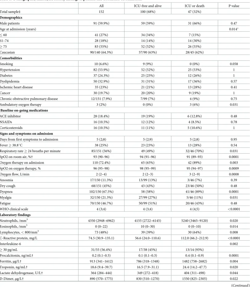
Table 1. Demographic, clinical, laboratory findings of patients and treatments on admission.
All ICU-free and alive ICU or death P value
Total sample‡ 152 100 (68%) 47 (32%)
Demographics
Male patients 91 (59.9%) 59 (59%) 31 (66%) 0.47
Age at admission (years) 0.014�
� 60 41 (27%) 34 (34%) 7 (15%) 61–74 28 (18%) 14 (14%) 14 (30%) � 75 83 (55%) 52 (52%) 26 (55%) Caucasian 90/140 (64.3%) 57/90 (63%) 28/45 (62%) Comorbidities Smoking 10 (6.6%) 9 (9%) 0 (0%) 0.058 Hypertension 82 (53.9%) 52 (52%) 25 (53%) 1 Diabetes 37 (24.3%) 25 (25%) 12 (26%) 1 Dyslipidemia 50 (32.9%) 31 (31%) 17 (36%) 0.57
Ischemic heart disease 35 (23%) 21 (21%) 13 (28%) 0.41
Cancer 30 (19.7%) 20 (20%) 9 (19%) 1
Chronic obstructive pulmonary disease 12/151 (7.9%) 7/99 (7%) 4 (9%) 0.75
Ambulatory oxygen therapy 3 (2%) 0 (0%) 3 (6%) 0.031
Baseline on-going medications
ACE inhibitor 28 (18.4%) 19 (19%) 6 (12.8%) 0.48
NSAIDs 16 (10.5%) 12 (12%) 4 (8.5%) 0.78
Corticosteroids 16 (10.5%) 11 (11%) 5 (10.6%) 1
Signs and symptoms on admission
Days from first symptoms to admission 5 (2;8) 5 (2;9) 5 (2;8) 0.95
Fever � 38.8˚C 38 (25%) 23 (23%) 13 (28%) 0.54
Respiratory rate � 24 breaths per minute 85/151 (56%) 49 (49%) 32/46 (70%) 0.031
SpO2 on room air, %† 93 (90–96) 94 (91–96) 91 (89–93) 0.0001
Oxygen therapy on admission 110 (72.4%) 65 (65%) 42 (89%) 0.003
SpO2 on oxygen therapy, % 96 (95–98) 98 (95–99) 95 (94–97) 0.0009
Oxygen flow, L/min 2 (2–4) 2 (2–3) 3 (2–9) 0.0008
Anosmia 17/150 (11.3%) 13/99 (13%) 3/46 (7%) 0.39
Dry cough 68/151 (45%) 43 (43%) 23/46 (50%) 0.48
Dyspnea 102/150 (67.5%) 58 (58%) 41/46 (89%) 0.0001
Myalgia 32/150 (21.3%) 27/99 (27%) 5/46 (11%) 0.031
Fatigue 70/150 (46.7%) 50/99 (51%) 20/46 (43%) 0.48
WHO clinical scale 4 (3;4) 4 (3;4) 4 (4;5) <0.0001
Laboratory findings Neutrophils, /mm3 4350 (2948–6962) 4155 (2722–6145) 5240 (3465–9120) 0.020 Eosinophils, /mm3 0 (0–22) 10 (0–30) 0 (0–10) 0.014 Lymphocytes, < 800/mm3 73 (48%) 39 (39%) 30 (64%) 0.008 C-Reactive protein, mg/L 74.5 (30.9–135.1) 56.6 (24.0–110.6) 112.0 (66.2–212.9) <0.0001 Interleukine-6 0.002 � 30 pg/mL 31/55 (56.4%) 17/38 (45%) 13/14 (93%) Procalcitonin, ng/mL† 0.2 (0.1–0.5) 0.1 (0.1–0.3) 0.4 (0.1–0.9) 0.0001 Ferritin,μg/L† 913 (341–1612) 786 (318–1348) 1482 (758–2682) 0.004 Troponin, ng/mL† 18.6 (9.4–39.7) 16.5 (7.9–31.1) 24.4 (14.2–47.7) 0.020
Lactate dehydrogenase, U/L† 364 (284–444) 349 (272–418) 404 (311–498) 0.044
D-Dimer,μg/L† 890 (570–1775) 830 (510–1270) 1550 (825–2305) 0.022
were 11 (6.75–14.5) and 6 (4–9) days, respectively. The estimated probability of ICU transfer or death by D14 was 32% (95% CI 25–40), the estimated probability of still needing non-inva-sive ventilation (NIV) or mechanical ventilation (MV), or being dead, at D14 was 27% (95% CI 20–35), while the estimated probability of being discharged alive by D14 was 58% (95% CI 49–66).
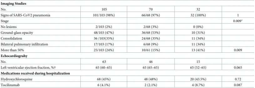
In univariable analysis, age at admission, chronic respiratory failure, respiratory rate � 24 breaths per minute, peripheral capillary oxygen saturation (SpO2) on room air, oxygen ther-apy on admission, SpO2 on oxygen, dyspnea, myalgia, WHO clinical scale, neutrophilia, eosi-nopenia, lymphopenia, CRP level, IL-6 level, procalcitonin, fibrinogen, serum ferritin, high-sensitivity cardiac troponin T, lactate dehydrogenase (LDH), D-dimer, and chest CT scan were associated with ICU transfer and/or death within 14 days (Table 1). For adjusted model development, the limiting number of events was 47 patients with ICU transfer or death within 14 days in the original sample. The multivariable model included age (� or > 60 years), respi-ratory baseline presentation (assessed by WHO scale levels from 3 to 5), CRP level and lym-phocytes count. Older age (OR 2.61, 95% CI 0.96–7.10), poorer respiratory presentation (OR 4.04 per 1-point increment on WHO scale, 95% CI 1.76–9.25) and higher CRP level (OR 1.63 per 100mg/L increment, 95% CI 0.98–2.71) were associated with an increased risk of ICU requirement or death, while lymphocytes count were associated with better outcome (OR 0.36 per 1000/mm3increment, 95% CI 0.13–0.99) (Fig 2, S2 Table inS1 File).Fig 2shows a forest plot of the multivariable models of COVID-19 patient’s outcomes. Internal and external vali-dation of the model was performed: the C-index (equivalent to AUC) was 0.80, 0.78 after cor-rection for over-optimism by resampling, and 0.78 on the external cohort (seeS1 Filefor further details and S1 Table inS1 Filefor description of the external cohort).
Table 1. (Continued) Imaging Studies
No. 105 70 32
Signs of SARS-CoV2 pneumonia 101/103 (98%) 66/68 (97%) 32 (100%) 1
Stage 0.009�
No lesions 2/103 (2%) 2/68 (3%) 0 (0%)
Ground-glass opacity 48/103 (47%) 36/68 (53%) 10 (31%)
Consolidation 36 /103(35%) 24/68 (35%) 11 (34%)
Bilateral pulmonary infiltration 17/103 (17%) 6/68 (9%) 11 (34%)
More than 50% 25/103 (24%) 10/61 (15%) 13 (41%) 0.009
Echocardiograhy
No. 63 46 15
Left ventricular ejection fraction, %† 65 (60–65) 65 (65–65) 65 (52–65) 0.065
Medications received during hospitalization
Hydroxychloroquine 68 (45%) 48 (48%) 20 (43.5%) 0.72
Tocilizumab 6 (4.1%) 2 (2.1%) 4 (8.7%) 0.087
ACE, angiotension-converting enzyme; NSAIDs, nonsteroidal anti-inflammatory drugs; SARS-CoV-2, severe acute respiratory syndrome coronavirus 2; WHO, World Health Organization; SpO2, peripheral capillary oxygen saturation. Data are median (IQR), n (%) or n/N (%). P values were calculated by Mann-Whitney U test, Fisher’s exact test, as appropriate.
‡5 of the 152 patients has incomplete follow-up for ICU transfer or death within 14 days
�Fisher’s exact test comparing all subcategories.
†: number of missing values for quantitative variables: SpO2n = 13, Procalcitonine n = 21, Ferritin n = 59, Troponin n = 22, LDH n = 46, D-Dimer n = 74, LVEF n = 2 among patients with echocardiography [for categorical variables, in case of missing values, the denominator in the table indicates the number of complete cases].
Fig 1. Flow chart of COVID-19 patients’ outcome. Maximum follow-up = 14 days.�at the time of analysis, 6 patients were still followed up for the study endpoint, 5
hospitalized in the medical ward and 1 in the ICU.
https://doi.org/10.1371/journal.pone.0240711.g001
Fig 2. Forest plot of multivariable analysis of COVID-19 patients’ outcome [black squares: Model for ICU transfer or death within 14 days of admission (main endpoint, analysis on 147 observations); gray triangles: Model for hospitalization status at day 14 (analysis on 146 observations); gray circles: Model for detailed status at day 14 (analysis on 146 observations)]. https://doi.org/10.1371/journal.pone.0240711.g002
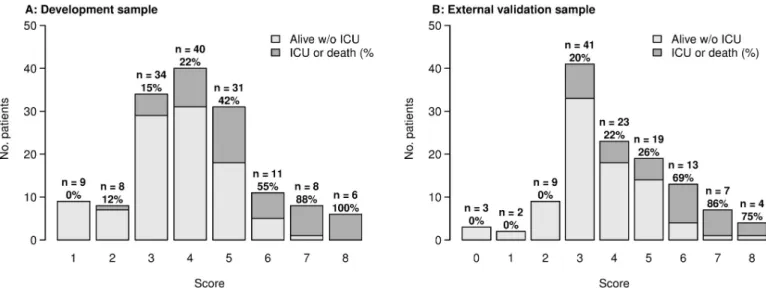
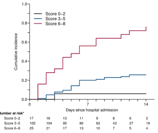
A tentative simplified scoring system was defined for the main endpoint (ICU transfer or death within 14 days of admission), for routine clinical field practice. To that aim, based on the linear predictor and the coefficients of the multivariable model, in an additive manner, 1 point was allo-cated for age above 60 years old; 1 point for oxygen therapy by nasal prongs or mask (WHO scale level 4); 3 points for high flow oxygen or NIV (WHO scale level 5); 1 point if 10 � CRP plasma level � 75 mg/L, 2 points if 75 � CRP � 150 mg/L, 3 points if CRP � 150 mg/L; 1 point if lympho-cytes count below 800/mm3 (SeeS1 File).Fig 3displays stratified risk according to each score. Therefore, we defined three risk groups: low (score 0–2), moderate (score 3–5), and high (score 6–8). Cumulative incidence for each of these groups is shown inFig 4. Overall, the estimated sensi-tivity of a score greater than 2 (moderate and severe risk groups) was 97% (95% CI 94–100), and the specificity of a score lower than 6 (low and moderate risk groups) was 94% (95% CI 89–98) for the main outcome. The positive predictive value for a high-risk score was 76% (95% CI 61–91), while the negative predictive value for a low risk score was 94% (95% CI 82–100).
At day 14, a total of 40 patients were still treated with NIV (n = 1) or MV (n = 7) ventilation,
or had died (n = 32), out of 146 evaluable patients. In univariable analysis, age at admission,
weight, chronic respiratory failure, respiratory rate � 24, SpO2 on room air, Oxygen therapy on admission, SpO2 on oxygen, dyspnea, myalgia, WHO clinical scale, neutrophils, eosino-phils, lymphocytes, platelets, CRP level, IL-6 level, procalcitonin, serum ferritin, high-sensitiv-ity cardiac troponin T, D-dimer, and chest CT-scan were associated with WHO scale � 5 within day 14. Multivariable analysis is represented inFig 2.
Eighty-four patients had been discharged by day 14, out of 146 evaluable patients. In uni-variable analysis, age at admission, respiratory rate < 24, SpO2 on room air, Oxygen therapy on admission, ageusia, dyspnea, WHO clinical scale, neutrophils, eosinophils, lymphocytes, platelets, CRP level, IL-6 level, procalcitonin, fibrinogen, serum ferritin, high-sensitivity car-diac troponin T, LDH, D-dimer, and chest CT scan were associated with discharge alive within 14 days. Multivariable analysis is represented inFig 2.
Discussion
The natural history and outcome of the COVID-19 patients initially hospitalized in a medical ward remain unpredictable. Currently, the main existing medical information stem from
Fig 3. Proportions of ICU transfer or death within 14 days after admission by risk score. Left panel A: development cohort. Right panel B: external validation
cohort.
China and prognostic factors of COVID-19 among European population are lacking. The most striking conclusions drawn by this study are (i) up to 35% of the COVID-19 patients hos-pitalized in a medical ward were transferred to ICU or died at day 14, (ii) we defined high-risk group of ICU transfer or death using a simplified scoring system from the multivariable mod-els including age, CRP level, lymphocytes count and WHO scale and (iii) we highlighted corre-lation between IL-6 level and extensive lesions in CT scan.
A clear and strong age gradient in death risk has been identified, increasing dramatically after 60 years [15]. Besides older age, comorbidities are also highlighted as key factors associ-ated with death [7,8,16]. Compared to the present study, retrospective Chinese cohorts popu-lation were younger (from 51 to 56 years) and had less comorbidities (up to 48%) [7,16]. Even with a median age of 77 years and more than 80% of comorbidity, our reported 21.9% mortal-ity rate lies within the 17.6–28.2% range extracted from other cohorts [7,8]. In contrast, the median time from symptoms onset to death in our population (11 days) is shorter than the 18.5 days previously reported [7], which can be ultimately the consequence of the higher risk profile of patients in the present study. Additionally, our ICU transfer rate (11.6%) was lower than the 26% described in Chinese cohorts [7,16]. In this regard, we must underline that our patients presented with less severe infection at baseline [7,16]. In addition, they were less eligi-ble to ICU admission, due to age and comorbidities. Beyond demographic and clinical charac-teristics, several laboratory features have been linked to a higher mortality. Studies identified a positive correlation with mortality for neutrophilia, lymphopenia, troponin, LDH and D-dimer levels [7,16]. Additionally, high levels of serum CRP, procalcitonin, and ferritin have also occasionally been associated with mortality [16,17]. In our cohort, two simple biomarkers from routine practice, lymphocytes count and CRP level, are independently associated with a
Fig 4. Cumulative incidence of ICU transfer or death by risk score. https://doi.org/10.1371/journal.pone.0240711.g004
worse prognosis. CRP level higher than 75 mg/L and lymphopenia below 800/mm3increased by two fold the odds of being transfer in ICU or death.
Herein, we provided for the first time a simplified scoring system which allows stratifying COVID-19 patients initially hospitalized in a medical ward, at low, intermediate, or high risk of ICU transfer or death. The score was validated with calibration evaluated both with an inter-nal resampling approach and by exterinter-nal validation on a cohort sample from a different hospi-tal. Based on the linear predictor of the multivariate model, age above 60 years, WHO scale, CRP level (10–75, 75–150, or > 150 mg/L), and lymphocytes count below 800/mm3 were included in the scoring system. A score equal or greater than 6 at baseline had a predicted probability of more than 60% to be transferred to ICU or dead by D14. In our regard, this high-risk patient profile should be monitored more closely and eventually considered for more aggressive treatment protocols than a patient with a score of less than 3. In a systematic review of the prediction models for diagnosis and prognosis of COVID 19 patients, Wynants et al identified ten prognostic models proposed by different Chinese teams [12]. By the time of this article writing, all these models were only available in pre-print and had not been peer-reviewed. They were exclusively based on small retrospectives cohorts, with most of them lack-ing an external validation cohort, or presentlack-ing a non-comparable small validation cohort. Nguyen et al [18] developed a 7-point score based on a retrospective analysis of 279 hospital-ized patients but without external validation. The strengths of the score presented here are its prospective nature and its external validation. In addition, its readily accessible variables make it easily reproducible in clinical practice.
Apart from CRP level and lymphocyte count, other significant findings from our study could be further used to refine the score. Chest CT scan is a useful diagnostic tool, especially for RT-PCR negative patients, but its role as a prognostic instrument is still unclear [19]. Herein, we pointed out that parenchymal involvement greater than 50% on chest CT scan at admission was associated with ICU transfer or death in 41% of cases. In parallel, high levels of serum IL-6 have been reported in moderate to severe cases of COVID-19 pneumonia [7,17]. IL-6 may result in increased alveolar-capillary blood-gas exchange dysfunction, especially impaired oxygen diffusion, and lead to pulmonary fibrosis and organ failure [20]. We were able to establish for the first time the correlation between IL-6 level and extensive parenchymal involvement on chest CT scan for ICU transfer or death.
Our study has some limitations. We presented models with both internal and external vali-dation. Discrimination of the model and of the simplified score for the main endpoint was consistent in the external cohort. Calibration assessment showed a slightly overestimated risk of event in the external cohort for those with higher scores. The external sample consisted of patients from a regional non-university hospital, which could explain the differences on catch-ment area and patient recruitcatch-ment. In the acute context of the first SARS-CoV-2 epidemic wave in France, we relied on a sample prospectively defined by consecutive eligible patients in the study center. Overall, the limited sample sizes of both development and validation samples require caution in interpreting results. Ideally, a sample size calculation at planning stage of the study should ensure sufficient collected data for predictive model development and valida-tion; approaches have been proposed to that aim [21,22]. Further external validation on larger prospective cohorts with planned sample sizes will be useful.
To our knowledge, this is the first prospective European cohort of COVID-19 non-critical inpatients and one of the largest standardized studies describing short term patients outcome. We provided a very simple and easily accessible score to estimate the risk of ICU transfer or death by day 14. In the context of the pandemic, this tool can help the management of patient flow, and also clinical trial design and therapeutic management.
Supporting information
S1 File. Supplementary material.
(DOCX)
Acknowledgments
DIMICOVID is De´partement de Me´decine Interne et Immunologie clinique groupe pour COVID-19à l’Hoˆpital Pitie´-Salpêtrière, APHP, Paris Sorbonne includes: Yves Allenbach, David Saadoun, Georgina Maalouf, Matheus Vieira, Alexandra Hellio, Jacques Boddaert, He´lène Gros, Joe Elie Salem, Olivier Benveniste, Patrice Cacoub, Ahlem Chaib, Nicolas Champtiaux, Aude Rigolet, Anne Simon, Ste´phane Barete, Jean-Charles Piette Perrine Guil-laume-Jugnot, Yasmina Ferfar, Mathieu Vautier, Se´golène Toquet-Bouedec, Christian de Gen-nes, Fanny Domont, Gae¨lle Leroux, Mathilde Leclercq, Chloe´ Comarmond, Anne-Claire Desbois, Nabiha Sbeih, Amine Ghembaza, Joana Alves-vieira, Hugues Gontier, Sofia Garabet-yan, Marion Larue, Andre´a Patissier, Elissone Sarkis, Sandrine Tramond, Roxana-Maria Bog-dan, Nicias Gorge, Benjamin Rossi, Marie Anne Bouldouyre, He´lène Guillot, Keito Le Goff, Leila Lefevre, Serge Barmo, Ana-Maria Cardamisa, Margot Hulin, Alexandre Lejoncour, Ce´line Anquetil, Bailly Laurent, Corti Le´onard, Gonc¸alo Boleto, Cindye Marques, Fe´lix Blanc, Charlotte Bouzbib, Sara Philonenkon, Violaine Foltz, Jeremy Rezai, Christiane Stern, Manon Allaire, Philippe Sultanik, Oussama Mouri, Alessandra Mazzola, Fre´de´rique Gandjbakhch, Eouard Larrey, Laure Gossec, Charlotte Tomeo, Vincent Mallet, Cle´mence Fron, Marika Rudler, Aline Lecleach, Bruno Fautrel, Pascal Lebray.
Author Contributions
Conceptualization: Yves Allenbach, David Saadoun, Olivier Benveniste, Patrice Cacoub. Data curation: Yves Allenbach, David Saadoun, Georgina Maalouf, Matheus Vieira,
Alexan-dra Hellio, Jacques Boddaert, He´lène Gros, Joe Elie Salem.
Formal analysis: Lucie Biard.
Investigation: Yves Allenbach, David Saadoun, Georgina Maalouf, Matheus Vieira. Methodology: Matthieu Resche Rigon, Cherifa Menyssa, Lucie Biard.
Software: Matthieu Resche Rigon, Cherifa Menyssa, Lucie Biard. Supervision: Patrice Cacoub.
Validation: Olivier Benveniste.
Writing – original draft: Yves Allenbach, David Saadoun, Georgina Maalouf, Matheus Vieira. Writing – review & editing: Jacques Boddaert, Matthieu Resche Rigon, Lucie Biard, Olivier
Benveniste, Patrice Cacoub.
References
1. World Health Organization. Corona-virus disease (COVID-19) outbreak. Accessed May 1, 2020.
https://www.who.int
2. Mizumoto K, Kagaya K, Zarebski A, Chowell G. Estimating the asymptomatic proportion of coronavirus disease 2019 (COVID-19) cases on board the Diamond Princess cruise ship, Yokohama, Japan, 2020. Euro Surveill 2020; 25(10):1–5.
3. Nishiura H, Kobayashi T, Miyama T, Suzuki A, Jung SM, Hayashi K, et al. Estimation of the asymptom-atic ratio of novel coronavirus infections (COVID-19). Int J Infect Dis 2020, Published online Feb 13.
4. Wu Z, McGoogan JM. Characteristics of and important lessons from the coronavirus disease 2019 (COVID-19) outbreak in China: summary of a report of 72 314 cases from the Chinese Center for Dis-ease Control and Prevention. JAMA 2020; Published online Feb 24.https://doi.org/10.1001/jama.2020. 2648
5. Guan WJ, Ni ZY, Hu Y, Liang WH, Ou CQ, He JX, et al. China Medical Treatment Expert Group for Covid-19. Clinical characteristics of coronavirus disease 2019 in China. N Engl J Med 2020; 382:1708– 1720.https://doi.org/10.1056/NEJMoa2002032PMID:32109013
6. Grasselli G, Pesenti A, Cecconi M. Critical care utilization for the COVID-19 outbreak in Lombardy, Italy: early experience and forecast during an emergency response. JAMA 2020; Published online Mar 13.https://doi.org/10.1001/jama.2020.4031.PMID:32167538
7. Zhou F, Yu T, Du R, Fan G, Liu Y, Liu Z, et al. Clinical course and risk factors for mortality of adult inpa-tients with COVID-19 in Wuhan, China: a retrospective cohort study. Lancet 2020; 395(10229):1054– 1062.https://doi.org/10.1016/S0140-6736(20)30566-3PMID:32171076
8. Cao J, Tu WJ, Cheng W, Yu L, Liu YK, Hu X, et al. Clinical Features and Short-term Outcomes of 102 Patients with Corona Virus Disease 2019 in Wuhan, China. Clin Infect Dis 2020; Published online Apr 2.
https://doi.org/10.1093/cid/ciaa243PMID:32239127
9. Guan W, Liang W, Zhao Y, Liang HR, Chen ZS, Li YM, et al. Comorbidity and its impact on 1590 patients with Covid-19 in China: A Nationwide Analysis. Eur Respir J 2020; Published online Mar 26.
https://doi.org/10.1183/13993003.00547–2020
10. Wang L, He W, Yu X, Hu D, Bao M, Liu H, et al. Coronavirus Disease 2019 in elderly patients: character-istics and prognostic factors based on 4-week follow-up. J Infect 2020; Published online Mar 30.https:// doi.org/10.1016/j.jinf.2020.03.019PMID:32240670
11. Coronavirus COVID-19 Global Cases by the Center for Systems Science and Engineering (CSSE). Accessed May 12, 2020.https://coronavirus.jhu.edu/map.html
12. Wynants L, Van Calster B, Bonten MMJ, Collins GS, Debray TPA, De Vos M, et al. Prediction models for diagnosis and prognosis of covid-19 infection: systematic review and critical appraisal. BMJ. 2020; 369:m1328.https://doi.org/10.1136/bmj.m1328PMID:32265220
13. WHO R&D Blueprint—COVID-19 Therapeutic Trial Synopsis Draft, February 18, 2020. Accessed May 1 2020.https://www.who.int/blueprint/priority-diseases/key-action/COVID-19_Treatment_Trial_ Design_Master_Protocol_synopsis_Final_18022020.pdf
14. Harrell F Jr, Regression Modelling Strategies ( 2nd Ed) 2015; Springer International Publishing, New York.
15. Verity R, Okell LC, Dorigatti I, et al. Estimates of the severity of coronavirus disease 2019: a model-based analysis. Lancet Infect Dis. 2020; pii: S1473-3099(20)30243-7. https://doi.org/10.1016/S1473-3099(20)30243-7PMID:32240634
16. Wu C, Chen X, Cai Y, Winskill P, Whittaker C, Imai N, et al. Risk Factors Associated With Acute Respi-ratory Distress Syndrome and Death in Patients With Coronavirus Disease 2019 Pneumonia in Wuhan, China. JAMA Intern Med 2020; Published online Mar 13.https://doi.org/10.1001/jamainternmed.2020. 0994PMID:32167524
17. Chen G, Wu D, Guo W, Cao Y, Huang D, Wang H, et al. Clinical and immunological features of severe and moderate coronavirus disease 2019. J Clin Invest. 2020; 130(5):2620–2629.https://doi.org/10. 1172/JCI137244PMID:32217835
18. Nguyen Yann, Corre Fe´lix, Honsel Vasco, Sonja C Vinciane Rebours, Fantin Bruno, et al. A nomogram to predict the risk of unfavourable outcome in COVID-19: a retrospective cohort of 279 hospitalized patients in Paris area, Annals of Medicine, 52:7, 367–375,https://doi.org/10.1080/07853890.2020. 1803499PMID:32723107
19. Rubin GD, Ryerson CJ, Haramati LB, Sverzellati N, Kanne JP, Raoofet S, et al. The Role of Chest Imaging in Patient Management during the COVID-19 Pandemic: A Multinational Consensus Statement from the Fleischner Society. Chest 2020; Published online Apr 7.https://doi.org/10.1016/j.chest.2020. 04.003.
20. Zhou Y, Fu B, Zheng X, Wang D, Zhao C, Qi Y, et al. Aberrant pathogenic GM-CSF+ T cells and inflam-matory CD14+CD16+ monocytes in severe pulmonary syndrome patients of a new coronavirus. Pre-print at BioRxiv 2020.https://doi.org/10.1101/2020.02.12.945576
21. Riley RD, Snell KIE, Burke D et al. Minimum sample size for developing a multivariable prediction model: Part I–Continuous outcomes. Statistics in Medicine 2019; 38:1262–75https://doi.org/10.1002/ sim.7993PMID:30347470
22. Riley RD, Snell KIE, Ensor J et al. Minimum sample size for developing a multivariable prediction model: Part I–binary and time-to-event outcomes. Statistics in Medicine 2019; 38:1276–96https://doi. org/10.1002/sim.7992PMID:30357870