ADAM8 as a drug target in pancreatic cancer
The MIT Faculty has made this article openly available.
Please share
how this access benefits you. Your story matters.
Citation
Schlomann, Uwe et al. “ADAM8 as a Drug Target in Pancreatic
Cancer.” Nature Communications 6 (2015): 6175.
As Published
http://dx.doi.org/10.1038/ncomms7175
Publisher
Nature Publishing Group
Version
Author's final manuscript
Citable link
http://hdl.handle.net/1721.1/106940
Terms of Use
Article is made available in accordance with the publisher's
policy and may be subject to US copyright law. Please refer to the
publisher's site for terms of use.
ADAM8 as a drug target in Pancreatic Cancer
Uwe Schlomann1,2, Garrit Koller1, Catharina Conrad2, Taheera Ferdous1, Panagiota Golfi1, Adolfo Molejon Garcia1, Sabrina Höfling1, Maddy Parsons3, Patricia Costa4, Robin Soper4,
Maud Bossard4, Thorsten Hagemann4, Rozita Roshani4, Norbert Sewald5, Randal R. Ketchem6, Marcia L. Moss7, Fred H. Rasmussen7, Miles A. Miller8, Douglas A. Lauffenburger8, David A. Tuveson9, Christopher Nimsky2, and Jörg W. Bartsch1,2,+
1King’s College London, Institute for Pharmaceutical Science and KCLDI, London SE1 9RT,
United Kingdom
2Department of Neurosurgery, Marburg University, Baldingerstr. 35033 Marburg, Germany 3Randall Institute, King’s College London, SE1 8RT
4Institute of Cancer and Inflammation, St. Mary’s, School of Medicine, John Vane Bldg,
Charterhouse Square, London W, UK
5Department of Organic Chemistry, Bielefeld University, 33615 Bielefeld, Germany 6Therapeutic Discovery, AMGEN Inc., Seattle, WA 98119, USA
7Biozyme Inc., Apex, North Carolina 27523, USA
8Department of Biological Engineering, Massachusetts Institute of Technology, Cambridge, MA,
02139
9Cold Spring Harbor Laboratory NY 11791, USA
Abstract
Pancreatic ductal adenocarcinoma (PDAC) has a grim prognosis with less than 5% survivors after 5 years. High expression levels of ADAM8, a metalloprotease-disintegrin, are correlated with poor clinical outcome. We show that ADAM8 expression is associated with increased migration and invasiveness of PDAC cells caused by activation of ERK 1/2 and higher MMP activities. For biological function, ADAM8 requires multimerisation and associates with β1-integrin on the cell surface. A peptidomimetic ADAM8 inhibitor, BK-1361, designed by structural modelling of the disintegrin domain, prevents ADAM8 multimerisation. In PDAC cells, BK-1361 affects ADAM8
Users may view, print, copy, and download text and data-mine the content in such documents, for the purposes of academic research, subject always to the full Conditions of use:http://www.nature.com/authors/editorial_policies/license.html#terms
+Address correspondence to: Jörg W. Bartsch, Marburg University, Department of Neurosurgery, Baldingerstr., 35033 Marburg, Germany, Tel: +49 6421 58 66753; Fax: +49 6421 58 66795, jbartsch@med.uni-marburg.de.
Author contributions
2Author contributions: Study was conceived by JWB, DAT, CN, DAL, TH, MP, MLM, NS, experiments were designed by JWB, GK, US, MLM; acquisition of all the data US, GK, CC, TF, PG, AMG, SH, PC, RS, MB, RR, RRK, FHR, MAM, the analysis and interpretation of data was performed by US, GK, CC, JWB, MLM, DAL, MAM, the final manuscript was prepared US, GK, CC, MLM, JWB, the whole study was supervised by JWB.
Author Manuscript
Nat Commun. Author manuscript; available in PMC 2016 September 07.
Published in final edited form as:
Nat Commun. ; 6: 6175. doi:10.1038/ncomms7175.
Europe PMC Funders Author Manuscripts
function leading to reduced invasiveness, and less ERK 1/2 and MMP activation. BK-1361 application in mice decreased tumour burden and metastasis of implanted pancreatic tumour cells and provides improved metrics of clinical symptoms and survival in a KrasG12D-driven mouse model of PDAC. Thus, our data integrate ADAM8 in pancreatic cancer signalling and validate ADAM8 as a target for PDAC therapy.
Introduction
Pancreatic ductal adenocarcinoma (PDAC) has the highest mortality rate of solid organ cancers with a 5-year survival rate below 5%1. This cancer type is remarkably homogenous in that 95% of PDAC originate from oncogenic mutations in the KRAS gene. Hence, representative authentic mouse models of PDAC with pancreas-specific expression of Kras have been generated2. In mice and man, KRAS mutations cause early stage pancreatic epithelial neoplasias (PanINs) with subsequent development of progressive PDAC. A hallmark of PDAC is the massive infiltration of tumour cells into the pancreas and
surrounding tissues including lymphatic organs, spleen and peritoneum and the concomitant metastasis to the liver and lungs3–6. Infiltration of pancreatic tumour cells depends critically on extracellular matrix (ECM) remodeling7, 8. Given the importance of the ECM in PDAC, the proteolytic release of membrane proteins (“shedding”) as well as ECM (e.g. collagens and fibronectin) degradation has previously been postulated to play a pivotal role in shaping the tumour microenvironment9, 10. Members of the Metzincin superfamily, Matrix
Metalloproteases (MMPs) and/or ADAM (A Disintegrin And Metalloproteinase) proteases have been described in these processes11. In particular, the contribution of ADAMs to extracellular remodeling12 and tumour growth, infiltration, metastasis and angiogenesis by shedding of membrane-associated proteins may be important9, 13, 14. In PDAC patient samples, elevated expression levels of ADAM8 (CD156a, MS2) have been identified vs. normal pancreatic tissues. In normal pancreas, ADAM8 expression is very low and restricted to the plasma membrane of ductal cells and, to a lesser extent, of islets and acinar cells. In PDAC tissues, ADAM8 is strongly expressed in tubular complexes and in cancer cells. Based on clinical data, high ADAM8 expression levels are associated with a poor patient prognosis, resulting in reduced survival and increased metastatic spread15.
ADAM8 is a proteolytically active member of the ADAM protease family originally described in inflammatory processes16–18 and subsequently in many systems of the body19. Increased expression of ADAM8 was observed in other neoplasias, such as high-grade glioma20, lung adenocarcinoma21, prostate cancer22, and more recently, in squamous head and neck cell carcinoma23, medulloblastoma24, osteosarcoma25 and breast cancer26 suggesting that ADAM8 plays an active role in tumour progression. Thus, understanding the functional role of ADAM8 in tumour biology is important.
ADAM8 is localized in a few distinct cell types and the analysis of ADAM8 deficient mice inferred dispensability for normal development and homoeostasis27, 28. ADAM8 is typically expressed at low levels, giving rise to the current hypothesis that it is functionally irrelevant for homeostasis unless induced by inflammatory stimuli17 or neoplasias. Once upregulated, ADAM8 can overlap with the substrate spectrum of ADAM10 and ADAM17,
Europe PMC Funders Author Manuscripts
two major shedding enzymes, and cleave proteins with immune functions such as Tumour Necrosis Factor receptor 1 (TNF-R128), L-Selectin29, CD2330, CXCL131, as well as cell adhesion proteins such as CHL132 thereby potentially modulating immune response or cell adhesion. Cleavage of other ADAM8 substrates such as Tie-2, Flt-1, VE-cadherin, Flk-1, EphB4, KL-1, CD31, and E-selectin33 or by cleavage of fibronectin12 may control tumour angiogenesis. Moreover, a role for ADAM8 in metastases34 and in cell invasiveness15, 20, 22 has been postulated, though the mechanism underlying these processes is unknown. ADAM8 is activated by autocatalysis in the trans-Golgi network (TGN)35 and, unlike other ADAMs, not by furine-like convertases. For in vivo activity, ADAM8 requires homophilic multimerisation of at least two ADAM8 monomers on the cell membrane. This specific interaction of ADAM8 monomers offers a potential strategy for blocking ADAM8 activity by preventing ADAM8 multimerisation in vivo. We have demonstrated previously that ADAM8-ADAM8 interactions critically depend on the disintegrin/cysteine-rich domain35. In addition, ADAM family members can bind to integrins in vitro via their disintegrin/ cysteine-rich domains36; as prototype, human ADAM15 contains a canonical RGD motif in the integrin-binding loop of the disintegrin domain37. However even for non-RGD
containing ADAMs such as ADAM9, integrin binding was demonstrated. ADAM9 binding to β1 integrin causes migration of melanoma cells38 and for ADAM8, binding to α9β1 was
shown in osteoclast turnover39 suggesting that these ADAM-integrin interaction have functional relevance.
Although ADAM8 has been associated with increased tumour cell migration, invasiveness and metastasis via a combination of catalytic, adhesion and cell signalling functions15, 20, no mechanistic data on ADAM8 in tumour progression, and specifically in PDAC are available. Here we provide evidence for an involvement of ADAM8 in cancer signalling and in tumour progression. Furthermore, we validate ADAM8 as a target in PDAC by
introducing a specific ADAM8 inhibitor.
Results
ADAM8 inhibition strategy
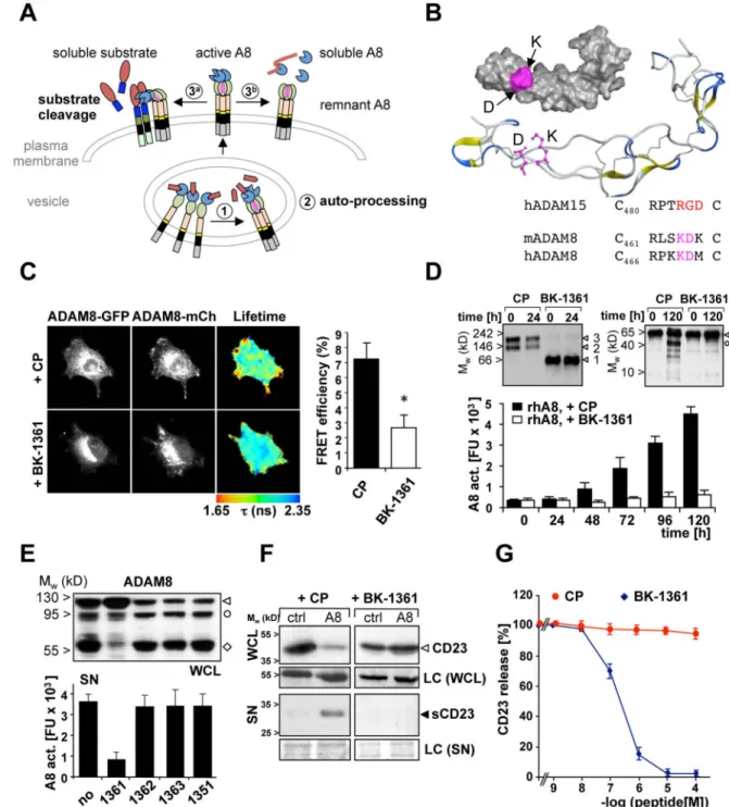
Cellular activation of ADAM8 occurs in two steps. The first is intracellular prodomain removal in vesicles while the second is metalloprotease (MP) domain removal from membrane-bound activated ADAM8 (Fig. 1A). Autocatalysis implies that ADAM8 multimerises (Fig. 1A) and that the ADAM8 disintegrin/cysteine-rich (DC) domain is critical for multimerisation as demonstrated previously by using an antibody directed against the DC domain35. To define the regions in the DC domain involved in ADAM8-ADAM8 interactions, homology modelling of the ADAM8 DC domain was performed based on the ADAM10 sequence derived from Janes et al.40 (Supplementary Fig. 1 and 2). We
hypothesised that an extended loop structure exposing the RGD-like positions of the “KDX” motif in the integrin binding loop (IBL) in human and mouse ADAM8 might be responsible for the observed interactions (Fig. 1B).
Europe PMC Funders Author Manuscripts
In mouse and human ADAM8, the amino acid residues K and D are exposed towards the outer aspect of the disintegrin (DI) domain thereby forming a potential contact interface (Fig. 1B, labelled in magenta). To generate a peptidomimetic compound, a series of cyclic peptides (6 aa) mimicking the motif “RLSKDK” of mouse ADAM8 in the IBL were generated. Amino acids R, L, or S were inserted in the peptide as D-amino acids to alter the conformational constraint of the KDK motif and to generate a potentially more stable peptide for in vivo work. The cyclic peptide sequence RLsKDK with “s” as D-serine named BK-1361 was most effective in blocking ADAM8-dependent cell adhesion in mouse and human cells with similar efficiencies (Supplementary Fig. 3) and ADAM8-ADAM8 interactions as shown by reduced FRET/FLIM efficiency. In contrast, a control peptide (CP, RLsADK; Fig. 1C) had no effect.
Multimerisation of ADAM8 was investigated by native gel electrophoresis (Fig. 1 D). Under native conditions, 100 ng of pro-ADAM8 associates in dimeric (~120 kD) and trimeric (~180 kD) complexes of pro-ADAM8, while in the presence of BK-1361, only monomers (~60 kD) were detected (Fig. 1D, left panel). Detection of dimers and trimers suggests that dimers associate by disintegrin domain (homophilic) interactions, whereas trimers could be formed by a different mode of interaction. At higher concentrations of recombinant ADAM8, we detected even larger complexes as a result of greater order multimerisation (Supplementary Figure 3E) in agreement with more than one interaction mode that results in ADAM8 complex formation. These interactions can be blocked by BK-1361 and we further analysed if prevention of complex formation affects ADAM8 activity in vitro.
Activation of pro-ADAM8 was detected over a time course of 120 h (Fig. 1D, right panel) but blocked by BK-1361 (Fig. 1D, bar graph) with an IC50 of 120 ± 19 nM. In a cell-based
assay, BK-1361 and other peptides (see table 1) were tested for their ability to inhibit ADAM8-dependent extracellular resulting in an active soluble MP domain (Figure 1 A, 3b). In cell lysates, presence of the remnant form indicates cellular processing of ADAM8 as seen with no or inactive control peptides (Fig. 1 E, upper panel). In cell supernatants, processing results in detectable activity of released ADAM8 MP ‥ BK-1361, but not other BK peptide variants, decreased this activity (Fig. 1 E, lower panel). BK-1361 blocks mouse and human ADAM8 in vitro with similar efficiencies, a prerequisite for testing BK-1361 in orthotopic PDAC models using human donor cells in mouse hosts.
We next tested BK-1361 and variants for their ability to inhibit shedding of CD23, a known substrate of ADAM830 in cell-based shedding assays (Fig. 1F and table 1). Co-transfection of ADAM8 with a tagged CD23 construct in COS7 cells resulted in significant shedding of CD23 as soluble 21 kD fragment (sCD23); sCD23 was detectable in supernatants when ADAM8 was co-expressed. In the presence of 500 nM BK-1361, sCD23 was undetectable, demonstrating that BK-1361 inhibits the in vivo shedding activity of ADAM8. ELISA assays were performed to determine [sCD23] vs. [BK-1361] with an IC50 of 182 ± 23 nM
for BK-1361; BK-1362 had no significant effect (Fig. 1G). We conclude that BK-1361 affects ADAM8-ADAM8 interactions thereby inhibiting cellular shedding and autocatalytic activation of ADAM8 in vitro and in a cell-based assay in a specific manner, as neither catalytic activities of ADAM 9, 10, 12, 17 nor MMP-2, -9, and -14 were inhibited in concentrations of up to 10 µM (Table 2).
Europe PMC Funders Author Manuscripts
ADAM8 induces migration/invasion of pancreatic cancer cells
As shown earlier, ADAM8 expression is correlated with invasiveness in vitro in various cell lines15,20,21. However, no functional in vivo data on ADAM8 in pancreatic malignancies are yet available. To establish cell lines for analysis of PDAC in vivo, we selected Panc1 (very low ADAM8) and AsPC-1 cells (high endogenous levels of ADAM8) as determined by Western Blot15 (Fig. 2A). Panc1 cell lines with a moderate over-expression of ADAM8 (Panc1_A8; NM_001109.4; Fig. 2B) and AsPC-1 cell lines with ADAM8 shRNA
knockdown constructs were generated. Microarray analysis of Panc1_ctrl vs. Panc1_A8 cells revealed that Panc1_A8 cells showed no off-target effects of the ADAM8 knockdown, as expression levels of >95% of genes were unchanged including genes encoding MMPs, other ADAMs, ADAMTS, and TIMP1-4 levels (Supplementary Fig. 4) Cellular localisation of ADAM8, analysed by CLSM, confirmed low ADAM8 expression in Panc1_ctrl cells compared to enhanced ADAM8 expression in Panc1_A8 cells. In Panc1_A8 cells, ADAM8 is localised in the cell membrane and membrane extrusions (Fig. 2B). In supernatants of different Panc1_A8 cell clones characterised (n=30), ADAM8 catalytic activity correlated with the ADAM8 dosage, determined by western blotting (Fig. 2C) and CD23 fluorescence assay (Fig. 2D). Migration and invasion behaviour of Panc1_ctrl cells was compared to Panc1_A8 cells in vitro (Fig. 2E and F). In wound-healing (“scratch”) assays, migration rates of Panc1_A8 cells were significantly increased (8 ± 3.8-fold) compared to Panc1_ctrl cells. Panc1_A8 cell migration is inhibited by BB-94, a metalloprotease inhibitor. In addition, application of BK-1361 reduced migration rates of Panc1_A8 cells significantly, but not to the same level as BB-94, suggesting that ADAM8 modulates other
metalloprotease activities accounting for migration above control levels. In accordance, invasion into different ECM substrates collagen I, collagenIV, fibronectin, and Matrigel was enhanced by ADAM8 (Fig. 2F).
ADAM8 increases extracellular MMP activity
A proteolytic activity matrix assay (PrAMA)41 was used for simultaneous detection of multiple activities in Panc1_ctrl and Panc1_A8 cells that could account for the observed invasiveness. Briefly, PrAMA is based on the knowledge of individual FRET-substrate MMP/ADAM cleavage signatures using purified enzymes41. Panels of FRET-substrate cleavage measurements can be used to infer a dynamic, quantitative and specific profile of MMP/ADAM proteolytic activities from complex enzyme mixtures such a supernatants and solubilised membranes (see M&M and Supplementary Fig. 5). PrAMA inference revealed increased activities of MMP-2 in supernatants and MMP-14 (MT1-MMP) in cell membranes of Panc1_A8 cells compared to Panc1_ctrl cells (Fig. 2G and Supplementary Fig. 5). Enhanced ADAM8 activities were detected in supernatants and membranes of Panc1_A8 cells. Gelatine zymography and Western blot for MMP-2 and MMP-14 confirmed increased activity of MMP-2 and higher membrane concentration of MT1-MMP (Fig 2H). However, elevated MMP activities in Panc1_A8 cells are not due to transcriptional activation of MMP-2 and MMP-14 (Supplementary Fig. 4).
We further investigated whether BK-1361 is able to affect ADAM8-dependent invasiveness and MMP secretion of Panc1_A8 cells (Fig. 2I and J). BK-1361 and peptide variants were tested for their ability to block invasiveness of Panc1_A8 cells (Fig. 2I). A dose-dependent
Europe PMC Funders Author Manuscripts
effect of BK-1361 on invasion of Panc1_A8 cells was observed. From control peptides, only BK-1364 had a slight effect on invasion. In parallel, we performed PrAMA assays in Panc1_A8 cells to evaluate MMP-2, MMP-14, and ADAM8 activities in the presence of BK-1361, BB-94, and the ERK1/2 inhibitor U0126, respectively (Fig. 2J). BK-1361 and BB-94 reduced activities of MMP-2, MMP-14, and ADAM8. ERK inhibition had a slight effect on MMP-2 activity and a greater effect on MMP-14 activity whilst ADAM8 activity was not affected. These findings argue for an ERK1/2- mediated effect on MMP-14, and, to a lesser extent, on MMP-2 activation. ADAM8 as regulator of ERK1/2 activation is not directly affected by U0126.
Our data suggest an effect of pharmacological ADAM8 inhibition on invasiveness of PDAC cells. To determine the effect of a genetic ADAM8 knockdown on cellular invasiveness, we selected AsPC-1 cells with high endogenous ADAM8 levels (Fig. 2A) and generated AsPC_1 cell clones carrying a stable knockdown of ADAM8 (sh_A8). Three representative cell clones from different sh_A8 constructs were analysed for ADAM8 expression (Fig. 3A-C), cell migration and invasion (Fig. 3 D,E, Supplementary Movie 1). Knockdown of ADAM8 in AsPC-1 cells caused a significant drop in cell migration depending on the gene dosage of ADAM8. Invasion of AsPC-1 cells was similarly affected by ADAM8 dosage (Fig.3E). BK-1361 treatment of wild-type AsPC-1 cells was similar to the genetic
knockdown of ADAM8 with 87 ± 3.5% inhibition (Fig. 3F). PrAMA assays were performed with AsPC-1_shCtrl ± BK-1361, and AsPC-1_shA8 cell clones 1 and 2 (Fig. 3G). Reduction of MMP-2 and MMP-14 activities were observed in AsPC-1_shA8 clones. In BK-1361 treated AsPC-1_shCtrl cells, MMP-2 was similarly affected, however, the effect on MMP-14 was less pronounced (Fig. 3G).
ADAM8 interacts with integrin β1 in pancreatic cancer cells
The membrane localisation of ADAM8 in Panc1_A8 cells suggests that ADAM8 is complexed with cellular integrins thereby enhancing cell migration and invasiveness. To investigate this, co-immunoprecipitation (co-IP) experiments were performed in Panc1 cells expressing either control or a tagged ADAM8 construct (ADAM8-BiPro).,As a result, β1 integrin, present in comparable amounts in Panc1_ctrl and Panc1_A8 cells, was co-immunoprecipitated with ADAM8 (Fig. 4 A,B,C). To analyse cellular ADAM8-β1 integrin and ADAM8-ADAM8 interactions, FRET/FLIM analyses were performed to detect FRET in cell lines expressing fusion proteins ADAM8-GFP and ADAM8-mCherry, respectively. ADAM8 multimerisation was detected in Panc1 cells (Fig. 4D&E), indicated by FRET efficiency of 7.5 ± 0.93%. In cells, complex formation and membrane localisation of ADAM8 in vivo involves β1 integrin, since treatment of Panc1 cells with a β1 integrin-blocking antibody (Fig. 4D and E) resulted in a significant drop to 3.1 ± 0.8% FRET efficiency. Moreover, MDA-MB-231 breast cancer cells lacking β1 integrin (Fig. 4F and G) show a significant change in cellular morphology whilst ADAM8 localisation in lamellipod structures is lost. Interestingly, administration of BK-1361 causes a similar change in cell morphology (Fig. 4 H and I). We conclude that β1 integrin knockdown or specific ADAM8 inhibition have similar effects on ADAM8 membrane localisation (Fig. 4J). In addition, areas of positive ADAM8-FRET in MDA-MB-231 cells were analysed for β1 integrin (Fig.
Europe PMC Funders Author Manuscripts
4 K and L). In most areas, activated β1 integrin was detected by antibody 12G10, suggesting that ADAM8 interaction and β1 integrin activation are correlated.
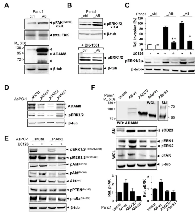
To analyse if ADAM8 interactions cause altered intracellular signalling, a MAP kinase array was used to screen for kinase phosphorylation in Panc1_ctrl and Panc1_A8 cells
(Supplementary Fig. 6). As potential downstream effectors of the observed ADAM8-β1 integrin interaction, we investigated phosphorylation of Focal adhesion kinase (FAK), ERK1/2, Akt, and p38γin Panc1_A8 vs. Panc1_ctrl cells. FAK was described as β1 integrin interacting protein42. In Western Blots, increased phosphorylation of FAK at residue Tyr397 correlates with ADAM8 expression levels in Panc1 cells (Fig. 5A). In addition to pFAK, we detected increased phosphorylation of ERK1/2 (p44/p42) in Panc1_A8 cells by western blotting using corresponding phospho-specific antibodies (3.4-fold ±0.2 Panc1_A8 vs. Panc1_ctrl, Fig. 5B). The observed increase in pERK1/2 was reduced in Panc1_A8 cells treated with BK-1361 (Fig. 5B). To correlate ERK1/2 phosphorylation with the observed invasiveness of Panc1 cells, matrigel invasion assays were performed in the presence of U0126 (Fig. 5C). U0126 blocked ERK1/2 phosphorylation in Panc1_ctrl and Panc1_A8 cells and resulted in decreased invasion of Panc1_A8 cells. In addition, AsPC-1_shCtrl and three AsPC-1_shA8 cell clones with different ADAM8 levels were analysed for pERK1/2 levels (Fig. 5 D). In cell clone AsPC-1_shA8/2, pERK1/2 levels were reduced by 2.9 ± 0.3 fold suggesting that ADAM8 expression levels are correlated with pERK1/2. In AsPC-1 cells, ADAM8 levels affect MEK1/2, p-Akt and c-Raf activation (Fig. 5E). In addition, a β1-integrin antibody that blocks activation was able to reduce pERK1/2 levels in Panc1_A8 cells, demonstrating that β1-integrin is required for ADAM8-dependent ERK1/2 activation (Supplementary Fig. 7).
To investigate whether the observed FAK and ERK1/2 activation depends on membrane bound ADAM8, Panc1 cells were transfected with wild-type ADAM8 (“A8 wt”), an ADAM8 construct lacking the cytoplasmic domain (“A8ΔCD”), or a soluble ADAM8 (“A8ecto”) construct (Fig. 5F). First, we confirmed that all constructs are catalytically active, as all three ADAM8 proteins shed CD23 (“sCD23”) from the cell membrane. Interestingly, neither ∆CD nor the ectodomain of ADAM8 were able to activate FAK and ERK1/2 (Fig. 5F), suggesting that intracellular signalling mediated by ADAM8 requires membrane localisation of ADAM8 and the presence of the cytoplasmic domain. As potential substrates for ERK activation i.e. the EGFR ligand family such as HB-EGF, EGF, or
amphiregulin, were screened (Supplementary Fig. 8). We have not identified significant EGFR ligand release, so that ADAM8-β1 integrin interactions might act independent from EGFR signalling.
Role of ADAM8 in PDAC and effect of BK-1361 in vivo
Based on our findings that ADAM8 inhibition blocks invasiveness and ERK1/2 signalling in PDAC cells, we used BK-1361 to validate ADAM8 as a therapeutic target in PDAC in vivo. Initially, acute and chronic toxicity analyses were performed in C57BL/6J mice. In single and repeated dose applications, doses of up to 10 µg/g body weight were well tolerated as mice showed no abnormalities, weight loss or motor performance over four weeks. After necropsy, organs investigated showed no abnormalities at histological level (Supplementary
Europe PMC Funders Author Manuscripts
Fig. 9). Due to the lack of acute and chronic toxicity, a dose of 10 µg/g body weight was applied daily for subsequent in vivo applications.
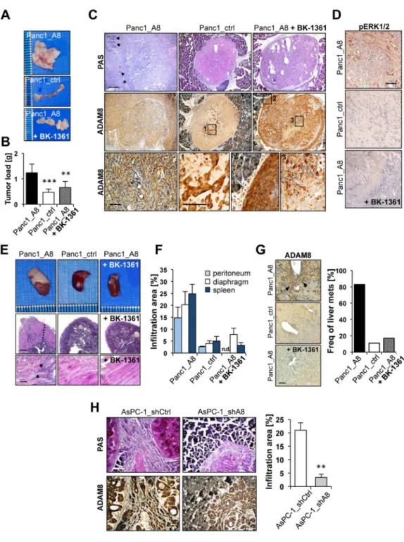
Initially, orthotopic injections of Panc1 cells into mouse pancreas were performed (Fig. 6A-G) in three cohorts (n=12 each); cohort 1 received Panc1_A8 cells, cohort 2 Panc1_ctrl cells, and cohort 3 Panc1_A8 cells followed by daily i.p. injection of 10 µg/g BK-1361. Mice were monitored for 12 days, by which time most of the mice injected with Panc1_A8 cells were moribund and reached endpoint criteria. In contrast, mice injected with Panc1_ctrl cells or Panc1_A8/BK-1361 treatment showed improved clinical parameters. At endpoint, pancreatic tumours formed from Panc1_ctrl cells were significantly smaller than from Panc1_A8 cells (Fig. 6A, B). Moreover tumours obtained from mice that received Panc1_A8 cells and daily injections of BK-1361 were significantly smaller. These data indicate that inhibition of ADAM8 reduced tumour load to almost the value of Panc1_ctrl derived tumours (0.42 grams for Panc1_ctrl vs. 0.62 for Panc1_A8/BK-1361). By histology, a significant invasion of Panc1_A8 cells into the pancreatic tissue was detected, whereas in tumours derived from Panc1_A8/BK-1361 cells, tumour masses embedded in matrigel were primarily localised to the implantation site even after 12 days, as the boundaries of pancreas and implanted tumour mass were still distinct (Fig. 6C). In addition, there were signs of necrosis inside the implanted tumour treated with BK-1361 (Fig. 6C), inferring that non-invasive Panc1 cells undergo necrotic changes. Moreover, ADAM8 levels in Panc1_ctrl cells located in the tumour were increased under hypoxic conditions. Co-staining for pERK1/2 was observed in infiltrative ADAM8-positive tumour cells (Fig. 6D, upper panel).
Metastasis and infiltration is the major cause for the observed morbidity in PDAC5, 6. Since ADAM8 was discussed in the context of infiltration and metastasis26,34, we investigated orthotopic mice for infiltration of close structures such as peritoneum, diaphragm and spleen and liver metastasis. From mice injected with Panc1_A8 cells, we found significant
infiltrates in adjacent organs (Fig. 6E&F). Macroscopic inspection and hematotoxylin/eosin (HE) stain of tissue sections revealed higher invasion into spleen and diaphragm of
Panc1_A8 injected mice (Fig. 6E). Analysis of infiltration areas showed enhanced invasive behaviour of Panc1_A8 cells vs. Panc1_ctrl and Panc1_A8/BK-1361 cells (Fig. 6F) in peritoneum, diaphragm and spleen.
ADAM8 staining of liver sections revealed occurrence of micrometastases with higher frequencies in Panc1_A8 implanted mice compared to Panc1_ctrl and Panc1_A8/BK-1361 (Fig. 6G). Metastases frequencies were markedly different between Panc1_A8, Panc1_ctrl, and Panc1_A8/BK-1361. Furthermore, the implantation of AsPC-1_shCtrl and
AsPC-1_shA8 cells was analysed (Fig. 6H). AsPC-1_shCtrl cells caused large streams of tumour cells invading the pancreatic tissue with an infiltration area of 21 ± 2.8 %. In contrast, AsPC-1_shA8 cells were located close to the injection site and showed less invasive behaviour with infiltration areas of 3.4 ± 1.2% (p<0.01). Thus, data derived from genetic ADAM8 knockdown support the results obtained with ADAM8 inhibition using BK-1361.
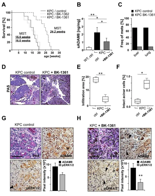
The therapeutic effect of ADAM8 inhibition in vivo was analysed in mice with genotype KrasLSL-G12D, Trp53R172H/+, PdxCre/+(KPC) 2, a genetically engineered PDAC mouse
Europe PMC Funders Author Manuscripts
model. Injections of BK-1361 were started around the onset of Pancreatic Intraepithelial Neoplasias (PanINs). KPC control groups received injections of either saline (as in a clinical setting) or control peptide (BK-1362). Control groups showed progression to PDAC with a median survival of 15.5 weeks for saline and 16 weeks for BK-1362. In contrast, BK-1361 treated KPC mice have extended median survival times of 24.2 weeks (Fig. 7A). The in vivo efficacy of BK-1361 was demonstrated by determining soluble ADAM8 levels (Fig. 7B). Lower frequencies of metastases in liver and lung were observed in BK-1361 treated KPC mice (Fig. 7C). Pancreas morphology in BK-1361 treated mice showed reduced infiltration areas in the pancreas compared to control mice (Fig. 7D and E), while the areas of intact acinar structures are increased (0.74% for BK-1361 treated vs. 0.18% for saline treated mice, Fig. 7F). Tumour progression was associated with increased staining for ADAM8 and pERK1/2 in control KPC mice. In BK-1361 treated KPC mice, ADAM8 and pERK1/2 staining is restricted to acinar structures, suggesting despite occurrence of neoplasias, tumour infiltration was reduced while the acinar architecture was more conserved (Fig. 7G). Staining intensities of pERK1/2 and ADAM8 is reduced in BK-1361 treated KPC mice (Fig. 7H), suggesting that in vivo, ADAM8 inhibition leads to reduced activation of pERK1/2.
Discussion
Our study links the available clinical data on ADAM8 expression in pancreatic cancer cell lines and in PDAC15 to mechanistic data on ADAM8 in PDAC tumour progression. Using a novel proof-of-concept ADAM8 inhibitor we demonstrate that ADAM8 inhibition in PDAC leads to reduction of tumour load, infiltration and metastasis in vivo by affecting
downstream signalling of ADAM8, thus further supporting the important role of ADAM8 in PDAC.
ERK1/2 signalling is considered a major pathway in PDAC, and the EGF/EGFR pathway has been established as an upstream effector of ERK1/2, so that PDAC development and progression is associated with overexpression of EGFR ligands and ADAM1743, 44. Aberrant EGF signalling is associated with ERK1/2 signalling in tumour proliferation and migration of pancreatic cancer cells45. Since ADAM8 is an active shedding enzyme, evidence of a function of ADAM8 in EGF/EGFR signalling was investigated. No ADAM8 dependent shedding of EGFR ligands such as EGF, epiregulin, amphiregulin and HB-EGF was detected in substrate screens (Supplementary Fig. 8). This suggests that ADAM8 could act independent from the EGF/EGFR pathway, as demonstrated for ADAM10 and
ADAM1714. Thus, it is likely that ADAM8 stimulates non-EGFR pathways for PDAC progression, in agreement with recent experimental observations that progression of PDAC is beyond sole EGF function46. For this hypothesis, we provide experimental evidence by demonstrating a link between ADAM8 and ERK1/2 signalling.
Our data suggest that ADAM8 interacts with β1 integrin, an essential signalling module in PDAC47. The β1-associated signalling pathways involving kinases such as FAK, p38, Akt and ERK1/2 are altered by expression of ADAM8. In particular, ERK1/2 activation is correlated with ADAM8 levels, as shown for cells either overexpressing ADAM8 or AsPC-1 cells bearing a genetic ADAM8 knockdown. In accordance with potent effects of ERK1/2 inhibitors PD98059 and U0126 on reducing Panc1 cell invasion, we propose an
Europe PMC Funders Author Manuscripts
induced ERK1/2 activation via the ADAM8 cytoplasmic domain, thereby regulating MMP activities more effectively than inhibitors for Akt and p38.
Although ADAM8 can cleave fibronectin12, the effect of ADAM8 on cell migration into different ECM substrates could better be explained by ERK1/2 activation in Panc1 cells that regulates the extracellular activities of MMPs. In accordance, increased extracellular activities of MMP-2 and MMP-14 were detected by PrAMA assays. MMP-2 was shown to promote PDAC significantly48,49 and MMP-2 inhibitors were exploited in Panc1-derived xenografted tumours, resulting in reduced tumour load50. Moreover, the role of MMP-14 has been investigated in PDAC and its role in invasion into collagen I, the most abundant ECM present in human PDAC, and its role in cancer invasion has been demonstrated8. MMP-2/MMP-14 induction however was not detected on transcriptional levels (Supplementary Fig. 4). Thus, ADAM8-dependent kinase activation such as FAK and ERK1/2 may increase the release of MMP-2 and -14, respectively. To support this notion, FAK and ERK1/2 were shown to regulate MMP release51, 52. MMP-9 is undetectable in Panc1 cells, so that the effect of FAK and ERK1/2 on MMP release is restricted to MMP-2 and MMP-14. Their activities are interlinked as pro-MMP-2 activation and MMP-14 processing are concomitant events53.
Xenograft data demonstrate that ADAM8 causes increased invasiveness and tumour growth in vivo. In cell culture, serum-dependent and –independent growth of Panc1 cells was not affected, suggesting that the observed growth relates to infiltrative growth. It is interesting to note that in the orthotopic PDAC model, non-invasive Panc1 cells (Panc1_ctrl or Panc1_A8 cells in mice treated with BK-1361) showed intratumoural hypoxia and necrosis. Hypoxia stimulates ADAM8 expression in a range of PDAC cell lines and Panc1 cells weakly in culture54, however our data demonstrated a strong induction of ADAM8 under hypoxic conditions in vivo. It is likely that ADAM8 stimulates angiogenesis under hypoxic conditions, as shown earlier in a model of retinopathy34.
It is remarkable how significantly ADAM8 contributed to tumour cell invasion and metastasis in the PDAC models. Consistent with a proposed role of ADAM8 in PDAC progression, we performed a chemotherapy study in KPC mice by initiating BK-1361 treatment at the onset of PanINs about 4 weeks of age2. Given the high degree of specificity reported for BK-1361 towards ADAM8 (see table 2 for details) it is likely that BK-1361 has selectivity for ADAM8 over other ADAM proteases. However, since BK-1361 mimics an integrin-binding motif, off-target effects may exist and could act on integrins, in particular on β1 integrin. In this respect, unwanted side effects of BK-1361 by inhibition of non-tumour cell located β1 integrin should be excluded. However, our in vivo data in PDAC mice suggest that off-target effects by BK-1361 may not affect the therapeutic benefit, given the prolonged survival of KPC mice treated with BK-1361 (median survival 15.5 weeks for saline vs. 24.2 weeks for BK-1361 treated KPC mice). In terms of percentage survival, ADAM8 inhibition in KPC mice was similarly effective as EGFR inhibition by erlotinib/ gemcitabine combination therapy43, considering the stringency of the PDAC model employed. For further clinical trials, a combination therapy might be beneficial with potentially additive effects of an ADAM8 inhibitory and an anti-EGFR therapy.
Europe PMC Funders Author Manuscripts
Our in vitro studies demonstrated that ADAM8 activity requires dimer or greater order multimerisation. As reported for ADAM12 and ADAM17, complex formation revealed ambiguous results, similar to our observations of ADAM8 dimers and trimers that were detectable in native gels. However, the precise mechanism of higher order aggregate formation remains to be elucidated and a number of domains have been implied in this process55,56. From the literature, different aggregation mechanisms of domains functionally also present in ADAM8 have been proposed and there may be several modes of
interaction55,56. We hypothesise that these interactions may occur sequentially, as the ADAM8 disintegrin domain interaction, blocked by BK-1361 prevents further complexes from forming. Since we detected dimers and trimers in the native gel at low concentrations of recombinant ADAM8, we interpret these data that dimers are formed by disintegrin domain interactions, whereas trimers could be formed by different interactions involving the EGF-like domain, as a model for ADAM17 multimerisation proposes55. Indeed, high concentrations of ADAM8 in vitro lead to higher order multimers such as aggregates of two dimers and/or two trimers suggesting the presence of such interactions apart from the disintegrin domain interactions (Supplementary Figure 3E). Given the effect of BK-1361 on preventing any ADAM8-ADAM8 interactions, we conclude that initial dimer formation is a critical step in ADAM8 multimerisation so that all higher aggregates are prevented from forming by BK-1361. For most ADAM family members, modelling of their disintegrin (DI) domains revealed a so-called C-fold scaffold57 that is inaccessible at the position of the IBL, as shown in the X-ray structure for ADAM1041. Homology modelling of the ADAM8 DI domain based on ADAM10 structural data reveals that the IBL is significantly shifted towards an extended loop structure as this was shown for ADAM2858. Initial proof for the functionality of ADAM8 binding peptides was obtained by blocking homophilic interactions in cell binding assays36 with EC50 values in the low nanomolar range using BK-1361 and
derived peptides. Since there is no homologous amino acid sequence in the IBL of other ADAM proteases, our approach combines the advantage of high specificity with little or no off-target effects as judged by the lack of toxicity of BK-1361. In conjunction with no expected side effects of ADAM8 inhibition as deduced from ADAM8 deficient mice28, 29, ADAM8 inhibition might be an effective therapy option in mouse PDAC that accurately mirrors human pathology. As first-in-class inhibitor, BK-1361 is structurally similar to cilengitide, an αv integrin binding angiogenesis inhibitor that raised no safety issues in
patients, is orally available but failed in phase III trials due to unwanted side effects. Even a short in vivo half-life of ~34 min for BK-1361 (Supplementary Fig 10), comparable to the half-life of cilengitide should not limit the therapeutic use of BK-1361, and provides a platform for further preclinical studies including alternative delivery routes and structure-activity relationship.
Materials and Methods
MiceK-rasLSL -G12D;Trp53R172H/+;PdxCre/+ (KPC) were described earlier2. After weaning, all mice were genotyped. Equal numbers of male and female mice were taken for analysis. All animal experiments were conducted in accordance with Home Office regulations under a
Europe PMC Funders Author Manuscripts
relevant project license (TH) and with the German Law on the protection of animals and were approved by the Local Government (Regierungspräsidium Giessen, JWB).
Cell lines
Panc1 and AsPC-1 cells were obtained from Sigma UK; HEK-293, COS-7 and MDA-MB-231 cells were purchased from ATCC. AsPC-1 cells were grown in RPMI medium; all other cell lines were grown in DMEM (Dulbecco’s Modified Eagle Medium, Invitrogen, Groningen, Netherlands), with all media containing 10% FCS, 1% Penicillin/Streptomycin, and 1% glutamine (Invitrogen, UK). For all experiments cells were kept in humidified atmosphere at 37°C/5% CO2.
Antibodies
Human recombinant ADAM8 ectodomain (1031-AD), recombinant ADAM9, ADAM10 and ADAM17, and MMPs 2 and 14, human Phospho-MAPK Array Kit (ARY002B) and Proteome Profiler soluble receptor array (ARY012) were obtained from R&D Systems (Abingdon, UK). For simultaneous detection of mouse and human ADAM8, a rabbit polyclonal antibody was purchased from Biorbyt (orb 4376, 1:1000). For
immunoprecipitation and native gel electrophoresis, an antibody against the ectodomain of ADAM8 (R&D Systems, AF1031, 1:1000), for detection of human ADAM8 in western blots, we used an antibody against the cytoplasmic domain of ADAM8 (AB19017;
Millipore, Watford, UK, 1:1000). MMP2 (R&D Systems, 1;1000), MMP9 (Genetex, Irvine, USA, 1:2000), MMP14 (R&D Systems, 1:1000), Birch pollen profilin (BiPro 4A6,
Antibody Facility, Braunschweig University, Germany, 1:2000) and Integrin β1 (sc-8978; Santa Cruz Biotechnology, USA, 1:500). Detection of CD23 was performed using an anti-HA antibody (3F10, Mannheim, Germany, 1:1000). Antibody 12G10 (Abcam, Cambridge, UK, 1:1000) was used to detect activated integrin β1. For blocking integrin β1, antibody P4C10 from Millipore (Watford, UK, 1:500) was used. Specific antibodies against the phosphorylated forms of ERK1/2 (T202/Y204; Cell Signaling, Hitchin, UK, 1:1000), Akt (Ser473; Cell Signaling, 1:1000 and Thr308; Cell Signaling, 1:1000), p38γ (T180/Y182; R&D, 1:1000), PTEN (Ser380, Cell Signaling, 1:1000), c-Raf (Ser 259, Cell Signaling, 1:1000), MEK1/2 (Ser217/221, Cell Signaling, 1:1000), and FAK (Y397; BD Biosciences, Oxford, UK, 1:1000) were used. Horseradish peroxidase (HRP) conjugates antibodies were purchased from Sigma and Southern Biotech (both UK, 1:2000) and fluorescent labeled antibodies were obtained from Invitrogen and Abcam (1:1000). The inhibitor tablets cOmplete w/o EDTA and PhosSTOP were purchased from Roche.
Peptides
Cyclic peptides (named BK-n) were synthesized by Peptide 2.0 (Canada). Purities were >97%. Peptide identities were verified by HPLC and MS. Fluroescent peptides contain 5-Carboxy-fluoresceine (FAM) as fluorophore and Dabcyl
([4-((4-(dimethylamino)phenyl)azo)benzoic acid]) as a quencher59. Dinitrophenyl labeled peptides were purchased from the UNC Chapel Hill peptide synthesis laboratory.
Oligonucleotides—Oligonucleotides for PCR and site-directed mutagenesis were synthesized by Sigma (UK).
Europe PMC Funders Author Manuscripts
Cloning of ADAM8 constructs
Full-length human ADAM8 cDNA was obtained by reverse transcription and PCR (hA8fw 5’ ATGCGCGGCCTCGGGCTCT, hA8Sto.as 5’ CTAGGGTGCTGTGGGAGCTCCG) from AsPC1 cells. The PCR product was cloned into the expression vector pTarget (Promega) and the sequences of the ADAM8 constructs were verified by DNA sequencing. The C-terminal tagged ADAM8 constructs (EGFP, mCherry and BiPro36) were generated by PCR using cloning primers (hA8SgfI 5’GAG GCG ATC GCC ATG CGC GGC CTC GGG CTC, hA8MluI 5’ GCG ACG CGT GGG TGC TGT GGG AGC TCC) and ligated into the pCMV6 expression vector (Origene) using the MluI and SgfI restriction sites, respectively. Transfection experiments
LTX Lipofectamine (Invitrogen) was used in cell transfection assays according to the manufacturer’s instructions. Cells were lysed 48h later in modified RIPA buffer (50 mM HEPES pH7,4; 150 mM NaCl; 1% NP40, 0.5% sodium deoxycholate; 0.1% SDS; 10 mM 1,10-phenanthroline; cOmplete EDTA free; PhosSTOP) and sonicated. A Bradford assay (Thermo Scientific) was used to determine the protein concentrations in whole cell lysates and in supernatants.
Generation of Panc1_A8 cells
Panc1 cells were co-transfected with a full-length ADAM8 construct (cloned in pTarget) and the pRFP-C-RS vector (Origene) encoding for red fluorescence protein (RFP). Control cells were transfected with RFP vector only. Twenty-four hours after transfection, cells were treated with the respective selection antibiotics (1 µg/ml puromycin and 1 mg/ml G418). Resistant RFP positive cells were isolated by fluorescence activated cell sorting (FACS). ADAM8 expression in single cell clones was analyzed by western blotting and qRT-PCR. Generation ADAM8 knockdown cells
AsPC-1 cells were stably transfected with HuSH shADAM8 constructs (TF314948, Origene) with the target sequence GCGGCACCTGCATGACAACGTACAGCTCA and selected with puromycine (1 µg/ml). Around twenty cell clones were selected and checked for successful knockdown of ADAM8. To generate control cell clones, a construct with a scramble shRNA sequence was used.
Quantitative PCR (qPCR) analysis
After reverse transcription using 1 mg of total RNA, quantitative PCR was performed using SYBR Green kits (Bioline, Luckenwalde, Germany) in a STEP-One Light cycler (ABI Systems, Weiterstadt, Germany). As a housekeeping control gene, acidic ribosomal gene XS13 was used. The primers were as follows: ADAM8: fw: 5’-ACA ATG CAG AGT TCC AGA TGC-3’; rev: 5’-GGA CCA CAC GGA AGT TGA GTT-3’; XS13: 5’-TGG GCA AGA ACA CCA TGA TG-3’; rev: 5’-AGT TTC TCC AGA GCT GGG TTG T-3’. ADAM8 ELISA
To determine soluble ADAM8 in mouse tissue, a commercial ADAM8 ELISA assay (Hoelzel Diagnostica, Cologne, Germany) was used according to the manufacturer’s
Europe PMC Funders Author Manuscripts
instructions. Pancreas tissues were homogenized in 10 volumes of ice-cold PBS containing 1mM EGTA and Complete Inhibitor Mix (Roche), clarified by centrifugation (10000 rpm, 10 min at 4°C). Supernatants were used for ELISA.
In vitro autocatalysis assay with ADAM8
Inactive recombinant human ADAM8 (Met1-Pro497) was obtained from R&D Systems (Abingdon, UK). ADAM8 autocatalytic activation assays were performed in 50 mM Tris, 10 mM CaCl2, 150 mM NaCl, pH 7.5 (TCN) buffer for the indicated times at 37 ° C. If
applicable, peptides were diluted 1:1000 in the assay. To avoid evaporation during longer incubation periods, the enzyme solutions were overlaid with mineral oil.
Native gel electrophoresis
To assess complex formation of recombinant pro-ADAM8, native gel electrophoresis was performed. SamplesA were dissolved in native buffer to retain protein complexes (0.02% Beta glycerophosphate, 0.02% Sodium orthovanadate, 0.04% EGTA, 0.6% HEPES, 0.03% EDTA, 0.58% Sodium chloride, 1.5% Dodecyl maltoside, 0.11% Sodium pyrophosphate decahydrate; Abcam, Cambridge, UK) with 100 ng pro-ADAM8 in the presence of either 200 nM control peptide (BK-1362) or BK-1361 were prepared and run on a 4-16 % native gel (Novex, Life Technologies, Darmstadt, Germany). As molecular weight marker, Novex Native Mark was used. Gels were blotted and stained with AF1031 (see antibody section). Activity assays with ADAM8
Activity of recombinant ADAM8 catalytic/disintegrin domain was monitored at 2 minute intervals using the fluorescent substrate Dabcyl-HGDQMAQKSK(5FAM)-NH259,
emulating the cleavage site of CD23, a physiological substrate for ADAM8 (Fourie et al., 2003). Assays were performed in a multi-well plate reader (Fluostar Optima, BMG Labtech, Offenburg, Germany) using an excitation wavelength of 485 nm and an emission of 530 nm, with activity expressed as fluorescence units per hour. The substrate concentration was 10 µM in Assay buffer (20 mM Tris, pH 8.0, 10 mM CaCl2, and 6 x 10⁻⁴% Brij-35). Reactions
were run in a 96-well black-coated plate. The concentration of ADAM8 enzyme was typically 10ng/reaction. For inhibition assays using the cyclic peptide inhibitor, concentrations varied from 10 nM up to 5 µM.
Determination of MMP/ADAM activities in PDAC cell lines
Proteolytic Activity Matrix Analysis (PrAMA)42 was performed as follows: . Panc1, AsPC-1 cells and derivates were cultured in duplicate using a 6-well plate for 24 h in serum-free DMEM medium without phenol red. Medium was removed, spun to remove cell fragments, and set aside for assaying. Cells were washed with PBS and then scraped from the plate and resuspended in a 1.5-ml tube in a cold solution of 0.25 M sucrose, 50 mM Tris, pH8, and a protease inhibitor mixture (Complete, Roche Applied Science). Cells were broken via pipetting up and down, and the suspension was spun at 13,000x g to pellet the membranes, which were resuspended and washed with sucrose buffer. After pelleting, membranes were resuspended in 200 µl of sucrose buffer/well of cells. Protein
concentrations were determined using the Bio-Rad BPA assay. The medium and membrane
Europe PMC Funders Author Manuscripts
suspension were tested for MMP/ADAM activity by using the proteolytic activity matrix analysis (PrAMA) technique developed by Miller et al. (2011) using substrates PEPDab005, PEPDab010, PEPDab008, PEPDab013 and PEPDab014, which varied in their specificities towards different ADAM family members and MMPs. Briefly, 12.5 µM substrate
concentrations in 60 µl of assay buffer (see above) were incubated with either 20 µl of medium or 10 μl of resuspended membranes. Fluorescence units versus time were monitored with a Fluostar BMG Optima using excitation and emission wavelengths of 485 and 530 nM, respectively. Specific protease activities were inferred with PrAMA by comparing the pattern of substrate cleavage rates for each sample to a matrix of known substrate
specificities for ADAM-8, ADAM-10, ADAM-17, MMP-2, and MMP-14 that were determined using purified enzymes (Miller et al. 2011). Before performing PrAMA, substrate cleavage rates were first converted to fold-change over control measurements as indicated. Each PrAMA experiment was repeated 3-times and values for activities are given as relative to defined control measurements (a.u.).
Immunofluorescence
MDA-MB-231, Panc1 cells and stable derivatives were grown on coverslips and were fixed with 3.7% paraformaldehyde. ADAM8 was detected in unpermeabilised cells by goat polyclonal antibody AF1031 (R&D Systems, Abingdon, UK) as primary antibody. As secondary antibody, we used goat-anti-rabbit-Cy3 (1:500, Sigma). Images were acquired by confocal microscopy imaging using a Nikon A1R microscope equipped with CFI Plan Fluor 40x oil objective. Images were captured and exported with NIS Elements software (Nikon) and presented as TIF files.
Immunoblotting and Immunoprecipitation experiment
Equal amounts of protein were loaded onto 10% reducing polyacrylamide gels. The proteins were transferred on nitrocellulose, unspecific binding sites were blocked (5% skimmed milk, 0.1% Tween-20 in PBS) and specific antibodies were used to detect the proteins of interest. An enhanced chemiluminescent (ECL) substrate (Thermo Scientific) for horseradish peroxidase (HRP) enzyme and a chemiluminescence reader (Intas, Goettingen, Germany) were used for visualization. Images in Figure 4 and 5 have been cropped for presentation. Full size images are presented in supplementary Figures 11 and 12.
For immunoprecipitation assays the cells were lysed in Co-IP buffer (50 mM HEPES pH 7,4; 150 mM NaCl2; 1% NP40; 0,5% sodium deoxycholate; 1,5 mM MgCl2; complete). The
sonicated lysates were centrifuged two times at full speed to remove cell debris. Cleared lysates were subjected to immunoprecipitation using specific antibodies in concentrations of 0.2 µg/ml (for anti-BiPro) and 2.5 µg/ml (for anti-ADAM8) overnight at 4° C. As unspecific binding controls, IgG was used instead of specific antibodies. After binding of precipitated material to Protein-G-sepharose and washing steps, the precipitated proteins were eluted by boiling in 2X SDS-loading buffer and analyzed by Western Blotting.
Gelatine zymography
Protein samples were prepared in non-reducing sample buffer without boiling before the experiment. Gelatin (0.1 %) was added in a separating gel to co-polymerize with
Europe PMC Funders Author Manuscripts
polyacrylamide (PAA). During electrophoresis, proteins are separated in the PAA gel, while SDS present in the gel preserves MMPs in an inactive state. After the run, gels were washed with renaturing buffer (2.5% Triton-X100; 2×30 minutes each) that resulted in partially renatured MMPs with restored activity. The gel was incubated in developing buffer (50 mM Tris, pH 7.5, 200 mM NaCl, 4 mM CaCl2, and 0.02% Brij-35; 30 minutes at RT, followed by 24 hours at 37oC in fresh developing buffer). Next day, the gel was dyed in Coomassie staining buffer for 1 hour followed by Coomassie destaining solution to visualise bands of active enzyme.
FRET/FLIM
Fluorescence lifetime imaging (FLIM) was performed and data analysed as described previously60. Fluorescence lifetime imaging capability was provided by time-correlated single photon counting electronics (Becker & Hickl, SPC 700). Widefield acceptor (mRFP) images were acquired using a CCD camera (Hamamatsu) at exposure times of <100ms. Data were analysed using TRI2 software (developed by Dr. Paul Barber). All histogram data are plotted as mean FRET efficiency from >12 cells per sample. Lifetime images of exemplary cells are presented using a pseudocolour scale whereby blue depicts normal GFP lifetime (i.e., no FRET) and red depicts reduced GFP lifetime (areas of FRET). Each experiment was repeated at least 3 times. ANOVA was used to test statistical significance between different populations of data.
Activated MAPK assay
The human Phospho-MAPK Array Kit (ARY002B; R&D Systems) was used to analyze the influence of ADAM8 on the activation state of altogether 26 mitogen-activated kinases (MAPK) and other serine/threonine kinases. The assay was used according to the manufacturer’s instructions. In brief, cells of the stable control PANC1 clone and the ADAM8 over expressing clone were lysed in lysis buffer 6, sonicated and a Bradford assay was used to determine the protein concentrations. Each array membrane was incubated in 2 mL Array Buffer 5 for 1 h at room temperature on a rocker. One mg of each lysate was mixed with Array Buffer 1 to a final volume of 1.5 ml. 20 μL reconstituted Detection Antibody Cocktail was added and the mixture was incubated for 1 h at room temperature. After removal of Array Buffer 5 from the membranes the lysate-Detection Antibody mixture was added to the membrane. The membranes were incubated at 4 °C on a rocker overnight. Each membrane was 3 x washed with 1x wash buffer (20 mL, 10 min each) and incubated with 2 mL Streptavidin-HRP (1:2000 in Array Buffer 5) for 30 min at room temperature on a rocker. After 3 repeating washing steps with 1x wash buffer (20 mL, 10 min each) an enhanced chemiluminescent (ECL) substrate (Thermo Scientific) for horseradish peroxidase (HRP) enzyme and an ECL camera (Intas, Goettingen, Germany) were used to detect the signals. The signals were quantified with the NIH ImageJ software package (freeware). ADAM8 Disintegrin Structure Modeling
A BLAST (a) search of the Protein Data Bank (b) using the ADAM8 sequence revealed the top structure hits as Russell's Viper Venom Metalloproteinase (2e3x)(c) and Vascular Apoptosis-Inducing Protein-2 (2ero)(d). 2E3X and 2ERO align structurally at 2.33 Å using the alignment in Supplementary Fig. 1. The structure of ADAM10 (2AO7)(e) was also
Europe PMC Funders Author Manuscripts
aligned even though it was eighteenth in the BLAST results. The percent identity matrix is shown in Supplementary Fig. 2.
Administration of cyclic peptide BK-1361 in mice
Lyophilised cyclic peptide was dissolved in sterile PBS to a concentration of 1 µg/µl. Since neither an acute (single injection) nor a chronic toxicity (repeated weekly injections over a total of four weeks) using a dosage of 10 µg/g body weight via intra-peritoneal route was observed based on organ histology after necropsy (Supplementary Fig. 9), this dosage regimen was administered throughout the experiments presented in this study. Equal volumes of sterile PBS were injected as control. For KPC mice, BK-1361 injections were started at 4 weeks of age and continued over 8 weeks. Mice were constantly monitored and were sacrificed when they reached endpoint criteria.
Orthotopic injections
Orthotopic pancreatic cancer tumours were implanted into 6-week old female CD1 nu/nu mice. Ketamine/xylazine (100/10 mg/kg) was used for anaesthesia. The left side of the mouse was shaved and the fur completely removed. The surgical area was sterilized with an iodine solution (Povidone-Iodine, Novaplus, Irving, TX) and a small incision (< 1 cm) was made through the skin and abdominal wall at the base of the spleen. The spleen was gently pulled through the incision, exposing the tail of the pancreas. The cell-Matrigel® solution (1×10⁶ cells in 50 µl) was injected into the tail of the pancreas. The Matrigel® was allowed to set (∼ 10 sec) and the needle gently removed from the pancreas and the area swabbed with iodine to devitalise any stray cells in the injection site. The pancreas and spleen were replaced in the abdomen and the incision site closed with 3-4 sutures (Ethilon 5-0 PS-3, Ethicon, Piscataway, NJ). The Matrigel® method of orthotopic tumour implantation has resulted in 100% tumour uptake with little evidence of extra-pancreatic leakage. Panc1 and AsPC-1 cell derived tumours were analysed 12 days after implantation.
Statistical Analysis
Invasion assays and western blot data were analyzed by one-way ANOVA. For in vivo experiments, two-way ANOVA using Shapiro-Wilk normality was used. For pairwise comparisons of tumour weights/volumes and tissue pathologies (acinar tissue and infiltration rates), including ImageJ analyses, Tukey-type linear contrast tests were used. Survival was estimated as a Kaplan-Meier survival curve, and the statistical analysis carried out using a log rank test for the censored data. Based on the obtained results, the data was considered not significant (P≥0.05), significant * (P≤0.05), highly significant ** (P≤0.01), or very highly significant *** (P≤0.001).
Supplementary Material
Refer to Web version on PubMed Central for supplementary material.
Acknowledgements
1JWB was supported by Cancer Research Technology (CRT) and the Adolf-Schmidtmann Foundation Marburg to CC, by Cancer Research UK (C18270/A12888 and C18270/A14355) to TH, Pancreatic Cancer Research Fund
Europe PMC Funders Author Manuscripts
(TH). The research leading to these results has received funding from the People Programme (Marie Curie Actions) of the European Union's Seventh Framework Programme FP7/2007-2013 under REA grant agreement no. 317445; we acknowledge Drs. L. Fletcher, I. Patzak, and J. Little (CRT) for their continuous support and many helpful discussions, S. Motzny for expert technical assistance, W. Schulz for IHC, S. Kramer for ADAM8-∆CD construct, Prof. A. Pagenstecher/Dr. M. Hofer (Marburg) for help with in vivo analyses. We furthermore acknowledge Zena Werb for the kind gift of a CD23-HA expression construct and Dr K. Bruce and Dr. S. Naus for critical reading of the manuscript.
Abbreviations used are
ADAM8 a disintegrin and metalloproteinase 8 Panc1 pancreatic cancer cell line Panc1 AsPC-1 pancreatic cancer cell line AsPC-1 Cha cyclohexylalanyl
BK-1361 cyclic peptide inhibitor of ADAM8 with the sequence: cyclo(RLsKDK) CLSM Confocal Laser-Scanning Microscopy
Cyt cytoplasmic tail
Dabcyl 4-(4-Dimethylaminophenylazo)benzoyl DI disintegrin
DC disintegrin/cysteine-rich DMSO dimethyl sulfoxide DNP dinitrophenyl
EGF Epidermal Growth Factor
EGFR Epidermal Growth Factor Receptor Fam 5-carboxyfluoresceine
FLIM fluorescence lifetime imaging FRET Foerster Resonance Energy Transfer KPC genetic PDAC mouse model MP metalloprotease
PanINs pancreatic epithelial neoplasias PAS Periodic Acid Schiffs
SN supernant
WCL Whole Cell Lysates
Europe PMC Funders Author Manuscripts
References
1. Erkan M, et al. The role of stroma in pancreatic cancer: diagnostic and therapeutic implications. Nat Rev Gastroenterol Hepatol. 2012; 9:454–467. [PubMed: 22710569]
2. Hingorani SR, et al. Preinvasive and invasive ductal pancreatic cancer and its early detection in the mouse. Cancer Cell. 2003; 4:437–450. [PubMed: 14706336]
3. Hezel AF, Kimmelman AC, Stanger BZ, Bardeesy N, Depinho RA. Genetics and biology of pancreatic ductal adenocarcinoma. Genes Dev. 2006; 20:1218–1249. [PubMed: 16702400] 4. Tuveson DA, Neoptolemos JP. Understanding metastasis in pancreatic cancer: a call for new clinical
approaches. Cell. 2012; 148:21–23. [PubMed: 22265397]
5. Haeno H, et al. Computational modeling of pancreatic cancer reveals kinetics of metastasis suggesting optimum treatment strategies. Cell. 2012; 148:362–375. [PubMed: 22265421] 6. Rhim AD, et al. EMT and dissemination precede pancreatic tumor formation. Cell. 2012; 148:349–
361. [PubMed: 22265420]
7. Kong X, Li L, Li Z, Xie K. Targeted destruction of the orchestration of the pancreatic stroma and tumor cells in pancreatic cancer cases: molecular basis for therapeutic implications. Cytokine Growth Factor Rev. 2012; 23:343–356. [PubMed: 22749856]
8. Shields MA, Dangi-Garimella S, Redig AJ, Munshi HG. Biochemical role of the collagen-rich tumour microenvironment in pancreatic cancer progression. Biochem J. 2012; 441:541–552. [PubMed: 22187935]
9. Murphy G. The ADAMs: signalling scissors in the tumour microenvironment. Nat Rev Cancer. 2008; 8:929–941. [PubMed: 19005493]
10. Hanahan D, Coussens LM. Accessories to the crime: functions of cells recruited to the tumor microenvironment. Cancer Cell. 2012; 21:309–322. [PubMed: 22439926]
11. Mochizuki S, Okada Y. ADAMs in cancer cell proliferation and progression. Cancer Sci. 2007; 98:621–628. [PubMed: 17355265]
12. Zack MD, et al. ADAM-8 isolated from human osteoarthritic chondrocytes cleaves fibronectin at Ala(271). Arthritis Rheum. 2009; 60:2704–2713. [PubMed: 19714641]
13. Moss ML, Bartsch JW. Therapeutic benefits from targeting of ADAM family members. Biochemistry. 2004; 43:7227–7235. [PubMed: 15182168]
14. Blobel CP. ADAMs: key components in EGFR signalling and development. Nat Rev Mol Cell Biol. 2005; 6:32–43. [PubMed: 15688065]
15. Valkovskaya N, et al. ADAM8 expression is associated with increased invasiveness and reduced patient survival in pancreatic cancer. J Cell Mol Med. 2007; 11:1162–1174. [PubMed: 17979891] 16. Yoshida S, Setoguchi M, Higuchi Y, Akizuki S, Yamamoto S. Molecular cloning of cDNA
encoding MS2 antigen, a novel cell surface antigen strongly expressed in murine monocytic lineage. Int Immunol. 1990; 2:585–591. [PubMed: 1982220]
17. Yoshiyama K, Higuchi Y, Kataoka M, Matsuura K, Yamamoto S. CD156 (human ADAM8): expression, primary amino acid sequence, and gene location. Genomics. 1997; 41:56–62. [PubMed: 9126482]
18. Schlomann U, Rathke-Hartlieb S, Yamamoto S, Jockusch H, Bartsch JW. Tumor necrosis factor alpha induces a metalloprotease-disintegrin, ADAM8 (CD 156): implications for neuron-glia interactions during neurodegeneration. J Neurosci. 2000; 20:7964–7971. [PubMed: 11050116] 19. Koller G, et al. ADAM8/MS2/CD156, an emerging drug target in the treatment of inflammatory
and invasive pathologies. Curr Pharm Des. 2009; 15:2272–2281. [PubMed: 19601829]
20. Wildeboer D, Naus S, Amy Sang QX, Bartsch JW, Pagenstecher A. Metalloproteinase disintegrins ADAM8 and ADAM19 are highly regulated in human primary brain tumors and their expression levels and activities are associated with invasiveness. J Neuropathol Exp Neurol. 2006; 65:516– 527. [PubMed: 16772875]
21. Ishikawa N, et al. ADAM8 as a novel serological and histochemical marker for lung cancer. Clin Cancer Res. 2004; 10:8363–8370. [PubMed: 15623614]
22. Fritzsche FR, et al. ADAM8 expression in prostate cancer is associated with parameters of unfavorable prognosis. Virchows Arch. 2006; 449:628–636. [PubMed: 17106710]
Europe PMC Funders Author Manuscripts
23. Zielinski V, et al. ADAM8 in squamous cell carcinoma of the head and neck: a retrospective study. BMC Cancer. 2012; 12:76. [PubMed: 22369429]
24. Zhang R, Yuan Y, Zuo J, Liu W. Prognostic and clinical implication of a disintegrin and metalloprotease 8 expression in pediatric medulloblastoma. J Neurol Sci. 2012; 323:46–51. [PubMed: 22959284]
25. Li Z, et al. Upregulation of a disintegrin and metalloprotease 8 influences tumor metastasis and prognosis in patients with osteosarcoma. Pathol Oncol Res. 2012; 18:657–661. [PubMed: 22215309]
26. Romagnoli M, et al. ADAM8 expression in invasive breast cancer promotes tumor dissemination and metastasis. EMBO Mol Med. 2014; 6:278–294. [PubMed: 24375628]
27. Kelly K, et al. Metalloprotease-disintegrin ADAM8: expression analysis and targeted deletion in mice. Dev Dyn. 2005; 232:221–231. [PubMed: 15580619]
28. Bartsch JW, et al. Tumor necrosis factor-alpha (TNF-alpha) regulates shedding of TNF-alpha receptor 1 by the metalloprotease-disintegrin ADAM8: evidence for a protease-regulated feedback loop in neuroprotection. J Neurosci. 2010; 30:12210–12218. [PubMed: 20826683]
29. Gómez-Gaviro M, et al. Expression and regulation of the metalloproteinase ADAM-8 during human neutrophil pathophysiological activation and its catalytic activity on L-selectin shedding. J Immunol. 2007; 178:8053–8063. [PubMed: 17548643]
30. Fourie AM, Coles F, Moreno V, Karlsson L. Catalytic activity of ADAM8, ADAM15, and MDC-L (ADAM28) on synthetic peptide substrates and in ectodomain cleavage of CD23. J Biol Chem. 2003; 278:30469–30477. [PubMed: 12777399]
31. Naus S, et al. Identification of candidate substrates for ectodomain shedding by the
metalloprotease-disintegrin ADAM8. Biol Chem. 2006; 387:337–346. [PubMed: 16542157] 32. Naus S, et al. Ectodomain shedding of the neural recognition molecule CHL1 by the
metalloprotease-disintegrin ADAM8 promotes neurite outgrowth and suppresses neuronal cell death. J Biol Chem. 2004; 279:16083–16090. [PubMed: 14761956]
33. Guaiquil VH, et al. ADAM8 is a negative regulator of retinal neovascularization and of the growth of heterotopically injected tumor cells in mice. J Mol Med (Berl). 2010; 88:497–505. [PubMed: 20119708]
34. Hernández I, Moreno JL, Zandueta C, Montuenga L, Lecanda F. Novel alternatively spliced ADAM8 isoforms contribute to the aggressive bone metastatic phenotype of lung cancer. Oncogene. 2010; 29:3758–3769. [PubMed: 20453887]
35. Schlomann U, et al. The metalloprotease disintegrin ADAM8. Processing by autocatalysis is required for proteolytic activity and cell adhesion. J Biol Chem. 2002; 277:48210–48219. [PubMed: 12372841]
36. Eto K, et al. Functional classification of ADAMs based on a conserved motif for binding to integrin alpha 9beta 1: implications for sperm-egg binding and other cell interactions. J Biol Chem. 2002; 277:17804–17810. [PubMed: 11882657]
37. Zhang XP, Kamata T, Yokoyama K, Puzon-McLaughlin W, Takada Y. Specific interaction of the recombinant disintegrin-like domain of MDC-15 (metargidin, ADAM-15) with integrin alphavbeta3. J Biol Chem. 1998; 273:7345–7350. [PubMed: 9516430]
38. Zigrino P, et al. Role of ADAM-9 disintegrin-cysteine-rich domains in human keratinocyte migration. J Biol Chem. 2007; 282:30785–30793. [PubMed: 17704059]
39. Rao H, et al. Alpha9beta1: a novel osteoclast integrin that regulates osteoclast formation and function. J Bone Miner Res. 2006; 21:1657–1665. [PubMed: 16995821]
40. Janes PW, et al. Adam meets Eph: an ADAM substrate recognition module acts as a molecular switch for ephrin cleavage in trans. Cell. 2005; 123:291–304. [PubMed: 16239146]
41. Miller MA, et al. Proteolytic Activity Matrix Analysis (PrAMA) for simultaneous determination of multiple protease activities. Integr Biol (Camb). 2011; 3:422–438. [PubMed: 21180771]
42. Lewis JM, Schwartz MA. Mapping in vivo associations of cytoplasmic proteins with integrin beta 1 cytoplasmic domain mutants. Mol Biol Cell. 1995; 6:151–160. [PubMed: 7540435]
43. Ardito CM, et al. EGF receptor is required for KRAS-induced pancreatic tumorigenesis. Cancer Cell. 2012; 22:304–317. [PubMed: 22975374]
Europe PMC Funders Author Manuscripts
44. Navas C, et al. EGF receptor signalling is essential for k-ras oncogene-driven pancreatic ductal adenocarcinoma. Cancer Cell. 2012; 22:318–330. [PubMed: 22975375]
45. Botta GP, Reginato MJ, Reichert M, Rustgi AK, Lelkes PI. Constitutive K-RasG12D activation of ERK2 specifically regulates 3D invasion of human pancreatic cancer cells via MMP-1. Mol Cancer Res. 2012; 10:183–196. [PubMed: 22160930]
46. Seton-Rogers S. Tumorigenesis: Pushing pancreatic cancer to take off. Nat Rev Cancer. 2012; 12:739. [PubMed: 23037449]
47. Grzesiak JJ, et al. Knockdown of the β(1) integrin subunit reduces primary tumor growth and inhibits pancreatic cancer metastasis. Int J Cancer. 2011; 129:2905–2915. [PubMed: 21491421] 48. Layton T, Stalens C, Gunderson F, Goodison S, Silletti S. Syk tyrosine kinase acts as a pancreatic
adenocarcinoma tumor suppressor by regulating cellular growth and invasion. Am J Pathol. 2009; 175:2625–2636. [PubMed: 19893036]
49. Grippo PJ, et al. Concurrent PEDF deficiency and Kras mutation induce invasive pancreatic cancer and adipose-rich stroma in mice. Gut. 2012; 61:1454–1464. [PubMed: 22234980]
50. Lu G, et al. Selection of peptide inhibitor to matrix metalloproteinase-2 using phage display and its effects on pancreatic cancer cell lines PANC-1 and CFPAC-1. Int J Biol Sci. 2012; 8:650–662. [PubMed: 22606046]
51. Segarra M, et al. Dual function of focal adhesion kinase in regulating integrin-induced MMP-2 and MMP-9 release by human T lymphoid cells. FASEB J. 2005; 19:1875–1877. [PubMed: 16260653] 52. Liu J, et al. BMP2 induces PANC-1 cell invasion by MMP-2 overexpression through ROS and
ERK. Front Biosci (Landmark Ed). 2012; 17:2541–2549. [PubMed: 22652796]
53. Stanton H, et al. The activation of ProMMP-2 (gelatinase A) by HT1080 fibrosarcoma cells is promoted by culture on a fibronectin substrate and is concomitant with an increase in processing of MT1-MMP (MMP-14) to a 45 kDa form. J Cell Sci. 1998; 111(Pt 18):2789–2798. [PubMed: 9718371]
54. Valkovskaya NV. Hypoxia-dependent expression of ADAM8 in human pancreatic cancer cell lines. Exp Oncol. 2008; 30:129–132. [PubMed: 18566576]
55. Lorenzen M, Trad A, Grötzinger J. Multimerisation of A disintegrin and metalloprotease protein-17 (ADAM17) is mediated by its EGF-like domain. Biochem Biophys Res Comm. 2011; 415:330–336. [PubMed: 22033402]
56. Stautz D, Leyme A, Vibo Grandal M, Albrechtsen R, van Deurs B, Wewer U, Kveiborg M. Cell-surface Metalloprotease ADAM12 is Internalized by a Clathrin- and Grb2-dependent Mechanism. Traffic. 2012; 13:1532–1546. [PubMed: 22882974]
57. Takeda S, Igarashi T, Mori H, Araki S. Crystal structures of VAP1 reveal ADAMs' MDC domain architecture and its unique C-shaped scaffold. EMBO J. 2006; 25:2388–2396. [PubMed: 16688218]
58. Bridges LC, Hanson KR, Tani PH, Mather T, Bowditch RD. Integrin alpha4beta1-dependent adhesion to ADAM 28 (MDC-L) requires an extended surface of the disintegrin domain. Biochemistry. 2003; 42:3734–3741. [PubMed: 12667064]
59. Moss ML, Rasmussen FH. Fluorescent substrates for the proteinases ADAM17, ADAM10, ADAM8, and ADAM12 useful for high-throughput inhibitor screening. Anal Biochem. 2007; 366:144–148. [PubMed: 17548045]
60. Parsons M, Messent AJ, Humphries JD, Deakin NO, Humphries MJ. Quantification of integrin receptor agonism by fluorescence lifetime imaging. J Cell Sci. 2008; 121:265–271. [PubMed: 18216331]
Europe PMC Funders Author Manuscripts
Figure 1. Extracellular ADAM8 processing and inhibition of ADAM8 activity
(A) ADAM8 processing: (1) autocatalytic prodomain (red) removal; (2) for active ADAM8; after membrane transport, ADAM8 cleaves membrane proteins (3a); alternatively, removal of a soluble MP (blue) domain (3b) leads to ECM cleavage and formation of remnant ADAM8. A putative interaction site (magenta) located in the disintegrin domain (DI, green). (B) Homology modelling of ADAM8 disintegrin/cysteine-rich (DC) domain based on homology to pdb file 2ao741 within the integrin-binding loop region of ADAM15. The amino acid motif “KD” is potentially accessible to peptidomimetics such as BK-1361 (see
Europe PMC Funders Author Manuscripts
table 1). (C) FRET analysis of ADAM8 monomers in the presence of BK-1362 (control peptide “CP”) and BK-1361 (500 nM, respectively) in Panc1 cells. Fluorescence lifetime (in nanoseconds “ns”) and FRET efficiency are calculated from >12 cells in 3 independent experiments. ANOVA was used as statistical test; data are presented as mean ± SEM; p-value *, p<0.05 (Student’s t-test). (D) left panel, complex of pro-ADAM8 (100 ng/lane) at 0 and 24 hours analysed by native gel electrophoresis and immunoblotting. With CP, pro-ADAM8 forms complexes (dimers and trimers, arrowheads 2 and 3); with BK-1361, only monomers of pro-ADMA8 are detected (arrowhead 1); right panel, activation of
recombinant pro-ADAM8 in vitro in the presence of CP and BK1361. Bar graph:
Autocatalytic activation of pro-ADAM8 ± BK-1361 (200 nM) in vitro. Pro-ADAM8 (100 ng) incubated for indicated times; Fluorescence activity using CD23 fluorogenic peptide monitored over 5 days in triplicates. “FU”, fluorescence units. (E) Effect of BK-1361 and control peptides (see table 1) on ADAM8 MP domain removal in ADAM8-BiPro transfected COS7 cells after 12 hours. Anti-BiPro antibody detects pro (triangle), mature (circle), and remnant (diamond) ADAM8 in cell lysates (WCL); soluble ADAM8 activity from processed ADAM8 is detected in cell supernatants (SN) by CD 23 peptide cleavage. (F) Test of BK-1361 for membrane-bound CD23 in WCL and soluble CD 23 (sCD23) in SNs. Cells were incubated with CP or BK-1361 (500 nM) for 12h. (G) Dose-dependent inhibition of CD23 shedding determined by ELISA (n=5) with mean values ± SD; IC50 value for
BK-1361: 182 ± 23 nM, whereas CP shows no significant inhibition of CD23 shedding.