ICTV Virus Taxonomy Profile: Caulimoviridae
Texte intégral
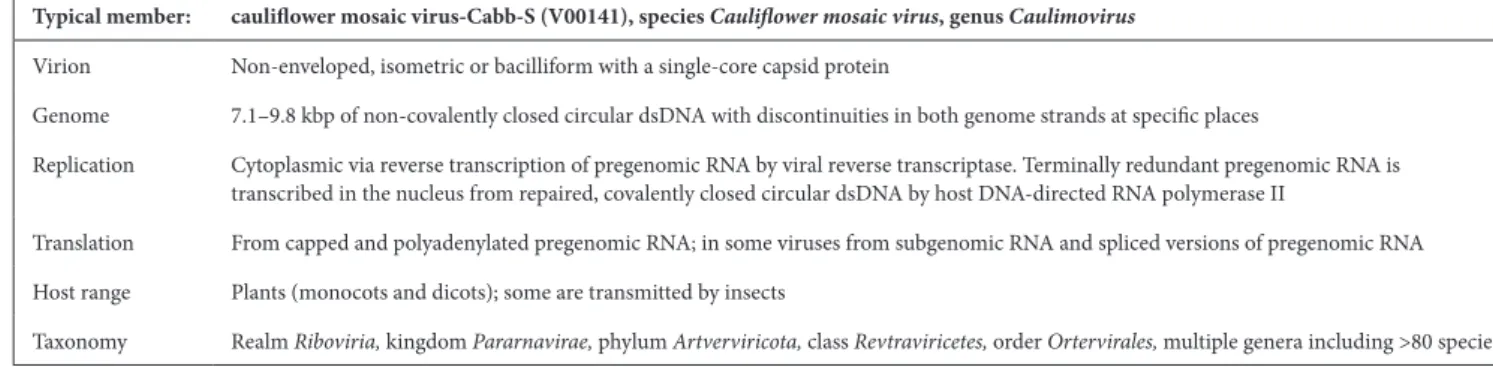
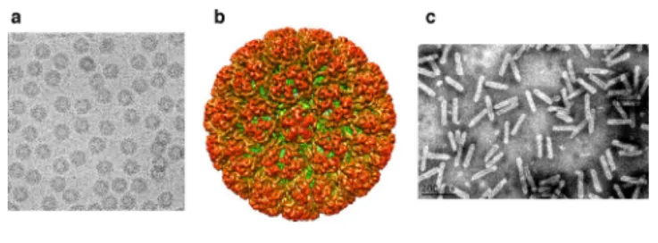
Figure
Documents relatifs
Typical member: citrus tristeza virus (U16304), species Citrus tristeza virus, genus Closterovirus Virion Non- enveloped, filamentous particles 650 to 2200 nm in length and 12 nm
Four distinct resonance modes were found for rectangular rings compared to the two modes seen in circular rings of identical width due to the presence of sharp corners and
Our first algorithm recursive grouping constructs the latent tree in a bottom-up fashion, by using information distances to add hidden nodes as neighbors to the existing
When recapitulating the distribution of EVEs in plant genomes, the known host range of exogenous viruses, and the phylogenetic relationships between caulimovirid OTUs and major
Considering that there are no examples of divided genomes in extant members of the Caulimoviridae, bipartite florendovirus genomes may therefore represent unsuccessful attempts in
La rotation sur deux ans de maïs associé aux légumineuses suivi de riz pluvial a été le système le plus rentable et le plus adopté sur tanety Les innovations paysannes
Après une étude détaillée basée sur une analyse du contenu des vignettes qui ont constitué notre corpus, nous avons confirmé que les personnages féminins dans
In general these doors are permitted only in fire separations that do not require a fire-resistance rating of more than h in buildings not more than three storeys in
