HAL Id: hal-02640324
https://hal.archives-ouvertes.fr/hal-02640324
Submitted on 28 May 2020
HAL is a multi-disciplinary open access archive for the deposit and dissemination of sci-entific research documents, whether they are pub-lished or not. The documents may come from teaching and research institutions in France or abroad, or from public or private research centers.
L’archive ouverte pluridisciplinaire HAL, est destinée au dépôt et à la diffusion de documents scientifiques de niveau recherche, publiés ou non, émanant des établissements d’enseignement et de recherche français ou étrangers, des laboratoires publics ou privés.
at the morphological and molecular level
Sebastian N. Politis, David Mazurais, Arianna Servili, Jose-Luis
Zambonino-Infante, Joanna J. Miest, Jonna Tomkiewicz, Ian A. E. Butts
To cite this version:
Sebastian N. Politis, David Mazurais, Arianna Servili, Jose-Luis Zambonino-Infante, Joanna J. Mi-est, et al.. Salinity reduction benefits European eel larvae: Insights at the morphological and molecular level. PLoS ONE, Public Library of Science, 2018, 13 (6), pp.e0198294. �10.1371/jour-nal.pone.0198294�. �hal-02640324�
Salinity reduction benefits European eel
larvae: Insights at the morphological and
molecular level
Sebastian N. Politis1*, David Mazurais2, Arianna Servili2, Jose-Luis Zambonino-Infante2, Joanna J. Miest3¤a, Jonna Tomkiewicz1, Ian A. E. Butts1¤b
1 National Institute of Aquatic Resources, Technical University of Denmark, DTU, Lyngby, Denmark, 2 Ifremer, Marine Environmental Science Laboratory UMR 6539, Plouzane´, France, 3 Helmholtz Centre for
Ocean Research, GEOMAR, Kiel, Germany
¤a Current address: Department of Life & Sports Sciences, University of Greenwich, Kent, United Kingdom
¤b Current address: Auburn University, School of Fisheries, Aquaculture and Aquatic Sciences, Auburn, Alabama, United States America
Abstract
European eel (Anguilla anguilla) is a euryhaline species, that has adapted to cope with both, hyper- and hypo-osmotic environments. This study investigates the effect of salinity, from a morphological and molecular point of view on European eel larvae reared from 0 to 12 days post hatch (dph). Offspring reared in 36 practical salinity units (psu; control), were compared with larvae reared in six scenarios, where salinity was decreased on 0 or 3 dph and in rates of 1, 2 or 4 psu/day, towards iso-osmotic conditions. Results showed that several genes relating to osmoregulation (nkcc2α, nkcc2β, aqp1dup, aqpe), stress response (hsp70,
hsp90), and thyroid metabolism (thrαA, thrαB, thrβB, dio1, dio2, dio3) were differentially
expressed throughout larval development, while nkcc1α, nkcc2β, aqp3, aqp1dup, aqpe,
hsp90, thrαA and dio3 showed lower expression in response to the salinity reduction.
More-over, larvae were able to keep energy metabolism related gene expression (atp6, cox1) at stable levels, irrespective of the salinity reduction. As such, when reducing salinity, an energy surplus associated to reduced osmoregulation demands and stress (lower nkcc, aqp and hsp expression), likely facilitated the observed increased survival, improved biometry and enhanced growth efficiency. Additionally, the salinity reduction decreased the amount of severe deformities such as spinal curvature and emaciation but also induced an edema-tous state of the larval heart, resulting in the most balanced mortality/deformity ratio when salinity was decreased on 3 dph and at 2 psu/day. However, the persistency of the pericar-dial edema and if or how it represents an obstacle in further larval development needs to be further clarified. In conclusion, this study clearly showed that salinity reduction regimes towards iso-osmotic conditions facilitated the European eel pre-leptocephalus development and revealed the existence of highly sensitive and regulated osmoregulation processes at such early life stage of this species.
a1111111111 a1111111111 a1111111111 a1111111111 a1111111111 OPEN ACCESS
Citation: Politis SN, Mazurais D, Servili A,
Zambonino-Infante J-L, Miest JJ, Tomkiewicz J, et al. (2018) Salinity reduction benefits European eel larvae: Insights at the morphological and molecular level. PLoS ONE 13(6): e0198294.https://doi.org/ 10.1371/journal.pone.0198294
Editor: Tzong-Yueh Chen, National Cheng Kung
University, TAIWAN
Received: February 20, 2018 Accepted: May 16, 2018 Published: June 13, 2018
Copyright:© 2018 Politis et al. This is an open access article distributed under the terms of the
Creative Commons Attribution License, which permits unrestricted use, distribution, and reproduction in any medium, provided the original author and source are credited.
Data Availability Statement: All relevant data are
within the paper and its Supporting Information file.
Funding: This study was part of the project Eel
Hatchery Technology for a Sustainable Aquaculture (EEL-HATCH) supported financially by
InnovationFund Denmark, Grant no. 5184-00093B. Sebastian N. Politis received travel grants from the COST Office (Food and Agriculture COST Action FA1205: Assessing and improving the quality of aquatic animal gametes to enhance aquatic
Introduction
Eels (Anguilla spp.) are euryhaline species that have adapted to cope with both, hyper- and
hypo-osmotic environments, likely due to regular salinity changes in their habitats (i.e. estuar-ies) and/or migrations between freshwater and marine environments to complete their life cycle [1]. Adult eels undertake a downstream (catadromous) reproductive migration towards oceanic spawning areas, while eel offspring undertake an upstream migration towards estua-rine or freshwater environments [1]. Unfortunately though, the natural populations and their reproductive potential have declined to a historical minimum, mainly due to climatic and anthropogenic pressures during the different phases of the eel life cycle [2]. The most exploited and negatively impacted populations today are the endangered American (A. rostrata) eel and
Japanese (A. japonica) eel [3–4] as well as the critically endangered European (A. anguilla) eel
[5].
In this context, actions have been taken to circumvent the eel decline, by minimizing fishery and pollution pressure through restocking and habitat restoration action plans [2]. Addition-ally, research is being conducted to advance knowledge on assisted reproduction and subse-quent early life history (ELH) rearing conditions, towards a sustainable aquaculture. After extensive efforts, breeding protocols using assisted reproductive technologies were developed for the Japanese eel, leading to the first production of eel leptocephali larvae in captivity [6]. The Japanese eel achievements formed the basis for eel research, which led to the development of artificial reproductive protocols of the American [7] and European eel [8–10]. Subsequent work sought to identify optimal environmental rearing conditions throughout the ELH of eels, such as temperature [11–13], light [14], and salinity [15–16].
Regarding salinity, most fish species are hyper-osmotic in freshwater, where plasma osmo-lality is higher than the environment and hypo-osmotic in seawater, where plasma osmoosmo-lality is lower than the environment [17]. Thus, in freshwater fish need to actively take up ions to counteract the diffusive ion loss and osmotic water gain, while in seawater they need to main-tain osmotic balance through a desalting process to counteract osmotic water loss [18]. Eel off-spring naturally occur in a hypo-osmotic environment in the ocean [19]. Interestingly though, it was shown that reducing salinity during ELH rearing, results in better growth and survival of Japanese eel larvae [15]. In some fish species, osmoregulatory organs such as gills or kidneys are not fully or not at all developed during ELH, thus embryos and early larvae control their ion balancevia chloride cells, commonly located in the yolk sac area and the epithelia covering
the larval body surface [20–21]. Those extra-branchial chloride cells (ionocytes) were also found to be located in the epithelium of the Japanese eel larval body surface and particularly abundant in the abdominal region, while forming multicellular complexes and influencing ionoregulation during ELH [22]. Furthermore, it was discovered that Japanese eel larvae can drink as early as the day of hatch, revealing that the role of gastro-intestinal osmoregulation starts earlier than previously anticipated [16,23,24]. The role and timing of the intestine and rectum in controlling ion balance was further confirmed by expression of osmoregulatory related genes such as Na+K+Cl-(nkcc) and Na+
Cl-(ncc) cotransporters in Japanese eel larvae
[16].
Moreover, a recent study, using a transcriptomic (microarray) approach allowed the identi-fication of a large number of genes involved with or affected by osmoregulatory changes dur-ing salinity adaptation in the European eel [25]. Furthermore, the European eel genome was recently sequenced and assembled [26], offering new perspectives for eel research in order to gain further knowledge regarding the molecular biology of this species. Thus, it is increasingly possible to follow targeted expression of genes involved in specific molecular mechanisms such as sodium potassium chloride ion cotransporters, which mediate the electroneutral resources. The need to harmonize and standardize
evolving methodologies, and improve transfer from academia to industry; AQUAGAMETE). Financial support for Ian A.E. Butts was partially provided by the Alabama Agricultural Experiment Station and the Hatch program of the National Institute of Food and Agriculture, US Department of Agriculture. The funders had no role in study design, data collection and analysis, decision to publish, or preparation of the manuscript.
Competing interests: The authors have declared
cotransport of Na+, K+and Cl-and are known to be involved in ion absorption and/or secre-tion as well as in cell volume homeostasis [27]. An additional mechanism of interest involves aquaporins, which are membrane proteins, forming pores to selectively facilitate rapid trans-port and exchange of water molecules in addition to diffusion through the plasma membrane [28]. Also an important mechanism controlling cellular homeostasis, is the molecular response to extrinsic (environmental) stressors. Here, heat shock proteins which are produced in all cel-lular organisms, regulate normal protein synthesis, and play an important role in an organ-ism’s health as they are expressed in response to stress in order to counteract physiological injury and reduce trauma [29]. A different molecular mechanism of interest is the thyroid endocrine system, as it has been shown to be involved in various physiological processes and contributes to homeostasis regulation [30]. Furthermore, the oxidative phosphorylation (OXPHOS) pathway represents a mechanism of importance, where genes associated to energy metabolism are today well known in teleost fishes [31].
In this context, this study investigates how salinity affects European eel larval biometry (such as morphology and growth), deformities and survival as well as expression profiles of genes related to processes involved in or affected by osmoregulation, such as ion transport [Na+K+2Cl-cotransporters (nkcc), aquaporins (aqp)], energy metabolism [mitochondrial ATP
Synthase F0 subunit 6 (atp6), cytochrome C oxidase 1 (cox1)], thyroid metabolism [thyroid
hormone receptors (thr), deiodinases (dio)] and stress response [heat shock proteins (hsp)].
We hypothesized that lower salinity towards a more iso-osmotic environment, already at a very early stage, would reduce stress and conserve energy due to lower cost for osmoregula-tion, resulting in higher survival and growth during the ELH stages but also facilitating a greater larval developmental potential. Thus, the main objective of this study was to determine the optimal condition for rearing European eel pre-leptocephalus larvae by decreasing salinity on 0 or 3 days post hatch (dph) and at rates of 1, 2 or 4 psu/day.
Material and methods
Ethics statement
All fish were handled in accordance with the European Union regulations concerning the pro-tection of experimental animals (Dir 86/609/EEC). Eel experimental protocols were approved by the Animal Experiments Inspectorate (AEI), Danish Ministry of Food, Agriculture and Fisheries (permit number: 2015-15-0201-00696). Briefly, adult eels were anesthetized using ethyl p-amino-benzoate (benzocaine) before tagging and handling. Endogenously feeding larvae of European eel were anesthetized prior to handling and euthanized prior to sampling by using tricaine methanesulfonate (MS-222). All efforts were made to minimize animal handling and stress.
Broodstock management
Female broodstock were wild-caught from lake Vandet (Denmark) or Lough Neagh (Ireland), while all males originated from a commercial eel farm (Stensgård Eel Farm A/S, Denmark). After collection, broodstock were transferred to an experimental facility of the Technical Uni-versity of Denmark, where they were maintained in ~1250 L polyethylene tanks equipped with a closed recirculation system, under a continuous flow rate of ~10–15 L min−1, low intensity light (~20 lux) and 12 h day/12 h night photoperiod. Acclimatization took place over two weeks, in order to reach a salinity of 36 psu and temperature of 20˚C. As eels naturally undergo a fasting period from the onset of the pre-pubertal silvering stage, they were not fed during this period. Prior to experimentation, the broodstock were anaesthetized (ethyl p-amino-benzoate, 20 mg L−1; Sigma-Aldrich Chemie, Steinheim, Germany), tagged with a passive inte-grated transponder, and length and weight recorded.
Gamete production, experimental design and conditions
To induce vitellogenesis females received weekly injections of salmon pituitary extract at 18.75 mg kg−1body weight (Argent Chemical Laboratories, Washington, USA) [10,32]. To stimu-late follicular maturation and induce ovulation, females received an additional injection of 17α,20ß-dihydroxy-4-pregnen-3-one (Sigma-Aldrich, St. Louis, MO, USA) at 2.0 mg kg−1
body weight [33]. Then, within 12–14 h, eggs were stripped from females. Males received weekly injections of human chorionic gonadotropin (hCG, Sigma Aldrich Chemie, Steinheim, Germany) at 150 IU/fish. Prior to fertilization, they were given another injection and milt was collected ~12 h after administration of hormone. Milt samples were pipetted into an immobi-lizing medium [34] and used for fertilization within 4 h of collection [35].
The experiment was repeated 3 times, each time using a different experimental cross. Eggs from each female were “crossed” with a sperm pool of several males to experimentally create the 3 experimental crosses. Eggs from each female were stripped into dry plastic containers and gametes were swirled together. Artificial seawater (20˚C and 36 psu) prepared by using reverse osmosis filtration (Vertex Puratek 100 gpd RO/DI, Vertex Technologies Inc., CA, USA) and salted with Red Sea Salt (Red Sea, Red Sea International, Eilat, Israel) was added for a gamete contact time of 5 min [35,36]. Eggs/embryos were then incubated in 15 L of the above described artificial seawater (18˚C and 36 psu) [13] and supplemented with rifampicin and ampicillin (each 50 mg L-1, Sigma-Aldrich, Missouri, USA) until hatch [37].
At hatch, larvae were randomly distributed (~1000 individuals per replicate) into 1 L glass beakers and reared throughout the endogenous feeding stage, from 0 to 12 dph, with no aera-tion [38]. Each beaker was filled with 800 mL of artificial seawater (18˚C and 36 psu) and sup-plemented with antibiotics as previously described [13,37]. Using a factorial approach, salinity treatments were step-wise adjusted beginning on 0 or 3 dph and at rates of 1, 2, or 4 psu/day, in order to reach a more iso-osmotic condition compared to full strength seawater (control), which was kept at 36 psu. This resulted in overall seven salinity treatments (con, 01, 02, 04, 31, 32 and 34) described inTable 1. All 3 experimental crosses where represented in 3 replicates for all 7 treatments, resulting in 63 experimental beakers. Each day, 400 mL were removed and replaced with pre-adjusted artificial seawater, in order to reach the desired salinity of each treatment, which was measured using a digital portable conductivity meter (WTW ProfiLine Cond 3110). All treatments underwent the same handling procedures. Rearing of embryos and larvae took place in darkness, while handling and sampling under low intensity (< 2.2μmol m-2s-1) light conditions [14].
Table 1. European eel (Anguilla anguilla) larvae reared in seven different salinity treatments; at 36 psu (con) and
in six further scenarios, where salinity was reduced on 0 or 3 days post hatch and at rates of 1, 2 or 4 psu/day (01, 02, 04, 31, 32 and 34) towards iso-osmotic conditions.
Age in days post hatch (dph)
Reduction ID 0 1 2 3 4 5 6 7 8 9 10 11 12 dph psu day-1 Salinity (psu)
Control con 36 0 1 01 35 34 33 32 31 30 29 28 27 26 25 24 23 2 02 34 32 30 28 26 24 22 20 18 16 4 04 32 28 24 20 16 3 1 31 36 35 34 33 32 31 30 29 28 27 26 2 32 36 34 32 30 28 26 24 22 20 18 16 4 34 36 32 28 24 20 16 https://doi.org/10.1371/journal.pone.0198294.t001
Data collection
Larval development (biometry), mortality, and gene expression of selected genes, correspond-ing to specific molecular mechanisms were followed from hatch and throughout the endoge-nous feeding stage (2, 4, 6, 8, 10, and 12 dph). All endogeendoge-nously feeding larvae of European eel were anesthetized using tricaine methanesulfonate (MS-222; Sigma-Aldrich, Missouri, USA) prior to digital imaging and euthanized post-sampling by using an MS-222 overdose [13]. All images were taken using a digital camera (Digital Sight DS-Fi2, Nikon Corporation, Japan) attached to a zoom stereomicroscope (SMZ1270i, Nikon Corporation, Japan), while NIS-Ele-ments D analysis software (Version 3.2) was used to analyze the larval images (Nikon Corpora-tion, Japan).
Mortality and biometry. Every day, dead larvae were counted and removed from all
experimental units. Additionally, all larvae at the end of the experiment as well as of all sam-pled larvae from each experimental unit were enumerated and recorded. Larval cumulative mortality was calculated as a percentage from hatch until 12 dph.
For analysis of larval biometry, ~10 larvae from each replicate (3×), cross, and treatment were randomly sampled at hatch and every second day until 12 dph. Larvae were digitally imaged (as described above) for later analyses, where total body and oil-drop area were mea-sured for each larva. Larval growth and oil-drop utilization were meamea-sured from the change in body and oil-drop area, respectively. Growth efficiency was measured by dividing the increase in body area by the corresponding decrease in oil-drop area. Moreover, larval deformities were classified according to [39] as spinal curvature, emaciation and pericardial edema (Fig 1).
Fig 1. Visualization of European eel (Anguilla anguilla) larval deformities. Normal (A), spinal curvature (B), emaciation (C), and pericardial
edema (D).
Gene expression. For molecular analysis, ~30 larvae from each replicate, experimental
cross, and treatment were randomly sampled at hatch and every second dph until the first-feeding stage. Those larvae were recorded, euthanized using MS-222, rinsed with deionized water, preserved in RNAlater Stabilization Reagent and kept at -20˚C following the procedure suggested by the supplier (Qiagen, Hilden, Germany). RNA was extracted using the NucleoS-pin1RNA Kit (Macherey-Nagel, Germany) following the manufacturer’s instructions. RNA concentration (264± 230 ng μl-1) and purity (260/280 = 2.13± 0.03, 260/230 = 2.23 ± 0.12) were determined by spectrophotometry using Nanodrop ND-1000 (Peqlab, Germany) and normalized to a common concentration of 100 ngμl-1with HPLC water. From the resulting total RNA, 680 ng were transcribed using the qScriptTMcDNA Synthesis Kit (Quantabio, Ger-many) according to the manufacturer’s instructions, including an additional gDNA wipe out step prior to transcription [PerfeCta1DNase I Kit (Quantabio, Germany)].
Theef1α and rps18 genes were chosen as housekeeping genes, since qBase+ software
revealed that these mRNA levels were stable throughout analyzed samples (M < 0.4); M gives the gene stability and M < 0.5 is typical for stably expressed reference genes [40]. The expres-sion levels of 16 target and 2 reference (ef1α, rps18) genes were determined by quantitative
real-time PCR (RT-qPCR), using specific primers (Table 2). Primers were designed using primer 3 software v 0.4.0 (http://frodo.wi.mit.edu/primer3/) based on cDNA sequences avail-able in GenBank Nucleotide, the European eel transcriptome database (EeelBase 2.0,http:// compgen.bio.unipd.it/eeelbase/) or the available European eel genome (https://www.ncbi.nlm. nih.gov/bioproject/PRJNA73577). All primers were designed for an amplification size ranging from 75 to 200 nucleotides and optimal Tm of 60˚C.
Expression of genes in each larval sample from 2 randomly selected replicates, from each parental cross, treatment, and larval age (2, 4, 6, 8, 10 and 12 dph) were analysed in two techni-cal replicates of each gene using the qPCR BiomarkTMHD system (Fluidigm) based on 96.96 dynamic arrays (GE chips) as previously described [41]. In brief, a pre-amplification step was performed with a 500 nM primer pool of all primers in TaqMan-PreAmp Master Mix (Applied Biosystems) and 1.3μL cDNA per sample for 10 min at 95˚C and then 14 cycles of 15 s at 95˚C and 4 min at 60˚C. Obtained PCR products were diluted 1:10 with low EDTA-TE buffer. The pre-amplified product was loaded onto the chip with SSofast-EvaGreen Supermix low Rox (Bio Rad) and DNA-Binding Dye Sample Loading Reagent (Fluidigm). Primers were loaded onto the chip at a concentration of 50μM. The chip was run according to the Fluidigm 96.96 PCR protocol with a Tm of 60˚C. The relative quantity of target gene transcripts was normal-ized and measured using theΔΔ Ct method [42]. Coefficient of variation (CV) of technical rep-licates was calculated and checked to be < 0.04 [40].
Statistical analyses
All data were analyzed using SAS statistical software (version 9.1; SAS Institute Inc., Cary, North Carolina). Residuals were tested for normality using the Shapiro-Wilk test and homoge-neity of variances was tested using a plot of residuals versus fit values (PROC GLOT, SAS Insti-tute 2003). Data were log10or arcsine square-root-transformed when data deviated from
normality or homoscedasticity [43]. The effect of salinity on larval deformities, growth rate, oil-drop utilization, and growth efficiency on 12 dph was determined using a series of one-way ANOVA models, where experimental cross was considered a random factor (SAS PROC MIXED; SAS Institute 2003). Tukey’s post-hoc analyses were used to compare least-squares means between treatments. Furthermore, statistical models were used to investigate salinity effects on larval body area, mortality and gene expression throughout early larval development (from 2 to 12 dph). Here, we analyzed the data using a series of repeated measures
mixed-model ANOVAs (PROC MIXED; SAS Institute 2003). Models contained salinity treatment and age main effects as well as the treatment× age interaction. Akaike’s (AIC) and Bayesian (BIC) information criteria were used to assess which covariance structure (compound symmetry, autoregressive order, or unstructured) was most appropriate [44]. Salinity treatment and age were considered fixed, whereas parental cross was considered random. Tukey’s post-hoc analy-ses were used to compare least-squares means between treatments. If a significant salinity× age interaction was detected, the model was decomposed into a series of reduced ANOVA models to determine the effect of salinity for each age. This was the case only forthrαA.
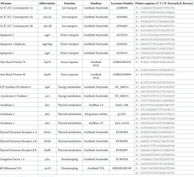
Table 2. Sequences of European eel,Anguilla anguilla primers used for amplification of genes by qRT-PCR. Primers were designed based on sequences available on
Genbank databases. The table lists accession number and corresponding database of target gene sequences.
Full name Abbreviation Function Database Accession Number Primer sequence (5’ 3’) (F: Forward; R: Reverse)
Na+K+2Cl-Cotransporter 1α nkcc1α Ion transport GenBank Nucleotide AJ486858 F: CCAAGGCTCAGATCTTCCTG R: TTTCCGAATGGTAACCGAAG Na+K+2Cl-Cotransporter 2α
nkcc2α Ion transport GenBank Nucleotide AJ564602 F: ACGTGGTTGGGTTTTCAGAG
R: GTGAGATCCCCAAAAGCAAA Na+K+2Cl-Cotransporter 2β nkcc2β Ion transport GenBank Nucleotide AJ564603 F: AGCCAAAGTGGTGGATGTTC R: TGTCAGCCTCTCCAGTTCCT
Aquaporin 3 aqp3 Water transport GenBank Nucleotide AJ319533 F: GCTCTCATGGCTTGTTCCTC
R: AAGGTCACAGTGGGGTTCAG Aquaporin 1 duplicate aqp1dup Water transport GenBank Nucleotide AJ564421 F: GAATTCCTGGCAACCTTTCA R: CAAGATGACCCAGACCCACT
Aquaporin e aqpe Water transport GenBank Nucleotide AJ784153 F: TGGGCAGCTGACAGTAACAG
R: AATCACCTGGTCCACAAAGC
Heat Shock Protein 70 hsp70 Stress response GenBank
WGS
AZBK01685255 F: TCAACCCAGATGAAGCAGTG R: GCAGCAGATCCTGAACATTG
Heat Shock Protein 90 hsp90 Stress response GenBank
WGS
AZBK01838994 F: ACCATTGCCAAGTCAGGAAC R: ACTGCTCATCGTCATTGTGC ATP Synthase F0 subunit 6 atp6 Energy metabolism GenBank Nucleotide NC_006531 F: GGCCTGCTCCCATACACATT R: GACTGGTGTTCCTTCTGGCA Cytochrome C Oxidase 1 cox1 Energy metabolism GenBank Nucleotide NC_006531 F: CTACTCCTCTCCCTGCCAGT R: CTTCTGGGTGGCCGAAGAAT
Deiodinase 1 dio1 Thyroid metabolism EeelBase 2.0 Eeel2-c186 F: AGCTTTGCCAGAACGACTGT
R: TTCCAGAACTCTTCGCACCT
Deiodinase 2 dio2 Thyroid metabolism Eel genome website g12347 F: GAAGAGGAGGATCGCCTACC
R: GCACTCTACCTCCGTCCAAA
Deiodinase 3 dio3 Thyroid metabolism EeelBase 2.0 Eeel-c22164 F: TACGGGGCGTATTTTGAGAG
R: GCTATAACCCTCCGGACCTC Thyroid Hormone Receptorα A thrαA Thyroid metabolism GenBank Nucleotide KY082904 F: GCAGTTCAACCTGGACGACT R: CCTGGCACTTCTCGATCTTC Thyroid Hormone Receptorα B thrαB Thyroid metabolism GenBank Nucleotide KY082905 F: GAAGCCTTCAGCGAGTTCAC R: ACAGCCTTTCAGGAGGATGA Thyroid Hormone Receptorβ B thrβB Thyroid metabolism GenBank Nucleotide KY082907 F: GAAGACTGAGCCCTGAGGTG R: AGGTAATGCAGCGGTAATGG Elongation Factor 1α ef1a Housekeeping GenBank Nucleotide EU407824 F: CTGAAGCCTGGTATGGTGGT R: CATGGTGCATTTCCACAGAC
40S Ribosomal S18 rps18 Housekeeping GenBank TSA GBXM01005349 F: TGACCGATGATGAGGTTGAG
R: GTTTGTTGTCCAGACCGTTG
Results
Mortality and biometry
European eel larval mortality (± SEM) until 12 dph, was highest (43 ± 10%) when larvae were reared at 36 psu (control) and significantly (p < 0.0001) lower in all salinity reduced treat-ments (Fig 2A). The statistically lowest larval mortality was observed when salinity was initially reduced on 0 dph and at 2 or 4 psu/day (treatments 02 and 04, respectively) as well as on 3 dph and at 4 psu/day (treatment 34).
Fig 2. Effect of salinity on European eel (Anguilla anguilla) larval mortality and biometry. Mortality (A), body area (B), growth (C), oil drop utilization (D) and
growth efficiency (E) from 2 to 12 days post hatch (dph) as well as occurrence of larval deformities such as spinal curvature (F), emaciation (G) and pericardial edema (H) on 12 dph. Values represent means (± SEM) among three crosses at each age and treatment. Different lower case letters represent significant statistical differences (p < 0.05).
Larval body area (±SEM), which increased until 12 dph, was found to be lowest (3.1 ± 0.2 mm2) when larvae were reared at 36 psu (control) and significantly (p < 0.0001) higher in all salinity reduced treatments (Fig 2B). Larvae developed the highest body area (3.8–4.0± 0.2 mm2) when salinity was initially reduced on 0 dph and at 2 or 4 psu/day (treatments 02 and 04, respectively) as well as on 3 dph and at 4 psu/day (treatment 34).
Larval growth (± SEM) throughout the endogenous feeding window was significantly (p < 0.0001) lower (0.16± 0.02 mm2per day) when larvae were reared at 36 psu (control) compared to all other treatments (Fig 2C). Highest growth (> 0.24± 0.02 mm2
/day) occurred when salinity was reduced on 0 dph and at 2 or 4 psu/day (treatments 02 and 04, respectively), as well as on 3 dph and at 4 psu/day (treatment 34). Oil drop utilization (± SEM) was signifi-cantly (p < 0.0001) lower (0.007± 0.0006 mm2/day) when salinity was reduced on 0 dph and at 4 psu/day (treatment 04) compared to all other treatments (Fig 2D). All salinity reduction treatments resulted in a growth efficiency (± SEM) that was significantly (p < 0.0001) higher compared to larvae reared at 36 psu (control; 0.72± 0.04), while highest growth efficiency (0.95± 0.04) occurred when salinity was reduced on 0 dph and at 4 psu/day (treatment 04; Fig 2E).
On 12 dph, the occurrence of larvae with spinal curvature (± SEM) were significantly (p < 0.0001) higher (56± 8%) when larvae were reared at 36 psu (control), compared to all salinity reduction treatments (Fig 2F), while lowest (4± 8%) occurrence of larvae with spinal curvature were detected when salinity was reduced on 0 dph and at 4 psu/day (treatment 04). Similarly, the occurrence of larvae with emaciation (± SEM) were significantly (p < 0.0001) higher (81± 12%) when larvae were reared at 36 psu (control), compared to all salinity reduc-tion treatments (Fig 2G), while lowest (6± 8%) occurrence of larvae with emaciation were detected when salinity was reduced on 0 dph and at 4 psu/day (treatment 04). On the contrary, when salinity was reduced on 0 dph and at 2 or 4 psu/day (treatments 02 and 04, respectively), as well as on 3 dph and at 4 psu/day (treatment 34), we observed larvae with a significantly (p < 0.0001) higher (> 73± 10%) number of pericardial edema compared to larvae reared at 36 psu (control;Fig 2H).
Gene expression
Osmoregulation. Na+K+2Cl-cotransporter regulation were affected by both larval age and salinity. Relative expression levels of the genes encoding fornkcc1α were stable (Fig 3A), whilenkcc2α and nkcc2β increased significantly (p < 0.0001) throughout larval development
and peaked at 12 dph (Fig 3C and 3E). Expression ofnkcc1α was significantly (p < 0.01)
reduced when salinity was decreased on 0 dph and at 1 or 4 psu/day (treatments 01 and 04, respectively) as well as on 3 dph and at 4 psu/day (treatment 34;Fig 3B) compared to the 36 psu control. Similarly, expression ofnkcc2β was significantly (p < 0.01) higher in the control
group than when salinity was decreased on 0 dph and 4 psu/day (treatment 04;Fig 3F), while no statistically significant effect of salinity was observed on expression levels ofnkcc2α
(Fig 3D).
Moreover, the relative expression of aquaporin 3 (aqp3) was stable (Fig 3G),aqp1dup
signif-icantly (p < 0.0001) decreased (Fig 3I), whileaqpe significantly (p < 0.0001) increased
throughout larval development (Fig 3K). Expression ofaqp3 was significantly (p = 0.005)
reduced when salinity was decreased on 0 dph and at 4 psu/day (treatment 04;Fig 3H) com-pared to no reduction (control) or the slowest reduction in treatment 31 (reduction on 3 dph and at 1 psu/day). Expression ofaqp1dup was also significantly (p < 0.002) higher when larvae
were reared at 36 psu (control) compared to when salinity was decreased on 0 dph and at 2 or 4 psu/day (treatments 02 and 04, respectively;Fig 3J). Similarly, the salinity reduction on 0
dph, irrespective of the reduction rate (treatments 01, 02 and 04;Fig 3L), caused a significant (p < 0.001) reduction in mRNA levels of the third aquaporin tested (aqpe).
Stress response. The relative expression of heat shock protein 70(hsp70) significantly
(p < 0.0001) increased throughout development, whilehsp90 experienced a significant
(p < 0.0001) decrease from 2 to 6 dph, but increased again beyond that; both reaching a peak on 12 dph (Fig 3M and 3O). The rate of reduction was the driver setting the expression pattern forhsp90, since reduction rates of 2 and 4 psu/day let to lowered mRNA levels
inde-pendent of the onset of the treatment (p < 0.001,Fig 3P). The expression response ofhsp70
Fig 3. Effect of age and salinity treatment on European eel (Anguilla anguilla) larval relative expression of selected genes. No significant salinity × age interaction
was detected for any gene. As such, over the entire experimental period, the main effects of age and salinity are displayed for genes encoding Na+K+2Cl-cotransporters
(A-F), aquaporins (G-L), and heat shock proteins (M-P). Values represent means (± SEM) among three crosses at each age and treatment. Different lower case letters represent significant statistical differences (p < 0.05).
showed a similar pattern tohsp90; however it was not statistically different among the
treat-ments (Fig 3N).
Thyroid metabolism. A significant (p < 0.001) age× salinity interaction was found for the relative expression of the thyroid hormone receptorthrαA (Fig 4A), whilethrαB and thrβB
were not significantly affected by salinity (Fig 4C and 4E). For the latter two genes, age was the main factor influencing gene expression. Expression levels ofthrαB increased throughout
ontogeny (Fig 4B), while expression ofthrβB increased from 2 to 4 dph and remained at steady
levels beyond that (Fig 4D; both p < 0.0001). Moreover, larval age and salinity influenced
Fig 4. Effect of age and salinity treatment on European eel (Anguilla anguilla) larval relative expression of selected genes. For thyroid hormone receptor αA
(thrαA), a significant salinity × age interaction was detected, thus the model was decomposed into a series of reduced ANOVA models to determine the effect of salinity
for each age (A). No significant salinity× age interactions were detected for all other genes. As such, over the entire experimental period, the main effects of age and salinity are displayed for genes relating to thyroid hormone receptors (B-E), deiodinases (F-K), and energy metabolism (L-O). Values represent means (± SEM) among three crosses at each age and treatment. Different lower case letters and asterisks represent significant statistical differences (p < 0.05).
genes involved in deiodination (dio1-3). Here, expression of dio1 and dio2 increased
through-out ontogeny (Fig 4F and 4H), while expression ofdio3 decreased from 2 to 6 dph and
increased again beyond that until 12 dph (Fig 4J; all p < 0.0001). However,dio3 was the only
gene in this functional group that was influenced by salinity, as its expression was reduced when salinity was decreased on 3 dph and at 2 psu/day (p < 0.02;Fig 4K).
Energy metabolism. Energy metabolism related genes (atp6, cox1) were found to be
steadily expressed within the endogenous feeding period (2 to 12 dph) and at all salinities, with no significant differences occurring among treatments (Fig 4L–4O).
Discussion
European eel is critically endangered with diminishing glass eel recruitment [2]. Thus, there is an urgent need to artificially produce offspring and improve survival during ELH of this spe-cies. Latest efforts have focused on extrinsic (environmental) factors such as light [14] and temperature [13,45], while only eggs and embryos have been assessed regarding salinity effects [36]. In the closely related Japanese eel, a major improvement in larval captive rearing has been attributed to reduced salinity regimes [15,46]. Therefore, this study investigated the effect of salinity on artificially produced European eel larvae (from a morphological and molecular point of view), to elucidate functionality and timing of osmoregulation related pro-cesses affecting ELH, in order to ultimately determine optimal conditions for rearing Euro-pean eel pre-leptocephalus larvae in aquaculture. To accomplish this, we reared offspring throughout the endogenous feeding window (0–12 dph) and compared larvae reared at 36 psu (control), which is close to the salinity occurring in the Sargasso Sea [19], with larvae reared under reduced salinity conditions. Here, salinity was decreased on 0 or 3 dph and at rates of 1, 2 or 4 psu/day, resulting in six different salinity treatments (01, 02, 04, 31, 32 and 34).
Mortality and biometry
Generally, we observed that reducing salinity, especially on 0 dph at 4 psu (fastest reduction) resulted in reduced mortality, increased body area, higher growth, reduced oil drop utilization and improved growth efficiency of the European eel larvae. Similar observations have been reported for Japanese eel, where better larval growth and survival were reported when reared in 50% reduced salinity [15]. Thereafter, it was shown that reducing seawater salinity (with an osmolality of ~1050 mOsm kg-1H2O) to ~50%, facilitated an iso-osmotic environment for eel
larvae with a tissue osmolality of 360 to 540 mOsm kg-1H2O [23]. Concurrently, it has been
argued that reducing salinity probably increased energy availability due to lower osmoregula-tory expenses, enabling the survival of weaker larvae, which would not survive in a high salin-ity environment [46]. Additionally, [47] reported increased spinal deformities at a high salinity level (42 psu). In our study, we observed the highest incidence of spinal curvature (> 50%) and emaciation (> 80%) when larvae were reared at 36 psu (control) compared to < 5% and < 10% respectively, when salinity was reduced on 0 dph and at 4 psu/day (treatment 04). How-ever, it has also been reported that rearing eel larvae at reduced salinity (< 33 psu), causes other deformities, such as pericardial edema and abnormal lower jaw [47]. The results of our study are in agreement with those findings, as we also observed an increased number of larvae with pericardial edema in the salinity reduced treatments. Thus, we demonstrate that reducing salinity was a tradeoff process, which improved survival and growth but also induced an edem-atous state of the larval heart. Hence, we consider that reducing salinity on 3 dph and at 2 psu/ day towards iso-osmotic conditions, results in a more balanced mortality/deformity ratio. However, the persistency of the edematous state of the larval heart and if or how it represents an obstacle in further larval development needs to be clarifiedvia future investigations.
Thyroid metabolism
In order to evaluate the underlying molecular backgrounds relating to the observed biometric changes, we targeted genes involved in the thyroid metabolism, which is directly linked to growth and development of fish [48]. The importance of the thyroid endocrine system, especially during metamorphosis, has been documented in the Japanese conger eel,Conger myriaster [49], while thyroid hormone treatment is applied to coordinate a synchronized metamorphosis in Japanese eels [50]. Moreover, it was shown that the thyroid hormone path-way is involved in the early life development of Japanese [51] as well as European eels and that genes encoding thyroid hormone receptors and deiodinases show sensitivity to extrinsic (envi-ronmental) stimuli, such as temperature [45]. In the current study we observed that an early and fast reduction of salinity from 0 dph onwards increasedthrαA levels, which is an
underly-ing molecular response that can potentially be correlated to growth efficiency, which was also highest in this treatment. Moreover, the expression ofdio3 was also influenced by the salinity
change, which would be in accordance with previous observations, where the outer ring deio-dination showed sensitivity to salinity in rainbow trout,Oncorhynchus mykiss [52]. Further-more, all genes relating to thyroid metabolism, investigated in this study, were affected by larval age and showed differentially expressed patterns throughout ontogeny, which are in accordance with previous reported results in European eel larvae [45] and thus further support the involvement of the thyroid endocrine system during ELH of this species.
Osmoregulation
To further support our findings, we followed the relative expression of genes involved in ion transport. Here, we targeted the NKCC subfamily of the chloride cation cotransporter gene family, which mediates the electroneutral cotransport of Na+, K+and Cl-ions and is known to be involved in ion absorption and/or secretion as well as in cell volume homeostasis [27]. A secretory (nkcc1) and an absorptive (nkcc2) subtype (with isoforms for each) have been
previ-ously identified in adult European eel [53]. Of these, thenkcc1α isoform showed a wide range
of tissue distribution, whereasnkcc1β was predominantly expressed in the brain [54]. More-over, thenkcc2α isoform was rather restricted to renal tissues, whereas the nkcc2β isoform
pre-dominated in the intestine and urinary bladder [55]. In our study, we observed thatnkcc1α
andnkcc2β expression decreased with salinity reduction, which indicates a downregulation of
the active Na+, K+, and Cl-transport. As this mechanism requires energy, a lowered transcellu-lar ion transport can probably lead to reduced cellutranscellu-lar energy consumption. Furthermore, the ionoregulatory ability of the larvae seemed to increase throughout ontogeny (i.e. increased expression ofnkcc2α and nkcc2β), which is probably coupled to the increasing functionality of
the associated tissue (kidney and gut respectively) during organogenesis.
Ionoregulation is tightly coupled to water flow across membranes, where aquaporins form pores to selectively facilitate rapid transport and exchange of water molecules [28]. In euroha-line fish, such as the sea-bass (Dicentrarchus labrax), aquaporins facilitate the water uptake in
the intestinal tract and the reabsorption of water in the kidney to counteract dehydration in response to high salinity [56]. Similarly, in relation to seawater acclimation, branchialaqp3
was downregulated, but intestinalaqp3 was unchanged [57], while renalaqp1, aqp1dup and aqpe were downregulated [58], but intestinalaqp1 and aqpe were upregulated [59] in European eel. Probably the high osmotic water loading through the gills and the associated excretion of large volumes of dilute urine (needed in freshwater) are no longer necessary in a saline envi-ronment (gill and renal expression downregulation), while on the contrary, the increased ingestion of seawater and the accompanied transcellular movement of large quantities of salts in the gastro-intestinal track, require concomitant changes in transcellular water transport
(intestinal expression upregulation) [60]. In our study we observed a down-regulation of
aqp1dup and the two aquaglyceroporins (aqp3, aqpe) when salinity was reduced early and fast
(from 0 dph and at 4 psu/day), which is an indication that larvae acclimated their hydromin-eral regulation in response to lower salinity by reducing water retention. However, as gills are not present and the kidney as well as the intestine are undeveloped and only gain functionality during this developmental period, the tissue specific functionality of this molecular mecha-nism in relation to salinity change and the potential sensitivity shift among developmental stages remain to be clarified.
Stress response
In order to analyze the cellular stress response in European eel larvae we targetedhsp70 and hsp90, which are expressed (in response to stress) to counteract physiological injury and
reduce trauma [13,29]. Thehsp function, is commonly associated but not restricted to heat
stress and has been recognized to have a more universal role in response to a number of stress-ors [29]. As such,hsps have been shown to be sensitive to changes in salinity, where lowest
lev-els ofhsps were found in Black sea bream (Mylio macrocephalus) reared in an iso-osmotic
salinity and highest levels when reared bellow (hyper-osmotic) or above (hypo-osmotic) the osmotic homeostasis [61]. A similar response was observed in our current study, where larval
hsp90 levels decreased in the treatments were salinity was reduced in 2 or 4 psu/day compared
to larvae reared in full strength seawater (36 psu). This suggests that eel larvae reared in iso-osmotic conditions are less stressed probably due to lower energetic costs in maintaining cellu-lar homeostasis.
Energy metabolism
Thereafter, we evaluated energy levels in European eel larvae in response to environmental salinity changes by targeting genes involved in the OXPHOS pathway and associated to energy metabolism in teleost fishes [31]. Here, the expression levels of ATP-synthase and cyto-chrome-c-oxidase were stable throughout the entire developmental period investigated and independent of salinity levels, suggesting that energy production was stable across all salinity treatments. As such, in the light of decreased osmotic demands and stress (i.e. lowerhsp, nkcc,
andaqp expression), European eel pre-leptocephalus larvae reared at iso-osmotic salinity
con-ditions seem to have a higher energy availability compared to larvae reared in full strength sea-water (36 psu), which can then be utilized more efficiently to increase growth and survival.
Conclusion
To summarize, this study morphologically and molecularly elucidated osmoregulation related processes and together with the consideration of the most balanced mortality/deformity ratio, we conclude that salinity reduction benefits European eel larvae in terms of lower mortality and improved growth efficiency, which is likely facilitated by an energy surplus associated to lower osmoregulation demands. Hence, the overall knowledge gained from this study adds to our understanding of underlying biological mechanisms during ELH of European eel and pro-vides a promising step towards optimized rearing conditions for European eel pre-leptocepha-lus larvae as well as in the strive for sustainable aquaculture of this species. Nevertheless, as the studied molecular parameters foreshadow an adequate subsequent larval development, likely due to a global lower osmoregulatory cost, further research is needed in order to verify the energetic developmental costs in contrasted salinity scenarios and evaluate the long-term mor-phological, molecular and physiological effects.
Supporting information
S1 File. Data.
(XLSX)
Acknowledgments
We would like to thank Johanna Sarah Kottmann, Elisa Benini and Maria Kru¨ger-Johnsen for broodstock management and assistance during experiments, Prof. Einar Eg Nielsen and Prof. Thorsten Reusch for providing laboratory space and Dorte Meldrup as well as Brian Klitgaard for assistance during laboratory work.
Author Contributions
Conceptualization: Sebastian N. Politis, David Mazurais, Arianna Servili, Jose-Luis
Zambo-nino-Infante, Joanna J. Miest, Jonna Tomkiewicz, Ian A. E. Butts.
Data curation: Sebastian N. Politis, Joanna J. Miest, Ian A. E. Butts. Formal analysis: Sebastian N. Politis, Ian A. E. Butts.
Funding acquisition: Sebastian N. Politis, Jonna Tomkiewicz, Ian A. E. Butts. Investigation: Sebastian N. Politis, Joanna J. Miest.
Methodology: Sebastian N. Politis, David Mazurais, Arianna Servili, Jose-Luis
Zambonino-Infante, Joanna J. Miest, Ian A. E. Butts.
Project administration: Jonna Tomkiewicz.
Resources: David Mazurais, Jose-Luis Zambonino-Infante, Jonna Tomkiewicz. Software: Jonna Tomkiewicz, Ian A. E. Butts.
Supervision: David Mazurais, Arianna Servili, Jose-Luis Zambonino-Infante, Joanna J. Miest,
Jonna Tomkiewicz, Ian A. E. Butts.
Validation: Sebastian N. Politis, David Mazurais, Arianna Servili, Jose-Luis
Zambonino-Infante, Joanna J. Miest, Ian A. E. Butts.
Visualization: Sebastian N. Politis.
Writing – original draft: Sebastian N. Politis.
Writing – review & editing: David Mazurais, Arianna Servili, Jose-Luis Zambonino-Infante,
Joanna J. Miest, Jonna Tomkiewicz, Ian A. E. Butts.
References
1. Tesch F-W. The Eel, Fifth ed. Blackwell Publishing, Oxford, UK.
2. ICES WGEEL REPORT 2017. ICES advisory committee. ICES CM 2017/ACOM:15. REF. ACOM, WGRECORDS, SSGEPD, FAO, EIFAAC & GFCM. Report of the Joint EIFAAC/ICES/GFCM Working Group on Eels (WGEEL). 3–10 October 2017. Kavala, Greece.http://ices.dk/sites/pub/Publication% 20Reports/Expert%20Group%20Report/acom/2017/WGEEL/wgeel_2017.pdf
3. Jacoby D, Casselman J, DeLucia M, Hammerson GA, Gollock M. Anguilla rostrata. The IUCN Red List of Threatened Species 2014: e.T191108A72965914.http://www.iucnredlist.org/details/191108/0
4. Jacoby D, Gollock M. Anguilla japonica. The IUCN Red List of Threatened Species 2014: e. T166184A1117791.http://www.iucnredlist.org/details/166184/0
5. Jacoby D, Gollock M. Anguilla anguilla. The IUCN Red List of Threatened Species 2014: e. T60344A45833138.http://www.iucnredlist.org/details/60344/0
6. Tanaka H, Kagawa H, Ohta H. Production of leptocephali of Japanese eel (Anguilla japonica) in captiv-ity. Aquaculture. 2001; 201: 51–60.
7. Oliveira K, Hable WE. Artificial maturation, fertilization, and early development of the American eel (Anguilla rostrata). Can J Zool. 2010; 88: 1121–1128.
8. Pedersen BH. Fertilisation of eggs, rate of embryonic development and hatching following induced mat-uration of the European eel Anguilla anguilla. Aquaculture. 2004; 237: 461–473.
9. Palstra AP, Cohen EGH, Niemantsverdriet PRW, van Ginneken VJT, van den Thillart GEEJM. Artificial maturation and reproduction of European silver eel: Development of oocytes during final maturation. Aquaculture. 2005; 249: 533–547.
10. Mu¨ller AV, McEvoy FJ, Tomkiewicz J, Politis SN, Amigo JM. Ultrasonographic predictors of response of European eels (Anguilla anguilla) to hormonal treatment for induction of ovarian development. Am J Vet Res. 2016; 77(5): 478–486.https://doi.org/10.2460/ajvr.77.5.478PMID:27111015
11. Okamura A, Yamada Y, Horie N, Utoh T, Mikawa N, Tanaka S, et al. Effects of water temperature on early development of Japanese eel Anguilla japonica. Fisheries Sci. 2007; 73: 1241–1248.
12. Ahn H, Yamada Y, Okamura A, Horie N, Mikawa N, Tanaka S, et al. Effect of water temperature on embryonic development and hatching time of the Japanese eel Anguilla japonica. Aquaculture. 2012; 330–333: 100–105.
13. Politis SN, Mazurais D, Servili A, Zambonino-Infante J-L, Miest JJ, Sørensen SR, et al. Temperature effects on gene expression and morphological development of European eel, Anguilla anguilla larvae. PLoS ONE. 2017; 12 (8), e0182726.https://doi.org/10.1371/journal.pone.0182726PMID:28806748
14. Politis SN, Butts IAE, Tomkiewicz J. Light impacts embryonic and early larval development of the Euro-pean eel, Anguilla anguilla. J Exp Mar Biol Ecol. 2014; 461: 407–415.
15. Okamura A, Yamada Y, Mikawa N, Horie N, Utoh T, Kaneko T, et al. Growth and survival of eel lepto-cephali (Anguilla japonica) in low-salinity water. Aquaculture. 2009; 296: 367–372.
16. Ahn H, Lee KM, Inokuchi M, Watanabe S, Okamura A, Tsukamoto K, et al. Observations of initial water ingestion and ion absorption in the digestive tract of Japanese eel larvae. Fish Sci. 2015; 81: 283–290.
https://doi.org/10.1007/s12562-014-0841-8
17. Marshall WS, Grosell M. Ion transport, osmoregulation, and acid–base balance, In: Evans D.H., Clai-borne J.B. (Eds.), The Physiology of Fishes, 3rd ed. CRC Press, New York. 2005; 177–230.
18. Evans DH. Teleost fish osmoregulation: what have we learned since August Krogh, Homer Smith, and Ancel Keys. Am J Physiol Regul Integr Comp Physiol. 2008; 295: 704–713.https://doi.org/10.1152/ ajpregu.90337.2008PMID:18525009
19. Castonguay M, McCleave JD. Vertical distributions, diel and ontogenetic vertical migrations and net avoidance of leptocephali of Anguilla and other common species in the Sargasso Sea. J Plankton Res. 1987; 9(I): 195–214.
20. Ayson FG, Kaneko T, Hasegawa S, Hirano T. Development of mitochondrion-rich cells in the yolk-sac membrane of embryos and larvae of tilapia, Oreochromis mossambicus, in fresh water and seawater. J Exp Zool. 1994; 270: 129–135.
21. Kaneko T, Hasegawa S, Takagi Y, Tagawa M, Hirano T. Hypoosmoregulatory ability of eyed-stage embryos of chum salmon. Mar Biol. 1995; 122: 165–170.
22. Sasai S, Kaneko T, Tsukamoto K. Extrabranchial chloride cells in early life stages of the Japanese eel,
Anguilla japonica. Ichthyol Res. 1998; 45(1): 95–98.
23. Lee KM, Yamada Y, Okamura A, Tsukamoto K, Kaneko T. Hyposmoregulatory ability and ion- and water-regulatory mechanisms during the leptocephalus stages of Japanese eel Anguilla japonica. Fish Sci. 2013; 79: 77–86.https://doi.org/10.1007/s12562-012-0576-3
24. Seo MY, Kuroki M, Okamura A, Tsukamoto K, Watanabe S, Kaneko T. Occurrence of larval and adult types of ion-secreting ionocytes in Japanese eel Anguilla japonica. Ichthyol Res. 2015; 62: 487–494.
https://doi.org/10.1007/s10228-015-0463-x
25. Kalujnaia S, McWilliam IS, Zaguinaiko VA, Feilen AL, Nicholson J, Hazon N, et al. Transcriptomic approach to the study of osmoregulation in the European eel Anguilla anguilla. Physiol Genomics. 2007; 31: 385–401.https://doi.org/10.1152/physiolgenomics.00059.2007PMID:17666525
26. Henkel CV, Burgerhout E, de Wijze DL, Dirks RP, Minegishi Y, Jansen HJ, et al. Primitive Duplicate Hox Clusters in the European Eel’s Genome. PLoS One. 2012; 7(2): e32231.https://doi.org/10.1371/ journal.pone.0032231PMID:22384188
27. Russell JM. Sodium-Potassium-Chloride Cotransport. Physiol Rev. 2000; 80(1): 211–276.https://doi. org/10.1152/physrev.2000.80.1.211PMID:10617769
28. Agre P. The aquaporin water channels. Proc Am Thorac Soc. 2006; 3(1): 5–13.https://doi.org/10.1513/ pats.200510-109JHPMID:16493146
29. Roberts RJ, Agius C, Saliba C, Bossier P, Sung YY. Heat shock proteins (chaperones) in fish and shell-fish and their potential role in relation to shell-fish health: a review. J Fish Dis. 2010; 33: 789–801.https://doi. org/10.1111/j.1365-2761.2010.01183.xPMID:20678104
30. Warner A, Mittag J. Thyroid hormone and the central control of homeostasis. J Mol Endocrinol. 2012; 49: 29–35.https://doi.org/10.1530/JME-12-00680952–5041/12/049–R29
31. Bermejo-Nogales A, Calduch-Giner JA, Pe´rez-Sa´nchez J. Unraveling the Molecular Signatures of Oxi-dative Phosphorylation to Cope with the Nutritionally Changing Metabolic Capabilities of Liver and Mus-cle Tissues in Farmed Fish. PLoS ONE 10(4): e0122889.https://doi.org/10.1371/journal.pone. 0122889PMID:25875231
32. Kagawa H, Tanaka H, Ohta H, Unuma T, Nomura K. The first success of glass eel production in the world: basic biology on fish reproduction advances new applied technology in aquaculture. Fish Physiol Biochem. 2005; 31: 193–199.https://doi.org/10.1007/s10695-006-0024-3PMID:20035458
33. Ohta H, Kagawa H, Tanaka H, Okuzawa K., Hirose K. Changes in fertilization and hatching rates with time after ovulation induced by 17,20β-dihydroxy-4-pregnen-3-one in the Japanese eel, Anguilla
japon-ica. Aquaculture. 1996; 139: 291–301.
34. Peñaranda DS, Pe´rez L, Gallego V, Barrera R, Jover M, Asturiano JF. European Eel Sperm Diluent for Short-term Storage. Reprod Domest Anim. 2010; 45: 407–415.https://doi.org/10.1111/j.1439-0531. 2008.01206.xPMID:18954399
35. Butts IAE, Sørensen SR, Politis SN, Pitcher TE, Tomkiewicz J. Standardization of fertilization protocols for the European eel. Aquaculture. 2014; 426–427: 9–13.
36. Sørensen SR, Butts IAE, Munk P, Tomkiewicz J. Effects of salinity and sea salt type on egg activation, fertilization, buoyancy and early embryology of European eel, Anguilla anguilla. Zygote. 2016; 24(1): 121–138.https://doi.org/10.1017/S0967199414000811PMID:25707438
37. Sørensen SR, Skov PV, Lauesen P, Tomkiewicz J, Bossier P, De Schryver P. Microbial interference and potential control in culture of European eel (Anguilla anguilla) embryos and larvae. Aquaculture. 2014; 426: 1–8.
38. Sørensen SR, Tomkiewicz J, Munk P, Butts IAE, Nielsen A, Lauesen P, et al. Ontogeny and growth of early life stages of captive-bred European eel. Aquaculture. 2016; 456: 50–61.
39. Kurokawa T, Okamoto T, Gen K, Uji S, Murasita K, Unuma T, et al. Influence of water temperature on morphological deformities in cultured larvae of Japanese eel, Anguilla japonica, at completion of yolk resorption. J World Aquacult Soc. 2008; 39: 729–735.
40. Hellemans J, Mortier G, De Paepe A, Speleman F, Vandesompele J. qBase relative quantification framework and software for management and automated analysis of real-time quantitative PCR data. Genome Biol. 2007; 8(2): R19.https://doi.org/10.1186/gb-2007-8-2-r19PMID:17291332
41. Miest JJ, Arndt C, Adamek M, Steinhagen D, Reusch TBH. Dietaryβ-glucan (MacroGard®) enhances survival of first feeding turbot (Scophthalmus maximus) larvae by altering immunity, metabolism and microbiota. Fish Shellfish Immun. 2015; 48: 94–104.
42. Livak KJ, Schmittgen TD. Analysis of relative gene expression data using real-time quantitative PCR and the 2−ΔΔCT Method. Methods. 2001; 25(4): 402–8.https://doi.org/10.1006/meth.2001.1262
PMID:11846609
43. Zar JH. Biostatistical Analysis. third ed. Prentice-Hall: New Jersey; 1996.
44. Littell RC, Milliken GA, Stroup WW, Wolfinger RD. SAS system for mixed models. Cary, North Carolina: SAS Institute Incorporated; 1996.
45. Politis SN, Servili A, Mazurais D, Zambonino-Infante J-L, Miest JJ, Tomkiewicz J, et al. Temperature induced variation in gene expression of thyroid hormone receptors and deiodinases of European eel (Anguilla anguilla) larvae. Gen Comp Endocr. 2018; 259: 54–65.https://doi.org/10.1016/j.ygcen.2017. 11.003PMID:29113916
46. Okamura A, Yamada Y, Mikawa N, Horie N, Tsukamoto K. Effect of salinity on occurrence of notochord deformities in Japanese eel Anguilla japonica larvae. Aquacult Int. 2016; 24: 549–555.https://doi.org/ 10.1007/s10499-015-9944-1
47. Okamoto T, Kurokawa T, Gen K, Murashita K, Nomura K, Kim S-K, et al. Influence of salinity on mor-phological deformities in cultured larvae of Japanese eel, Anguilla japonica, at completion of yolk resorption. Aquaculture. 2009; 293: 113–118.
48. Power DM, Llewellyn L, Faustino M, Nowell MA, Bjornsson BT, Einarsdottir IE, et al. Thyroid hormones in growth and development of fish. Comp Biochem Phys C. 2001; 130: 447–459.
49. Kawakami Y, Tanda M, Adachi S, Yamauchi K. cDNA cloning of thyroid hormone receptor betas from the conger eel, Conger myriaster. Gen Comp Endocrinol. 2003; 131: 232–240. PMID:12714004
50. Yamano K, Nomura K, Tanaka H. Development of thyroid gland and changes in thyroid hormone levels in Leptocephali of Japanese Eel (Anguilla japonica). Aquaculture. 2007; 270: 499–504.
51. Kawakami Y, Nomura K, Ohta H, Tanaka H. Characterization of thyroid hormone receptors during early development of the Japanese eel (Anguilla japonica). Gen Comp Endocr. 2013; 194: 300–310.https:// doi.org/10.1016/j.ygcen.2013.09.020PMID:24100168
52. Orozco A, Villalobos P, Valverde-R C. Environmental salinity selectively modifies the outer-ring deiodi-nating activity of liver and kidney in the rainbow trout. Comp Biochem Phys A. 2002; 131(2): 387–395.
https://doi.org/10.1016/S1095-6433(01)00490-1
53. Cutler CP, Cramb G. Molecular physiology of osmoregulation in eels and other teleosts: the role of transporter isoforms and gene duplication. Comp Biochem Phys A. 2001; 130: 551–564.
54. Cutler CP, Cramb G. Two isoforms of the Na+/K+/2Cl-cotransporter are expressed in the European eel (Anguilla anguilla). Biochim Biophys Acta. 2002; 1566: 92–103. PMID:12421541
55. Cutler CP, Cramb G. Differential expression of absorptive cation-chloride-cotransporters in the intesti-nal and reintesti-nal tissues of the European eel (Anguilla anguilla). Comp Biochem Physiol B. 2008; 149(1): 63–73.https://doi.org/10.1016/j.cbpb.2007.08.007PMID:17950642
56. Giffard-Mena I, Boulo V, Abed C, Cramb G, Charmantier G. Expression and localization of Aquaporin 1a in the sea-bass (Dicentrarchus labrax) during ontogeny. Front Physiol. 2011; 2(34): 1–13.https:// doi.org/10.3389/fphys.2011.00034PMID:21808622
57. Cutler CP, Martinez A-S, Cramb G. The role of aquaporin 3 in teleost fish. Comp Biochem Phys A. 2007; 148: 82–91.
58. Martinez A-S, Cutler CP, Wilson GD, Phillips C, Hazon N, Cramb G. Cloning and expression of three aquaporin homologues from the European eel (Anguilla anguilla): effects of seawater acclimation and cortisol treatment on renal expression. Biol Cell. 2005; 97: 615–627.https://doi.org/10.1042/ BC20040111PMID:15850452
59. Martinez A-S, Cutler CP, Wilson GD, Phillips C, Hazon N, Cramb G. Regulation of expression of two aquaporin homologs in the intestine of the European eel: effects of seawater acclimation and cortisol treatment. Am J Physiol-Reg I. 2005; 288: 1733–1743.
60. Madsen SS, Engelund MB, Cutler CP. Water Transport and Functional Dynamics of Aquaporins in Osmoregulatory Organs of Fishes. Biol Bull. 2015; 229: 70–92.https://doi.org/10.1086/BBLv229n1p70
PMID:26338871
61. Deane E, Kelly S, Luk J, Woo N. Chronic salinity adaptation modulates hepatic heat shock protein and insulin-like growth factor I expression in black sea bream. Mar Biotechnol. 2002; 4: 193–205.https:// doi.org/10.1007/s10126-001-0091-5PMID:14961280