HAL Id: hal-00946757
https://hal.univ-brest.fr/hal-00946757
Submitted on 14 Feb 2014
HAL is a multi-disciplinary open access
archive for the deposit and dissemination of
sci-entific research documents, whether they are
pub-lished or not. The documents may come from
teaching and research institutions in France or
abroad, or from public or private research centers.
L’archive ouverte pluridisciplinaire HAL, est
destinée au dépôt et à la diffusion de documents
scientifiques de niveau recherche, publiés ou non,
émanant des établissements d’enseignement et de
recherche français ou étrangers, des laboratoires
publics ou privés.
subsequent mortalities in juvenile Pacific oysters
Crassostrea gigas
Bruno Petton, Fabrice Pernet, René Robert, Pierre Boudry
To cite this version:
Bruno Petton, Fabrice Pernet, René Robert, Pierre Boudry. Temperature influence on pathogen
transmission and subsequent mortalities in juvenile Pacific oysters Crassostrea gigas. Aquaculture
environment interactions, 2013, 3, pp.257-273. �10.3354/aei00070�. �hal-00946757�
INTRODUCTION
Summer mortalities of oysters have been reported for decades in many countries throughout the world, but no strict or single pathological causal factor has been found (reviewed in Samain & McCombie 2008). These abnormal mortalities (affecting > 30% of the cultivated population) occur during summer months, when oysters are in sexual maturation. This phenom-enon reflects the effects of environmental influences, reproduction, stress, genetics, pathogens and
tem-perature (Samain & McCombie 2008). Summer mor-talities of oysters occur when seawater temperatures exceed 19°C, which is also the time of year when oys-ter energetic resources are lowest and energy de -mand and reproductive effort are highest (Soletchnik et al. 1997, 2006, Berthelin et al. 2000, Delaporte et al. 2006).
Since 2008, mass mortalities of young Crassostrea
gigas have notably affected all rearing sites along the
coasts of France where seawater temperatures reach 16 to 17°C (Bedier 2010, EFSA 2011). Young oysters
© The authors 2013. Open Access under Creative Commons by Attribution Licence. Use, distribution and reproduction are un -restricted. Authors and original publication must be credited. Publisher: Inter-Research · www.int-res.com
*Corresponding author. Email: fabrice.pernet@ifremer.fr
Temperature influence on pathogen transmission
and subsequent mortalities in juvenile Pacific oysters
Crassostrea gigas
Bruno Petton
1, Fabrice Pernet
1, 2,*, René Robert
1, Pierre Boudry
11Ifremer, UMR LEMAR 6539−Technopole de Brest-Iroise, BP 70 29280 Plouzané, France 2Ifremer, Laboratoire Environnement Ressources du Languedoc Roussillon, 34203 Sète, France
ABSTRACT: Since 2008, mass mortalities of 1-yr-old Crassostrea gigas associated with the ostreid herpesvirus OsHV-1 μVar have occurred along all the coasts of France when seawater tempera-ture reaches 16 to 17°C. The present study aimed to characterize the effect of temperatempera-ture on oys-ter survival in combination with OsHV-1 DNA quantification by standard real-time PCR and total vibrio population levels in oyster tissues. To examine the effect of seawater temperature on dis-ease transmission and related mortality of oysters, cohabitation experiments were conducted between healthy naïve oysters and oysters previously exposed to field conditions in areas where mortalities were occurring. Oysters initially maintained in controlled conditions (free of mortality and negative for OsHV-1), and then transferred to an area where high mortalities were occurring among farmed stocks, became infected with OsHV-1 and exhibited high loads of vibrios followed by significant mortalities. When previously exposed oysters were maintained indoors at 13.0°C for 40 d and then at 20.6°C, they exhibited no mortality, were negative for OsHV-1 detection, and did not transmit the disease to healthy oysters. Survival of previously exposed oysters maintained indoors at 8 temperatures ranging from 13.4 to 29.0°C varied from 25 to 48% and was negatively correlated with holding temperature. Concomitantly, survival of naïve cohabiting animals (62 to 98%) decreased with increasing seawater temperature until a plateau was reached between 16.2 and 21.9°C, and increased at higher temperatures. Therefore, the optimal temperature range for disease transmission from field-exposed to naïve animals was between 16.2 and 21.9°C. Our results suggest that a long-term period (40 d) at low temperature (13°C) may offer a method of mitigating mortalities in oysters that have been exposed to an infective environment.
KEY WORDS: Juvenile oyster · Pathology · Ostreid herpesvirus 1 · Survival · Temperature · Vibrios
O
PEN
PEN
(<1 yr) are decimated at levels ranging from 40 to 100%, depending on locations and batches, whereas older animals are generally much less affected (Per -net et al. 2010, 2012, Dégremont 2011). These recent mortality events represent the most serious crisis for the French oyster industry since the introduction of
C. gigas in the early 1970s. Although oyster
mortali-ties have mostly been reported in France during this period, several cases have also been noted in Ireland, the Channel Islands and the UK (EFSA 2011, Mar te -not et al. 2011, Renault 2011, Peeler et al. 2012). More recently, cases have also been reported in Australia and New Zealand (Renault et al. 2012). Results of diagnostic tests show that recent mortality events in France, Ireland and the UK are associated with the de tection of a particular genotype of the ostreid herpes virus 1 (OsHV-1) named μVar (Segarra et al. 2010).
Temperature is commonly one of the major trigger-ing factors of disease epizootics, especially for aquatic species. In France, OsHV-1 is generally de tected in dying oysters when seawater temperatures are >16 to 17°C (Pernet et al. 2012). Prior to 2008, the threshold temperature was 19°C (Samain & McCombie 2008). This change to the lower temperature threshold was synchronous with the rise in frequency of OsHV-1 μVar. Over the course of a single year, OsHV-1 is usu-ally first detected in the south of France, where tem-peratures are higher than in the north, and its de -tection follows a temporal south− north gradient of in creasing temperatures along the coast (EFSA 2011). Detection of OsHV-1 DNA and mortality in oysters generally occurs after a marked increase in mean daily seawater temperature (Garcia et al. 2011). A similar effect of temperature on OsHV-1-related mor-tality was reported in the field for juvenile oysters in Tomales Bay, USA (Burge et al. 2006), and under ex-perimental conditions (Sauvage et al. 2009). The influ-ence of temperature on OsHV-1 occurrinflu-ence and virus expression has also been demonstrated for Crasso
-strea gigas larvae (Le Deuff et al. 1996) and is
sus-pected for juveniles (Renault et al. 1995, Friedman et al. 2005, Burge et al. 2007, Sauvage et al. 2009).
The overall objective of the present study was to examine the effect of seawater temperature on dis-ease transmission and subsequent mortality of oys-ters. In contrast to most previous studies where dis-eased oysters were exposed to a specific pathogen by injection or cohabitation, oysters in the present study were simply exposed to field conditions where mass mortalities of farmed and wild oysters were occurring. The advantage of this infection protocol is that ex-posed oysters are naturally infected in the field;
therefore, if they contract a disease, then it is most probably caused by the same agents related to the mass mortality phenomenon. While OsHV-1 μVar is considered to trigger most mass mortality pheno -mena in oysters (Segarra et al. 2010, Schikorski et al. 2011b, Pernet et al. 2012), other pathogens such as vibrios probably also play a role (Paillard et al. 2004, Saulnier et al. 2010, Vezzulli et al. 2010). Our in -fection protocol takes these into account and aims to reproduce the natural infection process occurring in the field.
The first set of experiments (A1 and A2) was de
-signed to specifically investigate whether mortality of healthy oysters is influenced by cohabitation with oysters previously exposed to field conditions where mortalities are occurring and, thus, presumably in -fected. These experiments were conducted at 13.0 and 20.6°C, i.e. temperatures lower and higher than the generally accepted threshold values of 16 to 17°C above which mortalities usually occur. The second experiment (B) aimed to investigate whether disease transmission and related mortality of oysters are influenced by seawater temperature. Exposed oys-ters were placed in contact with naïve oysoys-ters at 8 temperatures ranging from 13.4 to 29.0°C, and ani-mals were regularly sampled for OsHV-1 DNA detection and total vibrio counts.
MATERIALS AND METHODS Collection and maintenance
Wild individuals were collected in Fouras (Ma -rennes- Oléron, France) in August 2008 and placed in mesh bags in February 2009 for transfer to Paim -pol (northern Brittany, France, 48° 48’ 24.49” N, 3° 0’ 22.84” W) until February 2010. Then, these animals were moved to the Ifremer grow-out farm located at Aber-Benoît (northern Brittany, France, 48° 34’ 29.976” N, 4° 36’ 18.378” W). These animals were exposed to disease during the spring of 2009 and suffered ca. 75% mortality.
In April 2010 (Expt A1, A2) and February 2011
(Expt B), 60 individuals were transferred to the Ifremer marine station located at Argenton (Brittany, France, 48° 31’ 16.320” N, 4° 46’ 01.998” W) for condi-tioning. These animals were held in 500 l flow-through tanks for 6 wk, with seawater at 19°C and en-riched with a phytoplankton mixture. Seawater was treated with UV radiation and filtered at 1 µm. The daily mixed diet consisted of Isochrysis affinis galbana (T-ISO) and Chaetoceros gracilis (1:1 in dry weight) at
a ration equivalent to 6% of the oyster dry weight. Once the oysters were reproductively mature, ga-metes from 13 males and 27 females, ob tained by stripping, were mixed in a 5 l jar at 50 spermatozoids oocyte−1on 9 June 2010 for Expt A and on 6 March
2011 for Expt B. The fertilized oocytes completed their embryonic development in 150 l tanks filled with 1 µm filtered and UV-treated seawater at 21°C for 48 h. The D-larvae were then collected and reared in flow-through rearing systems at 25°C (Rico-Villa et al. 2008). At the end of the pelagic phase (16 d), compe-tent larvae were collected on a 225 µm sieve and al-lowed to settle on cultch. Post-larvae were maintained in downwelling systems where they were continu-ously supplied with enriched seawater. After 10 d, the cultchless spat were collected on 400 µm mesh and reared at 25°C in downwellers for 90 d. In the larval and post-larval stages, the oysters were fed the same diet as the broodstock, at a concentration of 1500 µm3
µl−1(Rico-Villa et al. 2009). Throughout this time, the
oysters were free of any abnormal mortality and OsHV-1 DNA was not detected.
Experimental design
On 26 August 2010 (Expt A) and 6 July 2011 (Expt B), subsamples of juvenile oysters (ca. 2500 indi vi du als) were transferred to a farming area located in the Bay of Brest at Pointe du Chateau (48° 20’ 06.19” N, 4° 19’ 06.37” W) where mortalities were occurring among local oysters (Ifremer Obser-vatoire Conchylicole 2010, 2011, http:// wwz. ifremer. fr/ observatoire_conchylicole/ Resultats-nationaux/). Shell length of juvenile oysters varied between 15 and 30 mm, average whole body wet weight was 0.3 g and age was 3 mo. The experimental oysters were left exposed to field conditions for 16 d (Expt A) and 10 d (Expt B), and were then transferred back to the facilities in Argenton (Fig. 1). Mean seawater tem -peratures in the field during this exposure were 17.1 and 18.1°C for Expts A and B, respectively. These field-exposed oysters did not show significant mor-tality during the exposure periods. Subsamples of the exposed oyster batches were left in the field to exam-ine whether they suffered mortalities later (Fig. 1).
Expt A: effect of temperature and cohabitation between field-exposed and naïve oysters on mortality
The first objective of this experiment (A1) was to
investigate whether mortality of naïve oysters
(pre-sumably healthy oysters in which OsHV-1 DNA was not detected, see below for screening method) is influenced by cohabitation with oysters previously exposed to field conditions where mortalities were occurring (presumably infected), as a function of temperature. From 10 September 2010, exposed and naïve oysters were maintained separately or reared together in duplicate rectangular 23 l plastic tanks at 13.0 and 20.6°C for 40 d (Fig. 1A, Expt A1, 3
treat-ments [exposed alone, naïve alone and cohabitation] × 2 tanks × 2 temperatures = 12 experimental units). Naïve oysters maintained alone are hereafter re -ferred to as ‘control’ animals, whereas naïve oysters held in cohabitation with field-exposed animals are referred to as ‘challenged’ individuals. Naïve oysters were previously acclimated to the chosen ex peri -mental temperatures from 26 August 2010. Each tank contained 200 oysters. In the cohabitation treatment, 100 exposed oysters were placed on one side of the tank and 100 naïve oysters (‘challenged’) were placed downstream, on the opposite side. Each tank was supplied with 1 µm filtered seawater ex posed to UV irradiation flowing at 100 ml min−1.
The second objective of this experiment (A2) was to
investigate whether maintaining oysters at low tem-perature over a long-term period influences mortality of individuals previously exposed to field conditions (presumably infected) and disease transmission to naïve oysters. After 40 d at 13.0°C (Expt A1), the
oys-ters reared in cohabitation were suddenly exposed to a temperature of 20.6°C (Fig. 1A, Expt A2) and
main-tained at that temperature for 28 d. In parallel, some of the challenged oysters that had been grown in cohabitation were separated from the previously exposed oysters and maintained alone in separate tanks for 28 d. Concomitantly, groups of exposed and naïve oysters that had been held alone at 13°C for the first 40 d were, respectively, left alone until the end of the experiment at 13°C or exposed to 20.6°C (Fig. 1A, Expt A2).
Live and dead animals were regularly counted, and dead animals were removed from the tanks (see Fig. 1A for details of the time frame). Due to logistic limitations, OsHV1 analyses were conducted on se -lected samples only. OsHV-1 DNA detection was conducted at the start of Expt A on both exposed and naïve oysters, in duplicate samples consisting of 3 pooled individuals. Subsequently, detection of OsHV1 was only performed on oysters grown in co -habitation at 13.0 and 20.6°C. Three exposed and 3 naïve oysters were sampled after 3 d in each tank (n = 6 individuals for each status [exposed or naïve] and temperature). Finally, analyses of OsHV-1 were
also carried out on oysters grown in cohabitation at 13.0°C for the first 40 d, and then at 20.6°C. Five exposed and 5 naïve oysters were sampled after 41, 48 and 55 d in each tank (n = 10 individual oysters for each status and date of sampling). All OsHV-1 analyses for Expt A were performed by Labofarm (Lou -déac, France) using standard PCR methods (Pepin et al. 2008).
Expt B: effect of temperature on disease transmission and related mortalities
The objective of Expt B was to investigate whether disease transmission and related mortality of oysters are influenced by seawater temperature. This
cohabitation experiment was designed based on results ob -tained in Expt A, which showed that it was possible to transmit disease and related mortality to healthy
Crassostrea gigas spat in cohabitation experiments
using oysters infected by means of brief exposure to field conditions where mortalities were occurring. On 16 July 2011, these previously exposed oysters were placed randomly in duplicate rectangular 23 l plastic tanks in contact with naïve oysters at 8 differ-ent temperatures (13.4, 14.4, 15.4, 16.2, 17.5, 21.9, 26.9 and 29.0°C) (Fig. 1B). Naïve oysters had been previously acclimated at the desired experimental temperature from 6 July 2011. Then, 100 ex posed oysters were placed on one side of each tank, whereas another 100 naïve oysters were placed downstream, on the opposite side. Each tank was
Exposed Challenged : Exposed Control Exposed Challenged : Exposed Control 26/08 OsHV-1 10/09 Survival OsHV-1
Time frame and sampling (2010) 13/09 Survival OsHV-1 20/09 Survival 27/09 Survival 4/10 Survival 12/10 Survival 19 /10 Survival d-16 d0 d40 Thermal acclimation or field exposure d3 d10 d17 d25 d33 Cohabitation of exposed and naïve oysters at 2
temperatures Exposed (field) 13.0°C 20.6°C 17.1°C Naïve (indoor) d41 d48 d55 12/11 Survival d62 18/11 Survival d68 28/10 Survival OsHV-1 05 /11 Survival OsHV-1 20.6°C Challenged : Exposed Control 20.6°C Exposure of oysters maintained at 13°C to a temperature increase
× 2 replicate tanks for each treatment
A
1 20/10 Survival OsHV-1A
2Fig. 1. Crassostrea gigas. Schema showing experimental designs. Expts A1and A2investigated whether mortality of healthy
oysters (challenged) is influenced by cohabitation with oysters previously exposed to field conditions where mortalities were occurring and, thus, presumably infected. These experiments were conducted at 13.0 and 20.6°C. Expt B aimed to investi-gate whether disease transmission and related mortality of oysters are influenced by seawater temperature. Exposed oysters were placed in contact with naïve oysters (challenged) at 8 temperatures ranging from 13.4 to 29.0°C. Animals were
supplied with 1 µm filtered UV-irradiated seawater flowing at 100 ml min−1. These naïve oysters were
hereafter referred to as ‘challenged’ animals.
Live and dead animals were counted at the start of the experiment and after 2, 4, 6, 9, 13 and 16 d (Fig. 1B). Dead animals were removed from the tank at each count. Both exposed and challenged oysters were sampled at the start of the experiment and after 2, 4, 6 and 13 d for OsHV-1 DNA detection and bacte-rial analyses (n = 3 pooled oysters tank−1). For OsHV-1,
an additional sampling was conducted after 16 d.
Laboratory analyses
OsHV-1 DNA detection and bacterial analyses were conducted on 3 pooled oysters tank−1(n = 6 individuals
from each temperature treatment on each sampling day). OsHV-1 testing and DNA extraction were performed by IDHESA Bretagne Océane (Quim per,
France), and we carried out bacterial as sess ments (see below). For the analyses, oyster tissue (50 to 100 mg of flesh sample−1) was homogenized in sterile
artificial seawater diluent with a sterile pellet-pestle for 1 min (Pepin et al. 2008, Saulnier et al. 2010).
DNA quantification of herpesvirus OsHV-1
Total DNA was then extracted from an aliquot of tissue sample using a QIAgen QIAamp tissue mini kit according to the manufacturer’s protocol. The ex trac ted DNA was stored at −20°C prior to patho-gen de tection and quantification. The detection and quantification of OsHV-1 DNA was carried out using a previously published real-time PCR protocol (Pepin et al. 2008). Briefly, this protocol uses SYBR® Green chemistry with specific DPFor/ DPRev primers targeting the region of the OsHV-1 genome pre-dicted to encode a DNA polymerase catalytic sub-06/07 OsHV-1 Vibrios 16/07 Survival OsHV-1 Vibrios 01/08 Survival OsHV-1 Exposed (field) 13.4°C 14.4°C 15.4°C 16.2°C 17.5°C 21.9°C 26.9°C 29.0°C 18.1°C Challenged : Exposed Challenged : Exposed Challenged : Exposed Challenged : Exposed Challenged : Exposed Challenged : Exposed Challenged : Exposed Challenged : Exposed × 2 replicate tanks
Time frame and sampling (2011) 18/07 Survival OsHV-1 Vibrios 20/07 Survival OsHV-1 Vibrios 22/07 Survival OsHV-1 Vibrios 25/07 Survival 29/07 Survival OsHV-1 Vibrios d-10 d0 d16 Naïve (indoor) Thermal acclimation or field exposure d2 d4 d6 d9 d13
Cohabitation experiment of naïve and exposed oysters at 8 T°C
B
unit (Webb et al. 2007). The specificity and the sen-sitivity of the detection test using these primers were similar to those reported by Pepin et al. (2008) (data not shown). The method used in our study is the recommended method for reasons of availability, utility and diagnostic specificity and sensitivity for OsHV-1 detection (OIE 2012, www. oie. int/ fileadmin/ Home/ eng/ Health_standards/ aahm/ 2010/ 2.4.09_INF_ OSTREID _ HERPES. pdf). Results were ex pressed as detection frequencies when oysters were sampled individually (Expts A1, A2) or as viral DNA copy
number per milligram wet tissue when sampled oysters were pooled (Expt B).
Targeted detection of OsHV-1 μVar
The standard real-time PCR method using SYBR® Green cannot differentiate the OsHV-1 reference from OsHV-1 μVar. Therefore, specific primers were used to distinguish the ‘reference’ and the ‘μVar’ geno types by comparison with positive controls (EFSA 2011, Appendix B of Council Regulation 175/ 2010). These complementary OsHV-1 μVar- specific PCR analyses were performed on the samples (n = 5) in which OsHV-1 DNA had first been de tected. These samples were (1) the experimental oysters that were left exposed to field conditions for 16 d (Expt A) and 10 d (Expt B) and those that were used for the challenge experiments (n = 2 samples of pooled oys-ters). Also, complementary OsHV-1 μVar- specific PCR analyses were performed on challenged animals at 20.6°C in Expt A1after 3 d. It is noteworthy that in
2009 the OsHV-1 μVar had fully replaced the refer-ence OsHV-1 genotype in seed, presenting mortality at all French oyster production sites.
Quantification of vibrios
Tissue samples diluted in sterilized seawater (1:100) were spread on marine broth medium, thio-sulfate-citrate-bile salts-sucrose (TCBS) agar and CHROMagar in Petri dishes to quantify cultivable bacteria and, more specifically, vibrios. The bacterio-logical media used in this study were obtained from Difco Laboratories. These plates were incubated at 22.5°C for 48 h before counting the number of CFU. Only vibrios data collected on CHROMagar are pre-sented here because CHROMagar is somewhat more specific for vibrios than is TCBS (e.g. Di Pinto et al. 2011), and a corre lation with TCBS was found under the conditions used here.
Statistical analyses
Survival curves of oysters as a function of treatment (for Expt A: separate or in cohabitation), seawater temperature (2 levels for Expt A, 8 levels for Expt B) and status (previously exposed or unexposed to field conditions [naïve]) were compared using the Lifetest procedure in SAS 9.2 (SAS Institute). When differ-ences were detected, χ2 comparison tests were
applied to determine which treatment combinations differed significantly. A significant threshold of 0.05 was adopted for all statistical tests.
No statistical tests were performed on OsHV-1 data collected in Expts A1 and A2. Results concerning
OsHV-1 detection were very clear, and no further statistical analysis was performed. For Expt B, 3-way split-split plot ANOVAs were conducted to deter-mine differences in the quantity of herpesvirus OsHV-1 DNA and vibrios detected in oyster tissues, as a function of seawater temperature, status of oys-ters (previously exposed to field conditions or chal-lenged) and time (day of sampling). The unit of repli-cation was the tank in which the temperature was applied (n = 2 for each temperature). The main plots were seawater temperature levels (13.4, 14.4, 15.4, 16.2, 17.5, 21.9, 26.9 and 29.0°C), subplots were sta-tus levels (previously exposed and challenged) and sub-subplots were sampling time. Here we used a mixed linear model, which modeled not only the means of our data but also their variances and covari-ances. The need for covariance parameters arose because the experimental units on which the vari-ables were measured were grouped into clusters and repeated measurements were taken on the same experimental unit. The ‘repeated’ option was applied to the term ‘time’ to take into account temporal de -pendence (SAS 9.1.3, SAS Institute).
Where differences were detected, least-squared means multiple comparison tests were used to deter-mine which means were significantly different. When time interacted with temperature and status of oysters, regression models were used to examine the relationship between the tested variable and time for each temperature and each group of oysters. Residu-als were screened for normality using the expected normal probability plot and further tested using the Shapiro-Wilk test. Data on the quantity of herpes-virus OsHV-1 DNA and CFU of cultivable bacteria and vibrios were log(x + 1) transformed to obtain nor-mality of residuals and homogeneity of variances.
Regression models were used to examine the rela-tionships between temperature and final survival in Expt B for oysters exposed to field mortality and
oys-ters grown in cohabitation. Regression models were also used to examine the relationships between tem-perature and the time needed to reach peak values of OsHV-1 DNA and vibrios in challenged oysters. All temperature treatments were confounded (n = 8 tem-peratures). Finally, regression models were also used to investigate the synchrony between the onset of oyster mortality and outbreaks of OsHV-1 and vibriosis in challenged oysters. In particular, the time re -quired to observe at least 5% mortality was corre-lated with the time required to reach 104DNA copies
of OsHV-1 per mg or 104CFU of vibrios per 100 mg in
challenged oysters. The threshold was set at 104
DNA copies of OsHV-1 per mg because this is the value above which OsHV-1 is considered to be involved in mortality (Oden et al. 2011, Schikorski et al. 2011a). Additionally, 104 CFU of vibrios
corre-sponded to the maximum values that were found in challenged oysters (see ‘Results: Vibrios’). All tem-perature treatments were considered to gether (n = 16 tanks, 8 temperatures × 2 replicate tanks). Analyses were carried out using SAS 9.2 (SAS Institute).
RESULTS Survival
Survival of the oysters that had been left for a period at the field site in the Bay of Brest from 26 August 2010 (Expt A1and A2) or 6 July 2011 (Expt B)
was 32.0 and 50.0%, respectively, 16 and 10 d after the start of each of the laboratory experiments (d0; Fig. 1).
In Expt A1, survival of oysters maintained indoors
at 13.0°C for 40 d was 98.2%, irrespective of treat-ment (ex posed alone, control alone or cohabitation of ex posed and naïve animals; Fig. 2A,B). Similarly, sur-vival of control oysters at 20.6°C was 99.6% (Fig. 2B). In contrast, survival of field-exposed oysters main-tained at 20.6°C decreased rapidly as a function of time, reaching 10.2 and 24.8% after 40 d in cohabi -tation and separate (alone) conditions, respectively (Fig. 2A). Also, survival of challenged oysters at 20.6°C decreased moderately, reaching 71.2% after 40 d (Fig. 2B). Therefore, mortalities were recorded when seawater temperature was 20.6°C in oysters previously exposed to field conditions where mortal-ities were occurring and, to a lesser extent, in chal-lenged oysters. During Expt A2, no mortality was re
-corded in any treatment.
In Expt B, survival of oysters that had previously been exposed to field conditions was generally half
(37.9%) that of challenged animals, irrespective of temperature (79.1%; log-rank test, p < 0.001; Fig. 2C,D). Temperature influenced survival of both exposed and challenged oysters (log-rank tests, p < 0.001; Fig. 2C,D). In previously exposed oysters, the highest rate of decrease in survival and the lowest final survival (at the end of the experimental period) were observed when seawater tempera-tures ranged between 21.9 and 29.0°C (Fig. 2C,E). It is noteworthy that survival was only 50.2% in exposed oysters maintained between 21.9 and 29.0°C for 2 consecutive days, compared with the 93.5% recorded at 13.4 to 17.5°C (Fig. 2C). Final survival of previously ex posed oysters decreased as a function of seawater temperature, following a quadratic relationship (survival16d= 0.0014Temp2−
0.070Temp + 1.180; r2= 0.85, p = 0.007; Fig. 2E). In
contrast, in challenged oysters, the highest rate of decrease in survival and the lowest final survival were observed when seawater temperatures ranged between 16.2 and 21.9°C (Fig. 2D,E). As observed in previously ex posed oysters, final survival of chal-lenged oysters grown in cohabitation with exposed animals de creased as a function of seawater tem-perature, following a quadratic relationship (sur-vival16d = 0.0049Temp2 − 0.201Temp + 2.780; r2 =
0.88, p = 0.005; Fig. 2E). It is, however, noteworthy that at 13.4°C, the lowest temperature tested in this study, final survival of challenged oysters was 97.8% compared with only 48.0% in exposed ani-mals (Fig. 2E).
OsHV-1
At the start of Expts A and B, levels of OsHV-1 DNA in oysters previously exposed to field condi-tions in the Bay of Brest were 1.9 × 10⁸ and 1.7 ×
108 copies mg−1, respectively, which are higher
than the threshold values of 104 DNA copies mg−1
at which OsHV-1 is considered to be involved in mortality (Oden et al. 2011, Schikorski et al. 2011a, Pernet et al. 2012). In contrast, OsHV-1 DNA was not detected in naïve oysters at these times. There-fore, in both experiments, oysters became infected by OsHV-1 by means of brief exposure to field con-ditions for 10 or 16 d in areas where mortalities were occurring, whereas OsHV-1 DNA remained undetected in naïve oysters. The oysters exposed to field conditions and further used in Expts A and B were infected by OsHV-1. OsHV-1 μVar-specific PCR analyses performed on these samples detected OsHV-1 μVar genotype only.
Fig. 2. Crassostrea gigas. Survival of oysters previously exposed to field conditions (A,C) or naïve (B,D) as a function of time. Ex-posed and naïve oysters were maintained separately (‘alone’) or in cohabitation at 13.0 and 20.6°C (Expt A1: A, B), or in cohabitation
at 8 temperatures (Expt B: C, D). Panel E shows survival at the end of 16 d at 8 different temperatures of oysters previously exposed to field conditions or naïve oysters challenged by cohabitation with
In Expt A, OsHV-1 DNA became undetectable in previously exposed oysters after only 3 d at 13.0°C (0 out of 6 individuals sampled in each cohabitation tank). Meanwhile, OsHV-1 detection frequency was 100% in exposed oysters at 20.6°C (6 out of 6 individ-uals sampled in each cohabitation tank) and 50% in challenged oysters at 20.6°C (3 out of 6 individual oysters sampled in each cohabitation tank). Again, OsHV-1 μVar-specific PCR analyses performed on these samples only showed OsHV-1 μVar.
During Expt A2, OsHV-1 DNA remained
unde-tected in oysters in cohabitation that had been ini-tially maintained at 13.0°C and then suddenly ex -posed to 20.6°C (0 out of 40 individual oysters [both previously exposed and challenged] sampled in each cohabitation tank after 41, 47 and 55 d).
In Expt B, levels of OsHV-1 DNA in oysters varied as a function of the interaction of temperature, oyster status (exposed vs. challenged) and sampling time (Table 1, Fig. 3). In exposed oysters, OsHV-1 DNA de creased gradually as a function of time, irrespective of seawater temperature. At the end of the ex -perimental period, the level of OsHV-1 DNA was ~6.1 × 10⁴ copies mg−1 in exposed oysters at tem
-peratures ranging from 13.4 to 17.5°C (Fig. 3A to E), but OsHV-1 DNA was below 102 copies mg–1 in
exposed oysters at temperatures ≥21.9°C (Fig. 3F to H). Therefore, it seems that in creasing temperature
enhanced the rate of de crease in OsHV-1 DNA detected in previously ex posed oysters.
Levels of OsHV1 DNA in challenged oysters in -creased until they attained similar values to those observed in the previously exposed oysters, and de -creased thereafter irrespective of temperature (Fig. 3). OsHV-1 DNA concentrations in these challenged oysters peaked 2 to 13 d after contact with ex -posed animals. Interestingly, the time until the peak in OsHV-1 DNA occurred was negatively correlated with seawater temperature: time until peak (d) = 678.2Temp−1.73; r2= 0.68, n = 8, p = 0.012. Maximum
OsHV-1 DNA concentrations in challenged oysters were similar in the temperature range be tween 16.2 and 26.9°C. However, maximum OsHV-1 DNA con-centration in challenged oysters held at 29°C (the lowest value) was half that of animals held at 16.2°C (the highest value; Fig. 3). It is however noteworthy that there was no correlation between maximum OsHV-1 DNA concentration in challenged oysters and seawater temperature (data not shown).
Finally, the onset of mortality, expressed as the time elapsed before at least 5% mortality was ob -served, was not correlated with the time required to reach 104copies of OsHV-1 DNA mg−1in challenged
oysters (data not shown). In 13 out of 16 tanks, it took < 2 d to attain 104 copies mg−1 of OsHV-1 DNA in challenged oysters.
Vibrios
At the start of Expt B, the number of vibrio CFU detected on CHROMagar in oysters previously exposed to field conditions in the Bay of Brest was ~70 times higher than in naïve oysters (Fig. 4; p = 0.019). As the ex periment went on though, the number of vibrio CFU detected in oyster tissues varied as a function of the interaction of tem-perature, oyster status and samp ling time (Table 1, Fig. 4). In previously ex-posed oysters, the number of vibrio CFU varied as a function of time and temperature, but no consistent pattern emerged (Fig. 4). Overall, the number of vibrio CFU per 100 mg oyster flesh in previously exposed oysters varied from 1.9 × 10³ (after 13 d at 14.4 and
15.4°C) to 1.2 × 10⁵ after 6 d at 14.4°C.
In contrast to OsHV-1, the number of vibrio CFU in oysters previously
ex-Sources of variation OsHV-1 DNA Vibrios (CFU) df F p df F p Temp 7 3.32 0.057 7 1.00 0.495 Error A 8 8 S 1 9.05 0.017 1 19.38 0.002 Temp × S 7 0.95 0.522 7 0.48 0.824 Error B 8 8 Time 4 31.56 < 0.001 3 15.05 < 0.001 Temp × Time 28 1.40 0.133 21 4.15 < 0.001 S × Time 4 1.62 0.179 3 2.96 0.042 Temp × S × Time 28 2.32 0.003 21 2.05 0.020 Error C 64 48
Table 1. Crassostrea gigas. Expt B. Results of the 3-way split-split plot ANOVAs examining the effects of 8 temperatures (Temp) ranging from 13.4 to 29.0°C, oyster status (S: previously exposed or unexposed to field conditions [naïve]) and time on quantity of herpesvirus OsHV-1 DNA and CFU of vibrios detected in oyster tissues. All data were log(x + 1) transformed. Error A: Tank(Temp); Error B: S × Tank(Temp); Error C: Time × Tank(Temp), df = 32 or 24; S × Time × Tank(Temp), df = 32 or 24. Temperatures tested were 13.4, 14.4, 15.4, 16.2, 17.5, 21.9, 26.9 and 29.0°C. Oysters exposed to field conditions for 10 d were placed in contact with naïve animals in duplicate tanks for each tem-perature. OsHV-1 DNA detection and vibrio counts in oyster tissues were per-formed at the start of the experiment (data not included in mixed model ANOVAs) and after 2, 4, 6 and 13 d. For OsHV-1, an additional measurement
posed to field conditions did not de-crease with time (regression analyses be tween the number of vibrio CFU and time for each temperature and group of oysters, p > 0.075; Fig. 4).
As with the virus, the number of vibrio CFU in challenged oysters in-creased markedly until it attained similar values to those observed in previously exposed oysters; these re-sults were similar among the 8 tem-peratures tested in Expt B. The num-ber of vibrio CFU in challenged oysters peaked 2 to 6 d after contact with exposed animals. Interestingly, time until the peak was reached was 6 d at temp eratures ranging between 13.4 and 17.5°C compared with only 4 d at 21.9°C and 2 d when tempera-ture was 26.9 or 29°C. Therefore, time until the peak in vibrio CFU number was negatively correlated with sea-water temperature (time until peak [d] = −0.302Temp + 10.6; r2 = 0.95, n = 8, p < 0.001).
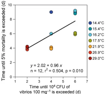
The onset of mortality, as expressed by the time elapsed before at least 5% mortality occurred, was significantly correlated with the time required to attain 104CFU of vibrios per 100 mg
oyster flesh in challenged oysters (Fig. 5). The relationship was linear, the slope was not significantly differ-ent from 1.0, and the constant was 2.0. This means that the onset of mortality followed the outbreak of vibriosis by 2 d and that a delay of 1 d in the vibrio-sis outbreak led to a delay of 1 d in mortality.
It is noteworthy that at 13.4, 14.4 and 29.0°C, OsHV-1 and vibrio num-bers peaked at roughly the same time (after 6 to 13 d at low temperatures,
Fig. 3. Crassostrea gigas. Temporal varia-tions in herpesvirus OsHV-1 DNA, detected in oysters previously exposed to field condi-tions (exposed, s) or naïve (challenged, y), maintained at 8 different temperatures (Expt B). Different letters indicate signifi-cant differences between means, calculated by a mixed linear model. SEM used by the
after only 2 d at 29°C) in challenged oysters. In contrast, OsHV-1 numbers peaked be fore vibrio numbers in challenged oysters when tempera-tures were be tween 16.2 and 26.0°C.
DISCUSSION Field infection
Pacific oysters that had been bred and reared under controlled condi-tions and then transferred for at least 10 d to a farming area where mortali-ties were occurring among surround-ing farmed oysters became positive for detection of OsHV-1 DNA μVar (Expts A and B) and contained high loads of vibrios (Expt B). As a result, groups of these ‘exposed’ animals suffered high mortalities and trans-mitted the disease to challenged oys-ters under controlled conditions.
Our study suggests that healthy oysters put in cohabitation with oys-ters that have high levels of OsHV-I DNA μVar and vibrios became in -fected by these pathogens, as previ-ously reported in other experiments with OsHV-1 (Schikorski et al. 2011b) or vibriosis (De Decker & Saulnier 2011). However, in contrast to these other studies, our diseased oysters were not injected with a specific pathogen, but were instead exposed to field conditions in which mortality was occurring. The advantage of our protocol is that we avoided high-dose intramuscular injection of pathogens, which may short-circuit some poten-tially important lines of defense and disturb normal host immune response
Fig. 4. Crassostrea gigas. Temporal variations in vibrio colonies plated on CHROM -agar, detected in oysters either previously exposed to field conditions (exposed, s) or naïve (challenged, y), maintained at 8 dif-ferent temperatures (Expt B). Difdif-ferent let-ters indicate significant differences between means, calculated by a mixed linear model.
against invaders (De Decker & Saulnier 2011). Ex -posed oysters were naturally in fected in the field, and the disease they contracted is clearly the one related to the mass mortality phenomenon that occurred in the field. However, the limitation of our method is that we cannot ascertain whether the ob -served effect of temperature on survival of exposed oysters and oysters that had been kept in cohabita-tion with them is associated with a sole pathogen, such as OsHV-1 μVar, because other pathogens (including virulent vibrios) may also have been contracted.
Horizontal transmission of disease and related mortality
Challenged oysters became infected by OsHV-1 μVar and showed significant mortality when sea -water temperatures were >13.4°C. Similarly, Schi -kor ski et al. (2011a) showed that it is possible to transmit OsHV-1 to healthy Crassostrea gigas juve-niles in cohabitation experiments using oysters experimentally infected by intramuscular injection of an OsHV-1 suspension. Mortality and detection of OsHV-1 DNA in oysters maintained in the Thau lagoon, but outside the farming area, was sporadic and coincided with currents coming from the farming area where mortality was occurring and OsHV-1
DNA was detected (Pernet et al. 2012). Together, these results suggest that horizontal transmission of OsHV-1 particles occurs from infected oysters toward healthy animals and coincides with mortality events both in laboratory and field conditions.
Vibrio levels in challenged oysters roughly dou-bled until they reached values similar to those of pre-viously exposed animals after only 2 to 6 d (Expt B). Evidence of successful transmission of vibriosis in cohabitation experiments with injected oysters was recently reported in Crassostrea gigas (De Decker & Saulnier 2011, De Decker et al. 2011). In our study, the timing of the onset of mortality events varied as a function of the timing of the outbreak of vibriosis. It is therefore possible that pathogenic strains of vibrios are involved in triggering oyster mortality in the field. For instance, in the Thau lagoon, mortalities always coincided with elevated detection frequency and quantity of OsHV-1 DNA and Vibrio splendidus in oysters, thus suggesting that both these pathogens are involved in triggering the mass mortality phe-nomenon (Pernet et al. 2012). Laboratory experi-ments have shown that V. splendidus alone can cause death in oysters (e.g. De Decker et al. 2011). Although the effect of co-infection by OsHV-1 and vibrios remains to be further investigated, these pathogens might have synergistic effects.
Survival of oysters previously exposed to field con-ditions in the Bay of Brest was 2 (Expt B) to 7 times (Expt A) lower than that of challenged animals. Sim-ilarly, Schikorski et al. (2011a) showed that survival of oysters experimentally infected by intramuscular injection of OsHV-1 was only ~10% compared with ~50% in animals that had cohabited with them at 22°C. These authors hypothesized that differences in susceptibility among injected animals and those put in cohabitation with them could primarily be ex -plained by the different routes of infection. Indeed, it is likely that some cohabitated oysters had not been exposed to OsHV-1, whereas oysters directly in -jected with the infectious virus suspension in the muscle are necessarily infected by this virus (Schikor -ski et al. 2011a). Alternatively, it was suggested that effective infection also depends on the quantity of viral particles in contact with oysters (Schikorski et al. 2011a). In our study, oysters were experimentally infected by means of exposure to field conditions where mortalities were occurring. Therefore, the lower survival of oysters previously exposed to field conditions compared with the survival of challenged oysters might reflect that there was a higher level of viral particles in contact with oysters in the field com-pared with that found in laboratory conditions.
Fig. 5. Crassostrea gigas. Relationship between the number of days required to observe at least 5% mortality and the number of days required to reach 104 CFU of vibrios per
100 mg oyster flesh in naïve oysters maintained in cohabitation with exposed animals. All temperature treatments were com-bined (n = 12 observations: 8 temperatures × 2 replicate tanks
Effect of temperature on disease and related mortality
This study clearly shows that seawater tempera-ture influences mortality among previously ‘exposed’ oysters as well as disease transmission to cohabitat-ing healthy animals. The influence of temperature on OsHV-1 occurrence and virus expression has previ-ously been demonstrated for Crassostrea gigas lar-vae (Le Deuff et al. 1996) and suggested for juveniles (Renault et al. 1995, Friedman et al. 2005, Burge et al. 2007, Sauvage et al. 2009). However, to our knowl-edge, the effect of low temperature (i.e. <13°C) has never been studied.
Effect of low temperature (≤13.4°C)
Oysters previously exposed to field conditions in the Bay of Brest, which were characterized by high levels of OsHV-1 μVar DNA, subsequently showed high mortalities in the laboratory when seawater temperatures were ≥13.4°C (Expts A and B). How-ever, at 13.0°C, survival of these exposed oysters was 96.6%, and OsHV-1 DNA was not detected after only 3 d in Expt A1. Additionally, when oysters of this
same treatment were exposed to thermal elevation up to 20.6°C after 40 d at 13.0°C, survivors of the first period showed no further mortality before the end of Expt A2(Day 68) and OsHV-1 DNA remained
unde-tected. All together, these results suggest that infec-tion was not necessarily irreversible, particularly when seawater temperature was reduced to ~13.0°C. Survival of oysters previously exposed to field con-ditions during Expt B was 48% at 13.4°C, compared with 100% at 13.0°C in Expt A and coincided with the detection of high levels of OsHV-1 DNA. There-fore, it is likely that another confounding factor inter-acted with temperature in its effect on this disease and the subsequent mortality. It is very possible that the prevalence of the disease in oysters transferred to the field between 6 and 16 July 2011 (Expt B) was higher than that of animals exposed between 26 Au-gust and 10 September 2010 (Expt A). Nevertheless, the effect of low temperatures on disease outbreak and subsequent mortalities of oysters previously ex-posed to field conditions needs further clarification.
Challenged oysters showed no mortality in our lab-oratory conditions when seawater temperatures were ≤13.4°C (100.0 and 97.8% survival in Expts A and B, respectively), and OsHV-1 DNA was not detected in oyster tissues sampled during Expt A. Additionally, when exposed to a thermal increase up
to 20.6°C after 40 d at 13.0°C, challenged and control oysters showed no mortality before the end of the experiment and OsHV-1 DNA was not detected (Expt A2). These results suggest that disease and
sub-sequent mortalities were not transmitted from ex -posed to challenged oysters when seawater tempera-ture was 13.0°C.
Although challenged oysters exhibited no mortality at low temperatures (≤13.4°C) in either Expt A or B, OsHV-1 DNA was detected at high copy numbers in oysters from Expt B (>106DNA copies mg−1) but not
from Expt A. Therefore, it seems that high quantities of OsHV-1 DNA do not necessarily lead to high mor-tality rate in oysters, as previously reported in oysters selected for improved survival to summer mortality (Dégremont 2011) and in animals cemented onto ropes at low density (Pernet et al. 2012). Additionally, this result suggests that OsHV-1 was transmitted from exposed to challenged oysters at 13.4°C. How-ever, it is likely that the virus was not pathogenic to these animals at low temperatures, even though OsHV-1 DNA was detected in their tissues. Indeed, PCR used to quantify OsHV-1 DNA did not specify whether the copies of viral DNA corresponded to in-fective viruses (enveloped virus particles), which are necessary to initiate the viral infection in host cells (Lyman & Enquist 2009). Finally, it is possible that conditions in Expt A were somewhat different from those in Expt B as a result of one or more uncontrolled parameters (i.e. prevalence of diseased animals after field exposure), which led to large differences in OsHV-1 detection in oyster tissues.
From a practical standpoint, this study showed that oysters previously infected with OsHV-1, maintained at 13.0°C for 40 d, exhibited a marked reduction in the viral DNA, showed no mortality and did not seem to transmit the virus to healthy oysters. Also, when these cold-acclimated and previously exposed oys-ters were reared for a further period at 20.6°C in lab-oratory conditions, OsHV-1 DNA remained unde-tected, the oysters exhibited no mortality and the disease was not transmitted to healthy cohabiting animals. Therefore, long-term holding at a low tem-perature (13°C) may offer a means for mitigating oys-ter mortality and the spread of the related disease. The long-term survival of these cold-acclimated, pre-viously infected oysters needs to be further evalu-ated, notably in the field where mortalities occurred, as it is possible that they are still susceptible to the disease. Although both experiments showed a posi-tive effect of low temperatures on the survival of healthy and infected oysters in cohabitation, varia-tion was high among the infected batches (100 vs.
48% survival in Expts A and B, respectively). Finally, this cold-acclimation protocol will need fine-tuning and validation at a larger scale.
Relationship between temperature and mortalities
Final survival of previously field-exposed oysters decreased with increasing seawater temperature, but reached a plateau between 21.9 and 29.0°C (Expt B). Although our study is the first to test a wide range of seawater temperatures on disease expres-sion and related mortality of oysters, a positive effect of high temperatures on OsHV-1 infection and oyster mortality has previously been suggested (Le Deuff et al. 1996, Sauvage et al. 2009). High temperature was reported to promote early proliferation of OsHV-1 particles in association with high and sudden mortal-ities in Crassostrea gigas larvae (Le Deuff et al. 1996); it was therefore suggested that viral replica-tion might be faster at 25 to 26°C than at 22 to 23°C. Survival of challenged oysters decreased as sea water temperatures rose, up to 16.2°C, where it re mained at a plateau until 21.9°C, above which it in -creased. Therefore, the optimal temperature range for disease transmission was between 16.2 and 21.9°C. This is in agreement with the fact that mass mortalities of 1-yr-old oysters usually start along the coasts of France when seawater temperature reaches 16°C during spring (Bedier 2010, Pernet et al. 2012). Also, the fact that mortality of challenged oysters de -creased at high temperatures (> 21.9°C) is in good agreement with recent field experiments conducted in the southern part of France, which showed that no mortality occurred in young oysters reared at temper-atures > 24°C (Bouquet et al. 2011, Pernet et al. 2012). In our study, oysters in cohabitation suffered signif-icant mortalities when seawater temperatures were 26.9 and 29.0°C, whereas no mortality occurred in young oysters held at temperatures > 24°C in the Thau lagoon (Pernet et al. 2012). Such a discrepancy between laboratory and field experiments may result from the confounding effects of factors other than temperature. Experimental conditions at the marine station might be more hostile for the young oysters already exposed to disease in the field than for those that remained in the field at the same time. Indeed, survival of oysters exposed to field conditions in the Bay of Brest from 4 July 2011, and brought back to the marine station at 21°C after 12 d, was markedly lower (only 29% on 25 July 2011) than that of control animals left in the field, where survival was 56% on 25 July 2011 (B. Petton et al. unpubl.).
The hydrodynamic connectivity between infected and healthy animals, which is a major driver of dis-ease epidemics in aquaculture (Gustafson et al. 2007, Kristoffersen et al. 2009, Salama & Murray 2011), was much higher in laboratory conditions: animals cohab-ited in a smaller and more confined environment in the laboratory than in the field, where the infected oysters were several metres away from the healthy ones. Alternatively, the biomass of infectious ani-mals, another important factor in explaining disease epidemics in aquaculture fish species (Salama & Murray 2011), may be higher in laboratory condi-tions, where 50% of the oysters were presumably infected with OsHV-1, than in the field. In support of these 2 hypotheses, survival of exposed and chal-lenged oysters cohabiting in laboratory conditions has also been seen to increase with seawater renewal and decrease as the biomass of infected oysters is increased (B. Petton et al. unpubl.).
Relationship between temperature, OsHV-1 and vibrios
Levels of OsHV-1 DNA detected in exposed oysters decreased gradually as a function of time, as re -ported in other recent studies (Dégremont 2011, Pernet et al. 2012). It is therefore likely that oysters in -fected with OsHV-1 eliminated viral DNA from their tissue. Alternatively, the surviving animals that remained in the tank at the end of the experiment may have had limited OsHV-1 replication.
The rate of OsHV-1 DNA decrease in oysters was accelerated by higher seawater temperatures in pre-viously exposed oysters: at the end of the experimen-tal period, the level of OsHV-1 DNA was >104copies
mg−1 at temperatures ranging between 13.4 and
17.5°C, whereas it was undetected at temperatures ranging between 21.9 and 29.0°C. High tempera-tures may promote antiviral activities in oysters. For instance, a positive correlation between seawater temperature and antiviral activity was reported in the abalone Haliotis rubra (Dang et al. 2012). In
Crassostrea gigas, antiviral activity is at its lowest
during the spring, in late April and early May, and increases markedly until August, when the highest values are reached (Olicard et al. 2005). These antivi-ral assays were performed on specimens collected in Marennes-Oléron, a temperate Atlantic coastal area where seawater temperatures generally vary from ~8°C during the winter to ~25°C during the summer. Alternatively, the greater elimination of viral DNA at higher temperatures likely reflects changes in
physiological rates of the host animal. For instance, physiological processes, e.g. assimilation, mainte-nance and structural growth, in poikilothermic mar-ine animals depend on seawater temperature. Within a speciesspecific temperature tolerance range, phy -sio logical rates increase exponentially with temp -erature, as described by the Arrhenius relation. For
Cras sostrea gigas, the lower temperature tolerance
boundaries for ingestion and respiration rates are both estimated to be 3°C, and the upper boundaries to be 25 and 32°C for ingestion and respiration rates, respectively (see Bourlès et al. 2009 for review). The application of this temperature tolerance range in the oyster dynamic energy budget model results in a slowing down of oyster growth when temperatures are > 25°C (Bourlès et al. 2009). Therefore, the greater elimination of viral DNA at higher tempera-tures coincides with higher respiration rate (catabo-lism) and lower ingestion and growth rates (ana -bolism), which may both contribute to reducing the replication of OsHV-1.
Levels of OsHV1 DNA in challenged oysters in creased over the experimental period until they at -tained values similar to those observed in previously exposed oysters, and decreased thereafter. For the first time, this study shows that the time required until peak values of OsHV-1 DNA were attained in challenged oysters decreased at higher seawater temperatures. Schikorski et al. (2011a) showed that peak values of OsHV-1 DNA were attained in oysters after 4 d of cohabitation at 22°C, which agrees with the results obtained in the present study at 21.9°C.
Although there was no correlation between temper-ature and the peak values of OsHV-1 DNA found in oysters grown in cohabitation, it should be noted that the lowest peak value of OsHV-1 DNA was found at 29.0°C, and coincided with higher survival. Lower peak values of OsHV-1 DNA in oysters held in cohab-itation at 29.0°C may reflect the lowering of ingestion rates that normally occurs in Crassostrea gigas at tem-peratures > 25°C (see Bourlès et al. 2009 for review). Indeed, a relationship between filtration rate and sus-ceptibility to viral disease could be responsible (Schikorski et al. 2011a). Alternatively, a lower peak value of OsHV-1 DNA in oysters in cohabitation held at 29.0°C may also reflect a higher antiviral activity in these animals (see previous discussion).
The number of vibrio CFU detected in oysters pre-viously exposed to field conditions showed no consis-tent patterns with regard to temperature treatment in our study. Higher vibrio abundances in seawater gen-erally occur at periods with higher temperatures (Vezzulli et al. 2010, Oberbeckmann et al. 2012). For
example, concentrations of vibrios in Mediterranean waters are correlated with sea- surface tempera-ture — with few vibrios being counted when temper-atures drop below 18°C and a sharp increase of abun-dance when temperatures are ≥22°C (Vezzulli et al. 2010). In our study, it is likely that the level of vibrio detected in oysters previously exposed to field condi-tions was mostly driven by the initial field exposure.
However, as observed for OsHV-1 DNA, the time required to attain peak values of vibrio CFU in chal-lenged oysters decreased with increasing seawater temperature. Therefore, high temperatures are likely to facilitate the transmission of vibrios from exposed animals to animals in cohabitation.
CONCLUSIONS
The present study set out to examine the thermal range for transmission of the disease responsible for the mass mortalities phenomenon in juvenile Pacific oysters. Our results show that the optimal tempera-ture range for disease transmission is between 16.2 and 21.9°C, which agrees well with field observa-tions. When seawater temperatures were <13.4°C, the disease was not transmitted to healthy oysters cohabiting with those transferred from the field. Finally, it seems that oysters previously infected with OsHV-1 could re cover after being kept at 13.0°C for 40 d. Therefore, our study suggests that long-term holding at low temperatures may offer a way to mitigate oyster mortalities.
Acknowledgements. The authors thank all the staff in volved
in oyster production at Ifremer Argenton. The authors also express their gratitude to P. Le Souchu for his technical assis-tance, to H. McCombie for revising the English version of this manuscript and 4 anonymous reviewers for their constructive comments. This work was supported by Ifremer (project ‘Sur-mortalité’ 2010-11 coordinated by N. Cochennec- Laureau) and is part of the GIGASSAT project funded by ANR-AGRO-BIOSPHERE No. ANR-12-AGRO-0001-01.
LITERATURE CITED
Bedier E (2010) Observatoire national Conchylicole. Rap-port 2009. Ifremer, Issy-les-Moulineaux
Berthelin C, Kellner K, Mathieu M (2000) Storage metabolism in the Pacific oyster (Crassostrea gigas) in relation to sum-mer mortalities and reproductive cycle (west coast of France). Comp Biochem Physiol B Biochem Mol Biol 125: 359–369
Bouquet al. Mille D, Blachier P, Oudot G and others (2011) Recherche de solutions zootechniques pour limiter les sur-mortalités — Bilan des suivis 2010. http: // creaa. pagesperso-orange. fr/Doc%20actualite/Rapport zoo technique 2010.pdf
Bourlès Y, Alunno-Bruscia M, Pouvreau S, Tollu G and oth-ers (2009) Modelling growth and reproduction of the Pacific oyster Crassostrea gigas: advances in the oyster-DEB model through application to a coastal pond. J Sea Res 62: 62−71
Burge CA, Griffin FJ, Friedman CS (2006) Mortality and herpesvirus infections of the Pacific oyster Crassostrea
gigas in Tomales Bay, California, USA. Dis Aquat Org 72:
31−43
Burge CA, Judah LR, Conquest LL, Griffin FJ and others (2007) Summer seed mortality of the Pacific oyster,
Crassostrea gigas Thunberg grown in Tomales Bay, Cali
-fornia, USA: the influence of oyster stock, planting time, pathogens, and environmental stressors. J Shellfish Res 26: 163−172
Dang VT, Speck P, Benkendorff K (2012) Influence of ele-vated temperatures on the immune response of abalone,
Haliotis rubra. Fish Shellfish Immunol 32: 732−740
De Decker S, Saulnier D (2011) Vibriosis induced by experi-mental cohabitation in Crassostrea gigas: evidence of early infection and down-expression of immune-related genes. Fish Shellfish Immunol 30: 691−699
De Decker S, Normand J, Saulnier D, Pernet F, Castagnet S, Boudry P (2011) Responses of diploid and triploid Pacific oysters Crassostrea gigas to Vibrio infection in relation to their reproductive status. J Invertebr Pathol 106: 179−191 Dégremont L (2011) Evidence of herpesvirus (OsHV-1) resistance in juvenile Crassostrea gigas selected for high resistance to the summer mortality phenomenon. Aqua-culture 317: 94−98
Delaporte M, Soudant P, Lambert C, Moal J, Pouvreau S, Samain JF (2006) Impact of food availability on energy storage and defense related hemocyte parameters of the Pacific oyster Crassostrea gigas during an experimental reproductive cycle. Aquaculture 254:571–582
Di Pinto A, Terio V, Novello L, Tantillo G (2011) Comparison between thiosulphate-citrate-bile salt sucrose (TCBS) agar and CHROMagar Vibrio for isolating Vibrio
para-haemolyticus. Food Contr 22: 124−127
EFSA (European Food Safety Association) (2011) Scientific opinion of the panel on animal health and welfare on a request from the European Commission on the increased mortality events in Pacific oysters Crassostrea gigas. EFSA 8: 1894−1953
Friedman CS, Estes RM, Stokes NA, Burge CA and others (2005) Herpes virus in juvenile Pacific oysters
Crass-ostrea gigas from Tomales Bay, California, coincides with
summer mortality episodes. Dis Aquat Org 63: 33−41 Garcia C, Thebault A, Degremont L, Arzul I and others
(2011) Ostreid herpesvirus 1 detection and relationship with Crassostrea gigas spat mortality in France between 1998 and 2006. Vet Res 42
Gustafson LL, Ellis SK, Beattie MJ, Chang BD and others (2007) Hydrographics and the timing of infectious salmon anemia outbreaks among Atlantic salmon (Salmo
salar L.) farms in the Quoddy region of Maine, USA, and
New Brunswick, Canada. Prev Vet Med 78: 35−56 Kristoffersen AB, Viljugrein H, Kongtorp RT, Brun E, Jansen
PA (2009) Risk factors for pancreas disease (PD) out-breaks in farmed Atlantic salmon and rainbow trout in Norway during 2003−2007. Prev Vet Med 90: 127−136 Le Deuff RM, Renault T, Gerard A (1996) Effects of
temper-ature on herpes-like virus detection among hatchery-reared larval Pacific oyster Crassostrea gigas. Dis Aquat Org 24: 149−157
Lyman MG, Enquist LW (2009) Herpesvirus interactions with the host cytoskeleton. J Virol 83: 2058−2066 Martenot C, Oden E, Travaille E, Malas JP, Houssin M
(2011) Detection of different variants of ostreid herpes -virus 1 in the Pacific oyster, Crassostrea gigas between 2008 and 2010. Virus Res 160: 25−31
Oberbeckmann S, Fuchs BM, Meiners M, Wichels A, Wilt-shire KH, Gerdts G (2012) Seasonal dynamics and mod-eling of a Vibrio community in coastal waters of the North Sea. Microb Ecol 63: 543−551
Oden E, Martenot C, Berthaux M, Travaillé E, Malas JP, Houssin M (2011) Quantification of ostreid herpesvirus 1 (OsHV-1) in Crassostrea gigas by real-time PCR: deter-mination of a viral load threshold to prevent summer mortalities. Aquaculture 317: 27−31
Olicard C, Didier Y, Marty C, Bourgougnon N, Renault T (2005) In vitro research of anti-HSV-1 activity in different extracts from Pacific oysters Crassostrea gigas. Dis Aquat Org 67: 141−147
Paillard C, Le Roux F, Borreg JJ (2004) Bacterial disease in marine bivalves, a review of recent studies: trends and evolution. Aquat Living Resour 17: 477−498
Peeler EJ, Reesea RA, Cheslettb DL, Geogheganb F, Powerb A, Thrusha MA (2012) Investigation of mortality in Pacific oysters associated with ostreid herpesvirus-1 μVar in the Republic of Ireland in 2009. Prev Vet Med 105: 136−143
Pepin JF, Riou A, Renault T (2008) Rapid and sensitive de -tection of ostreid herpesvirus 1 in oyster samples by real-time PCR. J Virol Methods 149: 269−276
Pernet F, Barret J, Marty C, Moal J, Le Gall P, Boudry P (2010) Environmental anomalies, energetic reserves and fatty acid modifications in oysters coincide with an excep-tional mortality event. Mar Ecol Prog Ser 401: 129−146 Pernet F, Barret J, Le Gall P, Corporeau C and others (2012)
Mass mortalities of Pacific oysters Crassostrea gigas re -flect infectious diseases and vary with farming practices in the Mediterranean Thau lagoon, France. Aquacult Environ Interact 2: 215−237
Renault T (2011) A review of mortality outbreaks in the Pacific oyster, Crassostrea gigas, reported since 2008 in various European Union Member States and the related implementation of Council Directive 2008/88/EC. Bul-letin OIE 4: 51−52
Renault T, Le Deuff RM, Cochennec N, Chollet B, Maffart P (1995) Herpes-like viruses associated with high mortality levels in larvae and spat of Pacific oysters, Crassostrea
gigas: a comparative study, the thermal effects on virus
detection in hatchery-reared larvae, reproduction of the disease in axenic larvae. Vet Res 26: 539−543
Renault T, Moreau P, Faury N, Pepin JF, Segarra A, Webb S (2012) Analysis of clinical ostreid herpesvirus 1 (Malaco-herpesviridae) specimens by sequencing amplified frag-ments from three virus genome areas. J Virol 86: 5942−5947
Rico-Villa B, Woerther P, Mingant C, Lepiver D, Pouvreau S, Hamon M, Robert R (2008) A flow-through rearing sys-tem for ecophysiological studies of Pacific oyster
Crass-ostrea gigas larvae. Aquaculture 282: 54−60
Rico-Villa B, Pouvreau S, Robert R (2009) Influence of food density and temperature on ingestion, growth and settle-ment of Pacific oyster larvae, Crassostrea gigas. Aqua-culture 287: 395−401
Salama NKG, Murray AG (2011) Farm size as a factor in hydrodynamic transmission of pathogens in aquaculture
➤
➤
➤
➤
➤
➤
➤
➤
➤
➤
➤
➤
➤➤
➤
➤
➤
➤
➤
➤
➤
➤
➤
➤
➤
➤
fish production. Aquacult Environ Interact 2: 61−74 Samain JF, McCombie H (2008) Summer mortality of Pacific
oyster Crassostrea gigas, the Morest project. QUAE/ Ifremer, Versailles
Saulnier D, De Decker S, Haffner P, Cobret L, Robert M, Garcia C (2010) A large-scale epidemiological study to identify bacteria pathogenic to Pacific oyster Crassostrea
gigas and correlation between virulence and
metallopro-tease-like activity. Microb Ecol 59: 787−798
Sauvage C, Pépin JF, Lapègue S, Boudry P, Renault T (2009) Ostreid herpes virus 1 infection in families of the Pacific oyster, Crassostrea gigas, during a summer mortality outbreak: differences in viral DNA detection and quan-tification using real-time PCR. Virus Res 142: 181–187 Schikorski D, Faury N, Pepin JF, Saulnier D, Tourbiez D,
Renault T (2011a) Experimental ostreid herpesvirus 1 infection of the Pacific oyster Crassostrea gigas: kinetics of virus DNA detection by q-PCR in seawater and in oys-ter samples. Virus Res 155: 28−34
Schikorski D, Renault T, Saulnier D, Faury N, Moreau P, Pepin JF (2011b) Experimental infection of Pacific oyster
Crassostrea gigas spat by ostreid herpesvirus 1:
demon-stration of oyster spat susceptibility. Vet Res 42
Segarra A, Pepin JF, Arzul I, Morga B, Faury N, Renault T (2010) Detection and description of a particular Ostreid herpesvirus 1 genotype associated with massive mortal-ity outbreaks of Pacific oysters, Crassostrea gigas, in France in 2008. Virus Res 153: 92−99
Soletchnik P, Razet D, Geairon P, Faury N, Goulletquer P (1997) Ecophysiology of maturation and spawning in oys-ter (Crassostrea gigas): metabolic (respiration) and feed-ing (clearance and absorption rates) responses at differ-ent maturation stages. Aquatic Living Res 10: 177–185 Soletchnik P, Faury N, Goulletquer P (2006) Seasonal
changes in carbohydrate metabolism and its relationship with summer mortality of Pacific oyster Crassostrea gigas (Thunberg) in Marennes-Oléron bay (France). Aquacul-ture 252:328–338
Vezzulli L, Previati M, Pruzzo C, Marchese A, Bourne DG, Cerrano C, the VibrioSea C consortium (2010) Vibrio infections triggering mass mortality events in a warming Mediterranean Sea. Environ Microbiol 12: 2007−2019 Webb SC, Fidler A, Renault T (2007) Primers for PCR-based
detection of ostreid herpes virus-1 (OsHV-1): application in a survey of New Zealand molluscs. Aquaculture 272: 126−139
Editorial responsibility: Megan La Peyre, Baton Rouge, Louisiana, USA
Submitted: January 7, 2013; Accepted: April 15, 2013 Proofs received from author(s): May 14, 2013


![Table 1. Crassostrea gigas. Expt B. Results of the 3-way split-split plot ANOVAs examining the effects of 8 temperatures (Temp) ranging from 13.4 to 29.0°C, oyster status (S: previously exposed or unexposed to field conditions [naïve]) and time on quantit](https://thumb-eu.123doks.com/thumbv2/123doknet/13779819.439552/10.918.93.555.879.1111/crassostrea-results-anovas-examining-temperatures-previously-unexposed-conditions.webp)