HAL Id: lirmm-02304820
https://hal-lirmm.ccsd.cnrs.fr/lirmm-02304820
Submitted on 3 Oct 2019
HAL is a multi-disciplinary open access
archive for the deposit and dissemination of
sci-entific research documents, whether they are
pub-lished or not. The documents may come from
teaching and research institutions in France or
abroad, or from public or private research centers.
L’archive ouverte pluridisciplinaire HAL, est
destinée au dépôt et à la diffusion de documents
scientifiques de niveau recherche, publiés ou non,
émanant des établissements d’enseignement et de
recherche français ou étrangers, des laboratoires
publics ou privés.
Monitoring phrenic nerve stimulation-induced breathing
via tracheal sounds
Xinyue Lu, David Guiraud, Serge Renaux, Thomas Similowski, Christine
Azevedo Coste
To cite this version:
Xinyue Lu, David Guiraud, Serge Renaux, Thomas Similowski, Christine Azevedo Coste. Monitoring
phrenic nerve stimulation-induced breathing via tracheal sounds. IFESS 2019 - RehabWeek 2019, Jun
2019, Toronto, Canada. �lirmm-02304820�
Monitoring phrenic nerve stimulation-induced breathing via tracheal sounds
Xinyue LU
1, 2, David GUIRAUD
1, Serge RENAUX
2, Thomas SIMILOWSKI
3, 4, Christine AZEVEDO
1 1INRIA, University of Montpellier, Montpellier, France
2
NeuroResp, Les Aires, France
3
INSERM, University of Sorbonne, UMRS1158, Paris, France
4APHP, Paris, France
xinyue.lu@inria.fr
Abstract: Diaphragm pacing (DP) is an efficient treatment to artificially restore respiratory function. Commercialized systems do not embed any respiratory monitoring function and cannot adapt patients’ electro-ventilation needs. To increase the performance and safety of these systems, in this study, an acoustic respiratory monitoring method based on a microphone is investigated. This method also captures stimulation signal so that it could generate an alarm for stimulation failure or an apnea. This could also help to optimize stimulation configurations individually for each patient. We have recorded the tracheal sound of one patient with tetraplegia implanted with a stimulator. Prom-ising preliminary results are a first step towards the vali-dation of the proposed monitoring algorithm of breathing events under phrenic nerve stimulation.
Keywords: respiratory monitoring, diaphragm pacing, phrenic nerve stimulation, tracheal sounds
Introduction
Central respiratory paralysis induces a dependence on arti-ficial ventilation. The paralysis can be congenital (e.g. Ondine’s syndrome) or acquired (e.g. upper spinal cord in-jury). The incidence of patients with cervical spinal cord injury on ventilation support is estimated around 12.7 per one million inhabitants, among which 6.5% requires long-term mechanical ventilation [1]. The number of patients with Ondine’s syndrome is about 100 in which 10%-15% patients have ventilatory dependence. In these situations, if phrenic nerves and diaphragm remain functional, dia-phragm pacing (DP) through electrical stimulation can pro-vide a more natural respiration instead of mechanical ven-tilation [2]. Furthermore, it allows an increased autonomy and reduces health costs, and, reduces infection risk, and significantly improves speech and olfactory sensation [2]– [4].
Even though DP has several advantages over the classical mechanical ventilation, it is not safe enough to maintain a full-time ventilatory assistance because of the absence of any " efficacy alarm" [5]. For example, DP systems alarms warn users of electronic failure, but, contrary to the case of mechanical ventilators, do not warn about ventilatory inef-ficiency. Indeed, DP systems do not embark any ventila-tory monitoring. As a result, patients cannot be left alone during the day and usually return to mechanical ventilation during the night for safety reasons. In addition, all existing systems work in open-loop; indeed, stimulation is deliv-ered with constant pre-defined parameters. It means that
stimulation intensity, pulse width and frequency are fixed at the installation of the implant, updated at each control visit, but do not adapt to patient's continuous situation evo-lution because of the absence of respiratory monitoring. To close the loop, an ambulatory respiratory monitoring solu-tion needs to be developed.
Classic respiratory monitoring methods, including nasal flow captors, pneumotachograph, oximetry, plethysmo-graph … are cumbersome and have limitations. As an al-ternative to these sensors we propose to use a microphone to capture the tracheal sounds. Indeed, during respiration, air turbulences in the upper air way make the around tissues to vibrate. This vibration induces the propagation of tra-cheal sounds so that recordings on the neck are possible [6]. Microphones are reduced in size, portable and have no electric contact with the user so no risk of interaction DP system.
Several studies proposed algorithms for apnea detection from breathing sounds [7]–[9], but those studies were fo-cused on sleep apnea, and few of them were used for real-time monitoring (treatment delay < 5 s). To adapt the use with patients with DP systems, we developed a real-time algorithm combining detections both in temporal and fre-quency domains for a better accuracy. Moreover, during recordings, the microphone captured the radio-frequency stimulation signals from the wireless implanted link, which are synchronized with respiratory sounds. Instead of con-sidering these signals as interferences, we propose to use them to verify if respiration is induced by the stimulation.
Electrical stimulator
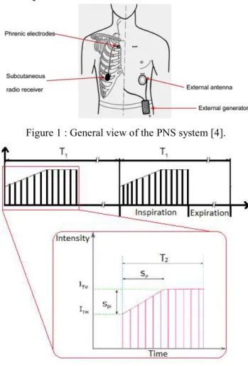
There are presently 3 DP systems available in the market: two intrathoracic phrenic nerve stimulation (PNS) systems: AtroStim® (Atrotech, Finland) and Mark IV Breathing Pacemaker (Avery Biomedical Devices, U.S.); one intra-muscular PNS system, NeuRx DPS® (Synapse Biomedi-cal, U.S.). The present work was carried out with AtroStim® PNS system shown in Figure 1.
At the installation of the device, stimulation parameters were set into the external controller. During stimulation, the controller sends power and stimulation commands to implanted radiofrequency receivers. According to the re-ceived commands, electrical pulses (stimulus) are sent through the quadripolar electrodes in alternative way. During the respiratory cycle, inspiration is an active move-ment whereas expiration is passive. Therefore, PNS is by nature limited to inspiration. Two cycles of stimulation sig-nals of the AtroStim® system are illustrated in Figure 2.
One breathing cycle lasts T1, including inspiration phase T2
(it is also the stimulation duration) and the expiration phase (T1-T2). Each red vertical line in this figure represents one
stimulation pulse which pulse-width is 200 µs. The mini-mum stimulation intensity (threshold) is ITH. And ITV is the
intensity for tidal volume, it is also the maximum stimula-tion intensity. During T2, the stimulation begins at the
in-tensity ITH, and increases at each pulse until ITV, then
re-mains at ITV until the end of T2. Except stimulation
pulse-width, all other stimulation parameters mentioned above are manually adjustable depending on patient's situation during and after implanted operation.
Figure 1 : General view of the PNS system [4].
Figure 2: Stimulation signal of AtroStim
A ramp stimulates muscle fibers progressively to avoid a too fast contraction of the diaphragm. This mimics the nat-ural recruitment of motor units during spontaneous inspi-ration. The inspiration begins when stimulation intensity reaches ITV.
Material and methods
An omni-directional microphone (pro-signal, ABM-705-RC) was used to record tracheal sounds. The microphone was inserted into a 3D printed bell-shape support to in-crease resonance. Ideally, the support should be positioned over the suprasternal notch for a best quality of signal [10]. This is not practical in tracheotomized patients, the reason for which we chose to attach the microphone just above the tracheotomy, outlined in red in Figure 3.
The signal from the microphone was first amplified (230 times) and filtered (100 Hz-1200 Hz, Band-pass, Butter-worth 2nd order) by a custom made analog card, then
con-verted to digital signal by an electronic development card (NUCLEO-F429ZI). Finally, the data were transmitted to a PC by USB link. The sampling frequency was set to 8500 Hz. All the recorded signals were post-processed with MATLABTM (MathWorks, Massachusetts, U.S.).
Figure 3 : Position of microphone
One participant agreed to participate in this observational study. This patient was equipped with an implanted AtroStim PNS device because of high cervical cord tetra-plegia following a ballistic lesion at the C2-C3 level. One recording of 30 s under stimulation was performed during a routine visit at the hospital. Patient’s usual stimu-lation configuration and parameters were unchanged. Signals analysis
Figure 4 : Recording under stimulation
One example of a recording under PNS is shown in Figure 4. Captured stimulation signals clearly appear as groups of regular peaks. These groups of peaks occur about every 3.5 s, corresponding to the respiratory rhythm (17 times/min) determined by patient's stimulation system.
Figure 5 : One enlarged cycle of recording under stimula-tion
One zoomed zone of the breathing cycle is shown in Figure 5. Each group of peaks lasts about 1.63 s within 40 peaks i.e. 25 Hz, corresponding to the stimulation frequency fixed in the stimulation system. As mentioned before, in-spiration is induced when stimulation intensity reaches ITV.
According to patient’s stimulation parameters, his inspira-tion should begin 0.64 s later than the first peak of stimu-lation. But as shown in Figure 4, in many cycles, another weak inspiration induced at the beginning of stimulation is present (identified by red cycles). These pre-inspirations may be caused by some of the first contractions of the dia-phragm muscle fibers. It could indicate a higher ITH having
been set.
Figure 6: Spectrum of tracheal sounds under stimulation For spontaneous respiration, respiratory frequency band from tracheal sounds vary depending on recording system, but could normally be detected in 200 Hz-2000 Hz [6]. And there is no obvious frequency difference between inspira-tion and expirainspira-tion [11]. With the recording system used in this work, natural breathing has the most information in 300 Hz-900 Hz. This frequency band differs from the one of induced inspiration.
The frequency spectrum of this recorded signal is plotted in Figure 6. Examples of induced inspiration, pre-inspira-tion, expirapre-inspira-tion, and stimulation events are labeled in dif-ferent colors, as well as some external noises. The expira-tion is always a passive movement, as in natural respira-tion, its frequency content is centered at 300 Hz-900 Hz. However, the frequency content of the induced inspiration can be divided into two frequency bands: one similar to the one of spontaneous respiration (300 Hz-900 Hz) and an-other one located in between 2000 Hz to 2500 Hz. This difference in higher frequency band could be a reference to identify the nature of inspiration.
Detection algorithm
Tracheal sounds recordings are processed in real-time with a delay of 0.22 s. This delay is within the acceptable alarm delay for stimulation system, which is around. As shown in the detection flow diagram (Figure 7), 3*1024 samples of recording is first filtered at 300 Hz to remove cardiac noises. Then the filtered signal is processed both in tem-poral and frequency domains:
In the temporal domain, the envelope of the signal is obtained by a low-pass filtering at wn = 0.01
rd.s-1. Then one minimum threshold is applied to
detect respiratory event. Once an event is de-tected, its center time is noted if it lasts more than 0.4 s.
In the frequency domain, the summation of fre-quency contents between 300 Hz and 600 Hz is calculated for every segment of 1024 samples. One minimum threshold is applied, but also a moving threshold depending on the average of power on previous, actual and next segments. As in temporal domain, the center time is noted for each detected event.Figure 7: Detection flow diagram
At the end, if one respiratory event is detected in both temporal and frequency domains, and if the time lag between these two detections (evaluated by their cen-ter times) is less than 1s, the event is considered as de-tected in the final result.
Results
The detection result is presented in Figure 8Erreur ! Source du renvoi introuvable.. The temporal detection result is shown in yellow, the frequency detection result is shown in purple and the final detection is shown in red. Each detection signal has two levels: a high level presents a detected respiratory event; a low level presents pause or apnea.
Figure 8: Example of a detection result
For this recording of 30 s, 9 induced inspirations and 8 ex-pirations are all detected successfully. Some noises at 2 s,
6 s, 17 s and 21 s are eliminated. And all pre-inspirations are not considerate as respiratory events.
Discussion
We have proposed a respiratory detection method for DP monitoring, which allows detecting breathing events and electrical stimulation signal. The results obtained with one recording on one patient are very promising.
Some short noises (shown in green circles in Figure 4) were considered as respiratory events by the frequency detection (presented in Figure 8). Temporal detection allows to elim-inate these noises are elimelim-inated by analyzing their dura-tions. On the other hand, temporal detection may be influ-enced by the stimulation signal, and this is when frequency detection can help because the inspiration frequency con-tent differs from the one of stimulation.
Furthermore, the captured stimulation signal could indicate a dysfunction of the pacing system and recorded tracheal sounds could give a feedback of the electro-ventilation quality. The synchronization of these two information could indicate if the stimulation settings (intensity, fre-quency ...) should be adapted. For example, in the present case, ITH may be set too high because some small
pre-in-spirations are induced at the beginning of the stimulation. Moreover, one possible application of this system during the implantation surgery would be to help verifying elec-trodes contacts and adjust stimulation settings instead of relying on the visual observation of diaphragm contrac-tions as it is assessed usually.
For the target population, the cardiac signal would also be an important vital indication. But in the present study, the microphone was located quite higher because of the trache-otomy, therefore cardiac sounds were too weak to be cap-tured. Tracheal sounds could not be recorded under me-chanical ventilation because air flow did not reach the mi-crophone which was placed above the tracheotomy. For the next study, we may place the microphone closer to the chest, or integrate it within the tracheotomy tube for a bet-ter flow sounds recording.
In the present work, only one short recording was done on one patient in a calm room, more recordings on different patients in various environments need to be analyzed to further validate the approach. Tracheal sounds may vary between different patients; it means that respiratory fea-tures could not be the same. A noise-reduction algorithm needs to be added because acoustic methods are always sensitive to noises. Besides, artificial intelligent methods could be studied because respiratory sounds and stimula-tion signals are so repetitive.
We believe that monitoring tracheal sounds could be a use-ful way beyond the very limited niche of patients with DP. Indeed, it would provide a non-invasive way to approxi-mate inspiratory flow that would be useful in all patients
requiring respiratory monitoring in acute situations (e.g. as a safety measure during the administration of morphine for acute pain) and in chronic situations (e.g. home mechanical ventilation).
References
[1] A. Quesnel, B. Veber, F. Proust, E. Agasse, F. Beuret Blanquart, and E. Verin, “What are the perspectives for ventilated tetraplegics? A French retrospective study of 108 patients with cervical spinal cord injury,” Ann. Phys. Rehabil. Med., vol. 58, no. 2, pp. 74–77, Apr. 2015.
[2] F. Le Pimpec-Barthes et al., “Diaphragm pacing: The state of the art,” J. Thorac. Dis., vol. 8, no. Suppl 4, pp. S376–S386, 2016.
[3] T. Similowski and J. Derenne, “Stimulation phrénique implantée,” Médecine thérapeutique, vol. 7, pp. 457–469, 2001.
[4] F. Le Pimpec-Barthes et al., “Intrathoracic phrenic pacing: A 10-year experience in France,” J. Thorac. Cardiovasc. Surg., vol. 142, no. 2, pp. 378–383, 2011.
[5] A. F. DiMarco, “Diaphragm Pacing,” Clin. Chest Med., vol. 39, no. 2, pp. 459–471, Jun. 2018. [6] T. Penzel and A. K. Sabil, “The use of tracheal
sounds for the diagnosis of sleep apnoea,” Breathe, vol. 13, no. 2, pp. e37–e45, 2017. [7] C. Kalkbrenner, M. Eichenlaub, S. Rüdiger, C.
Kropf-Sanchen, W. Rottbauer, and R. Brucher, “Apnea and heart rate detection from tracheal body sounds for the diagnosis of sleep-related breathing disorders,” Med. Biol. Eng. Comput., vol. 56, no. 4, pp. 671–681, 2017.
[8] S. Huq and Z. Moussavi, “Acoustic breath-phase detection using tracheal breath sounds,” Med. Biol. Eng. Comput., vol. 50, no. 3, pp. 297–308, 2012.
[9] A. Yadollahi, E. Giannouli, and Z. Moussavi, “Sleep apnea monitoring and diagnosis based on pulse oximetery and tracheal sound signals,” Med. Biol. Eng. Comput., vol. 48, no. 11, pp. 1087– 1097, 2010.
[10] P. Corbishley and E. Rodríguez-Villegas, “Breathing detection: Towards a miniaturized, wearable, battery-operated monitoring system,” IEEE Trans. Biomed. Eng., vol. 55, no. 1, pp. 196–204, 2008.
[11] Y. Nam, B. A. Reyes, and K. H. Chon,
“Estimation of Respiratory Rates Using the Built-in Microphone of a Smartphone or Headset,” IEEE J. Biomed. Heal. Informatics, vol. 20, no. 6, pp. 1493–1501, 2016.
![Figure 6: Spectrum of tracheal sounds under stimulation For spontaneous respiration, respiratory frequency band from tracheal sounds vary depending on recording system, but could normally be detected in 200 Hz-2000 Hz [6]](https://thumb-eu.123doks.com/thumbv2/123doknet/14338621.498939/4.892.464.807.230.580/spectrum-stimulation-spontaneous-respiration-respiratory-frequency-depending-recording.webp)
