HAL Id: inserm-03215564
https://www.hal.inserm.fr/inserm-03215564
Submitted on 3 May 2021
HAL is a multi-disciplinary open access
archive for the deposit and dissemination of
sci-entific research documents, whether they are
pub-lished or not. The documents may come from
teaching and research institutions in France or
abroad, or from public or private research centers.
L’archive ouverte pluridisciplinaire HAL, est
destinée au dépôt et à la diffusion de documents
scientifiques de niveau recherche, publiés ou non,
émanant des établissements d’enseignement et de
recherche français ou étrangers, des laboratoires
publics ou privés.
BACH1 and heme oxygenase-1 expression in
macrophages
Kritika Sudan, Vijith Vijayan, Kukuh Madyaningrana, Faikah Gueler,
Kazuhiko Igarashi, Roberta Foresti, Roberto Motterlini, Stephan Immenschuh
To cite this version:
Kritika Sudan, Vijith Vijayan, Kukuh Madyaningrana, Faikah Gueler, Kazuhiko Igarashi, et
al..
TLR4 activation alters labile heme levels to regulate BACH1 and heme oxygenase-1
ex-pression in macrophages.
Free Radical Biology and Medicine, Elsevier, 2019, 137, pp.131-142.
Contents lists available atScienceDirect
Free Radical Biology and Medicine
journal homepage:www.elsevier.com/locate/freeradbiomed
Original article
TLR4 activation alters labile heme levels to regulate BACH1 and heme
oxygenase-1 expression in macrophages
Kritika Sudan
a,1, Vijith Vijayan
a,1, Kukuh Madyaningrana
a, Faikah Gueler
b, Kazuhiko Igarashi
c,
Roberta Foresti
d, Roberto Motterlini
d, Stephan Immenschuh
a,∗aInstitute for Transfusion Medicine, Hannover Medical School, Carl-Neuberg-Str. 1, 30625, Hannover, Germany bDepartment of Nephrology, Hannover Medical School, Carl-Neuberg-Str. 1, 30625, Hannover, Germany
cDepartment of Biochemistry, Tohoku University Graduate School of Medicine, Seiryo-machi 2-1, Sendai 980-8575, Japan dINSERM U955, Team 12, Faculty of Medicine, University Paris Est, Creteil, France
A R T I C L E I N F O Keywords: BACH1 Carbon monoxide Heme Heme oxygenase-1 Inflammation Lipopolysaccharide Macrophages A B S T R A C T
Heme oxygenase (HO)-1, a stress-inducible enzyme that converts heme into carbon monoxide (CO), iron and biliverdin, exerts important anti-inflammatory effects in activated macrophages. HO-1 expression is mainly governed by a mutual interplay between the transcriptional factor NRF2 and the nuclear repressor BTB and CNC homology 1 (BACH1), a heme sensor protein. In the current study we hypothesized that alterations in the levels of intracellular labile heme in macrophages stimulated by lipopolysaccharide (LPS), a prototypical pro-in-flammatory Toll-like receptor (TLR)4 agonist, are responsible for BACH1-dependent HO-1 expression. To this end, labile heme was determined in both mouse bone marrow-derived macrophages (mBMDMs) and human monocyte-derived macrophages (hMDMs) using an apo-horseradish peroxidase-based assay. We found that LPS raised the levels of labile heme, depressed BACH1 protein and up-regulated HO-1 in mBMDMs. In contrast, in hMDMs LPS decreased labile heme levels while increasing BACH1 expression and down-regulating HO-1. These effects were abolished by the TLR4 antagonist TAK-242, suggesting that TLR4 activation triggers the signaling cascade leading to changes in the labile heme pool. Studies using mBMDMs from BACH1−/− and NRF2−/− mice revealed that regulation of HO-1 and levels of labile heme after LPS stimulation are strictly dependent on BACH1, but not NRF2. A strong interplay between BACH1-mediated HO-1 expression and intracellular levels of labile heme was also confirmed in hMDMs with siRNA knockdown studies and following inhibition of de novo heme synthesis with succinylacetone. Finally, CORM-401, a compound that liberates CO, counteracted LPS-dependent down-regulation of HO-1 and restored levels of labile heme in hMDMs. In conclusion, alterations of labile heme levels in macrophages following TLR4 stimulation play a crucial role in BACH1-mediated regulation of HO-1 expression.
1. Introduction
Heme oxygenase (HO) catalyzes the first and rate-limiting enzy-matic step of heme degradation to produce carbon monoxide (CO), iron and biliverdin, which is converted into bilirubin via biliverdin re-ductase [1–4]. HO-1, the inducible isoform of HO, provides antioxidant protection and exerts major immuno-modulatory functions in macro-phages by promoting anti-inflammatory effects [4,5]. For example, earlier work has demonstrated an increased susceptibility of HO-1 knockout mice to inflammation and that HO-1 controls interferon-β production in a conditional knockout mouse model lacking HO-1 in myeloid cells [6–8]. In addition, the HO-1 pathway is regulated via the
anti-inflammatory mediator interleukin (IL)-10 [9], and also inhibits the generation of pro-inflammatory markers such as TNF-α and IL-1β [6,10]. Importantly, specific interactions of HO-1 with its substrate heme appear to be critical for differentiation and inflammatory acti-vation of macrophages [11–14].
HO-1 is up-regulated by heme and numerous stress stimuli. Its ex-pression is governed by a complex network of signaling cascades and transcriptional regulators including BTB and CNC homology 1 (BACH1) and NRF2, which act as repressor and activator of the HO-1 gene, re-spectively [5,15]. In particular, BACH1 is a heme sensor protein that loses its repressing activity following increased intracellular heme le-vels and heme binding. Furthermore, high lele-vels of heme induce
https://doi.org/10.1016/j.freeradbiomed.2019.04.024
Received 15 February 2019; Received in revised form 22 March 2019; Accepted 20 April 2019
∗Corresponding author.
E-mail address:immenschuh.stephan@mh-hannover.de(S. Immenschuh).
1equally contributing authors.
Free Radical Biology and Medicine 137 (2019) 131–142
Available online 24 April 2019
0891-5849/ © 2019 The Authors. Published by Elsevier Inc. This is an open access article under the CC BY-NC-ND license (http://creativecommons.org/licenses/BY-NC-ND/4.0/).
degradation of BACH1 [16–18]. It is known that HO-1 expression by the prototypical pro-inflammatory activator lipopolysaccharide (LPS), a Toll-like receptor (TLR)4 activator, is regulated in coordination with BACH1 in human macrophages [19,20]. Although the underlying me-chanisms of this pathway are largely unknown, the crucial role of heme in the modulation of BACH1 suggests it could also be a central factor that determines BACH1/HO-1 regulation in inflammation.
The intracellular heme pool can be principally categorized into fractions of exchange-inert heme and labile heme [21–23]. In contrast to exchange-inert heme, which is covalently or non-covalently bound to hemoproteins (eg. cytochrome c and myoglobin), labile heme is redox-active heme that is loosely bound to proteins other than hemoproteins [22,24]. Notably, endogenous heme from the labile heme pool can be rapidly mobilized for signaling and other heme-dependent cellular processes and, thus, has also been designated regulatory heme [21,23,25,26]. Increased levels of heme in the circulation, due to he-molysis or tissue damage, have also been proposed to exert signaling functions by binding to pattern recognition receptors such as TLR4 in macrophages and inducing an adaptive immune response [27,28]. Conversely, whether TLR4 activation by inflammatory stimuli affects the homeostasis of intracellular heme has not been examined.
Here, we hypothesized that stimulation with LPS is accompanied by changes of the intracellular labile heme pool in macrophages resulting in modulation of BACH1 and HO-1. To address this possibility, we utilized cell culture models of murine bone marrow-derived phages (mBMDMs) and primary human monocyte-derived macro-phages (hMDMs) exposed to LPS, quantified labile heme using an apo-horseradish peroxidase (apo-HRP)-based enzymatic assay and de-termined the expression of HO-1, BACH1 and NRF2. Our data demon-strate that dynamic regulation of the labile heme pool is critically in-volved in BACH1-dependent HO-1 expression in LPS-stimulated primary mouse and human macrophages.
2. Materials and methods 2.1. Materials
Human sera, penicillin-streptomycin,L-glutamine, and
phosphate-buffered saline (PBS) were purchased from C.C.Pro GmbH (Oberdorla, Thuringia, Germany). RPMI-1640 was purchased from PAN-Biotech (Aidenbach, Passau, Germany) and accutase from Capricon Scientific (Ebsdorfergrund, Hesse, Germany). TAK-242 (CLI-09510), LPS (Escherichia coli 0111:B4), lipotechoic acid (LTA) (Staphylococcus aureus) was purchased from Invivogen (San Diego, CA, USA). Heme solution was prepared with hemin (H651-9) from Frontier Scientific (Logan, UT, USA), as described previously [29]. All other materials and reagents were obtained from Sigma-Aldrich (St. Louis, MO, USA) or Thermo Fischer Scientific (Waltham, MA, USA), unless otherwise in-dicated. CORM-401 and iCORM-401 were prepared as previously de-scribed [30].
2.2. Animals
BACH1−/− and NRF2−/− mice were of a C57BL/6J background. NRF2−/− mice, originally developed by Professor M. Yamamoto [31], were obtained from RIKEN BRC (Tsukuba, Japan) and bred with wild-type (WT) littermates (NRF2+/+). Mice were kept as previously de-scribed [32]. Approval for all experiments was given by the local an-imal protection committee (33.12-42502-04-14/1657). The German guidelines are in accordance with NIH guidelines for animal welfare. 2.3. Cell isolation and culture
Cell cultures were maintained under air/CO2 (19:1) at 100% hu-midity. Human peripheral blood mononuclear cells (PBMCs) were iso-lated from healthy donors as previously described [20]. Isolated PBMCs
were plated at a seeding density of 50 x 10ˆ6 cells in 10-cm polystyrene dishes (Greiner Bio-One, Frickenhausen, Germany) containing RPMI-1640 medium supplemented with 10% human serum. Media was changed on day 3 to remove non-adherent cells. On day 4 the differ-entiated macrophages were detached and seeded in 6- or 12-well plates for further experiments. mBMDMs were prepared from femurs and ti-bias of male C57BL/6J mice, as previously described [32]. The murine cell line RAW 264.7 was from American Type Culture Collection (Manassas, VA, USA) and cultured as described previously [33]. Treatment of cells with LPS (1μg/ml), LTA (5 μg/ml), heme (10 μM), succinylacetone (SA; 1 mM), TAK-242 (10μg/ml), iCORM-401 (50 μM), and CORM-401 (50μM), was performed in 1% sera conditions. 2.4. Quantitative real-time RT-PCR
Total RNA isolation, cDNA synthesis and RT-PCR along with the calculation for relative gene expression were performed as described previously [20]. Amplification was performed with pre-designed pri-mers listed inTable 1.
2.5. Western blot analysis
Western blotting was performed as previously described [20]. The primary antibodies (Abs) against BACH1, NRF2, and COX-2 (Abcam, Cambridge, UK) were used at a dilution of 1:1000. The primary Abs against HO-1 (Enzo Life Sciences, Farmingdale, NY, USA) and GAPDH (Santa Cruz Biotechnology, Dallas, TX, USA) were used at a dilution of 1:2000. Secondary Abs, polyclonal goat anti-rabbit IgG horseradish peroxidase (HRP) and polyclonal rabbit anti-mouse IgG HRP (Dako, Jena, Germany) were used at a dilution of 1:10,000. The clarity ECL chemiluminescent substrate (BioRad, Basel, Switzerland) was used for detection according to the manufacturer's instructions. Chemilumines-cent bands on autoradiograms were visualized in ChemiDocTM Touch Imaging System (Bio-Rad, Basel, Switzerland) and quantified using the ImageJ1.47v Gel Analysis program.
2.6. Immunofluorescence
Immunofluorescence staining was performed on hMDMs plated on coverslips in 12-well plates. After stimulation for 3 h, cells were washed once with PBS andfixed with 2% paraformaldehyde for 10 min at room temperature (RT). Cells were further permeabilized using 0.5% Triton X-100 for 5 min followed by blocking with 1% bovine serum albumin in PBS for 30 min at RT. Coverslips were incubated with primary Abs against BACH1 (Clone: 11D11C25; Biolegend, Koblenz, Germany) and NRF2 (sc13032, H-300; Santa Cruz Biotechnology, Dallas, TX, USA) at a dilution of 1:500 in PBS for 1 h at RT. Coverslips were then washed and incubated to secondary Abs conjugated with Alexa Fluor™ 488 (Invitrogen, Darmstadt, Germany) at a dilution of 1:800 in PBS for 1 h at RT. For visualization of nuclei, the cells were incubated with 4 ’,6-diamidino-2-phenylindole (DAPI) (Invitrogen, Darmstadt, Germany) at Table 1
List of primers used for TaqMan StepOnePlus Real-Time PCR System.
Gene of Interest Species Primer name
HO-1 Human Hs01110250_m1_HMOX1
COX-2 Human Hs00153133_m1_PTGS2
TNF-α Human Hs01113624_g1_TNF
NQO1 Human Hs00168547_m1_GAPDH
GAPDH Human Hs02758991_g1_GAPDH
HO-1 Mouse Mm00516005_m1_HMOX1
BACH1 Mouse Mm01344527_m1_Bach1
NRF2 Mouse Mm00477784_m1_Nfe2l2
NQO1 Mouse Mm01253561_m1_NQO1
a dilution of 1:1000 in PBS for 5 min followed by mounting with Mowiol-488. All images were acquired with an Olympus 1X81 fluor-escence microscope equipped with an Olympus MT10 camera. Images were analyzed using the ImageJ1.47v Gel Analysis program. The fluorescence intensity of the nucleus was measured and the following equation was used to calculate cellfluorescence: integrated density -(area offield x mean fluorescence of background). The average fluor-escent intensity values from three independent experiments are re-presented as arbitrary units.
2.7. Knockdown experiments
hMDMs were transfected in 6-well plates with 100 nM of BACH1 siRNA (ID: n259057) or HO-1 siRNA ID: s194530 purchased from Thermo Fischer Scientific, and control siRNA (Pre-designed validated AllStars Negative Control siRNA, Qiagen, Venlo, Netherlands) using ScreenFect A-plus transfection reagent (Incella, Eggenstein-Leopoldshafen, Germany) as previously described [34]. Cells were harvested 48 h after transfection and knockdown was verified by Western blot.
2.8. Apo-horseradish peroxidase-based (apo-HRP) assay for determination of labile heme
Intracellular levels of labile heme were determined with a method based on the reconstitution of apo-HRP as described previously [26]. Briefly, 10 μg of protein from each sample was added to the required amount of PBS to get an initial volume of 50μL. From a 5 nM heme stock solution, a heme standard curve of the following concentrations of 0.25, 0.5, 1, 1.5, 2.0 and 2.5 nM for a reaction volume of 100μL was calculated and prepared initially in 50μL. Next, 50 μL of 150 nM apo-HRP (APO-apo-HRP4C; BBI Solution, Gwent, UK), was added to all the samples and standards. The reconstitution reaction was carried out in a 96-well plate for 10 min at 4 °C. Following incubation, 10μl of each reaction was transferred to a new 96 well plate. The holo-HRP activity of the samples and standards were recorded by adding 200μl of TMB substrate (KEM-EN-TEC Diagnostics, Copenhagen, Denmark) and mea-suring the absorbance at 652 nm kinetically for 2 min on a BioTek Sy-nergy 2 plate reader. Unknown labile heme concentrations in the cell lysate calculated from the linear regression analysis of the heme stan-dard curve were expressed as percentage changes in relation to the labile heme levels in control unstimulated cells, which was taken as 100%.
2.9. Statistical analysis
Each result is a representative of at least three independent ex-periments. Statistical analysis was performed by Student's t-test or One-way ANOVA with post Bonferroni's test using GraphPad Prism 5 soft-ware. The data are represented as mean ± SD.
3. Results
3.1. Regulation of BACH1 and HO-1 by LPS correlates with alterations of the intracellular labile heme levels in mouse macrophages
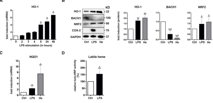
It has previously been shown that monocytes [19] and macrophages [20] stimulated with LPS exhibit changes in BACH1 and HO-1 expres-sion. To investigate whether and how the interplay of the heme sensor protein BACH1 with the master regulator of the antioxidant response, NRF2, may mediate HO-1 regulation in inflammatory macrophages, we utilized primary mBMDMs from WT C57BL/6J mice. In accordance with previousfindings [32], stimulation with LPS caused a time-de-pendent up-regulation of HO-1 mRNA levels in mBMDMs (Fig. 1A). A comparative study on the effects of LPS and the prototypical HO-1 in-ducer heme revealed that both stimuli up-regulated HO-1 protein
expression after 3 h (Fig. 1B). HO-1 induction was accompanied by down-regulation of BACH1 and concurrent up-regulation of NRF2 (Fig. 1B). In contrast, the pro-inflammatory inducible gene COX-2, which is not regulated by NRF2 and BACH1, was induced by LPS, but not by heme (Fig. 1B). Notably, up-regulation of NRF2 expression also correlated with induction of the classical NRF2-target gene, NQO1, which was also up-regulated by both LPS and heme stimulation (Fig. 1C).
Because BACH1 is known to be degraded in the presence of high levels of heme [16,35], we hypothesized that changes of the in-tracellular labile heme pool in response to LPS might regulate BACH1 and HO-1 in inflammatory macrophages. To assess this idea, we utilized an apo-HRP assay, which has previously been applied to measure in-tracellular labile heme [26]. Levels of labile heme were up-regulated in mBMDMs stimulated with LPS (Fig. 1D). As a control, treatment of cell cultures with exogenous heme led to increased levels of labile heme (data not shown). Taken together, these results indicate that labile heme levels and BACH1 are inversely regulated upon LPS stimulation in mouse macrophages.
3.2. LPS-dependent regulation of labile heme levels and HO−1 expression is abrogated in BACH1−/−, but not in NRF2−/− mBMDMs
To further evaluate the functional role of BACH1 in regulating HO-1 expression and the labile heme pool in LPS-stimulated macrophages, we performed studies in mBMDMs from BACH1−/− mice. Interestingly, expression of HO-1 and NRF2 (Fig. 2A) as well as levels of labile heme (Fig. 2B) in BACH1−/− mBMDMs were significantly higher under basal conditions in comparison to WT mBMDMs. In addition, in BACH1−/− mBMDMs, LPS failed to further increase HO-1 expression (Fig. 2A) and labile heme levels (Fig. -2B). In line with these results, the heme-dependent regulation of HO-1 and NRF2 was blunted (Fig. 2A). We compared these effects to those of mBMDMs from NRF2−/− mice. Notably, HO-1 and labile heme levels were down-regulated and BACH1 protein expression was increased under basal conditions in NRF2−/− mBMDMs compared to WT mBMDMs (Fig. 2C and D). Moreover, in NRF2−/− mBMDMs, LPS was still able to induce HO-1 and raise in-tracellular labile heme levels, similar to the regulatory pattern in WT mBMDMs (Fig. 2C and D) indicating that down-regulation of BACH1 is sufficient for the induction of HO-1. Likewise, both HO-1 and BACH1 were similarly regulated in NRF2−/− and WT mBMDMs upon treat-ment with exogenous heme (Fig. 2C). Independently, stimulation with the TLR2 agonist LTA induced a response similar to LPS in WT and NRF2−/− mBMDMs with decreased BACH1 and increased HO-1 pro-tein levels (Fig. 2C). In summary, these data indicate that absence of BACH1 affects the homeostasis of intracellular labile heme levels in macrophages with consequent effects on HO-1 regulation. Moreover, NRF2 and BACH1 expression appear to be mutually dependent on each other.
3.3. Regulation of labile heme and BACH1, but not NRF2, correlates with the LPS-dependent regulation of HO-1 in human macrophages
In our previous work [20] we have shown that LPS-stimulation of human macrophages leads to down-regulation of HO-1 expression and increased expression of BACH1, afinding that we replicate in the pre-sent study (Fig. 3A and B). These results are the opposite of those ob-tained in mouse macrophages; thus, we utilized the human model to further elucidate the role of BACH1, labile heme and NRF2 in the regulation of HO-1. If our hypothesis were correct, we would expect to find that labile heme levels are decreased by LPS. Indeed, con-comitantly with down-regulation of HO-1 and induction of BACH1 (Fig. 3B), we found markedly lower labile heme after LPS treatment (Fig. 3C). Noteworthy, the decrease in HO-1 expression was observed despite induction of NRF2 (Fig. 3B). As in mouse macrophages, NRF2 activation in human cells resulted in up-regulation of NQO1 (Fig. 3D).
Immunofluorescence analysis confirmed that LPS stimulation increased nuclear staining of both BACH1 and NRF2 (Fig. 3E) while stimulation with heme decreased the nuclear staining of BACH1 (Fig. 3E). Together with the data on mouse macrophages, these results strongly indicate that changes in labile heme and BACH1 expression, rather than NRF2 activation, dictate whether HO-1 is up-regulated or down-regulated during an inflammatory response in macrophages.
3.4. Pharmacological inhibition of TLR4 by TAK-242 abrogates LPS-dependent regulation of HO-1, BACH1, and labile heme levels
To investigate whether activation of TLR4 is directly implicated in the regulation of HO-1 and intracellular levels of labile heme, we ex-amined the effect of the pharmacological TLR4 antagonist TAK-242 in hMDMs treated with LPS. In control experiments we confirmed the expected action of TAK-242 by showing that the compound abrogated LPS-dependent TNF-α overexpression (Fig. 4A). Interestingly, TAK-242 also reversed the LPS-mediated down-regulation of HO-1 (Fig. 4B) and labile heme levels (Fig. 4C) as well as the increase in BACH1 and COX-2 expression (Fig. 4B). Conversely, TAK-242 did not significantly affect the induction of HO-1 or down-regulation of BACH1 elicited by heme (Fig. 4B). Of note, the LPS and heme-mediated induction of NRF2 was not affected by pretreatment with TAK-242 (Fig. 4B). In conclusion, these results indicate that changes in HO-1 and labile heme levels in response to LPS are a consequence of TLR4 activation.
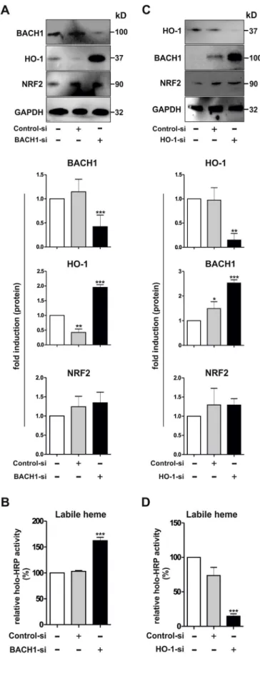
3.5. BACH1 controls HO-1 expression, and labile heme levels in hMDMs To further understand the functional interplay between BACH1 and labile heme levels, we performed a set of studies in cultured hMDMs, in which BACH1 was silenced via siRNA-mediated knockdown. As shown
inFig. 5A and B, lack of BACH1 resulted in higher HO-1 expression and increased intracellular levels of labile heme compared to control cells. Remarkably, HO-1 induction in BACH1-silenced macrophages was not mediated by NRF2, because induction of this transcription factor was not observed under these conditions. To further investigate the func-tional interaction of HO-1 with the intracellular labile heme pool, we also silenced HO-1 expression via siRNA-mediated knockdown in hMDMs. Knockdown of HO-1 (Fig. 5C) caused a significant decrease of intracellular levels of labile heme (Fig. 5D) and, a marked up-regulation of BACH1 without significantly affecting NRF2 (Fig. 5C). In summary, thesefindings indicate that BACH1 is crucial for the control of the labile heme pool in human macrophages, because a lower BACH1 protein is associated with higher labile heme despite increased HO-1 expression. In contrast, HO-1 knockdown is accompanied by enhanced BACH1 and significant reduction in labile heme. These results also suggest that the HO-1 pathway exerts an inhibitory action on BACH1 expression.
3.6. CO counters the effects of LPS on HO-1, BACH1, and labile heme levels CO is known to interact with heme [36–38] and could be the HO-derived product that affects the expression of BACH1 protein. To in-vestigate this possibility, the CO-releasing compound CORM-401 was applied to hMDMs and the levels of HO-1, BACH1, NRF2, and labile heme were measured. Interestingly, treatment with CORM-401 alone up-regulated HO-1, markedly down-regulated BACH1 below basal le-vels and caused a rise in labile heme (Fig. 6A and B). CORM-401 also increased NRF2 expression in hMDMs (Fig. 6A). These effects were much less pronounced when inactive CORM-401 (iCORM-401), which is depleted of CO, was used as a negative control. Because CO has been shown to regulate the inflammatory response in macrophages, we also examined if it changed the behavior of cells in the presence of LPS. Fig. 1. Regulation of HO-1, BACH1, NRF2, and labile heme levels in mBMDMs stimulated with LPS. (A) mBMDMs were treated with LPS (1μg/ml) for the indicated times and analyzed for HO-1 gene expression by RT-PCR. The house keeping gene GAPDH was used for normalization and the respectiveΔΔCT values are shown (n = 3). (B–C) mBMDMs were treated for 3 h with LPS or heme (10 μM), as indicated. (B) Total cell lysates were subjected to Western blot analyses and sequentially probed with antibodies against HO-1, BACH1, NRF2, COX-2, and GAPDH. A representative immunoblot and densitometric protein quantification of the indicated proteins normalized to GAPDH are shown (n = 6). (C) RNA isolated was analyzed for NQO1 gene expression by RT-PCR. The house-keeping gene GAPDH was used for normalization and the respectiveΔΔCT values are shown (n = 3). (D) mBMDMs were treated with LPS for 3 h and labile heme was determined using the apo-HRP assay as described in Materials and Methods. Total HRP activity in control unstimulated cells were taken as 100% and the relative changes in LPS-stimulated cells are shown (n = 3). Statistical significance of control versus treatment was determined by (A–C) One-way ANOVA with post Bonferroni's test and (D) Student's t-test : *p < 0.05, **p < 0.01, ***p < 0.001. Ctrl, control; He, heme.
Remarkably, CORM-401 reversed the LPS-dependent down-regulation of HO-1 and the increase in BACH1 and further enhanced HO-1 ex-pression in comparison to iCORM-401 (Fig. 6A). In addition, CORM-401 completely prevented the LPS-mediated down-regulation of labile
heme levels, which were further increased, compared to CORM-401 alone (Fig. 6B). NRF2 was higher in the presence of CORM-401 or LPS + CORM-401, and LPS-dependent COX-2 up-regulation was markedly reduced upon treatment with CORM-401 (Fig. 6A). In Fig. 2. Regulation of HO-1 expression, and labile heme levels in mBMDMs from BACH1−/− and NRF2−/− mice. (A–B) mBMDMs from wild-type (WT) and BACH1−/− mice were left untreated or treated with LPS (1μg/ml) or heme (10 μM) for 3 h, as indicated. (A) Representative immunoblot of WT and BACH1−/− mice (upper panel) and densitometric protein quanti fi-cation (lower panel) of the indicated proteins normalized to GAPDH are shown (n = 3). (B) Holo-HRP activity as an indicator of labile heme. Total HRP activity in WT untreated mBMDMs was taken as 100% and the relative changes in BACH1−/− mBMDMs are shown (n = 3). (C–D) mBMDMs from wild-type (WT) and NRF2−/− mice were left untreated or treated with LPS (1μg/ml), LTA (5μg/ml), or heme (10 μM) for 3 h, as indicated. (C) Representative immunoblot of WT and NRF2−/− mice (upper panel) and densitometric protein quantification (lower panel) of the indicated proteins normalized to GAPDH are shown (n = 3). (D) Holo-HRP activity as an indicator of labile heme. Holo-HRP activity in WT un-treated mBMDMs were taken as 100% and the relative changes in NRF2−/− mBMDMs are shown (n = 3). Statistical significance was determined by One-way ANOVA with post Bonferroni's test: untreated versus treated, *p < 0.05, **p < 0.01, ***p < 0.001, WT versus BACH1−/− or NRF2−/−; † p < 0.05, †† p < 0.01. Ctrl, control; He, heme.
conclusion, CO appears to counteract the effects of LPS in hMDMs via modulation of BACH1 expression, and labile heme levels.
3.7. Effect of inhibition of heme synthesis via SA on regulation of HO-1, BACH1, and labile heme in hMDMs
Inhibition of intracellular heme synthesis via the amino-levulinate alanine synthase (ALAS) blocking compound SA has previously been shown to regulate BACH1 expression in various cell lines [21,35]. Therefore, we also used SA to determine its effects on the labile heme pool in hMDMs in the absence or presence of LPS. Levels of labile heme in SA-treated hMDMs were markedly decreased under basal conditions (Fig. 7A). Pretreatment with SA down-regulated the expression of HO-1 and up-regulated that of BACH1 and NRF2 (Fig. 7B). Interestingly, the levels of labile heme, HO-1, BACH1 and NRF2 were not further changed in SA-treated hMDMs after LPS stimulation (Fig. 7A and B). These data indicate that newly synthesized heme contributes to the labile heme pool in unstimulated hMDMs, and its inhibition by SA affects the basal expression of HO-1, BACH1 and NRF2. Moreover, our results reveal that LPS becomes unable to modify these factors once the labile heme pool has been reduced by SA.
4. Discussion
Gene expression of HO-1 is governed by a complex network of signaling cascades and transcriptional regulators but the processes in-volved in controlling its expression during inflammation are poorly established. In the current study we show that the intracellular labile heme pool plays a critical regulatory role for BACH1-dependent HO-1 expression in LPS-stimulated macrophages.
In keeping with previousfindings by us and others [19,20], we corroborate in this study a counter-regulatory pattern of LPS-dependent BACH 1 and HO-1 expression in human and mouse macrophages. We attribute this peculiar response to the novel observation reported herein showing that LPS exerts an opposing effect on intracellular labile heme: in fact, heme levels are down-regulated in human macrophages re-sulting in accumulation of BACH1 and consequent HO-1 repression while the labile heme pool is raised in mouse cells leading to BACH1 disappearance and induction of HO-1. Why LPS causes a differential fluctuation in labile heme in the two cell types is unclear at present but our experiments using the TLR4 antagonist TAK-242 in human cells point to activation of TLR4 as directly responsible for the changes in heme levels as well as expressions of BACH1 and HO-1 (Fig. 8). We note that a detailed study on TLR4-dependent gene regulation in human and mouse macrophages has revealed extensive divergence in regulatory Fig. 3. Regulation of HO-1, BACH1, NRF2, and labile heme levels in hMDMs stimulated with LPS. (A) hMDMs were treated with LPS (1μg/ml) for the indicated times and analyzed for HO-1 gene expression by RT-PCR. The house keeping gene GAPDH was used for normalization and the respectiveΔΔCT values are shown (n = 3). (B) hMDMs were treated for 3 h with LPS, LTA (5μg/ml), or heme (10 μM) as indicated. Total cell lysates were subjected to Western blot analyses and sequentially probed with antibodies against HO-1, BACH1, NRF2, COX-2, and GAPDH. A representative immunoblot and densitometric values of the indicated proteins normalized to GAPDH are shown (n = 6). (C) Holo-HRP activity as an indicator of labile heme. Total HRP activity in control unstimulated cells were taken as 100% and the relative changes in LPS-stimulated cells are shown (n = 3). (D) hMDMs were treated for 3 h with LPS or heme (10μM) and the RNA isolated was analyzed for NQO1 gene expression by RT-PCR. The house-keeping gene GAPDH was used for normalization and the respectiveΔΔCT values are shown (n = 3). (E) hMDMs were treated with LPS or heme for 3 h and subjected to immunofluorescence analysis using Abs against BACH1 and NRF2 as described in Materials and Methods. A representative image (Bar 20μm) and the fluorescence intensity calculated from the images are shown as arbitrary units (n = 3). Statistical significance of control versus treatment was determined by One-way ANOVA with post Bonferroni's test: *p < 0.05, **p < 0.01, ***p < 0.001. Ctrl, control; He, heme.
patterns of orthologue genes in these cells, which appear to be linked with evolutionary-dependent structural differences in various gene promoters [39]. Accordingly, a number of functionally relevant di ffer-ences between the human and mouse HO-1 promoters have been
described, including a GT repeat in the proximal promoter region of the human HO-1 gene [40–42]. Remarkably, iNOS regulation also exhibits a different interspecies-specific expression pattern in murine and human macrophages [43,44]. Interestingly, our data highlight that this Fig. 4. Effect of the TLR4 antagonist TAK-242 on HO-1 regulation and labile heme levels in LPS-treated hMDMs. (A–B) hMDMs were treated with or without TAK-242 (10μM) for 30 min followed by LPS (1μg/ml) or heme (10 μM) sti-mulation for 3 h. (A) Total RNA isolated was analyzed for the expression of TNF-α. The values were normalized to the expression of GAPDH and the respectiveΔΔCT values are shown (n = 3). (B) Total cell lysates were subjected to Western blot analyses and sequentially probed with anti-bodies against HO-1, BACH1, NRF2, COX-2, and GAPDH. A representative immunoblot (upper panel) and densitometric protein quantification (lower panel) of the indicated proteins normal-ized to GAPDH are shown (n = 3). (C) hMDMs were treated with or without TAK-242 (10μM) for 30 min followed by LPS (1μg/ml) for an ad-ditional 3 h and assayed for labile heme levels. Total HRP activity in control untreated cells were taken as 100% and the relative changes in LPS and LPS + TAK-242 treated cells are shown (n = 3). Statistical significance was determined by One-way ANOVA with post Bonferroni's test; untreated versus treated: *p < 0.05, LPS versus TAK-242 + LPS; † p < 0.05. †† p < 0.01. Ctrl, control; He, heme.
divergence is evident only under TLR4 activation conditions since HO-1 and BACH1 regulation following exposure to exogenous heme is iden-tical in the two cell types and is not affected by TAK-242 in hMDMs.
The nuclear repressor BACH1 is known to control HO-1 expression together with the transcriptional activator NRF2. The results of our
study show that HO-1 gene expression in LPS-stimulated macrophages is primarily regulated via BACH1. This idea is supported by a series of experimental evidence demonstrating that: 1) even though NRF2 is activated by LPS in both human and mouse cells, HO-1 is decreased in correlation with BACH1 accumulation in hMDMs but is induced in as-sociation with BACH1 disappearance in mBMDMs; 2) in NRF2−/− mBMDMs, LPS still elicits up-regulation of HO-1 together with de-creases of BACH1 and 3) NQO1, a prototypical gene regulated by NRF2, is equally augmented by LPS in human and mouse cells. Notably, our currentfindings are in line with previous reports in which knockdown of BACH1 in human keratinocytes was specific for HO-1 regulation, whereas knockdown of NRF2 was associated with regulation of nu-merous inducible antioxidant and detoxification genes [45,46].
A central issue in our study is the significance of changes in labile heme levels in the macrophage response to inflammatory stimuli. In the current study endogenous labile heme was quantified using an enzy-matic apo-HRP assay previously described [21,26], which was speci fi-cally developed for detection of labile (or regulatory) heme [11,25,26]. In contrast to exchange-inert heme, which is primarily allocated to hemoproteins, regulatory heme is only loosely bound to non-hemo-proteins and can be readily mobilized for heme-dependent signaling and synthesis of hemoproteins [22,47]. Our data demonstrate that le-vels of labile heme are modulated in macrophages following LPS acti-vation, suggesting a mobilization of loosely-bound heme that subse-quently acts as a signal to regulate BACH1 and HO-1 expression. In addition, levels of labile heme change also in unstimulated macro-phages lacking either BACH1 or HO-1. It is intriguing to observe that in the absence of BACH1, mouse and human (in which BACH1 was si-lenced by siRNA) macrophages exhibit an increase in labile heme under basal conditions. This effect is evident despite induction of HO-1, which would be expected to degrade all excess heme available. Accordingly, silencing of HO-1 in human cells is accompanied by a sharp decrease in labile heme with marked overabundance of BACH1 protein. Thus, we are tempted to postulate that the labile heme pool is not accessible to HO for degradation, but serves as unique intracellular signal to regulate BACH1 expression. Whether this is a consequence of a higher affinity for or better access of BACH1 to labile heme compared to other heme binding proteins remains an open question. Concerning the origin of labile heme, our data using the inhibitor of ALAS SA indicate that newly synthesized heme substantially contributes to the maintenance of this regulatory heme pool.
It is important to note that regulation of intracellular labile heme levels is not only governed by degrading HOs and the heme-synthesizing enzyme ALAS, but also by various heme-binding proteins such as glutathione-S-transferases, heme-binding protein 23 and GAPDH [24,47–50]. Based on our currentfindings it is conceivable that upon inflammatory activation of macrophages, rapidly available heme is mobilized from the intracellular labile heme pool to provide the prosthetic group for the synthesis of various inducible hemoproteins including COX-2, iNOS, and NADPH oxidase-2 (NOX2) [43,51,52]. A minor fraction of so-called‘free’ heme may also exist even under phy-siological conditions as discussed by various authors [11,47,49]. Fig. 5. Effects of siRNA-mediated knockdown of BACH1 and HO-1 in hMDMs. Knockdown of (A–B) BACH1 and (C–D) HO-1 was performed with a siRNA-mediated approach in hMDMs as described in Materials and Methods. (A, C) Western blot analyses after (A) BACH1 knockdown or (C) HO-1 knockdown. A representative immunoblot (upper panel) and densitometric protein quanti-fication (lower panel) of the indicated proteins normalized to GAPDH are shown (n = 3). (B, D) HRP activity as an indicator of labile heme. Holo-HRP activity in control untransfected macrophages were taken as 100% and the relative changes in siRNA transfected macrophages are shown (n = 5). Statistical significance was determined by One-way ANOVA with post Bonferroni's test; untreated (−) versus treated (+): *p < 0.05, **p < 0.01, ***p < 0.001. Control-si, control siRNA with no known targets; Bach1-si, siRNA against BACH1; HO-1-si, siRNA against HO-1.
However, because‘free’ heme can be cytotoxic via the generation ex-cess reactive oxygen species (ROS), this issue is controversially dis-cussed. For example, in macrophages high concentrations of heme have been shown to cause necrotic cell death and heme has recently also been proposed to be an alarmin [28,53]. Ourfinding that HO-1 reg-ulation by exogenous heme was not blocked by the pharmacological TLR4 inhibitor TAK-242 in hMDMs suggests that TLR4-independent pathways may also be involved in mediating heme-dependent effects in macrophages, which adds to the on-going discussion on the potential mechanisms of heme signaling [27,54,55].
The data presented here support a dynamic and complex interplay among labile heme, BACH1 and HO-1. It appears also that metabolites of heme degradation may participate in the mechanistic regulation of the various factors examined. Our results point to the possibility that the HO-1 pathway restricts BACH1 protein since its silencing enhances the levels of this repressor and the HO-1 product CO, released from CORM-401, led to significant inhibition of BACH1 in unstimulated and LPS-challenged cells. In addition, CORM-401 not only counteracted, but
even over-compensated LPS-dependent down-regulation of HO-1 gene expression in hMDM. These effects are likely dependent on increased mobilization of labile heme observed after treatment with CO. Interestingly, enhanced cytosolic and nuclear labile heme mobilization has been reported also in Saccharomyces cerevisiae after exposure to the gaseous molecule NO [23]. Clearly, further experimental studies are required for a better understanding of the molecular mechanisms that are implicated in the interaction of CO, labile heme and BACH1 in macrophages.
Macrophages are key regulators of immune homeostasis and in-flammatory responses [56,57]. They exhibit phenotypical alterations ranging from inflammatory (also called M1) to anti-inflammatory (M2) macrophages as extremes of a continuous spectrum of activation in inflammation [58,59]. Notably, up-regulation of HO-1 has been asso-ciated with anti-inflammatory polarization of macrophages [60,61]. Thus, our currentfindings suggest that regulation of the cellular labile heme pool is critically involved in macrophage polarization via BACH1-dependent regulation of HO-1. Interestingly, loss of BACH1 in a mouse Fig. 6. Effect of CORM-401 on LPS-dependent HO-1 regulation and labile heme levels in hMDMs. hMDMs were treated with LPS (1μg/ml), iCORM-401 (50 μM), or CORM-401 (50μM) as indicated. (A) Total cell lysates were subjected to Western blot analyses and sequentially probed with antibodies against HO-1, BACH1, NRF2, COX-2, and GAPDH. A representative immunoblot (upper panel) and densitometric protein quantification (lower panel) of the indicated proteins normalized to GAPDH are shown (n = 3). (B) Holo-HRP activity as an indicator of labile heme. Holo-HRP activity in control cells were taken as 100% and the relative changes in treated cells are shown (n = 3). Statistical significance was determined by One-way ANOVA with post Bonferroni's test; untreated (−) versus treated (+): *p < 0.05, **p < 0.01, iCORM-401 (+) versus CORM-iCORM-401 (+); †† p < 0.01, ††† p < 0.001.
model of genetic BACH1 deficiency has previously been linked with M2 macrophage polarization in an in vivo model of colitis [62]. Moreover, administration of liposome-packed exogenous heme has been shown to provide protective therapeutic effects in a mouse model of myocardial infarction via reversing the M1 phenotype of inflammatory macro-phages into anti-inflammatory M2 macrophages [63] and similar ob-servations have recently also been reported in a dextran sodium sulfate-induced colitis model [64]. Finally, cellular levels of labile heme in macrophages may also be affected by alterations of extracellular heme in various in vivo situations such as hemolysis and/or tissue damage [27,65,66].
In conclusion, changes in intracellular labile heme pool are central
to the modulation of the BACH1-HO-1 axis in inflammatory macro-phages. Thesefindings may not only help to better understand macro-phage homeostasis during inflammatory responses, but may also direct towards the development of novel strategies for targeted anti-in-flammatory therapies based on controlled delivery of heme.
Acknowledgments
We would like to thank Anette Sarti and Sylvie Manin for technical assistance. This work was supported by funding from a PHC Procope/ DAAD Exchange Program between France and Germany (Project 57317676). Work in SI's laboratory is supported by the Deutsche Fig. 7. Effect of SA on regulation of HO-1, BACH1, NRF2, and labile heme levels in hMDMs. hMDMs were treated with and without SA (1 mM) for 24 h followed by exposure to LPS (1μg/ml) for 3 h. (A) Holo-HRP activity as an indicator of labile heme. Holo-HRP activity in control cells were taken as 100% and the relative changes in treated cells are shown (n = 3). (B) Total cell lysates were subjected to Western blot analyses and sequentially probed with antibodies against HO-1, BACH1, NRF2, and β-actin. A representative immunoblot (upper panel) and densitometric protein quantification (lower panel) of the indicated proteins normalized to β-actin is shown (n = 3). Statistical significance was determined by One-way ANOVA with post Bonferroni's test; untreated (−) versus treated (+): *p < 0.05, SA (−) versus SA (−): †p < 0.05.
Forschungsgemeinschaft (IM 20/4-1) and the European Union and the State of Niedersachsen project EFRE ZW6-85007634. RM and RF are supported by funding from INSERM and University Paris Est Créteil. References
[1] M.D. Maines, The heme oxygenase system: a regulator of second messenger gases, Annu. Rev. Pharmacol. Toxicol. 37 (1997) 517–554.
[2] R. Tenhunen, H.S. Marver, R. Schmid, The enzymatic conversion of heme to bilir-ubin by microsomal heme oxygenase, Proc. Natl. Acad. Sci. U.S.A. 61 (1968) 748–755.
[3] R.K. Kutty, R.F. Daniel, D.E. Ryan, W. Levin, M.D. Maines, Rat liver cytochrome P-450b, P-420b, and P-420c are degraded to biliverdin by heme oxygenase, Arch. Biochem. Biophys. 260 (1988) 638–644.
[4] S.W. Ryter, J. Alam, A.M. Choi, Heme oxygenase-1/carbon monoxide: from basic science to therapeutic applications, Physiol. Rev. 86 (2006) 583–650.
[5] A. Paine, B. Eiz-Vesper, R. Blasczyk, S. Immenschuh, Signaling to heme oxygenase-1 and its anti-inflammatory therapeutic potential, Biochem. Pharmacol. 80 (2010) 1895–1903.
[6] M.H. Kapturczak, C. Wasserfall, T. Brusko, M. Campbell-Thompson, T.M. Ellis, M.A. Atkinson, A. Agarwal, Heme oxygenase-1 modulates early inflammatory re-sponses: evidence from the heme oxygenase-1-deficient mouse, Am. J. Pathol. 165 (2004) 1045–1053.
[7] S. Tzima, P. Victoratos, K. Kranidioti, M. Alexiou, G. Kollias, Myeloid heme oxy-genase-1 regulates innate immunity and autoimmunity by modulating IFN-beta production, J. Exp. Med. 206 (2009) 1167–1179.
[8] K.D. Poss, S. Tonegawa, Reduced stress defense in heme oxygenase 1-deficient cells, Proc. Natl. Acad. Sci. U.S.A. 94 (1997) 10925–10930.
[9] T.S. Lee, L.Y. Chau, Heme oxygenase-1 mediates the anti-inflammatory effect of interleukin-10 in mice, Nat. Med. 8 (2002) 240–246.
[10] T.D. Hull, A. Agarwal, J.F. George, The mononuclear phagocyte system in home-ostasis and disease: a role for heme oxygenase-1, Antioxid. Redox Signal. 20 (2014) 1770–1788.
[11] S. Sassa, Why heme needs to be degraded to iron, biliverdin IXalpha, and carbon monoxide? Antioxid. Redox Signal. 6 (2004) 819–824.
[12] F.F. Dutra, M.T. Bozza, Heme on innate immunity and inflammation, Front. Pharmacol. 5 (2014) 115.
[13] V. Vijayan, F. Wagener, S. Immenschuh, The macrophage heme-heme oxygenase-1 system and its role in inflammation, Biochem. Pharmacol. 153 (2018) 159–167. [14] M. Haldar, M. Kohyama, A.Y. So, W. Kc, X. Wu, C.G. Briseno, A.T. Satpathy,
N.M. Kretzer, H. Arase, N.S. Rajasekaran, L. Wang, T. Egawa, K. Igarashi, D. Baltimore, T.L. Murphy, K.M. Murphy, Heme-mediated SPI-C induction promotes monocyte differentiation into iron-recycling macrophages, Cell 156 (2014)
1223–1234.
[15] J. Sun, H. Hoshino, K. Takaku, O. Nakajima, A. Muto, H. Suzuki, S. Tashiro, S. Takahashi, S. Shibahara, J. Alam, M.M. Taketo, M. Yamamoto, K. Igarashi, Hemoprotein Bach1 regulates enhancer availability of heme oxygenase-1 gene, EMBO J. 21 (2002) 5216–5224.
[16] K. Ogawa, J. Sun, S. Taketani, O. Nakajima, C. Nishitani, S. Sassa, N. Hayashi, M. Yamamoto, S. Shibahara, H. Fujita, K. Igarashi, Heme mediates derepression of Maf recognition element through direct binding to transcription repressor Bach1, EMBO J. 20 (2001) 2835–2843.
[17] K. Igarashi, T. Kurosaki, R. Roychoudhuri, BACH transcription factors in innate and adaptive immunity, Nat. Rev. Immunol. 17 (2017) 437–450.
[18] K. Igarashi, M. Watanabe-Matsui, Wearing red for signaling: the heme-bach axis in heme metabolism, oxidative stress response and iron immunology, Tohoku J. Exp. Med. 232 (2014) 229–253.
[19] T. Miyazaki, Y. Kirino, M. Takeno, S. Samukawa, M. Hama, M. Tanaka, S. Yamaji, A. Ueda, N. Tomita, H. Fujita, Y. Ishigatsubo, Expression of heme oxygenase-1 in human leukemic cells and its regulation by transcriptional repressor Bach1, Cancer Sci. 101 (2010) 1409–1416.
[20] M.J. Dorresteijn, A. Paine, E. Zilian, M.G. Fenten, E. Frenzel, S. Janciauskiene, C. Figueiredo, B. Eiz-Vesper, R. Blasczyk, D. Dekker, B. Pennings, A. Scharstuhl, P. Smits, J. Larmann, G. Theilmeier, J.G. van der Hoeven, F.A. Wagener, P. Pickkers, S. Immenschuh, Cell-type-specific downregulation of heme oxygenase-1 by lipopolysaccharide via Bach1 in primary human mononuclear cells, Free Radic. Biol. Med. 78 (2015) 224–232.
[21] X. Yuan, N. Rietzschel, H. Kwon, A.B. Walter Nuno, D.A. Hanna, J.D. Phillips, E.L. Raven, A.R. Reddi, I. Hamza, Regulation of intracellular heme trafficking re-vealed by subcellular reporters, Proc. Natl. Acad. Sci. U.S.A. 113 (2016) E5144–E5152.
[22] A.R. Reddi, I. Hamza, Heme mobilization in animals: a metallolipid's journey, Acc. Chem. Res. 49 (2016) 1104–1110.
[23] D.A. Hanna, R.M. Harvey, O. Martinez-Guzman, X. Yuan, B. Chandrasekharan, G. Raju, F.W. Outten, I. Hamza, A.R. Reddi, Heme dynamics and trafficking factors revealed by genetically encodedfluorescent heme sensors, Proc. Natl. Acad. Sci. U.S.A. 113 (2016) 7539–7544.
[24] I. Hamza, H.A. Dailey, One ring to rule them all: trafficking of heme and heme synthesis intermediates in the metazoans, Biochim. Biophys. Acta 1823 (2012) 1617–1632.
[25] S. Granick, P. Sinclair, S. Sassa, G. Grieninger, Effects by heme, insulin, and serum albumin on heme and protein synthesis in chick embryo liver cells cultured in a chemically defined medium, and a spectrofluorometric assay for porphyrin com-position, J. Biol. Chem. 250 (1975) 9215–9225.
[26] H. Atamna, M. Brahmbhatt, W. Atamna, G.A. Shanower, J.M. Dhahbi, ApoHRP-based assay to measure intracellular regulatory heme, Metallomics 7 (2015) 309–321.
[27] R.T. Figueiredo, P.L. Fernandez, D.S. Mourao-Sa, B.N. Porto, F.F. Dutra, L.S. Alves, M.F. Oliveira, P.L. Oliveira, A.V. Graca-Souza, M.T. Bozza, Characterization of heme as activator of Toll-like receptor 4, J. Biol. Chem. 282 (2007) 20221–20229. [28] M.P. Soares, M.T. Bozza, Red alert: labile heme is an alarmin, Curr. Opin. Immunol.
38 (2016) 94–100.
[29] S. Janciauskiene, S. Tumpara, M. Wiese, S. Wrenger, V. Vijayan, F. Gueler, R. Chen, K. Madyaningrana, R. Mahadeva, T. Welte, S. Immenschuh, J. Chorostowska-Wynimko, Alpha1-antitrypsin binds hemin and prevents oxidative activation of human neutrophils: putative pathophysiological significance, J. Leukoc. Biol. 102 (2017) 1127–1141.
[30] S.H. Crook, B.E. Mann, A.J. Meijer, H. Adams, P. Sawle, D. Scapens, R. Motterlini, [Mn(CO)4{S2CNMe(CH2CO2H)}], a new water-soluble CO-releasing molecule, Dalton Trans. 40 (2011) 4230–4235.
[31] K. Itoh, T. Chiba, S. Takahashi, T. Ishii, K. Igarashi, Y. Katoh, T. Oyake, N. Hayashi, K. Satoh, I. Hatayama, M. Yamamoto, Y. Nabeshima, An Nrf2/small Maf hetero-dimer mediates the induction of phase II detoxifying enzyme genes through anti-oxidant response elements, Biochem. Biophys. Res. Commun. 236 (1997) 313–322. [32] A. Thorenz, K. Derlin, C. Schroder, L. Dressler, V. Vijayan, P. Pradhan,
S. Immenschuh, A. Jorns, F. Echtermeyer, C. Herzog, R. Chen, S. Rong, J.H. Brasen, C. van Kooten, T. Kirsch, C. Klemann, M. Meier, A. Klos, H. Haller, B. Hensen, F. Gueler, Enhanced activation of interleukin-10, heme oxygenase-1, and AKT in C5aR2-deficient mice is associated with protection from ischemia reperfusion in-jury-induced inflammation and fibrosis, Kidney Int. 94 (2018) 741–755. [33] V. Vijayan, E. Baumgart-Vogt, S. Naidu, G. Qian, S. Immenschuh, Bruton's tyrosine
kinase is required for TLR-dependent heme oxygenase-1 gene activation via Nrf2 in macrophages, J. Immunol. 187 (2011) 817–827.
[34] V. Vijayan, T. Srinu, S. Karnati, V. Garikapati, M. Linke, L. Kamalyan, S.R. Mali, K. Sudan, A. Kollas, T. Schmid, S. Schulz, B. Spengler, T. Weichhart, S. Immenschuh, E. Baumgart-Vogt, A new immunomodulatory role for peroxisomes in macrophages activated by the TLR4 ligand lipopolysaccharide, J. Immunol. 198 (2017) 2414–2425.
[35] Y. Zenke-Kawasaki, Y. Dohi, Y. Katoh, T. Ikura, M. Ikura, T. Asahara, F. Tokunaga, K. Iwai, K. Igarashi, Heme induces ubiquitination and degradation of the tran-scription factor Bach1, Mol. Cell. Biol. 27 (2007) 6962–6971.
[36] L.E. Otterbein, F.H. Bach, J. Alam, M. Soares, H. Tao Lu, M. Wysk, R.J. Davis, R.A. Flavell, A.M. Choi, Carbon monoxide has anti-inflammatory effects involving the mitogen-activated protein kinase pathway, Nat. Med. 6 (2000) 422–428. [37] R. Motterlini, L.E. Otterbein, The therapeutic potential of carbon monoxide, Nat.
Rev. Drug Discov. 9 (2010) 728–743.
[38] R. Motterlini, R. Foresti, Biological signaling by carbon monoxide and carbon monoxide-releasing molecules, Am. J. Physiol. Cell Physiol. 312 (2017) C302–C313.
Fig. 8. Schematic summary. The interplay of labile heme with HO-1 regula-tion in LPS-treated macrophages as demonstrated in this study (see Discussion for details).
[39] K. Schroder, K.M. Irvine, M.S. Taylor, N.J. Bokil, K.A. Le Cao, K.A. Masterman, L.I. Labzin, C.A. Semple, R. Kapetanovic, L. Fairbairn, A. Akalin, G.J. Faulkner, J.K. Baillie, M. Gongora, C.O. Daub, H. Kawaji, G.J. McLachlan, N. Goldman, S.M. Grimmond, P. Carninci, H. Suzuki, Y. Hayashizaki, B. Lenhard, D.A. Hume, M.J. Sweet, Conservation and divergence in Toll-like receptor 4-regulated gene expression in primary human versus mouse macrophages, Proc. Natl. Acad. Sci. U. S. A. 109 (2012) E944–E953.
[40] E.M. Sikorski, T. Hock, N. Hill-Kapturczak, A. Agarwal, The story so far: molecular regulation of the heme oxygenase-1 gene in renal injury, Am. J. Physiol. Renal. Physiol. 286 (2004) F425–F441.
[41] M. Exner, E. Minar, O. Wagner, M. Schillinger, The role of heme oxygenase-1 promoter polymorphisms in human disease, Free Radic. Biol. Med. 37 (2004) 1097–1104.
[42] N. Yamada, M. Yamaya, S. Okinaga, K. Nakayama, K. Sekizawa, S. Shibahara, H. Sasaki, Microsatellite polymorphism in the heme oxygenase-1 gene promoter is associated with susceptibility to emphysema, Am. J. Hum. Genet. 66 (2000) 187–195.
[43] C. Bogdan, Nitric oxide and the immune response, Nat. Immunol. 2 (2001) 907–916.
[44] J. Mestas, C.C. Hughes, Of mice and not men: differences between mouse and human immunology, J. Immunol. 172 (2004) 2731–2738.
[45] A.K. MacLeod, M. McMahon, S.M. Plummer, L.G. Higgins, T.M. Penning, K. Igarashi, J.D. Hayes, Characterization of the cancer chemopreventive NRF2-de-pendent gene battery in human keratinocytes: demonstration that the KEAP1-NRF2 pathway, and not the BACH1-NRF2 pathway, controls cytoprotection against electrophiles as well as redox-cycling compounds, Carcinogenesis 30 (2009) 1571–1580.
[46] T.W. Kensler, N. Wakabayashi, S. Biswal, Cell survival responses to environmental stresses via the Keap1-Nrf2-ARE pathway, Annu. Rev. Pharmacol. Toxicol. 47 (2007) 89–116.
[47] D. Chiabrando, F. Vinchi, V. Fiorito, S. Mercurio, E. Tolosano, Heme in pathophy-siology: a matter of scavenging, metabolism and trafficking across cell membranes, Front. Pharmacol. 5 (2014) 61.
[48] S. Immenschuh, V. Vijayan, S. Janciauskiene, F. Gueler, Heme as a target for therapeutic interventions, Front. Pharmacol. 8 (2017) 146.
[49] H.M. Girvan, A.W. Munro, Heme sensor proteins, J. Biol. Chem. 288 (2013) 13194–13203.
[50] E.A. Sweeny, A.B. Singh, R. Chakravarti, O. Martinez-Guzman, A. Saini, M.M. Haque, G. Garee, P.D. Dans, L. Hannibal, A.R. Reddi, D.J. Stuehr, Glyceraldehyde-3-phosphate dehydrogenase is a chaperone that allocates labile heme in cells, J. Biol. Chem. 293 (2018) 14557–14568.
[51] W.L. Smith, D.L. DeWitt, R.M. Garavito, Cyclooxygenases: structural, cellular, and molecular biology, Annu. Rev. Biochem. 69 (2000) 145–182.
[52] B.M. Babior, NADPH oxidase: an update, Blood 93 (1999) 1464–1476.
[53] G.B. Fortes, L.S. Alves, R. de Oliveira, F.F. Dutra, D. Rodrigues, P.L. Fernandez, T. Souto-Padron, M.J. De Rosa, M. Kelliher, D. Golenbock, F.K. Chan, M.T. Bozza, Heme induces programmed necrosis on macrophages through autocrine TNF and ROS production, Blood 119 (2012) 2368–2375.
[54] K.A. Nath, J.D. Belcher, M.C. Nath, J.P. Grande, A.J. Croatt, A.W. Ackerman, Z.S. Katusic, G.M. Vercellotti, Role of TLR4 signaling in the nephrotoxicity of heme and heme proteins, Am. J. Physiol. Renal. Physiol. 314 (2018) F906–F914. [55] J.D. Belcher, C. Chen, J. Nguyen, L. Milbauer, F. Abdulla, A.I. Alayash, A. Smith,
K.A. Nath, R.P. Hebbel, G.M. Vercellotti, Heme triggers TLR4 signaling leading to endothelial cell activation and vaso-occlusion in murine sickle cell disease, Blood 123 (2014) 377–390.
[56] D.M. Mosser, J.P. Edwards, Exploring the full spectrum of macrophage activation, Nat. Rev. Immunol. 8 (2008) 958–969.
[57] S. Gordon, The macrophage, Bioessays 17 (1995) 977–986.
[58] Y.C. Liu, X.B. Zou, Y.F. Chai, Y.M. Yao, Macrophage polarization in inflammatory diseases, Int. J. Biol. Sci. 10 (2014) 520–529.
[59] F.O. Martinez, S. Gordon, The M1 and M2 paradigm of macrophage activation: time for reassessment, F1000Prime Rep 6 (2014) 13.
[60] N. Weis, A. Weigert, A. von Knethen, B. Brune, Heme oxygenase-1 contributes to an alternative macrophage activation profile induced by apoptotic cell supernatants, Mol. Biol. Cell 20 (2009) 1280–1288.
[61] Y. Naito, T. Takagi, Y. Higashimura, Heme oxygenase-1 and anti-inflammatory M2 macrophages, Arch. Biochem. Biophys. 564 (2014) 83–88.
[62] A. Harusato, Y. Naito, T. Takagi, K. Uchiyama, K. Mizushima, Y. Hirai, Y. Higashimura, K. Katada, O. Handa, T. Ishikawa, N. Yagi, S. Kokura, H. Ichikawa, A. Muto, K. Igarashi, T. Yoshikawa, BTB and CNC homolog 1 (Bach1) deficiency ameliorates TNBS colitis in mice: role of M2 macrophages and heme oxygenase-1, Inflamm. Bowel Dis. 19 (2013) 740–753.
[63] T. Ben-Mordechai, D. Kain, R. Holbova, N. Landa, L.P. Levin, I. Elron-Gross, Y. Glucksam-Galnoy, M.S. Feinberg, R. Margalit, J. Leor, Targeting and modulating infarct macrophages with hemin formulated in designed lipid-based particles im-proves cardiac remodeling and function, J. Control. Release 257 (2017) 21–31. [64] H. Kayama, M. Kohyama, D. Okuzaki, D. Motooka, S. Barman, R. Okumura,
M. Muneta, K. Hoshino, I. Sasaki, W. Ise, H. Matsuno, J. Nishimura, T. Kurosaki, S. Nakamura, H. Arase, T. Kaisho, K. Takeda, Heme ameliorates dextran sodium sulfate-induced colitis through providing intestinal macrophages with nonin-flammatory profiles, Proc. Natl. Acad. Sci. U.S.A. 115 (2018) 8418–8423. [65] F.F. Dutra, L.S. Alves, D. Rodrigues, P.L. Fernandez, R.B. de Oliveira,
D.T. Golenbock, D.S. Zamboni, M.T. Bozza, Hemolysis-induced lethality involves inflammasome activation by heme, Proc. Natl. Acad. Sci. U. S. A. 111 (2014) E4110–E4118.
[66] R. Martins, S. Knapp, Heme and hemolysis in innate immunity: adding insult to injury, Curr. Opin. Immunol. 50 (2018) 14–20.