Characterization of murine Vyl,V66 T cells
by David Gerber S.B., Mathematics M.I.T. (1987)Submitted to the Department of Biology in Partial Fulfillment of
the Requirement for the Degree of
Doctor of Philosophy in Biology
at the
Massachusetts Institute of Technology June 199,f6 f Signature of Author ... ... ..--. ... Department of Biology
May 13, 1996
Certified by ...-..- .... ... negawa Dr. Susum Tonegawa Professor of Biology Thesis Supervisor A ccepted by ... "- . ... .. -. .. ... Dr. Frank Solomon i= . Chairman, Biology Department Graduate CommitteeCharacterization of murine Vy1,V86 T cells
by David GerberABSTRACT
The work described in this thesis began with an attempt to determine whether murine yS T cells respond to mycobacterial antigens. To this end we generated a panel of •S T cell hybridomas following in vitro stimulation of purified y8 cells with mycobacterial purified protein derivative (PPD). 21 hybridomas that secreted IL-2 upon mAb
mediated TCR crosslinking were chosen for further study. The hybridomas were primarily of the Vyl,V66 class, as all hybridomas contained an in frame Vyl-Jy4
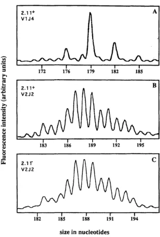
rearrangement, and the majority contained an in frame V56-J81 rearrangement. Junctional sequences of the y and 8 chain genes were diverse. The hybridomas were found to secrete IL-2 spontaneously, and only modest increases in IL-2 secretion were observed upon addition of PPD. The high background level of IL-2 secretion complicates investigation of the antigenic specificity of the hybridomas.
The constitutive IL-2 secretion could be blocked by mAbs that bind to the CD3/y8TCR complex, suggesting that IL-2 secretion depends on TCR-mediated activation. To identify additional molecules, and potential cellular TCR ligands, involved in the hybridoma activation, we generated mAbs that block hybridoma IL-2 secretion following immunization of hamsters with hybridoma cells. All mAbs that specifically blocked IL-2 secretion of the Vyl,V86 hybridomas either recognized an epitope of the y• TCR, or recognized the avt3 vitronectin receptor. Chapters 3 and 4 of this thesis describe characterization and expression analysis for anti-Vyl and anti-av13 mAbs respectively.
To characterize primary Vyl,V56 T cells, particularly with regard to activation status and response to mycobacterial antigens, we generated mice transgenic for the y and 5 genes that encode the Vy1,V86 yS TCR of one of the hybridomas. These transgenic mice (A-2m) contained increased numbers of Vyl cells in the thymus and peripheral lymphoid organs. A-2m transgenic cells appeared to be in a resting state in vivo as determined by analysis of expression of surface activation markers and proliferative status, however, the cells could be activated by mAb mediated TCR crosslinking in the presence of modest amounts of recombinant IL-2. A-2m transgenic cells did not display a convincing response to mycobacterial antigens, including PPD, Hsp 65 and isopentenyl pyrophosphate. Other notable features of the transgenic mice include the ability of the transgenes to rescue development of CD4+CD8+ thymocytes when crossed into the Rag1 deficient background, and the constitutive expression of the IL-2 receptor
P
chain by the A-2m transgenic cells.Submitted to the Department of Biology on April 16, 1996 in partial fulfillment of the requirements for the Degree of Doctor of Philosophy in Biology
Thesis Supervisor: Dr. Susumu Tonegawa
Acknowledgements:
First, I thank my parents for all of the love, support and encouragement that they have given me throughout the years. This thesis is dedicated to them. I also thank all of my brothers and sisters for their love and encouragement, and particularly my sister Monica for companionship and making my life much easier and happier during the last two years of this undertaking.
I thank Dr. Philip S. Perlman for giving me my start in science too many years ago. I am very grateful to Joy Yang in the Hynes lab for her useful
experimental suggestions and for all of the reagents that she generously gave me. I also thank Craig Morita in the Brenner lab for providing me with reagents.
I am most grateful to Shu Huang for her excellent work and for the friendship and support she has given me over the past several years. It is my greatest luck to work with her, and without her help and encouragement this thesis would not have been possible. I thank Pablo Pereira for being a wonderful colleague and friend, and for teaching me so much about science. I thank Juan Lafaille for all of his help and advice and friendship over the past eight years. I thank Toshi Sasaoka for teaching me everything I know about mice, and for helping me to make transgenics, and for everything he has done for the lab in general-I really appreciate it. I thank Linda Graziadei for helping me type in references, and Jie Shen, Joe Delaney and Patrik Kunzler for helping me to enter the computer age.
I thank all members of the Tonegawa lab for all of the help that they have given me throughout the years and for creating a unique atmosphere in which to work. I am particularly grateful for my friendships with Shu, Jie, Linda, Haydn, Pablo, Antonio, Juan, Ming, Veronica, Heather, Kumiko, and Ken.
Finally, I thank Susumu for taking me as his student, for giving me the freedom to work on whatever I wanted and providing me the opportunity to learn so much, for support and encouragement and for letting me work and develop at my own pace.
Table of contents
Chapter 1
Introduction
2
1. Foreword 2
:2. General features of y8 T cells 2
Nomenclature of y and 5 gene segments 2
y8 T cell subsets 3
Determination of murine ap and y8 T cell lineages 4
Thymic selection of murine y8 T cells 5
Extrathymic selection of murine 7y T cells 7
3. Specificity of y8 cells 8
Classical MHC proteins 8
Non classical MHC proteins 9
Mycobacterial antigens 10
Superantigens 12
Interaction with antigen 12
4. y8 T cell function 13
Potential immune functions 13
y6 cells and infectious disease 14
y8 cells and cancer 15
Development of intestinal epithelium 16
5. Conclusion 16
6. Table of murine Vy gene segment nomenclatures 17
6. References for chapter 1 18
Chapter 2
Generation of y8 T cell hybridomas
31
1. Abstract 31
2. Introduction 32
3. Materials and Methods 34
4. Results 37
Generation ,of y8 T cell hybridomas 37
PCR and sequencing analysis 37
Spontaneous reactivity of Vyl,V86 hybridomas 38 Inhibition of spontaneous IL2 secretion
by anti-CD3/TCR mAbs 39
6. Figures, tables and legends 42 Figure 1 legend 42 Figure 1 43 Table I legend 44 Table 1 45 Table 2 legend 46 Table 2 47 Figure 2 legend 48 Figure 2 49 Figure 3 legend 50 Figure 3 51
7. References for Chapter 2 52
Chapter 3
Ontogenic development and
tissue distribution of Vyl- expressing y8
T lymphocytes in normal mice
55
1. Abstract 55
2. Introduction 56
3. Materials and Methods 58
4. Results 63
Characterization of the 2.11 mAb 63
Ontogeny of y8 T cells expressing the
Vyl-Cy4 gene product in the thymus 64
Tissue distribution of Vyl-Cy3 expressing y8 T cells 65 Vyl-Cy4 expressing i-IELs originate outside
the thymus 66
The proportion of Vyl+ cells among y6 i-IELs is
independent of the antigenic load of the gut 68 Expression of Vyl-Cy4 i-IELs in different mouse strains 68
5. Discussion 70
6. Figures, tables and legends 76
Table 1 76 Table 2 77 Table 3 78 Table 4 79 Figure 1 legend 80 Figure 1 81 Figure 2 legend 82 Figure 2 83 Figure 3 legend 84 Figure 3 85 Figure 4 legend 86
Figure 4 87
Figure 5 legend 88
Figure 5 89
7. References for chapter 3 90
Chapter 4
Expression of av and P33 integrin chains
on murine lymphocytes
98
1. Abstract 98
2. Introduction 99
3. Materials and Methods 101
4. Results 105
Characterization of anti-vitronectin receptor
monoclonal antibodies 105
Inhibition of substrate binding 106
Expression of av and 33 chains on
primary lymphocytes 107
Expression of av and 33 chains on
CD4-CD8- thymocytes 108
5. Discussion 109
6. Figures, tables and legends 112
Table 1 112 Figure 1 legend 113 Figure 1 114 Figure 2 legend 115 Figure 2 116 Figure 3 legend 117 Figure 3 118 Figure 4 legend 119 Figure 4 120 Figure 5 legend 121 Figure 5 122 7. References to chapter 4 123
Chapter 5
Generation and characterization of Vyl,V86
transgenic mice
126
1. Abstract 126
:2. Introduction 127
3. Materials and Methods 128
Generation of Vy1,V86 transgenic mice 131
Expression of transgenes 131
B cell and a: TW cell populations appear normal
in Vyl transgenic mice 132
Presence of both y2F and 810.1 transgenes rescues CD4+CD8+ development in Rag1
deficient mice 133
A-2m transgenic cells have resting
phenotype in vivo 133
A-2m transgenic cells express CD122 and
proliferate in response to high IL-2 concentration 134 A-2m transgenic cells show modest response
to mycobacterial antigens 135
5. Discussion 136
6. Figures and figure legends 139
Figure 1 legend 139 Figure 1 140 Figure 2 legend 141 Figure 2 142 Figure 3 legend 143 Figure 3 144 Figure 4 legend 145 Figure 4 146 Figure 5 legend 147 Figure 5 148 Figure 6 legend 149 Figure 6 150 Figure 7 legend 151 Figure 7 152 Figure 8 legend 153 Figure 8 154
7. References for chapter 5 155
Conclusions and future directions
157
Appendices
162
Appendix 1
Rearrangement and expression of Vy1,
Appendix 2
'On somatic recombination in the central
nervous system of transgenic mice
170
Chapter 1: Introduction
Foreword
During the search for genes encoding the ap T cell receptor, a third rearranging gene designated y was discovered (1, 2). Antibodies made against peptides derived from the y sequence allowed identification of a distinct TCR designated y8 (3). The 8 gene was later cloned based on its position within the TCR a locus (4). Subsequent studies identified a distinct, new class of T cells that express the y• receptor and are designated y8 T cells (5-7).
Unlike ap T cells whose function in cellular immunity was outlined long before the cloning of the ap TCR, y• T cells were identified based on the serendipitous isolation of the gene encoding the TCR y chain, and therefore, their role in the immune system was unknown from the outset. Since their initial description, a vast effort has been directed towards understanding the function of y8 cells. Despite the accumulation of a large body of literature describing various features of y6 cells, the function of this class of T cells remains unknown. This introduction will highlight some of the
fundamental information that is known about murine and human •8 cells. The first section will describe some general features of y8 T cells. The second section will discuss the specificity of •S T cells, and the last section will address the function of y8 T cells. A small amount of material described in the body of the thesis has been incorporated into the introduction for the sake of
completeness.
General features of
y8
T cells
Nomenclature of y and 8 gene segments
The genes encoding y and 8 TCR chains in both mice and man were identified by a number of groups at different times, and therefore, various different nomenclatures for y and 8 TCR genes are present in the literature. This can cause considerable confusion as the same gene segments are often referred to by different names in different publications. In the case of the
murine y locus, it is particularly confusing as there are four different nomenclatures based on 1) order of identification, and publications by 2) Garman et al., (8) 3) Iwamoto et al., (9) and 4) Pelkonen et al., (10) These nomenclatures are listed in a table at the end of this introduction. The nomenclature based on order of identification is used in this thesis. More recently, a new WHO-IUIS nomenclature for all TCR segments has been
devised(1 1).
y8 T cell subsets
In mice, there are 5 major subsets of y8 T cells that can be distinguished
by such parameters as Vy gene usage, junctional diversity, time of appearence
in ontogeny, tissue localization and thymus dependence. The earliest T cells to develop in the thymus are the Vy5V81 subset (12), which appear as early as embryonic day 15. The TCR of this class of cells is entirely homogeneous, encoded by y and 8 chains with canonical sequences (13). By one week after birth, no Vy5 cells remain in the thymus, and in adult mice these cells
specifically colonize the epidermis of the skin (12, 13). The second wave of T cells to develop in the thymus are the Vy6V81 (14) cells which also have a single canonical TCR. Upon leaving the thymus, these cells specifically colonize the epithelium of uterus, vagina and tongue (15). The next two y8 subsets to appear in the thymus are the Vy4 and Vyl populations(16, 17). Vy4 cells have diverse TCRs based on the utilization of different V8 chains and junctional diversity of both the y and 8 chains. These cells are present in the
peripheral immune system, and can be found in blood, lymph nodes and spleen. Vyl cells also have diverse TCRs that utilize different 8 chains and exhibit junctional sequence diversity. Along with Vy4 cells, Vyl cells
constitute the major class of y8 cells in the peripheral immune system of mice
(18). Unlike Vy4 cells, a large population of Vyl cells is present in the
intestinal epithelium, and these Vyl intestinal intraepithelial lymphocytes can develop extrathymically (18). The final class of murine 78 T cells are the
Vy7 cells which have diverse TCRs, specifically colonize the intestinal
epithelium and for the most part, develop extrathymically (19-22).
In humans there are two major subsets of y8 T cells defined by their use of two different 8 chains. Rearrangements of V82 to D83 and Vy1.8 or Vy9 to
the Jyl cluster can be found in the thymus early in ontogeny (23, 24). Cells utilizing these chains are designated the V82 subset. Predominant
rearrangements that occur later in thymic ontogeny include rearrangements of V81 to D81 or D82 and rearrangements of Vy gene segments from the Vy2 family to the Jy2 cluster (23, 24). Cells that use these TCR chains are referred to as the V81 subset. After birth, the V82 cells comprise a minority of yS cells in the thymus and are predominant in the blood. The percentage of V52 cells in the blood increases with age. In contrast, after birth, the V81 cells are
predominant in the thymus and are a minority of y8 cells in the blood, with their percentage in blood decreasing with age (25-29).
Determination of murine a3 and y8 T cell lineages
Several models have been proposed to explain the segregation of xpC and
y8 T cell lineages. Rearrangements of y and 8 chain genes and surface
expression of y and 8 chains precede those of a and
3
chains during thymic ontogeny (30, 31). In addition, non-productive rearrangements of y chain genes are frequently observed in xp T cells. These facts led to the proposal thatap T cells develop from T cell precursors that failed to express a functional y8
TCR (31, 32). In this model, complete y and 8 TCR gene rearrangements initially occur in common precursors at a time when only partial (D-J)
rearrangements occur at the
P
locus. Generation of a functional y8 TCR would inactivate the recombination machinery and commit the cell to the ySlineage. Failure to productively rearrange both y and 8 chain genes would allow the cell to proceed to further rearrangement of
P
and a chain genes and potential commitment to the ap lineage. Studies of transgenic mice that express rearranged y and 8 transgenes undermined this model that would predict that expression of y and 8 transgenes should disrupt ap T celldevelopment. In contrast to this prediction, a first set of y8 transgenics to be generated displayed normal development of ap cells (33). Transcripts of the transgenes were not present in the ap cells of these mice, leading to the notion that y and/or 8 chain genes have a silencer element that blocks their expression in cells of ap lineage. A second set of mice which had shorter transgenes displayed disrupted ap T cell development. It was concluded that these transgenes lacked the silencer element (34). These findings led to the
:notion that transcriptional silencing of y and 8 genes is involved in commitment of precursor cells to the ap lineage. An a silencer was also :identified that could function in commitment to the y8 lineage (35).
Another model proposed that the 8 locus cannot rearrange in ap lineage cells. This model was based on the finding that the 8 locus present on circular DNAs generated from Vca-Jcz rearrangement was almost exclusively in the germline configuration (36). Yet another model proposed that commitment to the ap lineage involves site-specific deletion of the 8 locus by
recombination between two elements (8rec and Jca) (37, 38). Two recent reports that describe the occurrence and characterization of 8 chain gene rearrangements in cup lineage cells are incompatible with these two models (39, 40).
Several approaches including the expression of y and 5 transgenes in the SCID mouse background (41) and injections of bone marrow cells (42) or purified y8 cells (43) into SCID mice have suggested the involvement of y• cells in development of 0xp lineage cells to the CD4+CD8+ double positive stage. However, characterization of 8 gene deficient mice has shown that presence of functional 8 chains is not required for development of pP T cells (44). Similarly, y8 T cells can develop normally in mice deficient for the TCR
3
gene (45).
Based collectively on the above cited reports, it is currently believed that
y8 and ap cells emerge from common precursors and that lineage
commitment is not controlled at the level of rearrangement of the TCR genes. Rather, it is believed that lineage commitment is either based on gene silencing or competition between the different TCRs or pre-TCRs. The
potential role of y8 cells in pcP development remains unclear.
Thymic selection of murine y8 T cells
In order to create a functional repertoire of p43 T cells that can recognize foreign antigens in the context of self MHC and ignore self antigens, ap
thymocytes undergo processes of positive and negative selection. Although in most cases the ligands for murine y8 cells are unknown, several studies have addressed the issue of thymic selection of y8 T cells. Studies of the sequences of rearranged Vy5, Vy6 and V81 genes from PCR amplified genomic DNA
from fetal thymocytes and epithelial T cells showed that there is greater junctional diversity among non-productive than among productive
rearrangements. This observation suggested the existence of cellular selection for cells expressing the canonical TCRs (46). However, analysis of Vy -Jy and V&-D-J8 rearrangements in mice deficient for the C8 region did not support this conclusion. In these mice, which cannot assemble functional 78 TCRs, the relevant junctional sequences were comparably homogeneous to those
described in normal mice (44). This finding favored the existence of a
molecular mechanism (molecular constraint model) to limit the diversity of these junctional sequences. It is likely that rearrangements of these segments are greatly limited by molecular constraint, and that in addition, some
cellular selection for canonical TCRs occurs.
Stronger evidence for thymic selection of y6 T cells came from studies of y7 transgenic mice. In one study, mice transgenic for genes encoding the Vy4
y6 TCR expressed by the KN6 hybridoma that recognizes the TL gene, T22 (47,
48), which is expressed in H-2b mice but not in H-2d, mice provided evidence for negative selection of the transgenic 7y T cells (49). The KN6 transgenes were crossed into the H-2b and H-2d backgrounds, and it was found that these mice had similar numbers of transgenic thymocytes, but the number of
transgenic cells was 10-fold lower in the spleens of the H-2b mice than spleens of H-2d mice. The transgenic cells derived from the H-2b mice were also shown to be larger in size and lower in TCR density than those from H-2d mice. In addition, 7y T cells from H-2b mice did not proliferate in response to H-2b stimulator cells, although they could proliferate in the presence of exogenous IL-2, indicating that KN6 transgenic cells are rendered anergic in the H-2b background (49). A second study of mice transgenic for the genes encoding the Vy4 y7 TCR from the clone G8, which also recognizes a TL gene product also provided evidence for negative selection of Vy4 y6 T cells (50).
Studies of mice derived from crossing the transgenics described above into the 02m-deficient background provided evidence for positive selection of
y8 T cells (51, 52). Transgenic cells with high TCR density were present in the
thymus of transgenic 02m-deficient H-2b and H-2d mice, but they did not populate peripheral lymphoid tissues. The transgenic cells did not proliferate strongly in response to H-2b stimulator cells, even in the presence of
were essentially all positive for J11d expression, while only half of the
transgenic thymocytes were J11d positive in the normal background. As J11d is a marker for immature thymocytes, it appeared that expression of 32m and thus MHC class I type molecules is essential for maturation of the transgenic thymocytes. Thus at least some y8 T cells undergo processes of thymic
selection reminiscent of those described for ap T cells.
Extrathymic selection of murine y8 T cells
Extrathymic selection of y8 T cells has been suggested for two different classes of cells, the y8 pulmonary resident lymphocytes and the y8 intestinal
intraepithelial lymphocytes (i-IELs). In one series of experiments, it was shown that two 78 TCR sequences termed BID and GxYS are expressed by a large fraction of pulmonary resident lymphocytes from BALB/c mice and
BALB.B mice but not from C57BL/6 mice (53-55). The BID and GxYS TCRs were found in (BALB/c x C57BL/6) Fl hybrids and also in athymic BALB/c mice. Productively rearranged genes capable of encoding BID and GxYS TCRs were found among thymocytes of all strains tested, including C57BL/6 (54). Collectively, these studies concluded that expression of the BID and GxYS TCRs in the lungs requires extrathymic positive selection of cells expressing these receptors mediated by a molecule(s) that is present in BALB/c and absent in C57BL/6 and is not a classical MHC molecule.
The y8 i-IELS are a major population of y8 cells in mice. Unlike the case of peripheral lymphoid organs in which y8 cells are a small minority of total T cells, among i-IELs, cells expressing the y8 TCR are equally abundant as cap T cells. The extrathymic development of y8 i-IELs has been suggested by their presence in nude mice and their Thyl negative phenotype. Studies of i-IELs revealed that the percentage of i-IELs that express the V84 chain varies from 20% to 50% among different strains of mice (56). In addition, F1 hybrids
between the high and low expressors retained the high expression phenotype. Analysis of normal and thymectomized chimeras generated by injection of bone marrow from F1 mice into mice of the parent backgrounds indicated that the V84 cells were positively selected by host cells in the high
backgrounds and that this selection does not require a thymus. Analysis of V64 expression among i-IELs from recombinant inbred strains, and strains
with recombinant H-2 loci, revealed that the selection process that leads to the VM4-high phenotype is dependent on a gene linked to the MHC class II region and requires I-E expression.
More recently, the issue of whether development of the i-IEL 78 subset is truly extrathymic has been questioned. Analysis of euthymic and
thymectomized Rag2 deficient mice that were reconstituted with bone marrow from H-2-matched, Rag2+ donors showed that while y5 i-IELs can develop in the thymectomized recipients, they are present in significantly greater numbers in the euthymic mice (57). Furthermore, recent studies found a dramatic decrease in the number of y8 i-IEL in mice thymectomized 24 to 72 hrs before birth compared to euthymic controls (58, 59). These studies suggest that presence of a thymus or thymically derived T cells could be important in y8 i-IEL development.
Specificity of
y6
T cells
Classical MHC proteins
The majority of c4p T cells recognize peptide antigens presented by MHC class I and class II molecules. cap cells generally express one of the two co-receptor molecules, CD4 and CD8, which are involved in recognition of class
II and class I molecules, respectively. One of the hallmarks of most y8 cells is
their CD4-CD8-, double negative phenotype. The fact that y8 cells do not express the co-receptors for class I and class II molecules suggests that similar recognition of class I or class II molecules is not a feature of 78 T cells. Indeed the high degree of alloreactivity between individuals with different MHC class I and class II alleles that is a striking feature of ap T cells is not observed for y8 T cells, again suggesting that recognition of class I and class II proteins is not a general feature of y5 T cells. Nevertheless, some murine and human y8 T cells have been shown to respond to MHC class I and class II molecules. In mice, a y8 T cell line was isolated from draining lymph node cells one week after footpad immunization with allogeneic spleen cells in complete Freund's adjuvant. This cell line was shown to respond to H-2Dk (60). In addition, a y8 T cell clone was generated following repeated stimulation of lymph node cells from nude mice with allogeneic spleen cells. This clone was shown to
respond to I-Ek (61)and several lines of evidence suggested that the recognition did not require peptide presentation (62).
In humans, T cell clones that respond to the class I molecules, HLA-A2 (63) or HLA-A24 (64), were generated following culture of purified CD4-CD8-healthy donor-derived peripheral blood lymphocytes in the presence of allogeneic stimulator cells. The recognition of the HLA-A2 molecules
appeared to be peptide-dependent, because mutations in the A2 molecule that affect the response of A2 restricted ap cells also affected the response of the Y8 cells (63). Several human y5 T cell clones have been isolated that respond to various class II alleles. In one study, a panel of alloreactive y• T cell clones that recognize different HLA-DR alleles was generated (65). Other reports describe the isolation of human y8 T cell clones specific for peptides from tetanus toxoid in the context of HLA-DRw53 (66) or HLA-DR4 (67). These studies suggest that peptide antigens can be recognized in the context of MHC molecules by y8 cells, however the nature of peptide presentation could differ between ap and y5 cells.
Despite these examples, it appears that in general, y8 cells do not recognize classical MHC proteins. Surveys of large numbers of murine y8 hybridomas (68) and human y8 T cell clones activated in limiting dilution culture (69) showed that the vast majority fail to respond to classical MHC proteins. In addition, •8 T cell responses can not generally be inhibited by antibodies against classical MHC proteins, again suggesting that recognition of classical MHC proteins is not typical of y8 T cells (70).
Non classical MHC proteins
In addition to the classical MHC class I and class II proteins, the MHC locus encodes a variety of MHC-like proteins. In mice, as described above, Two y8 T cell hybridomas were shown to respond to TL gene products (47, 48, 60, 71). TL gene products resemble class I molecules and were initially
identified on leukemic T cells. Various TL gene products are also expressed on normal thymocytes and on epithelial cells in the intestine (72, 73). The specific expression of several TL-encoded proteins on intestinal epithelial cells that are in close proximity to the y8 i-IELs raises the interesting possibility that y• i-IELs recognize antigens presented by TL-encoded proteins (73). It has
been reported that one TL-encoded protein, T23b, can bind peptides. Furthermore, a murine y8 T cell line was identified that responds to a
synthetic Glu:Tyr copolymer presented by the T23b protein (74). The generality of murine y8 recognition of antigens presented by TL-encoded products has yet to be established.
The human MHC locus also encodes a number of class I-like molecules, some of which may be related to the TL-encoded proteins in mice. So far direct evidence for recognition of TL-like proteins by human y8 T cells has not been obtained. CD1 molecules are another subset of class I-like glycoproteins that require 32m for stable cell surface expression (75, 76). In humans, some
ap cells respond to CD1b while y8 clones have been identified that respond to
CD1c (77-79). acc cells have been shown to respond to the mycobacterial lipid, mycolic acid, presented by the CD1b molecule (79). Therefore, it is possible that some human y8 T cells also respond to lipid antigens in the context of CD1c molecules. However, the frequency of ap cells and y8 cells that recognize CD1 appears to be very low.
So far, antigen presenting molecules that activate a large fraction of y8 T cells have not been identified. It does not appear that y8 cells generally
respond to classical MHC proteins, and examples of y8 reactivity to MHC-like proteins also seem to be exceptions. Since, alloreactivity is not a feature of y8
cells, it is likely that antigen presenting molecules for y8 cells are
non-polymorphic. It is also possible that most y8 cells do not respond to presented antigens. Instead they could directly associate with foreign antigens or cell surface proteins expressed on stressed or damaged cells.
Mycobacterial antigens
The reactivity of human y5 T cells to mycobacterial antigens is well established. Several y8 T cell clones derived from a BCG immune donor (80), patients with tuberculoid leprosy (81), or patients with rheumatoid arthritis
(66) were initially shown to respond to recombinant mycobacterial heat shock
proteins. Later, it was shown that y8 T cells from healthy subjects with no discernible prior exposure to mycobacterial antigens also display a strong response to heat killed mycobacteria or mycobacterial extracts in the presence of antigen presenting cells. It was also realized that mycobacterial heat shock
proteins were not the major antigenic components for the y- cells (82-84). In one study, half of the peripheral blood y8 cells from some donors were shown
to respond to killed mycobacteria, while only a minority of resulting y8 lines also responded to PPD or mycobacterial Hsp 65 (82). The major human Vy2V82 y8 T cell antigens present in mycobacterial extracts were initially found to reside in a low molecular weight fraction (2-10 kD) and were shown to be protease resistant and phosphatase sensitive (83, 84). The chemical nature of some of these novel antigens has been recently described. In one case, a y derivative of thymidine triphosphate was shown to stimulate Vy2V52 ,y T cells (85), however, the chemical nature of this compound was not determined. Another study showed that Vy2V62 y6 cells repond to synthetic monoalkyl phosphates such as monoethyl phosphate (86). Subsequently, isopentenyl pyrophosphate and related prenyl phosphate derivatives were purified from mycobacterium tuberculosis, and shown to be potent antigens for Vy2V82 y8 T cells (87). Recently the requirement for
presentation of these antigens was analyzed, and it was found that a non-conventional extracellular presentation pathway that does not require
antigen uptake, antigen processing or expression of classical MHC molecules is necessary for the 7y response (88).
In mice, the reactivity of y6 T cells to mycobacterial antigens is not as well established. Initially it was reported that CD4-CD8- T cells accumulate in the draining lymph nodes of mice immunized with killed mycobacteria (89). These cells were not directly shown to be y8 T cells, and since that report, even with the availability of anti-y6 TCR mAbs, it has not been possible to directly show accumulation of y6 cells in mice following exposure to mycobacterial
antigens. In another study, a panel of hybridomas obtained by fusing
thymocytes from newborn mice or splenocytes from adult mice with BW5147 cells were shown to produce IL-2 in response to mycobacterial purified
protein derivative (PPD) (68, 90). Many of these hybridomas also responded to spleen cells incubated with recombinant mycobacterial Hsp 65 or peptide 180-196 from mycobacterial Hsp 65 (68, 91). A lesser response to the corresponding peptide from murine Hsp 63 was also observed (91). These hybridomas were primarily of the Vy1V66 class and exhibited extensive junctional diversity (92). One problem with these studies was the high background of constitutive IL-2 secretion by the hybridomas. Specific responses to antigens could only be
observed under certain conditions, rendering interpretation of these experiments questionable.
In one study, yS resident pulmonary lymphocytes were shown to be activated by aerosolized PPD (93, 94), and another report showed that Vy5V81 dendritic epidermal cells respond to heat stressed keratinocytes (95),
indicating possible recognition of heat shock proteins. In general, several lines of evidence have suggested recognition of mycobacterial and
endogenous Hsps by murine y8 cells, however this response to mycobacterial antigens is not as clearly delineated as that of the human Vy2V82 cells to low molecular weight antigens.
Superantigens
Superantigens were first identified for (p cells as antigens that stimulate all ap cells expressing particular V3 chains regardless of a chain use and junctional sequence. Such antigens are presented by MHC class II proteins and bind to non-diverse elements of certain V3 chains and to a non-polymorphic region of the class II molecule. Staphylococcal enterotoxin A (SEA) is a
superantigen for (p cells that has also been shown to be a superantigen for
Vy2 cells (96, 97). In this case, the SEA is also presented by class II molecules
and binds to a non-diverse region of the Vy2 chain.
Many observed y8 responses to such antigens as microbial extracts, heat shock proteins and tumor cells, involve y8 subsets with particular 8 and /or y chains and extensive TCR junctional diversity, suggesting that these
responses may be determined by superantigens (70). Hopefully, with the identification of presenting molecules and the further determination of the structure of antigenic components and their interactions with the y8 TCR and presenting molecules, this issue will be resolved.
Interaction with antigen
In addition to the above examples, many instances of y8 stimulation by viral, bacterial, parasitic and tumor cell antigens have been described.
Certainly y8 cells have the capacity to recognize a broad spectrum of antigens which can differ dramatically in chemical nature, including peptides, low
molecular weight phosphorylated compounds and superantigens. It is quite likely that antigen recognition by y5 cells is fundamentally different from that of ap cells. Indeed recent comparison of the CDR3 structures of the known antigen receptors concluded that the y8 receptor is more similar in this respect to immunoglobulins than to the ap TCR (98). This raises the possibility that in general '8 cells recognize unprocessed, conformationally dependent antigens without the requirement for conventional presentation. y6 cells could therefore represent a class of cells whose antigen recognition is similar to B cells and whose response is more aligned with a cellular immune
response.
y6
T cell function
Potential immune functions
yS T cells share a number of features with ap T cells that imply various potential roles for y8 cells in immune responses. First, y8 cells have been shown to have cytolytic activity (99-101), and can lyse target cells using the same cytolytic molecules, perforin and serine esterases 1 and 2, as other
cytolytic cells (102-104). Indeed, y8 i-IELs from some strains have been shown to have constitutive cytolytic activity (105-107). In addition, some y8 cells have been shown to express the Fc receptors expressed by NK cells that mediate antibody based cellular cytotoxicity (108, 109). Thus y8 cells may have the
capacity to kill infected or stressed host cells, and to kill invading pathogens. Second, y8 T cells have been shown to secrete a variety of lymphokines (reviewed in (70)), and recently one report described a parallel Thl/Th2 dichotomy in CD4+ ap cells and y8 cells. In this study, the reponse of 'y8 cells
to the two pathogens, L. monocytogenes and Nippostrongylus brasiliensis was analyzed. L. monocytogenes has previously been shown to elicit an ap Thl response typified by secretion of IL-2 and IFN-y, while N. brasiliensis has been
shown to induce an ap Th2 type response characterized by secretion of IL-4, IL-5 and IL-10. Priming with L. monocytogenes produced y6 T cells that
secreted the Thl lymphokine, IFN-y, while priming with N. brasiliensis induced y8 cells that secrete the Th2 cytokine, IL-4. The peak response of the 78 cells occurred prior to the •cp response, suggesting that the y8 cells are
involved in the primary immune reaction and could have an incipient role in determining the Thl/Th2 outcome of the response (110).
Two other studies suggested a regulatory role for yS cells in determining the immune response. In one study, T cells from TCR y8 knock out mice were shown to be deficient for antigen induced IFN-y production following
infection with M. bovis BCG (111). It was concluded that interactions between
y8 and c(p T cells are essential for IFN-y production in response to that
antigen. In the other study, regulation of IgE responses following exposure to inhaled antigen was shown to depend on antigen dependent CD8+ y8 T cells
(112). In this case suppression of the IgE component of the anti-OVA response following inhalation of OVA could be induced in naive recipients by transfer of as few as 5 X 10² y8 cells. These "suppressor cells" were shown to produce INF-y, and it was suggested that they function by regulating the response of CD4+ ap T cells. Since y8 cells are present in such low numbers in the
peripheral lymphoid organs of mice and man, it is an attractive idea that they have a regulatory role in the immune response, rather than functioning directly as effector cells.
y8 cells and infectious disease
The finding that y8 cells are present in a variety of epithelial tissues prompted the theory that '8 cells act as a first line of defense against invading pathogens. One indication that y8 T cells function in the immune response to
pathogens is the increased numbers of y8 cells that are frequently observed following infection (reviewed in (70, 113, 114)). In one early study,
accumulation of y6 T cells was found in the lesions of leprosy and
leishmaniasis patients (81). Subsequently, increases in y8 T cell numbers have been observed in rodents and humans following exposure to a variety of infectious agents including bacteria, viruses and parasites. One report described expansion of Vy2 cells in the blood of humans infected with
Salmonella (115). Similar increases in the numbers of y8 T cells have been
observed in patients infected with Epstein Barr virus (116), human immunodeficiency virus (117, 118), and in patients with Plasmodium
falciparum (119) and Schistosomiasis (120) parasitic infections. Increases in y8
following i.p. infection with Salmonella (121) and L. monocytogenes (122, 123) or following intranasal infection with influenza virus (124). Increases in y6 T cell numbers have also been observed in mice infected with the parasites
Trypanosoma cruzi and Plasmodium chabaudi.
More direct evidence for the function of y8 cells in response to infectious agents was achieved through experiments with mice depleted of y, T cells. Mice depleted of yT T cells following administration of anti-y8 TCR mAb cleared Listeria from spleen and liver more slowly than untreated controls (122, 125, 126). In two additional studies, y8 depleted mice infected with
Leishmania major were shown to have larger lesions than non-depleted
controls (127) and y6 depleted mice were shown to be more susceptible to lethal Salmonella infection than y7+ controls (113). Experiments with y7
knock out mice also provided evidence for the role of y6 cells in the immune response against pathogens. In one study, 7y deficient mice were shown to be unable to form granulomas in the liver in response to infection with Listeria.
(128) Instead, these mice tended to form liver abscesses. Granuloma
formation is part of the normal response that controls Listeria infection, therefore, this study suggested that yT cells are essential for the generation of certain immune responses. Although the precise functional role of y8 cells in the immune responses to pathogens has not been defined, collective evidence from infected patients and mice exposed to a variety of infectious agents suggests the involvement of y5 cells in the immune response against infectious diseases.
y8 cells and cancer
Various lines of evidence suggest that y5 T cells may perform a surveillance function against tumors. In mice y6 cells were shown to
proliferate in response to B cell lymphomas (129). More recently, protection against T cell leukemias was observed in mice transgenic for a VylJy4Cy4 cDNA (130). In humans y8 cells derived from children with Burkitt's lymphoma have been shown to lyse autologous tumor cells, and an
interaction was observed between the y8 TCR and the surface Ig of the tumor
(131). In addition, there are numerous reports of Vy2V62 T cells responding to
cell lytic activity have been isolated from patients with Burkitt's lymphoma or acute lymphoblastic leukemia in complete remission (135). Furthermore,
y8 T cells have been found among tumor infiltrating lymphocyte populations
in lung carcinoma, renal carcinoma, Wilm's tumor, melanoma and sarcoma
(136-138). In the cases of the lung carcinoma and renal carcinoma, the
infiltrating y8 cells were shown to have autologous tumor cell lytic activity, however, not all tumor infiltrating y8 cells have been found to have specific lytic activity. Further study is required to establish that y8 cells play a
protective role against tumors.
Development of intestinal epithelium
As discussed above, y8 cells are present in a number of epithelial tissues, however their function at these sites has not been determined. One recent study addressed the role of y8 i-IELs by comparing the intestinal epithelium in TCR8 deficient and TCR3 deficient mice (139). The y8 deficient mice were found to have reduced generation of crypt cells, a reduction in the ability of crypt cells to migrate to the ends of villi, and a downregulation of MHC class II expression on intestinal epithelial cells. These effects were not observed in
p3 deficient mice. This work suggests that y8 i-IELs have a regulatory role in
the generation and differentiation of intestinal epithelial cells.
Conclusion
Despite the large amount of knowledge about y8 T cells that has been generated since the discovery of the y gene, a clear understanding of their function has remained elusive. This may be partly due to the fact that, so far, many of the lines of investigation into the function of y8 cells have been biased by already established notions of how the immune system works. While the inability to discover an essential function for y8 cells, and the lack of a dramatic phenotype in mice deficient for the TCR8 gene may be viewed as discouraging, the evolutionary conservation of y8 T cells argues for their
function. Hopefully, such work as the recent characterization of the novel low molecular weight antigens for human Vy2V82 y8 T cells will lead to identification of unique functions of this class of T cells.
oý p
"
o " ( 0 CD CD r". i-1* 0,J €' A NJ A Ar
n -C)~ NJ A1 C) C) C) C) C) 0~3 C) NJ NJ I-r A C 03 C 03 NJ· NJ NJ C) C)Dcrr
oH nXn
M
0 OO E:5
C) 0C3 C) -ee C/IReferences:
1. Saito, H., D. M. Kranz, Y. Takagaki, A. C. Hayday, H. N. Eisen, S.
Tonegawa 1984. Complete primary structure of a heterodimeric T-cell receptor deduced from cDNA sequences. Nature 309: 757-762.
2. Kranz, D.M., H. Saito, M. Heller, Y. Takagaki, W. Haas, H. N. Eisen, S. Tonegawa 1985. Limited diversity of the rearranged T cell gamma gene.
Nature 313: 2752-55.
3. Brenner, M.B., J. McLean, D. P. Dialynas, J.L. Strominger,
J.
A. Smith, F.L. Owen,
J.
G. Seidman, S. Ip, F. Rosen, M. S. Krangel 1986. Identification of aputative second T-cell receptor. Nature 322: 145-49.
4. Chien, Y.H., M. Iwashima, K. B. Kaplan, J. F. Elliott, M. M. David. 1987.
A new T-cell receptor gene located within the alpha locus and expressed early
in T-cell differentiation. Nature 327: 677-82.
5. Bank, I., R. A. DePinho, M. B. Brenner, J. Cassimeris, F. W. Alt, L.
Chess 1986. A functional T3 molecule associated with a novel hererodimer on the surface of immature human thymocytes. Nature 322: 179-81.
6. Lanier, L.L., N. A. Federspiel,
J.
J. Ruitenberg, J. H. Phillips,J.
P. Allison, D. Littman, A. Weiss 1987. The T cell antigen receptor complex expressed on normal peripheral blood CD4-, CD8- T lymphocytes. J. Exp. Med.165: 1076-94.
7. Lew, A.M., D. M. Pardoll, W. L. Maloy, B.
J.
Fowlkes, A. Kruisbeek, S-F. Cheng, R. N. Germain, J. A. Bluestone, R. H. Schwartz, J. E. Coligan 1986. Characterization of T cell receptor gamma chain expression in a subset of murine thymocytes. Science 234: 1401-5.8. Garman, R.D., P. J. Doherty, D. H. Raulet 1986. Diversity,
rearrangement, and expression of murine T cell gamma genes. Cell 45: 733.
9. Iwamoto, A., F. Rupp, P. S. Ohashi, C. L. Walker, H. Pircher, R. Joho,
H. Hengartner, T. W. Mak 1986. T cell specific y genes in C57BL/10 mice: sequence and expression of new constant and variable region genes. J. Exp.
Med. 163: 1203.
10. Pelkonen, J., A. Traunecker, K. Karjalainen 1987. A new mouse TCR Vy gene that shows remarkable evolutionary conservation. EMBO J. 6: 1941.
11. WHO-IUIS nomenclature sub-committee on TCR designation 1995. A
special issue on nomenclature, sequences and comparisons of human and mouse T-cell receptor genes. Immuno-genetics 42: 451-540.
12. Havran, W.L., J. P. Allison 1988. Developmentally ordered appearance of thymocutes expressing different T-cell antigen receptors.
Nature 335: 443-45.
13. Asarnow, K.M., W. A. Kuziel, M. Bonyhadi, R. E. Tigelaar, P. W.
Tucker, J. P. Allison 1990. Limited diversity of y/8 antigen receptor genes of
Thy-1+ dendritic epidermal cells. Cell 55: 837-47.
14. Ito, K., M. Bonneville, Y. Takagaki, N. Nakanishi, O. Kanagawa, E. Krecko, S. Tonegawa 1989. Different y8 T-cell receptors are expressed on thymocytes at different stages of development. Proc. Natl. Acad. Sci. USA 86:
631-35.
15. Itohara, S., A. B. Farr, J.
J.
Lafaille, M. Bonneville, Y. Takagaki, W. Haas, S. Tonegawa 1990. Homing of a y/8 thymocyte subset with homogenous T-cell receptors to mucosal epithelium. Nature 343: 754-57.16. Itohara. S. N. Nakanishi, O.K., R. Kubo, S. Tonegawa 1989.
Monoclonal antibodies specific to native murine T-cell receptor y/8: analysis of y/8 T-cells during thymic ontogeny and in peripheral lymphoid organs.
Proc. Natl. Acad. Sci. USA 86: 5094-98.
17. Takagaki, Y., N. Nakanishi, I. Ishida, O. Kanagawa, S. Tonegawa 1989.
T cell receptor y and 8 genes preferentially utilized by adult thymocytes for the surface exrpession. J. Immunol. 142: 2112-21.
18. Pereira, P., D. Gerber, S. Y. Huang, S. Tonegawa 1995. Ontogenic
development and tissue distribution of Vyl-expressing y/8 T lymphocytes in normal mice. J. Exp. Med. 182: 1921-30.
19. Bandeira, A., S. Itohara, M. Bonneville, O. Burlen-Defranoux, T.
Mota-Santos, A. Coutinho, S. Tonegawa. 1991. Extrathymic origin of intestinal intraepithelial lymphocytes bearing T-cell antigen receptor y78. Proc. Natl.
Acad. Sci. USA 88: 43-47.
20. Bonneville, M., C. A. Janeway Jr., K. Ito, W. Haser, I. Ishida, N. Nakanishi, S. Tonegawa 1988. Intestinal intraepithelial lymphocytes are a
21. Takagaki, Y., A. Decloux, M. Bonneville, S. Tonegawa 1989. Diversity of y8 T-cell receptors on murine intestinal intraepithelial lymphocytes.
Nature 339: 712-14.
22. Kyes, S., E. Carew, S. R. Carding, C. A. Janeway Jr., A. Hayday 1989. Diversity in T-cell receptor y gene usage in intestinal epithelium. Proc. Natl. Adac. Sci. USA 86: 5527-31.
23. Krangel, M.S., H. Yssl, H., C. Brocklehurst, H. Spits 1990. A distinct wave of human T cell receptor y8 lymphocytes in the early fetal thymus: Evidence for controlled gene rearrangement and cytokine porduction. J. Exp. Med. 172: 847-59.
24. McVay, L.D., S. R. Cardig, K. Bottomly, A. C. Hayday 1991. Regulated expression and structure of T cell receptor gamma delta transcripts in human thymic ontogeny. EMBO J. 10: 83-88.
25. Casorati, G., G. De Libero, A. Lanzavecchia, N. Migone 1989.
Molecular analysis of human y8 clones from thymus and peripheral blood. J. Exp. Med. 170: 1521-35.
26. Parker, C.M., V. Groh, H. Band, S. Porcelli, C. Morita, M. Fabbi, D. Glass, J. L. Strominger, M. B. Brenner 1990. Evidence for extrathymic changes in the T cell receptor y/8 repertoire. J. Exp. Med. 171: 1597-1612.
27. Triebel, F., Faure, F., Graziani, M., Jitsukawa, S., Lefranc, M.P., & Hercend, T. 1988. A unique V-J-C-rearranged gene encodes a gamma protein expressed on the majority of CD3+ T cell receptor-alpha/beta- circulating lymphocytes. J Exp Med 167: 694-9.
28. Borst, J., Wicherink, A., Van, D.J., De, V.E., Comans, B.W.,
Wassenaar, F., Van, Den, Elsen, & P 1989. Non-random expression of T cell receptor gamma and delta variable gene segments in functional T lymphocyte clones from human peripheral blood. Eur J Immunol 19: 1559-68.
29. Bottino, C., Tambussi, G., Ferrini, S., Ciccone, E., Varese, P., Mingari, M.C., Moretta, L., & Moretta, A. 1988. Two subsets of human T lymphocytes expressing gamma/delta antigen receptor are identifiable by monoclonal antibodies directed to two distinct molecular forms of the receptor. J Exp Med 168: 491-505.
30. Raulet, D.H., R. D. Garman, H. Y. Saito, S. Tonegawa 1985.
Developmental regulation of T-cell receptor gene expression. Nature 314:
31. Pardoll, D.M., B. J. Fowlkes, J. A. Bluestone, A. Kruisbeck, W. L.
Maloy, J. E. Coligan, R. H. Schwartz 1987. Differential expression of two distinct T-cell receptors during thymocyte development. Nature 326: 79-81.
32. Allison, J.P., L. L. Lanier 1987. The T-cell antigen receptor gamma
gene: rearrangement and cell lineages. Immunol. Today 8: 293-296.
33. Ishida, I., S. Verbeck, M. Bonneville, S. Itohara, A. Berns, S. Tonegawa 1990. T-cell receptor y8 and y transgenic mice suggest a role of a y gene silincer
in the generation of ca T-cells. Proc. Natl. Acad. Sci. USA 87: 3067-71. 34. Bonneville, M., I. Ishida, P. Mombaerts, M. Katsuki, S. Verbeek, A. Berns, S. Tonegawa 1989. Blockade of cap T cell development by TCR y8 transgenes. Nature 342: 3067-71.
35. Winoto, A., D. Baltimore 1989. ap lineage-specific expression of the a
T-cell receptor gene by nearby silencers. Cell 59: 649-55.
36. Winoto, A., D. Baltimore 1989. Separate lineages of T cells expressing
the ap and y8 receptors. Nature 338: 430-432.
37. De Villartay, J.-P., R. D. Hockett, D. Coran, S. J. Korsmeyer, D. I. Cohen 1988. Deletion of the human T-cell receptor 8-gene by a site-specific
recombination. Nature 335: 170-174.
38. Hockett, R.D., J. -P. deVillartay, K. Pollock, D. G. Poplack, D. I. Cohen, S.
J.
Korsmeyer 1988. Human T-cell antigen receptor (TCR) 8-chain locus andelements responsible for its deletion are within the TCR a-chain locus. Proc.
Natl. Acad. Sci. USA 85: 3067-3071.
39. Livak, F., H. T. Petrie, I. N. Crispe, D. G. Schatz 1995. In-frame TCR 8
gene rearrangements play a critical role in the ap3/,8 T cell lineage decision.
Immunity 2: 617-627.
40. Nakajima, P., J. P. Menetski, D. B. Roth, M. Gellert, M.
J.
Bosma 1995. V-D-J rearrangements at the T cell receptor 8 locus in mouse thymocytes of the ap lineage. Immunity 3: 609-621.41. Iwashima, M., M. M. Davis, Y-h, Chien 1991. A 7/8 T cell receptor heterodimer induces the expression of CD4 and CD8 in thymocytes.
42. Shores, E.W., S. O. Sharrow, I. Uppenkamp, A. Singer 1990. T cell receptor-negative thymocytes from SCID mice can be induced to enter the CD4/CD8 differentiation pathway. Eur. J. Immunol. 20: 69-77.
43. Lynch, F., E. M. Shevach 1993. y8 T cells promote CD4 and CD8 expression by SCID thymocytes. Int. Immunol. 5: 991-995.
44. Itohara S., P.M., J. Lafaille, J. Iacomini, A. Nelson, A. R. Clarke, M. L. Hooper, A. Farr, S. Tonegawa 1993. T cell receptor 8 gene mutant mice: independent generation of acc T cells and programmed rearrangements of y6 TCR genes. Cell 72: 337-348.
45. Mombaerts, P., A. R. Clarke, M. A. Rudnicki, J. Iacomini, S. Ithohara,
J. L. Lafaille, L. Wang, Y. Ichikawa, R. Jaenisch, M. L. Hooper, S. Tonegawa 1992. Mutations in T-cell antigen receptor genes ac and
P
block thymocyte development at diffeent stages. Nature 360: 225-231.46. Lafaille, J.L., A. Decloux, M. Bonneville, Y. Takagaki, S. tonegawa 1989. Junctional sequences of T cell receptor y8 genes: implication for y8 T cell lineages and for a novel intermediate of V(D)-J joining. Cell 339: 857-70.
47. Bonneville, M., K. Ito, E. G. Krecko, S. Itohara, D. Kappes, I. Ishida, O. Kanagawa, D. A. Janeway Jr., D. B. Murphy, S. Tonegawa 1989. Recognition of a self MHC TL region product by y6 T cell receptors. Proc. Natl. Acad. Sci.
USA 86: 5928-32.
48. Ito, K., L. Van Kaur, M. Bonneville, S. Hau, D. B. Murphy, S.
Tonegawa. 1990. Recognition of the product of a novel MHC TL region gene (2 7b) by a mouse y8 T cell receptor. Cell 62: 549-56.
49. Bonneville, M., I. Ishida, S. Itohara, S. Verbeek, A. Berns, O
Kanagawa, W. Haas, S. Tonegawa 1990. Self-tolerance to transgenic y8 T cells by intratyhmic inactivation. Nature 344: 163-65.
50. Dent, A.L., L. A. Matis, F. Hooshmand, S. M. Widacki, J. A. Bluestone, S. M. Hedrick, 1990. Self-reactive y6 T cells are eliminated in the thymus. Nature 343: 714-19.
51. Pereira, P., M. Ziljstra, J. McMaster, J. M. Loring, R. Jaenisch, S. Tonegawa 1992. Blockade of transgenic of y8 T cell development in 32-microglobulin deficient mice. EMBO 11: 25-31.
52. Wells, F.B., S. J. Gahm, S. M. Hedrick, J. A. Bluestone, A. Dent, L. A. Matis 1991. Requirement for positive selection of y5 receptor bearing T cells. Science 253: 903-5.
53. Sim, G.K., A. Augustin 1990. Dominantly inherited expression of BID, an invariant undiversified T cell receptor y chain. Cell 61: 397-405.
54. Sim, G.K., A. Augustin 1991. Dominant expression of the T cell receptor BALB invariant y (BID) chain in resident pulmonary lymphocytes is due to selection. Eur. J. Immunol. 21: 859-61.
55. Sim, G.K., A. Augustin 1991. Extrathymic positive selection of 78 T
cells.
J.
Immunol. 146: 2439-45.56. L. Lefrancois, R.L., J. Mayo, J. A. Bluestone, T. Goodman. 1990.
Extrathymic selection of TCR y8+ cells by class II major histocompatibility complex molecules. Cell 63: 333-40.
57. Rocha, B., P. Vassalli, D. Guy-Grand 1994. Thymic and extrathymic
origins of gut intraepithelial lymphocyte populations in mice. J. Exp. Med.
180: 681-686.
58. Lefrancois, L., S. Olson 1994. A novel pathway of thymus-directed T
lymphocyte maturation. J. Immunol. 153: 987-995.
59. Lin, T., G. Matsuzaki, H. Kenai, K. Nomoto 1994. Progenies of fetal
thymocytes are the major source of CD-CD8+~ intestinal intraepithelial lymphocytes early in ontogeny. Eur. J. Immunol. 24: 1785-1791.
60. Bluestone, J.A., R.
Q.
Cron, M. Cotterman, B. A. Houlden, L. A. Matis, 1988. Structure and specificity of T cell receptor y/8 on majorhistocompatibility complex antigen-specific CD3+, CD4-, CD8- T lymphocytes.
J.
Exp. Med. 168: 1899-1916.61. Matis, L.A., A. M. Fry, R.
Q.
Cron, M. M. Cotterman, R. F. Dick, J. A. Bluestone 1989. Structure and specificity of a class II MHC alloreactive y8 T cell receptor heterodimer. Science 245: 746-49.62. Schild, H., N. Mavaddat, C. Litzenberger, E. W. Ehrich, M. M. Davis, J. A. Bluestone, L. Matis, R. K. Draper, Y-h Chien 1994. The nature of major histocompatibility complex recognition by y6 T cells. Cell 76: 29-37.
63. Spits, H., Paliard, X., Engelhard, V.H., & de, V.J. 1990. Cytotoxic
activity and lymphokine production of T cell receptor (TCR)- alpha beta+ and TCR-gamma delta+ cytotoxic T lymphocyte (CTL) clones recognizing HLA-A2 and HLA-A2 mutants. Recognition of TCR-gamma delta+ CTL clones is affected by mutations at positions 152 and 156. J Immunol 144: 4156-62.
64. Ciccone, E., Viale, O., Pende, D., Malnati, M., Battista, F.G., Barocci, S., Moretta, A., & Moretta, L. 1989. Specificity of human T lymphocytes
expressing a gamma/delta T cell antigen receptor. Recognition of a
polymorphic determinant of HLA class I molecules by a gamma/delta clone.
Eur
J
Immunol 19: 1267-71.65. Flament, C., A. Benmerah, M. Bonneville, F. Triebel, F. Mami-Chouaib 1994. Human TCR-y/8 alloreactive response to HLA-DR molecules.
Comparison with response of TCR-oc/f molecules. J. Immunol. 153: 2890-2904.
66. Holoshitz, J., Vila, L.M., Keroack, B.J., McKinley, D.R., & Bayne, N.K. 1992. Dual antigenic recognition by cloned human gamma delta T cells. J Clin Invest 89: 308-14.
67. Kozbor, D., G. Trinchieri, D. S. Monos, M. Isobe, G. Russo et. al. 1989.
Human TCR-y+/8 +, CD8+ T lymphocytes recognixe tetanus toxoid in an
MHC-restricted fashion. J. Exp. Med. 169: 1847-1851.
68. O'Brien, R.L., Happ, M.P., Dallas, A., Palmer, E., Kubo, R., & Born, W.K. 1989. Stimulation of a major subset of lymphocytes expressing T cell receptor gamma delta by an antigen derived from Mycobacterium
tuberculosis. Cell 57: 667-74.
69. Kabelitz, D., A. Bender, S. Schondelmaier, M. L. Da Silva Lobo, O. Janssen 1990. Human cytotoxic lymphocytes V. Frequency and specificity of
y5+ cytotoxic lymphocyte precursors activated by allogeneic or autologous
stimulator cells. J. Immunol. 144: 1288-94.
70. Haas, W., P. Pereira, S. Tonegawa 1993. Gamma/delta cells. Ann. Rev. Immunol. 11: 637-85.
71. Houlden, B.A., L. A. Matis, R. Q. Cron, S. M. Widacki, D. Brown, C. Pampeno, D. Meruelo, J. A. Bluestone 1990. A TCR gamma/delta cell
recognizing a novel TL-encoded gene product. Cold Spring Harbor Symp. 54: 45-55.
72. Hershberg, R., Eghtesady, P., Sydora, B., Brorson, K., Cheroutre, H., Modlin, R., & Kronenberg, M. 1990. Expression of the thymus leukemia
antigen in mouse intestinal epithelium. Proc Natl Acad Sci U S A 87: 9727-31. 73. Wu, M., van, K.L., Itohara, S., & Tonegawa, S. 1991. Highly restricted expression of the thymus leukemia antigens on intestinal epithelial cells. J
Exp Med 174: 213-8.
74. Vidovic, D., Roglic, M., McKune, K., Guerder, S., MacKay, C., & Dembic, Z. 1989. Qa-1 restricted recognition of foreign antigen by a gamma delta T-cell hybridoma. Nature 340: 646-50.
75. Bleicher, P.A., S. P. Balk, S. J. Hagen, R. S. Blumberg, T. J. Flotte, C Terhorst 1990. Expression of murine CD1 on gastrointestinal epithelium. Science 250: 679-682.