HAL Id: tel-02864765
https://tel.archives-ouvertes.fr/tel-02864765
Submitted on 11 Jun 2020
HAL is a multi-disciplinary open access archive for the deposit and dissemination of sci-entific research documents, whether they are pub-lished or not. The documents may come from teaching and research institutions in France or abroad, or from public or private research centers.
L’archive ouverte pluridisciplinaire HAL, est destinée au dépôt et à la diffusion de documents scientifiques de niveau recherche, publiés ou non, émanant des établissements d’enseignement et de recherche français ou étrangers, des laboratoires publics ou privés.
Exploration of altered molecular pathways involved in
pathophysiology of LMNA-cardiomyopathy
Blanca Morales Rodríguez
To cite this version:
Blanca Morales Rodríguez. Exploration of altered molecular pathways involved in pathophysiology of LMNA-cardiomyopathy. Cardiology and cardiovascular system. Sorbonne Université, 2018. English. �NNT : 2018SORUS327�. �tel-02864765�
Sorbonne Université
École doctorale Complexité du vivant
Centre de recherche en Myologie Sorbonne Université Inserm UMRS 974 - Institut de MyologieExploration of molecular pathways involved in
pathophysiology of LMNA-cardiomyopathy.
Blanca Morales Rodríguez
Presented and defended the 7th September 2018 Invited members: Dr. Philippe Beauverger (Thesis co-supervisor Sanofi) Dr. Véronique Briand (Thesis co-supervisor Sanofi) PhD jury composition: Dr. Antoine Muchir (Thesis co-supervisor) Dr. Gisèle Bonne (Thesis co-supervisor) Prof. Lucie Carrier (Reviewer) Dr. Jeremy Fauconnier (Reviewer) Dr. Annachiara De Sandre Giovannoli (Examinator) Prof. Onnik Agbulut (Sorbonne Université representative)
Abstract
Dilated cardiomyopathy is characterized by enlargement of the left ventricular chamber, compromising cardiac contractility and ultimately resulting in poor left ventricular function. Mutations in LMNA gene, encoding nuclear A-type lamins, have been identified in patients presenting dilated cardiomyopathy. This pathology, referred to as LMNA-cardiomyopathy, is an anatomic and pathologic condition associated with muscular and electrical dysfunction of the heart, often leading to heart failure-related disability. Although early initiation of treatments may delay progression and prolong the pre-transplantation phase of the disease, more definitive therapies for LMNA-cardiomyopathy await better mechanistic understanding of the molecular basis to develop specific treatments. The main aim of my thesis was to decipher molecular pathways implicated in the development of the disease, specially focusing on calcium homeostasis and oxidative stress.
To have a better understanding of the LMNA-cardiomyopathy, I used the
LmnaH222P/H222P mouse model. In the first part of this thesis I showed an
increased oxidative stress levels in the hearts of LmnaH222P/H222P mice,
associated with a decrease of the key cellular antioxidant glutathione. Oral administration of N-acetyl cysteine (NAC), a glutathione precursor, led to a marked improvement of glutathione content, a decrease in oxidative stress markers including protein carbonyls and an improvement of left ventricular structure and function in a model of LMNA-cardiomyopathy.
The second part of this thesis aims to investigate the abnormal elevated cardiac expression level of sarcolipin (SLN), which is an inhibitor of the sarco/endoplasmic reticulum (SR) Ca2+ ATPase (SERCA) in the hearts of
LMNA-cardiomyopathy inducing an overexpression in cardiac cell lines as well
as in wild type mice. Simultaneously, hypothesizing a pathological effect of SLN overexpression, I used RNA interference to inhibit its expression. These findings suggest that sarcolipin is a critical regulator of SERCA in LMNA-cardiomyopathy.
Collectively, these results provide molecular insights into LMNA-cardiomyopathy and open novel therapeutic avenue for this debilitating cardiac disease.
Résumé
Les cardiomyopathies dilatées sont caractérisées par un affaiblissement du muscle ventriculaire cardiaque gauche (et droit dans les cas les plus sévères) induisant une diminution de la force de contraction. Des mutations du gène LMNA codant pour les lamines de type A ont été identifiées comme responsables d’une forme de cardiomyopathie dilatée, i.e.
cardiomyopathie-LMNA. Elle est caractérisée par des modifications anatomiques associées à une
dysfonction musculaire mais aussi électrique du cœur, menant à une incapacité liée à l’insuffisance cardiaque.
Même si une prise en charge précoce peut retarder la progression de la maladie, aucun traitement curatif n’est disponible pour ces patients. Il apparaît donc indispensable d’avoir une meilleure compréhension mécanistique des bases moléculaires de la maladie afin de développer des traitements plus spécifiques.
Le but de mon travail a été de déchiffrer les mécanismes moléculaires impliqués dans la mise en place de la maladie, en se concentrant sur le stress oxydatif et l'homéostasie calcique.
Afin d’avoir une meilleure compréhension de la cardiomyopathie liée à
LMNA, nous avons utilisé le modèle murin LmnaH222P/H222P. D’un côté nous nous sommes intéressés au stress oxydant, nous avons montré une augmentation des niveaux de stress oxydatif dans le cœur des souris porteuses de la mutation LMNA, associée à une diminution du glutathion antioxydant cellulaire clé. L'administration orale de N-acétyl cystéine (NAC), précurseur du glutathion, a entraîné une augmentation du taux de glutathion, une diminution des marqueurs de stress oxydatif incluant les protéines carbonyles et les peroxydes lipidiques et une amélioration de la structure et de la fonction ventriculaire gauche dans un modèle de
cardiomyopathie-
LMNA.
D’un autre coté nous avons observé un niveau anormalement élevé de l'expression de la sarcolipine (SLN) cardiaque, qui est un inhibiteur de la pompe Ca2+ ATPase du réticulum sarcoplasmique (SERCA). J’ai donc étudié son implication dans l'instauration de la cardiomyopathie-LMNA en induisant sa surexpression in vitro et in vivo. Simultanément, dans l'hypothèse d'un rôle délétère de la surexpression de SLN, nous avons inhibé l’expression de la protéine en utilisant un ARN d’interférence délivré par AAV9. De plus, afin de disposer d’un modèle in vitro, nous avons développé deux lignées cellulaires cardiaques sur-exprimant la SLN. Nos résultats suggèrent que la SLN pourrait être un régulateur essentiel de la pompe SERCA dans le muscle cardiaque dans le cadre de la cardiomyopathie-LMNA.
Collectivement, nos résultats fournissent des possibles pistes thérapeutiques pour la cardiomyopathie-LMNA.
Abbreviations
A. ACE: angiotensin converting enzyme AAV: adeno associated virus ADLD: Autosomal Dominant Leukodystrophy. AKT or PKB: protein kinase B AP: action potential ARVC: arrhythmogenic right ventricular cardiomyopathy AV: atrio-ventricular aWRN: atypical Werner syndrome B. BAF: Barrier-to-Autointegration Factor BMP: bone morphogenic protein Bpm: beats per minute BSA: bovine serum albumin BW: body weight C. Ca2+: calcium CAM: calmodulinCaMKII or CaMK2 : Ca2+/calmodulin-dependent protein kinase II
CAT: catalase CICR: calcium induce calcium release CMT: Charcot-Marie-Tooth disease CSQ : calsequestrin D. DCM: dilated cardiomyopathy DMD: Duchenne muscular dystrophy DNA: deoxyribonucleic acid E. E-C coupling: excitation contraction coupling EDMD: Emery-Dreifuss muscular dystrophy ERK1/2: extracellular signal-related kinases 1/2 ESCs: embryonic stem cells F. FBS: fetal bovine serum FPLD: familial lipodystrophy of Dunnigan type FS: fractional shortening
G. GFP: green fluorescent protein GPX: gluthathione peroxidase GSH: gluthathione H. H2O2 : hydrogen peroxydase HCM: hypertrophic cardiomyopathy HF: heart failure HGPS: Hutchinson-Gilford Progeria Syndrome I. Ig: immunoglobulin INM: inner nuclear membrane IVS: inter ventricular septum J. JNK : c-Jun N-terminal kinase K. KASH: Klarsicht, ANC-1, Syne Homology KDa: Kilodaltons KI: knock-in KO: knock-out L. LADs: lamina-associated domains LBR: Lamin B receptor L-CMD: LMNA-related congenital muscular dystrophy LGMD: Limb-Girdle Muscular Dystrophy LINC: linker of nucleoskeleton and cytoskeleton. LMNA-DCM: LMNA-cardiomyopathy L-type channels: long-lasting type channels LV: left ventricle LVEDD: left ventricular end diastolic diameter LVESD: left ventricular end systolic diameter LVM: left ventricular mass LVFS: left ventricular fractional shortening M. MAPK: mitogen-activated protein kinase Mg2+: magnesium MOI: multiplicity of infection mTOR: mammalian target of rapamycin
N. NAC: N-acetyl-cysteine Na+: sodium NADPH: Nicotinamide-Adenine-Dinucleotide-Phosphate NCX: sodium-calcium exchanger NE: nuclear envelope NETs: nuclear envelope transmembrane proteins NL: nuclear lamina NOX: NADPH oxidase NPCs: nuclear pore complexes O. O2-: superoxide anion OH: hydroxyl radical ONM: outer nuclear membrane P. PCR: polymerase chain reaction PIP2: phosphatidyl-inositol 4,5 biphosphate PKA: protein kinase A PKC: protein kinase C PKG: protein kinase G PLN: phospholamban PW: left ventricular posterior wall R. RCM: restrictive cardiomyopathy RNA: ribonucleic acid ROS : reactive oxygen species RyR: Ryanodine receptor S. SA node: Sinus-atrial node SCD: sudden cardiac death SDS: sodium dodecyl sulphate SERCA: sarcoplasmic reticulum calcium ATPase Sh: short hairping SIRT6: sirtuin 6 SLN: sarcolipin SOD: superoxide dismutase SR: sarcoplasmic reticulum SUN: Sad1 and UNC-84 T.
X. XPG: xeroderma pigmetosum group A. W. WT: wild type
Table of contents
Abstract ... 1 Résumé ... 3 Abbreviations ... 5 Table of contents ... 9 Preamble ... 11 Chapter 1: Introduction ... 15 1. Cardiac muscle ... 15 1.1 The heart and its functions ... 15 1.2 The cardiac muscle cell ... 18 2. Cardiomyopathies ... 25 2.1 Definition ... 25 2.2 Classification ... 25 3. LMNA pathologies ... 31 3.1 LMNA-cardiomyopathy ... 31 3.2 Laminopathies ... 34 3.3 Lamins ... 37 3.4 Animal models ... 45 4. Cellular and molecular alterations of cardiomyopathies: special focus on LMNA-cardiomyopathy. ... 49 4.1 Altered signalling pathways in LmnaH222P/H222P and therapeutic approaches ... 49 4.2 Other therapeutical approaches ... 53 4.3 Sarco(endo)plasmic reticulum calcium ATPase and its inhibitors 57 Chapter 2: Results ... 67 N-acetyl cysteine alleviates oxidative stress and protects mice from dilated cardiomyopathy caused by mutations in nuclear A-type lamins gene ... 69 Activation of sarcolipin expression links mutations in A-type lamins genes to cardiomyopathy ... 97 Chapter 3: Discussion, conclusions and recommendations ... 129 1. Oxidative stress and lamins: new therapeutic avenues? ... 129 2. Sarcolipin: key regulator of pathological Ca2+ homeostasis in LMNA cardiomyopathy? ... 135 3. Cross- link between defective lamins, increased oxidative stress and calcium imbalance. ... 139 3.1 Dysfunctional lamin lead to oxidative stress ... 141 3.2 Dysfunctional lamins lead to an altered calcium homeostasis ... 143 3.2 Increased oxidative stress lead to dysfunctional lamins and calcium imbalance ... 1443.4 Altered calcium homeostasis alters lamin functions and induces oxidative stress ... 146 Chapter 4: Bibliography ... 149 Table of illustrations ... 174 Table of tables ... 175
Preamble
Dilated cardiomyopathy caused by LMNA mutations is an aggressive disease of the heart, with no curative treatment to date. The two aims of my thesis were 1) to assess the oxidative stress and 2) to understand the impact of calcium regulators imbalance in dilated cardiomyopathy caused by LMNA mutations.
This doctoral thesis is divided into three parts:
§ A general introduction to the scientific topics addressed in the dissertation,
§ Two peer-reviewed articles, which document and discuss my scientific work. One of the peer-reviewed article focuses on the role of oxidative stress and the second one focus on the role of sarcolipin (SERCA inhibitor) overexpression in the pathology. § A concluding chapter summarizing the principal outcomes of my work.
Introduction
Chapter 1: Introduction
1. Cardiac muscle
1.1 The heart and its functions
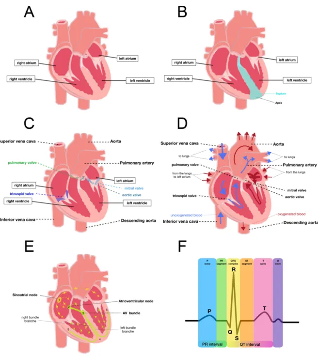
The heart is the first organ to form during development. It is responsible for pumping the blood and therefore, transporting nutrients, oxygen and hormones to the body. The heart is divided into four chambers: two atria (upper chambers) and two ventricles (lower chambers) that ensure the blood pumping (Figure 1A-C). To assure normal circulation, the heart presents different valves in each chamber, which prevent the backward blood flow. These valves act as one-way inlets of blood on one side of a ventricle and one-way outlets of blood on the other side of a ventricle. Each valve has three flaps (i.e., tricuspide), except the mitral valve, which has two flaps. The four valves are: 1) pulmonary valve located between the right ventricle and the pulmonary artery and allows the blood flow from the heart to the lungs, 2) tricuspid valve between the right chambers, 3) mitral valve between the left chambers, and 4) aortic valve between the left ventricle and the aorta, which carry blood from the heart to the body (Figure 1A-C). 1.1.1 Cardiac contraction The cardiac contraction can be separated in systole and diastole. Systole is the period when ventricles contract, allowing pressure to rise. It begins when ventricles contract and ends when ejection stops. Diastole begins when ejection ceases as ventricles relax. Ventricular contraction causes ejection of blood through the pulmonary valve, into the pulmonary arteries. This allows blood to get oxygenated and to come back to the left atrium via the pulmonary veins. During systole, arterial blood reaches its peak (systolic blood pressure)
normally between 90 to 120 mmHg. Then the atrium contracts and blood enters the left ventricle through the mitral valve. When left ventricle is loaded with blood, the mitral valve closes and the contraction of the ventricle allows the blood flow through the aortic valve to the aorta. Blood comes back to the heart through the superior and inferior vena cava into the right atrium. When the right atrium contracts, the blood passes thought the tricuspid valve into the relaxed right ventricle (Figure 1D)(Phibbs, 2007).
1.1.2 Conduction system of the heart
Effective functioning of the heart requires that all parts of the separate chambers contract in a synchronic movement. This is achieved by an efficient conduction system (Figure 1E) composed of: 1) sinoatrial node (SA node) located in the right atrium at the entry point of the superior vena cava. Being the natural pacemaker of the heart, the SA node initiates heartbeats and determines heart rate. Electrical impulses from the SA node spread throughout both atria and stimulate them to contract; 2) atrioventricular node (AV node) located at the opposite side of the right atrium compared to the SA node (near the AV valve). It serves as an electrical gate to the ventricles. It delays the passage of the electrical impulses to the ventricles. This delay allows the total ejection of the blood from the atria into the ventricles before they contract. The AV node receives signals from the SA node and sends them to the bundle of His; 3) bundle of His located in the septum is divided into right and left branches, which conduct the impulses through the apex. The electrical impulse is then passed into Purkinje fibres turning upwards and spreading the signal through the ventricular myocardium.
Figure 1: Heart anatomy and physiology A. Cardiac chambers. B. Cardiac anatomy. C. Cardiac valves. D. Cardiac circulation. E. Conduction system F. Electrogrardiogram.
Electrocardiography allows recording cardiac electrical activity using electrodes placed on the skin. It comprises several waves and intervals: 1) the P wave corresponds to atrial depolarization; 2) the PR interval is the time between the first deflection of the P wave and the first deflection of the QRS complex; 3) the QRS wave complex is composed of: the Q wave that corresponds to depolarization of the inter-ventricular septum, the R wave that reflects the depolarization of the main mass of the ventricles and the S wave that signs the final depolarization of the ventricles at the base of the heart; 4) the ST segment is the time between the end of the QRS complex and the start of the T wave. It reflects the period of zero potential between ventricular depolarization and repolarization; 5) the T wave represents ventricular repolarization and 6) the U wave is only visible when the heart rate falls below 65 bpm. The source of it is unknown, three theories regarding its origin are: delayed repolarisation of Punkinje fibres, prolonged repolarisation of mid-myocardial and after-potentials resulting from mechanical forces in the ventricular wall (Figure 1F)(Ashley and Niebauer, 2004; U Wave basic patterns - LITFL ECG Library ).
1.2 The cardiac muscle cell
The mammalian heart is composed of several cell types: cardiomyocytes, fibroblasts, endothelial cells and perivascular cells (Zhou and Pu, 2016). Contracting more than 3 billions times during average lifespan in human, cardiomyocytes are the most physically energetic cells in the body, and therefore contain an important number of mitochondria. Cardiomyocytes are highly specialised cells to assure their functions: 1) the plasma membrane (known as sarcolemma) has invaginations called T-tubules for the fast propagation of action potential; 2) the cytoplasm (known as sarcoplasm)
contains a high number of mitochondria; 3) a specialised endoplasmic reticulum (known as sarcoplasmic reticulum) that contains a high concentration of calcium; 4) and the contractile apparatus composed of myofibers consisting of sarcomeres (Figure 2A).
1.2.1 General overview of cardiomyocytes components A. Sarcolemma
Going from the outside to the inside of the cell, the first specialised structure is the plasma membrane: the sarcolemma. It serves as a barrier for diffusion and contains membrane proteins, including receptors and ion channels (Figure 2B). The sarcolemma forms two specialised regions: the intercalated disks and the transverse tubules. The intercalated disks are structures that allow communication and anchoring between adjacent cardiomyocytes. Intercalated disks are composed of several types of junctions: gap junctions, adherence junctions and desmosomes. Transverse tubules (also called T-tubules) are invaginations of the sarcolemma that allow the propagation of a fast action potential through the membrane deep into the cytoplasm and contain high density of L-type channels. These invaginations allow the rapprochement of L-type channels and the sarcoplasmic reticulum, more especially to the Ryanodine receptors (RyR). Other calcium channels implicated in the excitation contraction coupling (E-C coupling) are also located in this membrane as the sodium-calcium exchanger (NCX) (Walker and Spinale, 1999).
Calcium channels located in the sarcolemma:
§ L-type channels: also known as dihydropyridine receptors, are voltage dependent calcium (Ca2+) channels composed of different subunits. They are heterotetrameric polypeptides complexes containing α1, α2/δ, β subunits. α1, is the main functional component, which contains the voltage sensor and two of its transmembrane segments form the pore of the canal. α2/δ, β are auxiliary subunits implicated in modifying the gating properties of the channel, as well as the channel expression level. The combination of all subunits allows the depolarization-induced calcium influx into the cytosol, which is essential for the calcium-induce-calcium release (CICR) process. The close proximity of L-type channels and RyR2 allows the calcium influx across the plasma membrane to trigger the massive release of calcium from the SR stores through RyR2 opening. L-type channels are manly localised in the T-tubules facing the SR assuring the fast passage of the Ca2+ to the cytosol (Bodi et al., 2005; Kamp and Hell, 2000).
§ NCX: Sodium-calcium exchanger channel is a key actor of cardiac relaxation, located at the sarcolemma, including at the T-tubules. It is responsible of the extrusion of the cytosolic Ca2+ via a transport of three sodium ions (Na+) in exchange for one Ca2+. Under physiological conditions, NCX removes the same amount of Ca2+ that entered the cell through L-type channels in order to maintain cellular Ca2+ balance. In the heart, the isoform 1 is mostly expressed. NCX activity can be regulated by Na+, Ca2+, protons, phosphatidyl-inositol 4,5 biphosphate (PIP2) in the membrane, protein kinase A (PKA) and exogenous agents. (Ottolia et al., 2013; Schulze et al., 2003).
B. Sarcoplasmic reticulum
The sarcoplasmic reticulum (SR) is a specialised endoplasmic reticulum that serves as a source and internal storage of cytosolic Ca2+ required for excitation-contraction coupling. SR contains proteins that are essential for calcium homeostasis as the ryanodine receptor (RyR), the sarcoplasmic reticulum calcium ATPase (SERCA) and his two regulators phospholamban (PLN) and sarcolipin (SLN).
§ RyR: Ryanodine receptor mediates intracellular Ca2+ release during contraction, being a key actor of calcium induce-calcium release (CICR). In the heart, the most expressed isoforms is RyR2, but other isoforms have been detected. It is a homotetramer regulated directly or indirectly by L-type channels, Ca2+, magnesium (Mg2+), protein kinase A (PKA), FK506 binding proteins (FKBP12 and 12.6), calmodulin (CaM), Ca2+/calmodulin-dependent protein kinase II (CaMKII), calsequestrin (CSQ), triadin and junctin (Lanner et al., 2010).
§ SERCA: Sarco-endoplasmic calcium ATPase is responsible for muscle relaxation due to Ca2+ re-uptake. In heart, the most expressed isoform is the SERCA2a. Its ATPase activity assures the transport of two Ca2+ ions per ATP molecule. It is composed by a catalytic subunit with usually ten transmembrane spans and four intracellular loops in which is located the ATP and the phosphorylation sites (Sweadner and Donnet, 2001). The activity of this pomp is regulated by two small proteins: phospholamban (PLN) and sarcolipin (SLN) (Periasamy and Kalyanasundaram, 2007). PLN and SLN have a similar structure and roles but their inhibitory procedures are different.
◊ PLN: Phospholamban is a 52-amino-acids protein (6 KDa) that associates into pentamers to interact and inhibit SERCA by lowering
its Ca2+ affinity. It can exclusively bind to SERCA when there is a low Ca2+ concentration. Its activity is controlled by the phosphorylation of its Serine16 residue via PKA or its Threonine17 residue via CaMKII and calcium concentration. (Asahi et al., 2003a). Upon phorphorylation, PLN is released from SERCA, allowing calcium to be pumped back into the SR thus leading to a decrease in cytoplamic calcium.
◊ SLN: Sarcolipin is a 31-amino-acids protein that interacts and inhibits SERCA by lowering its Vmax. SLN can bind to SERCA at any cytosol calcium concentration. It has been shown to be an important mediator of muscle thermogenesis due to its interaction with the SR ATPase pump allowing the ATP hydrolysis but decreasing the Ca2+ transport (Bal et al., 2012; Pant et al., 2016).
C. Contractile apparatus
The contractile apparatus formed by sarcomeres is a highly organised array of myofilament proteins composed primarily of thick myosin, thin actin filaments and tropomyosin-troponin complex. The overlapping of these proteins gives to the cardiac muscle its striated appearance. Sarcomeric myosins convert the chemical energy of ATP into mechanical energy upon binding to actin thing filaments and promote sarcomere shortening (Figure 2D).
1.2.2 Cardiomyocyte contraction
The contractility of cardiomyocyte is under the control of a spatially defined meshwork of ion channels and exchangers that control Ca2+ entry. The cardiac contraction is initiated by electrical impulse known as action potential (AP) coming from the SA node inducing a depolarization of the sarcolemma. The AP can be separated in five different phases: 1) phase 0 depolarization; 2) phase 1 early repolarization; 3) phase 2 plateau phase; 4) phase 3 repolarization; and 5) phase 4 resting phase, described in Figure 2C. The depolarization propagates throughout the cardiomyocyte reaching the T-tubules, inducing the opening of the L-type channel and calcium conductance. The influx of calcium passing through L-type channels that reaches the sarcoplasm activates the Ryanodine channels. The activation results in an immediate release of large amounts of Ca2+ from the sarcoplasm (Fabiato, 1983). The increase of cytosolic Ca2+ concentration allows the binding of Ca2+ to troponin C, resulting in a shift of troponin I affinity from the actin filament to troponin C. This induces a shift of the troponin-tropomyosin complex away from the actin-myosin binding site. The shift allows myosin to swing towards the thin filament (consuming ATP). This conformational change generates a force moving the thin filament relative to the thick filament. This cycle will continue until the Ca2+ is removed from the cytoplasm by active, energy-dependent means (by SERCA activity) or by exhaustion of ATP stores (Walker and Spinale, 1999).
Figure 2: Cardiac cellular structure and components. A Cardiac muscle fiber. B. Schematic representation of a cardiomyocyte, and its calcium channels. C. Action potential. D. Sarcomere composition
2. Cardiomyopathies
2.1 Definition
Cardiomyopathies are a group of cardiac disorders characterized by a cardiomyocytes dysfunction and tissue-wide remodelling of the myocardium leading to functional decline. The prevalence arises with the age due to the age-related changes that can induce pressure overload. Distinct techniques as echocardiography1, electrocardiography2 and cardiac magnetic resonance imaging3 allow the characterisation of these pathologies. Features like severity, distribution, extent of myocardial hypertrophy, thickening of valves, ventricular dilatation, myocardial fibrosis and detection of infiltrates can be determined using these techniques and are essential for diagnosis (Elliott et al., 2008; McKenna et al., 2017).
2.2 Classification
The classification of cardiomyopathies has always been a source of debate. The first classification based on structural and functional changes was proposed by Goodwin in 1961, including congestive cardiomyopathy, hypertrophic cardiomyopathy and constrictive cardiomyopathy (Goodwin et al., 1961). Since then, worldwide cardiac societies propose regularly classifications for these pathologies. Currently four subgroups of cardiomyopathies are defined: 1) hypertrophic cardiomyopathy (HCM), 2) 1 Echocardiography: non-invasive ultrasound technique allowing the visualisation of the pumping function, the chamber size and the velocity of blood flow of the heart. 2 Electrocardiography: non-invasive technique recording the electrical activity of the heart by electrodes attached to the surface of the skin.
restrictive cardiomyopathy (RCM), 3) arrhythmogenic right ventricular cardiomyopathy (ARVC) and 4) dilated cardiomyopathy (DCM). Each phenotype is subclassified depending on the causes of the pathology (Figure 3) (Charron et al.).
Figure 3: Cardiomyopathies: actual classification and prevalence. A. Cardiomyopathies
classification adapted from Elliot et al 2008. B. Representation of the European cardiomyopathy prevalence in 2018 (3208 patients from EURObvational research program).
A.
B.
2.2.1 Hypertrophic cardiomyopathy
Hypertrophic cardiomyopathy (HCM) is characterized by an increased left ventricular (LV) wall thickness that is not solely explained by abnormal loading conditions. The LV wall thickness is ≥13mm and presents a normal or increased ejection fraction. The cardiomyocytes are hypertrophied, disorganised and separated by areas of interstitial fibrosis. HCM is a pathology affecting both children and adults and is the major cause of sudden death in teenagers and elite athletes. HCM is the most common cardiomyopathy and can be inherited or acquired. It is commonly acquired in an autosomal dominant pattern of inheritance but other patterns have been described. The mutations of different genes lead to HCM but genes encoding for proteins of the sarcomere as myosin-binding protein C, myosin heavy chain, cardiac troponin I, α-tropomyosin, cardiac α-actin, myosin light chains 2 and 3 and cysteine and glycine-rich protein 3 are responsible of most reported cases (Marian and Braunwald, 2017; 2014).
2.2.2 Restrictive cardiomyopathy
Restrictive cardiomyopathy (RCM) is characterized by an increased myocardial stiffness that leads to impaired ventricular filling. RCM affects either one or both ventricles but their size is not affected until late stages of the pathology. It is frequently accompanied of arrhythmias and conductions disturbances. RCM is the least common cardiomyopathy and it can be inherited in an autosomal dominant manner or acquired. Most cases of RCM are acquired, however some mutations are recurrent affecting the genes encoding for Troponin T and I, α-actin and β-myosin heavy chain (Muchtar et al., 2017).
2.2.3 Arrhythmogenic right ventricular cardiomyopathy
Arrhythmogenic right ventricular cardiomyopathy (ARVC) is characterized by fibro-fatty replacement of the right ventricular myocardium leading to paroxysmal ventricular arrhythmias and a predisposition to sudden cardiac death (SCD)(2nd most common cause of SCD in children, young adults and athletes). ARVC manifests with electrocardiographic abnormalities as syncope or ventricular arrhythmias. Sustained ventricular tachycardia and/or ventricular arrhythmia lead to SCD. In a cellular level cardiomyocytes are progressively necrotic and frequently accompanied by inflammation, fatty infiltration and replacement of cardiomyocytes. ARVC it is typically inherited in an autosomal dominant manner, with variable penetrance and expression. Most cases of ARVC are due to mutations on the genes encoding for the proteins located in the desmosome (junction plakoglobin, desmoplakin, phakophilin-2, desmoglein-2, desmocollin-2). However mutations in non-desmosomal genes such as genes encoding for cardiac ryanodine receptor 2, transforming growth factor β-3, the nuclear transmembrane protein 43 and desmin had also be reported to induce ARVC. (Basso et al., 2018; Corrado et al., 2017; Mestroni and Sbaizero, 2018; Priori et al., 2015)
2.2.4 Dilated cardiomyopathy
Dilated cardiomyopathy (DCM) is characterized by an enlargement and an impaired systolic function of left or both ventricles. DCM can include atrial and/or ventricular arrhythmias and conduction defects. It is a major risk factor for developing heart failure (HF) and sudden death can occur at any stage of the disease. DCM is the second most common cardiomyopathy (Figure 3B). DCM can be attributed to genetic alterations, main genetic causes responsible of the pathology are summarised in table 1. The majority of the
inherited DCM is autosomal dominant. Inherited DCM is genetically heterogeneous due to mutations of proteins forming the sarcomeres, the Z disc, the dystrophin complex, the cytoskeleton, the desmosomes, the sarcoplasmic reticulum and cytoplasm, the ion channels, the mitochondria, the extracellular matrix, the lysosomes, the nucleus and the nuclear envelope. Within all familial forms of DCM, LMNA-cardiomyopathy is the second most common pathology (Hershberger et al., 2010) and is the focus of my work. Patients with LMNA-cardiomyopathy have been reported to have a worse clinical prognosis than DCM patients carrying different pathologic DCM-associated gene mutations (McNally and Mestroni, 2017; Pinto et al., 2016; Wang et al., 2017). Medical terms
Ejection fraction Percentage of blood ejected out from the heart chamber with each beat. Bradyarrhythmia Reduced heart rate: Sinus rhythm with a resting heart rate of 60 beats per minute or less.
Atrioventricular block
Interruption or delay of electrical conduction from the atria to the ventricles due to conduction system abnormalities in the AV node or the His-Purkinje system.
Atrial arrhythmia Abnormal heart rate (60-100 bpm): being too fast (tachyarrhythmia) or too slow (bradyarrhythmia).
Atrial fibrillation Irregular and uncoordinated contraction. Absence of P waves with an atrial rate of 350-600 beats/min, irregular ventricular rhythm, ventricular rate 100-180 beats/min Conduction system disease Disruption of the electrical impulses of the heart. It can be due to a defect in the sinoatrial node, intermodal tract, atrioventricular node right or left bundle branches or the bachman’s bundle. The three main types of conduction system disease are the heart block, long QT syndrome and the bundle branch block.
Genetic dilated cardiomyopathies Affected organ(s) Disease/affected gene Predominant cardiac phenotype familial DCM % - Titin (TTN) ~ 20-25 % - Lamin A/C (LMNA) ~ 6 % - Myosin heavy chain (MYH7) ~ 4 % - Troponin T (TNNT2) ~ 2 % - Myosin-binding protein C (MyBPC3) ~ 2 % - RNA-binding modif-20 (RBM20) ~ 2 % - Myopalladin (MYPN) ~ 2 % - Sodium channel alpha unit (SCN5A) ~ 2 % - BaCl2-associated athanogene 3 (BAG3) ~ 2 % - Phospholamban (PLN) ~ 1 % Neuromuscular disorders - Duchenne muscular dystrophy (DMD) - Becker muscular dystrophy (DMD) - Myotonic dystrophy or Steinert myopathy (DMPK) Syndromic diseases - - Mitochondrial diseases (mtDNA or nuclear genes) Tafazin (TAZ)
Acquired dilated cardiomyopathies Group Causing agent Drugs - Antineoplastic drugs - Psychiatric drugs - Other drugs Toxic and overload - Ethanol - Cocaine, amphetamines, ecstasy - Other overload - Iron overload Nutritional deficiency - Selenium deficiency - Thiamine deficiency - Zinc and copper deficiency - Carnitine deficiency Electrolyte disturbance - Hypokalaemia, hypophosphatemia Endocrinology - Hypo- and hyper-thyroidism - Cushing/Addison disease - Phaecromocytoma - Acromegaly - Diabetes mellitus Infection - Viral - Bacterial - Mycobacterial - Fungal - Parasitic Auto-immune diseases Organ specific - Giant-cell myocarditis - Inflammatory DCM (non-infectious myocarditis) Not organ specific - Polymyositis/dermatomyositis Peripartum Table 1: Aetiology of dilated cardiomyopathies (adapted from Pinto et al 2016).
3. LMNA pathologies
3.1 LMNA-cardiomyopathy
LMNA-cardiomyopathy is characterized by a left ventricular dilatation
and/or reduced systolic function (left ventricular ejection fraction <50%). It is frequently accompanied by significant conduction system disease and arrhythmias. The conduction system disease commonly precedes development of DCM. It is characterized by: 1) sinus bradycardias, 2) sinus node arrests with junctional rhythms, 3) a high frequency of atrioventricular blocks, 4) atrial fibrillation and 5) ventricular arrhythmias. Sudden cardiac death (SCD) due to the ventricular arrhythmias occurs frequently, often before the development of DCM (30% of the cases). First signs of LMNA-cardiomyopathy usually appear at mid-age (20’s 30’s) usually with conduction system disease or atrial arrhythmia but some patients are asymptomatic. A gradual worsening of the pathology occurs leading to a full penetrance at the age of 60-70 years (Hasselberg et al., 2018; Hershberger and Morales, 2016; Hershberger et al., 2010; Wang et al., 2017).
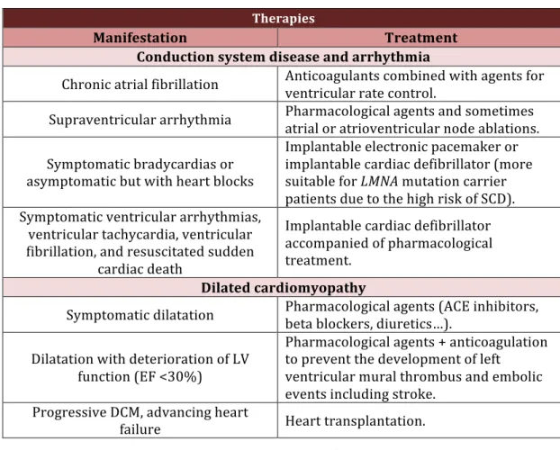
No curative treatment is currently available for LMNA-cardiomyopathy patients, but research on mouse models have identified different potential therapeutic targets (Worman, 2018), some being currently tested in clinics (NCT02057341). Current clinical management strategies for LMNA-cardiomyopathy patients are the ones used for patients with other cardiomyopathies. Individual symptoms of the pathology are treated to maintain the cardiac function compiled in table 2. The management of LMNA-cardiomyopathy is focused on treatment of conduction system disease, arrhythmias, and DCM (Hershberger and Morales, 2016), the patients commonly require implantable cardiac defibrillators in order to prevent SCD
Therapies
Manifestation Treatment
Conduction system disease and arrhythmia
Chronic atrial fibrillation Anticoagulants combined with agents for ventricular rate control. Supraventricular arrhythmia Pharmacological agents and sometimes atrial or atrioventricular node ablations.
Symptomatic bradycardias or asymptomatic but with heart blocks Implantable electronic pacemaker or implantable cardiac defibrillator (more suitable for LMNA mutation carrier patients due to the high risk of SCD). Symptomatic ventricular arrhythmias, ventricular tachycardia, ventricular fibrillation, and resuscitated sudden cardiac death Implantable cardiac defibrillator accompanied of pharmacological treatment. Dilated cardiomyopathy
Symptomatic dilatation Pharmacological agents (ACE inhibitors, beta blockers, diuretics…).
Dilatation with deterioration of LV function (EF <30%) Pharmacological agents + anticoagulation to prevent the development of left ventricular mural thrombus and embolic events including stroke. Progressive DCM, advancing heart failure Heart transplantation. Table 2: Current treatments for LMNA-cardiomyopathy.
In addition to LMNA-cardiomyopathy, LMNA mutations have been described in patients with severe forms of arrhythmogenic right ventricular cardiomyopathy (Quarta et al., 2012), restrictive cardiomyopathy (Paller et al., 2018) and in patients with an increased risk of thromboembolic complications (van Rijsingen et al., 2013).
LMNA mutations are not only responsible for cardiac diseases but also
lead to a broad spectrum of pathologies called laminopathies (described in 3.2.). It is important to note that the cardiac involvement in laminopathies is not exclusive to LMNA-cardiomyopathy. It is also present in patients with Emery-Dreifuss muscular dystrophy, Limb girdle muscular dystrophy,
Progeria syndromes and lipodystrophies. The main characteristics of the cardiac involvement in these pathologies are summarised in table 3.
Laminopathies with heart involvement
Pathology Cardiac phenotype Ref
Dilated cardiomyopathy with conduction defects
Left ventricle dilatation, systolic dysfunction, atrioventricular conduction block, arrhythmia, congestive heart failure (Taylor et al., 2003) Emery Dreifuss muscular dystrophy Atrioventricular conduction block, arrhythmia, systolic dysfunction, congestive heart failure (Zhang et al., 2015) Limb girdle muscular dystrophy Atrioventricular block, progressive left ventricle dysfunction, arrhythmia (Chang et al., 2010) Variant progeroid syndrome with right ventricular cardiomyopathy Right atrium and ventricle dilatation,
tricuspid valve dilatation (Alastalo et al., 2015) Atypical progeroid syndrome with cardiomyopathy Right ventricle dilatation, arrhythmia, tricuspid valve regurgitation (Guo et al., 2016) Familial partial lipodystrophy of Dunningan type 2 Left ventricle dilatation, systolic dysfunction, atrioventricular block, complete left bundle branch block (Andre et al., 2015) Lipodystrophy with hypertrophic cardiomyopathy Left ventricle hypertrophy, aortic valve
calcification, stenosis and regurgitation (Duparc et al., 2009) Charcot-Marie-Tooth type 2
axonal neuropathy Left ventricle dilatation, systolic dysfunction, (Duparc et al., 2009) Severe metabolic syndrome
caused by a ZMPSTE24 mutation
Left ventricle dilatation, systolic 16
dysfunction, ventricular extra systole (Galant et al., 2016)
3.2 Laminopathies
Laminopathies are a wide spectrum of pathologies caused by mutations in genes encoding for nuclear lamins and associated proteins. They are commonly divided in 4 groups depending on the affected tissue: 1) striated muscle, 2) adipose tissue, 3) nervous system and 4) accelerated aging syndromes.
During the 90’s the first mutations of the genes responsible of laminopathies were identified. In 1994 Bione et al. reported a mutation in
EMD gene leading to X-linked Emery–Dreifuss muscular dystrophy (EDMD),
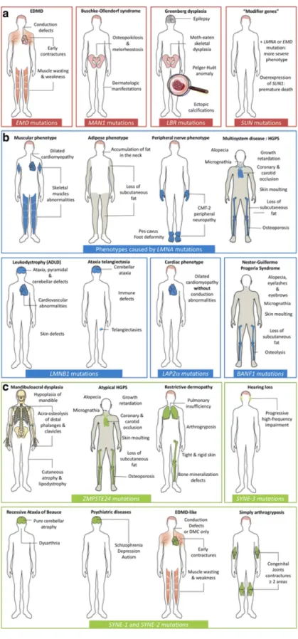
not long after Bonne et al. reported the first LMNA mutation leading to autosomal dominant forms of EDMD (Bione et al., 1994; Bonne et al., 1999). Since then, more than 460 mutations in LMNA gene have been reported (http://www.umd.be/LMNA/). Mutations on the genes encoding for other nuclear proteins as emerin, nesprins,… lead to similar alterations. The main mutations and the general characteristics of these pathologies are summarised in Figure 4. For this work, a special focus on LMNA gene mutations will be done.
The large spectrum of LMNA-laminopathies can be classified into four groups with some phenotypic overlapping 1) striated muscles diseases including LMNA-DCM-CD, EDMD, LMNA-related congenital muscular dystrophy (L-CMD) and limb-girdle muscular dystrophy type 1B (LGMD1B), 2) diseases affecting adipose tissue including familial partial lipodystrophy of Dunnigan type (FPLD), 3) peripheral neuropathy associated with demyelination of motor neurons such as axonal neuropathy Charcot-Marie-Tooth type 2B1 (CMT2B1), and 4) premature aging syndromes which include Hutchinson-Gilford progeria syndrome (HGPS) and atypical Werner syndrome (aWRN) (Bonne et al., 1999; Fatkin et al., 1999; Muchir et al., 2000; Shackleton et al., 2000; Sandre-Giovannoli et al., 2002; Chen et al., 2003; Eriksson et al.,
2003). Among all LMNA-laminopathies approximately 60% of the mutations involve striated muscle phenotype (Bertrand et al., 2011).
3.2.1 Striated muscle laminopathies
Emery-Dreifuss muscular dystrophy (EDMD) is a genetically heterogeneous disorder with X-linked, autosomal dominant and autosomal recessive forms. It is characterized by 1) early contractures of the Achilles tendons, elbows and post-cervical muscles, 2) slow progressive muscle wasting and weakness and 3) a cardiomyopathy usually presenting as cardiac conduction defects (Bonne et al., 1999) (Vigouroux and Bonne, 2013).
Limb-Girdle Muscular Dystrophy (LGMD) is a heterogeneous group of disorders that can be acquired in a dominant or recessive manner. It can present or not a cardiac alteration. In particular, LGMD associated with atrioventricular conduction disturbances (LGMD1B) is characterized by symmetrical weakness starting in the proximal lower limb muscles, and gradually proximal upper limb muscles also become affected. Mostly all patients present dysrhythmias and atrioventricular conductions defects. (van der Kooi et al., 1997; Muchir et al., 2000).
LMNA-associated congenital muscular dystrophy (L-CMD) is a myopathy
due to de novo mutations. It appears in first year of life and can have different severity degrees. The main characteristics of this pathology are a selective axial weakness and wasting of the cervico-axial muscles, a limb muscle involvement and ventilator difficulties (needing a ventilator support) (Quijano-Roy Susana et al., 2008). Cardiac involvement was reported in some cases (i.e. severe ventricular arrhythmias associated with sudden cardiac death).
Figure 4: Summary of all known human diseases caused by mutations in genes coding for nuclear envelope components. The diversity of
phenotypes induced by
mutations in genes encoding nuclear envelope components, as well as the tissues affected by these, is illustrated and organized according to the localization of the mutated protein: (a) inner nuclear membrane, (b) nuclear lamina, and (c) outer nuclear membrane components. EDMD:
Emery-Dreifuss Muscular
Dystrophy, HGPS: Hutchinson-Gilford Progeria Syndrome, CMT: Charcot-Marie-Tooth Disease, ADLD: Autosomal Dominant Leukodystrophy.
Extracted from: Nuclear
envelopathies: a complex LINC between nuclear envelope and pathology (Janin et al., 2017)
3.3 Lamins
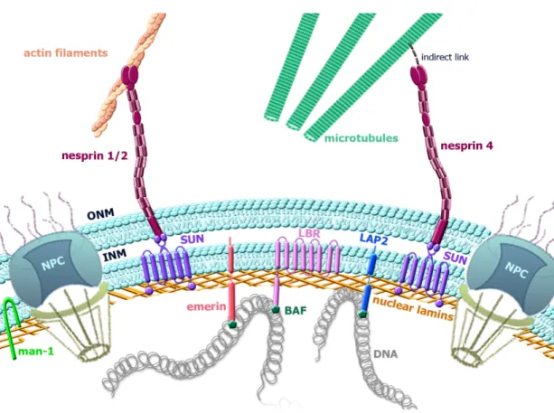
Lamins are the major components of the nuclear lamina (NL). The NL is a protein meshwork underlying the inner aspect of the inner nuclear membrane known to serve as major structural component of the nucleus (Figure 5). Lamins provide a platform for the binding proteins and chromatin. They have varied roles in the cell, implicated in nuclear mechanical stability, chromatin organisation, DNA repair/replication, transcription, mediating cellular signalling and cytoskeletal interactions. Figure 5: Schematic diagram of the nuclear envelope (NE) organization. NE is composed by an outer nuclear membrane (ONM) and an inner nuclear membrane (INM) joining at the nuclear pore complexes. Inserted into the NE, integral proteins MAN-1, SUNs, Emerin, LBR, LAP2, Nesprins display various interactions connecting the nucleoskeleton to the cytoskeleton.
3.3.1 Structure and genetics determinants of lamin proteins
Lamins are the only intermediated filaments located in the nucleus. As all intermediate filaments proteins, they possess an α-helical coiled-coil central rod domain, composed of heptad repeats of amino acids, bordered by a globular amino-terminal “head” domain and a carboxyl-terminal “tail” domain. This latter comprises an (Ig)-like domain and a nuclear localization sequence (Dhe-Paganon et al., 2002; Frangioni and Neel, 1993; Krimm et al., 2002; Parry et al., 1986). After being processed, lamins form coiled-coil dimers (Aebi et al., 1986) that interact longitudinally thought head-tail association to form a long polar polymer that can further assemble laterally forming 10nm intermediate filament proteins (Fisher et al., 1986; McKeon et al., 1986; Goldman et al., 1986). Recent studies are however challenging this view and described lamins as being 3.5nm filaments (Harapin et al., 2015; Turgay et al., 2017). This controversy would need further clarification.
Based on their sequences and structural properties nuclear lamins can be separated in two types: A-type and B-type lamins (Gerace, L, 1978), which form two separated filaments networks. A-type lamins are expressed in most differentiated somatic cells (Dechat et al., 2010) while B-type lamins are constitutively expressed. In mammals three genes encode for lamins: LMNA,
LMNB1 and LMNB2. LMNA is located to chromosome 1q21.2 (Wydner et al.,
1996), it encodes for A-type lamins, which major isoforms are lamin A and C (Lin and Worman, 1993). These proteins are identical for their first 566 amino acids and vary in their carboxyl terminal tails due to alternative splicing. Lamins are synthesized as precursors that are post-translationally processed (Beck et al., 1990; Dhe-Paganon et al., 2002; Farnsworth et al., 1989; Maske et al., 2003; Nigg et al., 1992; Varela et al., 2005). Minor A-type lamins isoforms have been described such as lamin C2 and lamin AΔ10 (Nakajima and Abe,
1995). The most abundant B-type lamins are lamin B1 and B2 respectively encoded by LMNB1 and LMNB2, both being constitutively expressed. LMNB2 encodes also lamin B3 by alternative splicing, which is restricted to the male germ line (Burke and Stewart, 2013; Gruenbaum et al., 2005). 3.3.2 Functional roles of lamin proteins A. Nuclear envelope support
The nuclear envelope (NE) is a porous interface that separates the nucleus and the cytoplasm in eukaryotic cells (Aaronson and Blobel, 1975; Watson, 1955). Structurally this lipid bilayer consist in an outer nuclear membrane (ONM) and an inner nuclear membrane (INM) both enclosing the perinuclear space but joining at sites occupied by nuclear pore complexes (NPCs). NPCs are protein-based channels controlling the movement of molecules form the nucleus to the cytoplasm and vice versa. INM and ONM despite having continuous border they present several structural and functional differences.
The ONM is continuous with the endoplasmic reticulum membrane and shares some functions and structural features (Hetzer, 2010). Despite the continuity between this two membranes ONM contains several specific nuclear envelope transmembrane proteins (NETs) essential for the interaction with the cytoskeleton (Crisp et al., 2006; Starr and Fridolfsson, 2010; Gant Luxton and Starr, 2014). The INM interacts with the nucleoplasm through more than 100 NETs. Among all these proteins some are implicated in the assembling of nuclear factors that regulate transcription, cell division and DNA repair (Starr, 2011). In a general point of view INM and its associated NETs have two main roles, the interaction NE-chromatin and the interaction NE-NL
NL started to be studied for their roles during mitosis (Moir et al., 2000; Panorchan et al., 2004) but have been demonstrated to be implicated in the maintaining of normal nuclear morphology and its composition (Gundersen and Worman, 2013). As structural components of the NL, A-type lamins confer viscosity and stiffness, while B-type lamins confer elasticity to the nucleus (Lammerding et al., 2006; Pajerowski et al., 2007). In addition to that, different mutations of lamins induce misshapen nuclei, nuclear pore clustering, mislocalisation of NETs and aberrant intranuclear foci, demonstrating the important role played by lamins in structural the structural nuclear support (Broers et al., 2004; Lammerding et al., 2004; Sullivan et al., 1999). B. Lamin and chromatin NL participates in chromatin organization, DNA repair and transcriptional regulation. Chromatin organization Lamins can organize and regulate chromatin position within the NE. They can interact in a direct or indirect manner (via lamin-binding proteins) with the chromatin (Camozzi et al., 2014; Gruenbaum and Foisner, 2015; Perovanovic et al., 2016). These dynamic interactions are essential to guide the spatial folding of the chromosomes (depending on the cell cycle state) and do not occur in all cell types identically (Guelen et al., 2008; Therizols et al., 2014). The direct interaction between lamins and chromatin occurs via specific genomic regions called lamina-associated domains (LADs). Contrarily indirect interactions can take place thanks to two families of INM integral proteins: LEM domain proteins family and Lamin B receptor proteins family. LEM domain proteins family is composed by: LAP2, emerin and MAN 1
domain, characterized by a 40 residues segment that interacts the Barrier-to-Autointegration Factor proteins (BAF). BAF proteins bind to double strand DNA and histones (Zheng et al., 2000). Through interactions with BAF proteins, LEM proteins contribute to the tethering of genomic regions to nuclear periphery, connecting interphase chromosomes to the nuclear lamina, thereby intervening in global nuclear organization. It is interesting to note that there is a 90% overlap between the LEM associated regions and LADs, suggesting that LEM proteins may contribute to LAD establishment or maintenance (Barton et al., 2014, 2015).The function of LEM proteins and their interactions with BAF and lamins are highly conserved through evolution, suggesting their essential role in scaffolding the NE.
Lamin B receptor proteins (LBR) are another family of INM integral proteins that contribute to the interactions of the nuclear lamina with the chromatin. These proteins bind to lamin B and chromatin during some cell phases (Olins et al., 2010). Their terminal domains give them their two main functions. The N-terminal domain tethers chromatin to the nuclear periphery, thus contributing to the shape of interphase nuclear architecture, while the transmembrane domains exhibit sterol reductase activity (Nikolakaki et al., 2017). DNA repair A-type lamins are implicated in: 1) the recruitment of the cell repair factor p53-binding protein (Liu et al., 2005; Varela et al., 2005), 2) in the protection against DNA damaging agents inducing single or double-stranded breaks (Liu et al., 2005; Masi et al., 2008; Richards et al., 2011), 3) in the control of ROS production and the sensitivity to oxidative stress (Richards et al., 2011) 4) in the regulation of Sirtuin 6 (SIRT6) activities (Ghosh et al., 2015) and 5) in the regulation of DNA repair foci positional stability (Mahen et al., 2013).
Mutations on A-type lamins have been associated with defective DNA repair. The progeria Zmpste24−/− mouse model presents increased DNA damage, chromosome aberrations and higher sensitivity to DNA-damaging agents. In addition to that, the recruitment of some implicated-DNA-repair-proteins as Rad51 or 53p binding protein 1(53BP1) is impaired due to the mutation (Liu et al., 2005). Not only the recruitment of the 53BP1 is altered but all the stress signalling pathway marked by an upregulation of p53 target genes inducing a delayed checkpoint response and defective DNA repair (Varela et al., 2005; Masi et al., 2008). Contrarily the absence of lamin leads to 53BP1 degradation by the proteasome, showing its important role in the repair control (Gonzalez-Suarez et al., 2009). The absence of lamin induces slowed double strands breaks, with non-homologous end joining (Redwood et al., 2011).
The defects of DNA repair can also be highlighted in Zmpste24−/− mouse model by an increased sensitivity to γ-irradiation (80% of the KO mice died compared to the 20% of the WT)(Liu et al., 2005). In fibroblasts caring the A-type lamin-R527H mutation, the impairment of DNA repair was evidenced by an increased chromosome damage and the higher percentage of residual g-H2AX foci (corresponding to unrepaired DNA-damage sites) (Masi et al., 2008). The fibroblast studies also showed an aberrant accumulation of Xeroderma Pigmentosum group A (XPA), a unique nucleotide excision repair protein (Liu et al., 2008) being partially responsible of the DNA repair deficiencies.
Transcription regulation
Nuclear lamins can modulate gene expression by direct interaction with chromatin or by sequestrating transcriptional regulators at the nuclear periphery (Andrés and González, 2009; Reddy et al., 2008). During cells
differentiation, several genes located in LADs move away the NL and become activated or towards the NL being repressed (Therizols et al., 2014). Nevertheless different studies showed that the NL is not essential for this gene repression but rather increases the robustness of the repression (Yáñez-Cuna and van Steensel, 2017). In addition to that nuclear lamin can inhibit RNA polymerase II activity, (Spann et al., 2002).
Lamins also interact with some transcriptional regulators. A-type lamins have been shown to interact with Rb, Gcl, Mok2, cFos, and Srebp1, affecting gene expression either by a sequestration of these factors or by influencing the assembly of core transcriptional complexes (González et al., 2008; Kumaran et al., 2002; Malhas et al., 2009).
C. Mediating nucleo-cytoskeletal connections
Interactions with the cytoskeleton are essential for some of the nuclear lamin functions: nuclear positioning, nuclear shape and mechanosensing (Chang et al., 2015). These interactions involve proteins located in the INM and in the ONM among which KASH (Klarsicht, ANC-1, and Syne homology) and SUN (Sad1 and UNC-84) proteins, which form the linker of nucleoskeleton and cytoskeleton termed the LINC complex (Crisp et al., 2006; Gant Luxton and Starr, 2014; Starr and Fridolfsson, 2010).
Carboxyl terminus of A-type lamins interact with the LINC complex via SUN proteins in the nucleoplasm (Crisp et al., 2006; Haque et al., 2006). SUN-proteins are located in the INM and are encoded by SUN1 to SUN5, being SUN1 and SUN2 the main isoforms. SUN-proteins bind through the perinuclear space to KASH proteins interacting with cytoskeleton. The mammalian KASH proteins also called nesprins are located in the ONM, they are encoded by four different genes: SYNE1, SYNE2, SYNE3 and SYNE4. Among all the isoforms
obtained by alternative splicing nesprin 1α and nesprin 2β interact with lamins (and emerin).
In mutant cells lacking lamin A, the mobility of the SUN proteins is increased, proving that lamin A plays a role in anchoring SUN proteins to INM (Östlund et al., 2009). In addition to that, during development B-type lamins play a role in SUN proteins anchoring (Chang et al., 2015). Other groups confirmed this anchoring function, reporting instability of TAN lines in cells lacking lamin A and nuclear migrations defects in cells with a weakened SUN-lamin interaction (Bone et al., 2014; Folker et al., 2011). Other factors also contribute to the SUN proteins anchoring because some cellular models lacking lamin present a normal SUN protein localization (Chang et al., 2015).
D. Mediating cellular signaling pathways
For almost 20 years now, it is known that proteins of the NL can modulate different signalling pathways due to their interactions with chromatin, cytoskeleton, LINC complex... These pathways have shown to be defective (altered/increased/decreased) in different laminopathies, explaining the important role of lamins. A-type lamins are involved in signalling pathways affecting cell growth, survival, migration and differentiation, including mitogen-activated protein kinase (MAPK) and mammalian target of rapamycin (mTOR) pathways (Andrés and González, 2009; Bakay et al., 2006; Muchir et al., 2007a, 2007b).
More detailed signalling pathways implicated in LMNA-cardiomyopathy are described in 4.1.
The use of animal models has allowed a better understanding of these signalling pathways and has opened perspectives for new therapeutic approaches.
3.4 Animal models
To gain insight in the striated muscle laminopathies, different knock-out (KO), knock-in (KI) and transgenic mouse models have been developed.
While the use of KO mouse models are essential for the functional understanding of lamins, KI and transgenic mouse models allowed to understand the pathology and tissue specificity. All the actual striated muscle mouse models are presented in table 4 and only the striated muscle Lmna-models are described below.
3.4.1 KO mouse model
The first animal model created was the Lmna KO mouse (Lmna-/-). Even if
there are no reported cases of human lacking lamin A/C (except an individual with haploinsuficiency of LMNA and foetus that died in gestation with an homozygous premature-stop-codon), this mouse model has been pivotal to determine the A-type lamins roles (Bonne et al., 1999; van Engelen et al., 2005; Muchir et al., 2003). This model has been one of the most used to understand the mechanics of lamins. Lmna-/- mice present a dilated
cardiomyopathy with conductions defects and skeletal myopathy similar to the one observed in patients with Emery-Dreifuss Muscular Dystrophy (Sullivan et al., 1999). Lmna-/- mice at only 2 weeks of age present important
growth defects and a reduced lifespan dying between 6 and 8 weeks of age. The heterozygous mice do not present any symptoms at early age but develop
atrio-ventricular conduction defects with atrial and ventricular arrhythmias (Wolf et al., 2008). However, even if this model was considered as “null” for many years, it was shown that the mice express a truncated form of Lmna (deletion of exons 8 to 11). The protein was detected at really low levels (mRNA and protein) but could act as a toxic molecule (gain-of-function) or as an hypoactive protein (loss-of-function) explaining the phenotype observed in this mice (Jahn et al., 2012). Three KO mice were latter developed: the Lmna GT-/- mice (Kubben et al., 2011) the Lmna Δ/Δ (Kim and Zheng, 2013) and the Zp3-Lmna (Solovei et al., 2013). The mice present severe growth retardation and cardiac developmental defects, myocytes hypertrophy and have a reduced life expectancy. They highlighted the implication of lamins in muscle development after birth (Kubben et al., 2011; Kim and Zheng, 2013; Solovei et al., 2013). While the use of KO mouse models are essential for the functional understanding of lamins, the KI and transgenic mouse model allowed to understand the instauration of the pathology and its tissue specificity.
3.4.2 KI and transgenic mouse model
The KI and transgenic mouse were developed from mutations found in patients with different Emery-Dreifuss Muscular Dystrophy. Two KI mouse models were developed almost simultaneously. Arimura et al developed the
LmnaH222P/H222P, created from a mutation found in patients with classical forms of EDMD. This model is characterized by a dilated cardiomyopathy with conductions defects and a progressive skeletal impairment leading to a premature death between 6 to 9 months for male and 7 to 13 for females. The cardiomyopathy is characterized by a left ventricular enlargement with a reduced fractional shortening and conductions defects with frequent sinoatrial blocks and ventricular extrasystoles (Arimura et al., 2005). The
signalling pathways (presented below). The skeletal phenotype had been less explored in those mice but similar signalling pathways are altered. Mounkes et
al developed the LmnaN195K/N195K mouse model, created from a missense
mutation found in patients with dilated cardiomyopathy. The mice present a cardiac phenotype but no signs of skeletal muscle impairment. The cardiomyopathy is more severe than the one observed in LmnaH222P/H222P mouse, and its accompanied by arrhythmias responsible of a premature death at 3 months of age (Mounkes et al., 2005). Wang et al developed the LmnaM371K transgenic mouse model. This mutated lamin alters development that explains the increased prenatal loss and induces a severe cardiac phenotype that leads to a premature death at 2 to 7 weeks (Wang et al., 2006).
Bertrand et al developed a KI mouse model harbouring a LMNA-CMD;
LmnaΔK32/ΔK32. This model exhibits altered striated muscle maturation and severe metabolic defects leading to a premature death at 2 weeks of age. Lamin proteins expression is reduced and delocalized, showing that the localization of lamins could be important for tissue maturation (Bertrand et al., 2012). The heterozygous LmnaΔK32/+ mice do not present any initial phenotype with metabolic defects and die between 10 and 20 months of age. Nevertheless these mice presented a cardiac phenotype caused by the lamin A/C haploinsufficiency (Cattin et al., 2013).
The B-type mouse models develop different laminopathies such as Pelger-Huët anomaly, HEM-Greenberg skeletal dysplasia and ichthyosis reflecting the important role of lamin B in development. However these models no do not present a pathological muscular phenotype (Cohen et al., 2008; Shultz et al., 2003).
Model
Features Reference
Lmna models
KO Lmna -/- EDMD and DCM-CD (Sullivan et
al., 1999)
C.KO LmnaGT-/- Cardiomyocyte hypertrophy, skeletal muscle
hypotrophy and metabolic defects.
(Kubben et al., 2011)
C.KO LmnaΔ/Δ Cardiomyocyte hypertrophy, skeletal muscle
hypotrophy and metabolic defects.
(Kim and Zheng, 2013)
C.KO Zp3-Lmna Cardiomyocyte hypertrophy, skeletal muscle hypotrophy and metabolic defects.
(Solovei et al., 2013)
KI LmnaH222P/H222P
AD-EDMD and DCM-CD (Arimura et
al., 2005) KI LmnapN195K/pN19 5K DCM-1A -CD (Mounkes et al., 2005) Tr Lmnap.M371K/p.M37 1K EDMD and DCM (Wang et al., 2006)
KI LmnaΔK32/ΔK32 L-CMD, defective skeletal and cardiac muscles
maturation and metabolic defects.
(Bertrand et al., 2011)
KI LmnaΔK32/+ DCM (Cattin et
al., 2013)
KI LmnaL530P/L530P Progeroid syndrome with growth retardation
and heart underdevelopment and degeneration.
(Mounkes et al., 2003)
Emd models
KO Emd -/-
Muscle regeneration defects (Melcon et
al., 2006)
KO Emd -/-
Motor coordination abnormalities, atrio-ventricular conduction defects
m
(Ozawa et al., 2006)
KASH models
KO Δ/Δ KASH
DCM-CD (Puckelwa
rtz et al., 2010)
Table 4: Mouse models for laminopathies.