Publisher’s version / Version de l'éditeur:
Journal of Agricultural and Food Chemistry, 58, 3, pp. 1462-1468, 2010-01-11
READ THESE TERMS AND CONDITIONS CAREFULLY BEFORE USING THIS WEBSITE.
https://nrc-publications.canada.ca/eng/copyright
Vous avez des questions? Nous pouvons vous aider. Pour communiquer directement avec un auteur, consultez la
première page de la revue dans laquelle son article a été publié afin de trouver ses coordonnées. Si vous n’arrivez pas à les repérer, communiquez avec nous à PublicationsArchive-ArchivesPublications@nrc-cnrc.gc.ca.
Questions? Contact the NRC Publications Archive team at
PublicationsArchive-ArchivesPublications@nrc-cnrc.gc.ca. If you wish to email the authors directly, please see the first page of the publication for their contact information.
NRC Publications Archive
Archives des publications du CNRC
This publication could be one of several versions: author’s original, accepted manuscript or the publisher’s version. / La version de cette publication peut être l’une des suivantes : la version prépublication de l’auteur, la version acceptée du manuscrit ou la version de l’éditeur.
For the publisher’s version, please access the DOI link below./ Pour consulter la version de l’éditeur, utilisez le lien DOI ci-dessous.
https://doi.org/10.1021/jf903485k
Access and use of this website and the material on it are subject to the Terms and Conditions set forth at
Determination of thiols in yeast by HPLC coupled with LTQ-Orbitrap
mass spectrometry after derivatization with
p-(hydroxymercuri)benzoate
Rao, Yulan; Xiang, Bingren; Bramanti, Emilia; D’Ulivo, Alessandro; Mester,
Zoltan
https://publications-cnrc.canada.ca/fra/droits
L’accès à ce site Web et l’utilisation de son contenu sont assujettis aux conditions présentées dans le site LISEZ CES CONDITIONS ATTENTIVEMENT AVANT D’UTILISER CE SITE WEB.
NRC Publications Record / Notice d'Archives des publications de CNRC:
https://nrc-publications.canada.ca/eng/view/object/?id=96ea2d9e-4c57-4755-966c-2333fe3697a1 https://publications-cnrc.canada.ca/fra/voir/objet/?id=96ea2d9e-4c57-4755-966c-2333fe3697a1
Determination of thiols in yeast by HPLC coupled with LTQ-Orbitrap mass spectrometry after derivatization with p-hydroxymercurybenzoate
Yulan Rao,a,b Bingren Xianga, Emilia Bramantic, Alessandro D’Ulivoc, and Zoltan Mester*b a Center for Instrumental Analysis, China Pharmaceutical University, Nanjing 210009, China
b Institute for National Measurement Standard, National Research Council Canada, Ottawa, Ontario K1A 0R6, Canada
c Italian National Research Council-Istituto per i Processi Chimico-Fisici, Laboratory of Instrumental Analytical Chemistry, Via G. Moruzzi 1, 56124 Pisa, Italy
* Author to whom correspondence should be addressed. Tel: 613-993-5008. Fax: 613-993-2451. E-mail: zoltan.mester@nrc.ca
ABSTRACT
A liquid chromatography method with mass spectrometric detection has been developed for the simultaneous determination of six thiols in the sulfur metabolic pathway, including cysteine (Cys), homocysteine (HCys), glutathione (GSH), cysteinyl-glycine (Cys-Gly), γ-glutamyl-cysteine (Glu-Cys), and s-adenosyl-homocysteine (AdoHcy). Tris(2-carboxyethyl) phosphine (TCEP) was used as the reducing reagent and p-hydroxymercurybenzoate (PHMB) as the derivatization reagent. Thiols were extracted from three mg of yeast using water in an ultrasonic bath. The absolute detection limits for the compounds studied were in the sub-picomole range. It was found that AdoHcy, Cys, GSH, Cys-Gly, Glu-Cys, and very little HCys were present in the selenium enriched yeast sample studied, and GSH, Glu-Cys, very little AdoHcy, Cys, and Cys-Gly were present in three bakery yeasts.
Introduction
Endogenous low molecular weight thiols, such as glutathione (γ-glutamyl-cysteinyl-glycine, GSH), cysteine (Cys) and homocysteine (HCys) and their corresponding disulfides (glutathione disulfide (GSSG), cystine and homocystine) are critical cellular components. In the sulfur metabolic pathway, Cys and γ-glutamyl-cysteine (Glu-Cys) are endogenous precursors of GSH. The biosynthesis of the tripeptide GSH occurs in two steps, the first of which is the synthesis of Glu-Cys catalyzed by γ-glutamylcysteine synthetase. The addition of a glycine residue is then catalyzed by glutathione synthetase. The degradation of glutathione is catalyzed by γ-glutamyl transpeptidase, and results in the formation of cysteinyl-glycine (Cys-Gly) (1). HCys is formed from endogenous s-adenosyl-homocysteine (AdoHcy). HCys is an intermediate in the metabolism of methionine, and it can either be catabolized to Cys or remethylated to methionine (2, 3). There is also auto-oxidation of HCys to homocystine (4).
Thiols circulate as free thiols, disulfides, -NO, -OH derivatives, and protein-bound complexes. It is widely accepted that thiols participate in the processes of metabolism, antioxidant defense and drug detoxification, and alterations in the concentrations and the ratio of free thiols and disulfides can be recognized as biomarkers of the metabolism and redox status in biochemical, physiological, pharmacological and toxicological studies. Therefore, analytical methods for the simultaneous detection and quantification of these thiols in biological samples are of great importance.
The main challenges in the assay of thiols in biological samples are due to their unfavorable physicochemical properties. Thiols are highly polar and water soluble compounds, and it’s usually difficult to separate them from complex biological matrices without derivatization. Most of the thiols are present at low concentrations in biological samples, and a selective and sensitive detection system for the determination of these compounds is often required. Thiols are also labile due to the high reactivity of the free sulfhydryl (-SH) group and convert easily to disulfides via an auto-oxidative process (5). Therefore, it is important to stabilize these -SH groups in biological samples immediately after sample collection to prevent conversion of thiol compounds.
The most common step to stabilize the thiols is to derivatize them with a suitable derivatization reagent, which can react instantaneously and quantitatively under suitable conditions. Usually the derivatization complexes are more hydrophobic than thiols, so separation of the derivatization complexes from other polar compounds in the biological matrices becomes much easier.
Many derivatization reagents for the stabilization of thiols have been described in the literature. The most widely used ones are N-ethylmaleimide and its analogs, but they have some disadvantages. The primary fluorescent adducts of the thiol derivatives are unstable and are converted into the secondary fluorophores at the N-C=O position of maleimide. A prolonged reaction time is required to completely convert the primary fluorophore into the secondary one; otherwise, two peaks for fluorophores are observed in the chromatogram (6). The combination of a thiol compound with Nethylmaleimide would form a new chiral center on the Nethylmaleimide side of the thiol adduct. If a chiral center already exists in a thiol compound, such as L-cysteine, two pairs of diastereomeric derivatives are generated, and two peaks would show up as a result (7, 8).
Ellman’s reagent (5, 5’-dithiobis-2-nitrobenzoic acid, DTNB) is widely used for the analysis of thiols via the detection of the Ellman’s anion (ES-) generated in the reaction. But another Ellman-based species (ESO2-) generated by a side reaction would be an interferant and the side reaction would lead to an overestimation of the thiol concentration (9). Another widely used derivatization reagent is Sanger reagent (2, 4-dinitro-l-fluorobenzene DNFB, DNPF, FDNB), but it has been reported that cyst(e)ine and γ-glutamyl-cyst(e)ine in lichen Pseudevernia furfuracea couldn’t be detected after derivatization with Sanger reagent. These thiols were detected after labelling with monobromobimane (mBBr), but this method is complicated and time-consuming (10).
Some other derivatization reagents described in the literature such as 2-chloro-1-methylquinolinium and 2-Chloro-1-methylquinolinium tetrafluoroborate (CMQT) were synthesized by the authors themselves in the lab (11, 12) and are not commercially available.
For this study, an organic mercury probe, p-hydroxymercury benzoate (PHMB), was chosen as the derivatization reagent. PHMB has the advantage of only reacting with one sulfhydryl group, while some other mercury reagents such as divalent mercury ions (Hg2+) have the potential to react with two sulfhydryl groups or existing disulfide bonds (13). Based on this characteristic, PHMB has been served as a mass spectrometry tag for counting the number of sulfhydryl groups and disulfide bonds in peptides and proteins (14). PHMB has also been applied to the protein quantification, and it improves the detection sensitivity of ovalbumin significantly in comparison to the use of sulfur as naturally occurring elemental tag (15). The PHMB derivatization reaction is very fast. It has been reported that PHMB reacts with –SH groups at room temperature in less than 90 seconds with high affinity and specificity. Thiol-PHMB complexes maintain the solubility of the non-complexed peptides (16). Thiol-PHMB complexes are stable for the length of the working day if kept at room temperature, or 3 months if stored at -20°C (17). Due to the unique isotope envelope of the mercury tag, the actual identification of the thiols is greatly simplified. The mercury in PHMB can also be detected by atomic fluorescence spectrometry (AFS) or inductively coupled plasma mass spectrometry (ICP-MS). A method based on AFS using PHMB labeling for the quantification of thiols in urine was published recently, and it showed good sensitivity (18).
Disulfides contain no –SH, and can’t be derivatized with PHMB directly therefore, a reducing reagent is needed to reduce the disulfides to free thiols. Several reducing agents have been described in the literature: sodium borohydride (NaBH4), dithiothreitol (DTT), 2-mercaptoethanol, and phosphine derivatives, such as tris(2-carboxyethyl) phosphine (TCEP) and tri-n-butylphosphine (TBP). In this study, TCEP was selected as the reducing reagent based on efficiency, reaction time, water solubility, and stability in both acidic and basic solutions (19-23).
After derivatization, high-performance liquid chromatography (HPLC) coupled with a mass spectrometry (MS) was used as the analytical system. Finally, the developed method was applied to the analysis of thiols in yeast.
One author has reported the use of 25 % ethanol (v/v) to extract GSH from yeast (24), but most of the previous studies focused on extraction of thiols in water using an ultrasonic bath which requires a long extraction time (at least 3 hours) (25, 26). The major limitation
in using such a long extraction time for thiols is the possibility of changes to the original sample composition due to thiol oxidation or degradation.
The extraction of Se-thiols by accelerated solvent extraction (ASE) has also been reported (24), but whether thiols can withstand such a high temperature (100 °C) is doubtful.
An ultrasonic probe (27) and glass beads (28, 29) have also been used for the disruption of yeast cells.
In previous studies, large amounts, 250 mg to 300 mg of yeast sample were used (25, 26, 30-32). In this study, three mg of yeast was extracted for 10 min in an ultrasonic bath, and more than 97.4 % of thiols in the yeast were extracted. The sample processing procedure was simple and fast, and using such a small amount of sample could be an advantage when studying the metabolomics of yeast. In summary, our extraction method was efficient and practical in terms of sample amount, simplicity and extraction time.
MATERIALS AND METHODS Reagents and solutions
AdoHcy, Cys, HCys, GSH, Cys-Gly, Glu-Cys, cystine, homocystine, GSSG, TCEP and PHMB (p-(hydroxymercuric) benzoic acid, sodium salt) were purchased from Sigma–Aldrich Chemical Co. (Oakville, ON, Canada).
Methanol was HPLC grade and was obtained from EMD Chemicals Inc. (Darmstadt, Germany). Formic acid and ammonium hydroxide were purchased from Anachemia Canada Inc. (Montreal, QC, Canada). High purity water was generated using a MILLI-Q-ADVANTAGE (A10) system from Millipore Corporation (Saverne, Alsace, France). Stock solutions of AdoHcy, Cys, HCys, GSH, Cys-Gly, Glu-Cys (1 mmol/L) were prepared in water, and cystine, homocystine, GSSG (1mmol/L) were prepared in 0.1 mol/L hydrochloric acid to improve their solubility. A stock solution of TECP (40 mmol/L) was prepared in water. A stock solution of PHMB (10 mmol/L) was prepared by dissolving the sodium salt in 0.1 % NH4·OH.
Samples
Selenium enriched yeast, bread machine yeast, instant yeast and active dry yeast were obtained locally.
Sample pretreatment
About three mg of yeast was accurately weighed in an eppendorf tube, 600 µl of water was added, and the sample was vortex-mixed for 1 min. After extraction for 10 min in an ultrasonic bath, the sample was centrifuged (12000 rpm, 5 min), and 250 µl of the supernatant was transferred to an eppendorf tube.
Derivatization procedure
50 µl of TECP (5 mmol/L) was added to the tube and the sample was vortex-mixed for 10 s. After incubation at room temperature for 15 min, 50 µl of PHMB (3.5 mmol/L) was added. 30 min later, the sample was centrifuged (12000 rpm, 5 min), and 10 µl of the supernatant was analyzed by LC-MS.
High performance liquid chromatography and tandem mass spectrometry
Samples were analyzed using an 1100 series HPLC system from Agilent Technologies, Inc. (Palo Alto, CA, USA). The HPLC consisted of an autosampler, a quaternary pump, a Zorbax SB-C8 (2.1×100 mm, 3.5 µm) column and a guard column (Zorbax SB-C8, 2.1×12.5 mm, 5 µm). The mobile phase flow-rate of 0.2 ml/min was achieved with an elution gradient composed of solvent A (0.1 % formic acid in water) and of solvent B (0.1% formic acid in methanol). The gradient was as follows: 10 % solvent B for 1 min; 9 min linear increase up to 30 % solvent B; 10 min linear increase up to 90% solvent B; 7 min linear decrease to 10 % solvent B; 10 % B hold for 8 min. The injection volume was 10 µl and the total analysis time was 35 min.
Detection was performed on a LTQ-Orbitrap mass spectrometer manufactured by Thermo Fisher Scientific Inc. (Bremen, Germany) which was equipped with an electrospray ion source. Samples were analyzed using both the Iontrap and the Orbitrap mass spectrometers. The Orbitrap was operated at resolution settings of 15000, 30000, 60000 and 100000. Full scan spectra over the m/z range 335-685 were acquired in the positive ion mode. The voltage on the electrospray needle was set to 3 kV. The capillary temperature was set to 300°C. The sheath and auxiliary gases were nitrogen at flow rates of 20 and five arbitrary units, respectively. The tube lens voltage was set to 95 V, and capillary voltage was set to 39 V. Ions with m/z of 385, 444, 458, 630, 501, 573, which correspond to the protonated molecular ions of AdoHcy, and PHMB derivatized Cys (Cys-PHMB), HCys (HCys-PHMB), GSH (GSH-PHMB), Cys-Gly (Cys-Gly-PHMB), Glu-Cys (Glu-Cys-PHMB) were extracted from the full-scan spectra. Data were processed using Xcalibur software (Thermo Fisher Scientific, Inc., Bremen, Germany) version 2.0.7.
Figures of merit of the analytical protocol LOD
The LOD was defined as the analyte concentrations calculated by using three times the standard deviation of the intensity of the noise observed when a blank solution is injected, divided by the slope of the calibration curve. LODs for both the standard solutions and yeast samples were determined.
Linear range
Two series of calibration samples each covering a range of 0.5–15 µmol/L for each thiol were prepared by mixing known amounts of the thiol standards with water and yeast. Five point calibration curves were obtained by least-squares linear regression analysis of the thiol peak area ratio vs the thiol concentration in the calibration sample. The slopes of the calibration curves for water and yeast extract were compared to evaluate matrix effects. Precision and accuracy
Six replicate yeast samples were spiked with thiols (5 µmol/L) and analyzed to obtain the precision of the method, expressed as %RSD. Accuracy was assessed by determining yeast samples spiked with thiols (5 µmol/L) and expressed as (mean observed concentration) / (spiked concentration) ×100%.
Stability study
A yeast sample was spiked with thiols (3 µmol/L of each thiol), derivatized, and injected multiple times over 46 hours, and the peak areas were compared.
Application to yeast samples
The proposed method was applied to the determination of total thiols in selenium enriched yeast and three bakery yeast samples. A standard addition method was used for quantification, and thiol standards were added before extraction.
Results and discussion LC/MS analysis
Figure 1 shows the mass specta of Cys-PHMB at various mass resolutions. Because of the unique isotope envelope of the mercury tag, the actual identification of the thiols was greatly simplified. Figure 2 shows representative LC/MS chromatograms of a mixture of standards (Figure 2a) and a selenium enriched yeast sample (Figure 2b).
Study of the concentration of TCEP and PHMB
Derivatization of disulfides (1 ml of a mixture of cystine, homocystine, GSSG) with a concentration of 5 µmol/L each) was performed by mixing the solutions with 50 µl of TCEP with concentrations of 12, 20, and 28 mmol/L as the reducing reagent. The mixtures were incubated for 10 min, and then mixed with 100 µl of PHMB (1-8 mmol/L) and allowed to stand for 30 min. The best combination of peak shapes and derivatizaton efficiency was obtained with 20 mmol/L of TCEP and 7 mmol/L of PHMB, which were chosen for future experiments.
Study of the reduction time
Derivatization of disulfides (1 ml of a mixture of cystine, homocystine, GSSG, at a concentration of 5 µmol/L each) was performed by mixing the solutions with 50 µl of TCEP (20 mmol/L) as the reducing reagent for varying incubation for times, and then adding 100 µl of PHMB (7 mmol/L) and letting the mixture stand for 30 min.
As can be seen in Figure 3, reduction times between 1 and 20 min do not influence the response of the mercury complexes formation significantly. A 15 min incubation time for the reduction phase was used for subsequent derivatizations. These results are consistent with the literature (21), where it has been reported that TCEP reduction of HCys is complete within a minute or so at room temperature.
Optimization of the extraction of thiols from yeast
The overall goal for the extraction method development was to be able to process a large number of samples simultaneously with a typical samples size in the low milligram range.
Initially, water with or without the addition of methanol was used as the extraction solvent for the extraction of thiols from yeast. It was found that the addition of methanol for the extraction of the highly polar thiol compounds provided no additional benefit.
No significant differences were observed among these approaches. The ultrasonic bath was chosen for further use, since smaller amounts of sample could be treated and more samples could be extracted at the same time, compared to the ultrasonic probe method. Different sonication times (0-90 min, with ice in the ultrasonic bath) were studied, and no significant change with time was observed. Ten minute was then chosen as the sonication time for subsequent extractions.
Extraction efficiency was also determined. Three replicate yeast samples were extracted three times in an ultrasonic bath for ten min with water as the extracting solvent. Thiols were determined in each of the three extracts of each sample. The results showed that more than 97.4 % of the thiols in yeast were extracted in the first step. (97.4 -100% first step, 0-2.6 % second step, 0-0.3 % third step)
The sample processing procedure was simple and fast, and the oxidation of thiols could be minimized. Small amounts of yeast sample (three mg) can be used for each analysis, which will be an advantage when this method is applied to metabolic studies.
Figures of merit for the analytical protocol
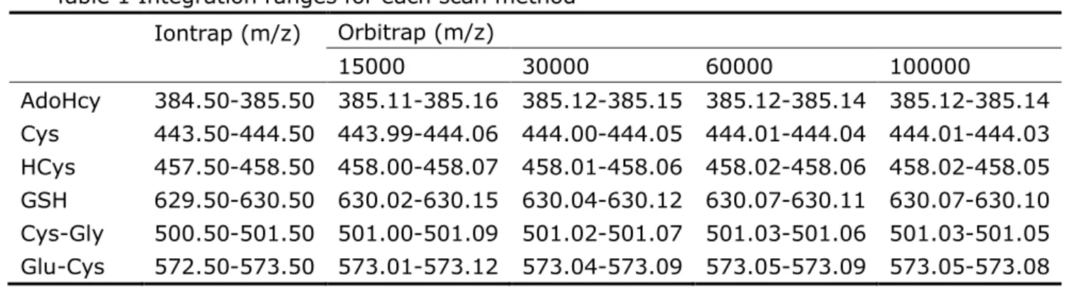
The sensitivity of the Iontrap and Orbitrap were compared in terms of LOD. The integration windows for each of the scan methods are shown in Table 1, and the LODs of thiols both in standard solutions and yeast samples are shown in Table 2. The results show that, for most of the analytes, the Orbitrap provided better LODs than the Iontrap, though the differences are not large. The advantage of high mass resolution became obvious when the complex yeast extracts were analyzed. The Orbitrap, at resolution 30000, was chosen for use in the rest of the study.
The absolute LODs for the thiols were in the sub-picomole range, and they were better than the LC-UV methods (11, 12, 33), most of the MS based detection methods (5, 34-39), and the results via fluorescence measurement (40, 41).
The relationship between detector response and thiol concentrations was demonstrated, and the calibration curves were linear up to 15 μmol/L for the compounds studied.
The precision of the method was established by analysis of six replicate yeast samples, and RSDs were no more than 9 %. Extraction yields for 6 thiols ranged from 95 to 107 %. In order to study the effect of matrices two different sets of calibration samples were prepared by adding the reduced thiols to water and to yeast. The slopes of the calibration curves were compared to evaluate the matrix effects. No significant differences between calibration slopes in the water and yeast matrices were observed. It may be concluded that the analysis of thiols in yeast is not influenced significantly by the sample matrix. This finding is consistent with the literature (19).
As shown in Figure 4, the thiols after derivatization were stable at room temperature over a time period of 46 hours. It has been reported in the literature that thiol-PHMB complexes are stable for the length of the working day if kept at room temperature (17). Figure 4 also shows the instrument variation over a two day period.
The results of yeast analysis
The quantification of thiols in yeast samples was carried out using selenium enriched yeast and three bakery yeast samples (machine yeast, instant yeast, active dry yeast).
Standard addition was used for the quantification of thiols. The results of measured concentrations of thiols in yeast are shown in Table 3.
It is known that selenium is a cofactor of glutathione peroxidase (GPX), one of the important antioxidant enzymes, and that Saccharomyces cerevisiae has three GPX homologues (42). However, the role of selenium in thiol metabolism in yeast has not been studied. The concentration of total GSH (reduced and oxidized) in bakery yeasts was in the range of 14.63- 18.34 µmol/g, and it was consistent with the value reported in literature (43) (about 16.27 µmol/g). Interestingly, we found that the concentration of GSH decreased by 3-4 times and Glu-Cys by about 1/3 in selenium enriched yeast. Both these peptides are substrates of -glutamyl transferase (GGT), an enzyme involved in transport and metabolism of GSH (44). GGT converts Glu-Cys to Cys and GSH to Cys-Gly, and Cys-Gly is converted to Cys and Gly by L-Cys-Gly dipeptidase (45). This could explain the increase of Cys-Gly and Cys concentration. AdoHCy and HCys are involved in the S-adenosyl-methionine (SAM) cycle, which comprehends the reactions that produce, consume, and regenerate SAM. In the first step of this cycle, the SAM-dependent methylases (EC 2.1.1) that use SAM as a substrate produce AdoHCy as a product (46). AdoHCy is hydrolysed to HCys and adenosine by s-adenosylhomocysteine hydrolase (EC 3.3.1.1) and the HCys recycled back to methionine and methionine converted back to SAM, completing the cycle (47). The concentration of AdoHCy and HCys increased in selenium enriched samples: very little HCys was found in the selenium enriched yeast and no HCys was found in the bakery yeasts. In the literature (48), no HCys could be detected in yeast either, and there was a rapid increase of HCys in response to cadmium treatment. It was reported that the concentration of HCys in yeast was about 20 times less than the concentration of Cys (49). The amount of HCys in other biological samples (such as urine and plasma) was also quite low, compared with Cys (the concentration ratio between Cys and HCys ranging from 14 : 1 to 41:1) (5, 11, 34, 38, 50). According to this ratio, the concentration of HCys should be 11 – 33 nmol/g which would be at or near the LOD (2 nmol/g). It has been reported that about 70% of the homocysteine in human plasma is associated with plasma proteins (51). The possibility and degree of protein binding of HCys in yeast remains to be investigated.
These results might suggest an involvement of selenium in thiol metabolism, not limit to its role as an enzyme cofactor or as an S-analogue in S-containing amino acids. Thus, the developed methodology can be fundamental for future metabolic studies.
Abbreviations USed
Cys: cysteine; HCys: homocysteine; GSH: glutathione; Cys-Gly: cysteinyl-glycine; Glu-Cys: γ-glutamyl-cysteine; AdoHcy: s-adenosyl-homocysteine; TCEP: Tris(2-carboxyethyl) phosphine; PHMB: p-hydroxymercurybenzoate; GSSG: glutathione disulfide; GPX: glutathione peroxidase; GGT: -glutamyl transferase; SAM: s-adenosyl-methionine.
Safety
Literature cited
1. Meister, A.; Anderson, M. E., Glutathione. Annu. Rev. Biochem. 1983, Vol. 52, 711-760. 2. Harvey Mudd, S.; Finkelstein, J. D.; Irreverre, F.; Laster, L., Homocystinuria: An enzymatic defect. Science 1964, 143 (3613), 1443-1445.
3. Medina, M. A.; Urdiales, J. L.; Amores-Sanchez, M. I., Roles of homocysteine in cell metabolism: Old and new functions. Eur. J. Biochem. 2001, 268 (14), 3871-3882.
4. Ramakrishnan, S.; Sulochana, K. N.; Lakshmi, S.; Selvi, R.; Angayarkanni, N., Biochemistry of homocysteine in health and diseases. Indian J. Biochem. Biophys. 2006, 43 (5), 275-283.
5. Seiwert, B.; Karst, U., Simultaneous LC/MS/MS determination of thiols and disulfides in urine samples based on differential labeling with ferrocene-based maleimides. Anal. Chem. 2007, 79 (18), 7131-7138.
6. Shimada, K.; Mitamura, K., Derivatization of thiol-containing compounds. Journal of Chromatography B: Biomedical Applications 1994, 659 (1-2), 227-241.
7. Srinivas, N. R.; Mamidi, R. N. V. S., Bioanalytical considerations for compounds containing free sulfhydryl groups. Biomed. Chromatogr. 2003, 17 (5), 285-291.
8. Jemal, M.; Hawthorne, D., High performance liquid chromatography/ionspray mass spectrometry of N-ethylmaleimide and acrylic acid ester derivatives for bioanalysis of thiol compounds. Rapid Commun. Mass Spectrom. 1994, 8, 854-857.
9. Russell, J.; McKeown, J. A.; Hensman, C.; Smith, W. E.; Reglinski, J., HPLC determination of biologically active thiols using pre-column derivatisation with 5,5'-dithio-(bis-2-nitrobenzoic acid). J. Pharm. Biomed. Anal. 1997, 15 (11), 1757-1763. 10.Kranner, I.; Grill, D., Determination of glutathione and glutathione disulphide in lichens: A comparison of frequently used methods. Phytochemical Analysis 1996, 7 (1), 24-28. 11. Kusmierek, K.; Glowacki, R.; Bald, E., Analysis of urine for cysteine, cysteinylglycine, and homocysteine by high-performance liquid chromatography. Anal. Bioanal. Chem 2006, 385 (5), 855-860.
12. Kusmierek, K.; Bald, E., Reduced and total glutathione and cysteine profiles of citrus fruit juices using liquid chromatography. Food Chem. 2008, 106 (1), 340-344.
13. Zaluzec, E. J.; Gage, D. A.; Watson, J. T., Quantitative assessment of cysteine and cystine in peptides and proteins following organomercurial derivatization and analysis by matrix-assisted laser desorption ionization mass spectrometry. J. Am. Soc. Mass Spectrom. 1994, 5 (5), 359-366.
14. Guo, Y. F.; Chen, L. Q.; Yang, L. M.; Wang, Q. Q., Counting sulfhydryls and disulfide bonds in peptides and proteins using mercurial ions as an MS-tag. J. Am. Soc. Mass Spectrom. 2008, 19 (8), 1108-1113.
15. Kutscher, D. J.; Busto, M. E. D.; Zinn, N.; Sanz-Medel, A.; Bettmer, J., Protein labelling with mercury tags: fundamental studies on ovalbumin derivatised with p-hydroxymercuribenzoic acid (pHMB). J. Anal. At. Spectrom. 2008, 23 (10), 1359-1364. 16. Bramanti, E.; Lucchesini, S.; D'Ulivo, A.; Lampugnani, L.; Zamboni, R.; Spinetti, M. C.; Raspi, G., Selective determination of thiolic proteins by hydrophobic interaction chromatography coupled with on-line cold vapour atomic fluorescence spectrometry. J. Anal. At. Spectrom. 2001, 16 (2), 166-171.
Speciation and quantification of thiols by reversed-phase chromatography coupled with on-line chemical vapor generation and atomic fluorescence spectrometric detection: Method validation and preliminary application for glutathione measurements in human whole blood. Clin. Chem. 2005, 51 (6), 1007-1013.
18. Tang, L.; Chen, F.; Yang, L.; Wang, Q., The determination of low-molecular-mass thiols with 4-(hydroxymercuric)benzoic acid as a tag using HPLC coupled online with UV/HCOOH-induced cold vapor generation AFS. J. Chromatogr. B. Analyt. Technol. Biomed. Life. Sci 2009, 877 (28), 3428-3433.
19. Krijt, J.; Vackova, M.; Kozich, V., Measurement of homocysteine and other aminothiols in plasma: Advantages of using tris(2-carboxyethyl)phosphine as reductant compared with tri-n-butylphosphine. Clin. Chem. 2001, 47 (10), 1821-1828.
20. Sack, R.; Willi, A.; Hunziker, P. E., Determination of total glutathione in cell lysates by high performance liquid chromatography with o-phthalaldehyde precolumn derivatization in the presence of tris(2-carboxyethyl)-phosphine. J. Liq. Chromatogr. Related. Technol 2000, 23 (19), 2947-2962.
21. Cole, D. E.; Lehotay, D. C.; Evrovski, J., Simplified simultaneous assay of total plasma homocysteine and methionine by HPLC and pulsed integrated amperometry. Clin. Chem. 1998, 44 (1), 188-190.
22. Han, J. C.; Han, G. Y., A procedure for quantitative determination of tris(2-carboxyethyl)phosphine, an odorless reducing agent more stable and effective than dithiothreitol. Anal. Biochem. 1994, 220 (1), 5-10.
23. Getz, E. B.; Xiao, M.; Chakrabarty, T.; Cooke, R.; Selvin, P. R., A comparison between the sulfhydryl reductants tris(2- carboxyethyl)phosphine and dithiothreitol for use in protein biochemistry. Anal. Biochem. 1999, 273 (1), 73-80.
24. Xiong, Z. Q.; Guo, M. J.; Guo, Y. X.; Chu, J.; Zhuang, Y. P.; Zhang, S. L., Efficient extraction of intracellular reduced glutathione from fermentation broth of Saccharomyces cerevisiae by ethanol. Bioresour. Technol. 2009, 100 (2), 1011-1014.
25. Garcia-Reyes, J. F.; Dernovics, M.; Ortega-Barrales, P.; Fernandez-Alba, A. R.; Molina-Diaz, A., Accurate mass analysis and structure elucidation of selenium metabolites by liquid chromatography electrospray time-of-flight mass spectrometry. J. Anal. At. Spectrom. 2007, 22 (8), 947-959.
26. Encinar, J. R.; Ouerdane, L.; Buchmann, W.; Tortajada, J.; Lobinski, R.; Szpunar, J., Identification of water-soluble selenium-containing proteins in selenized yeast by size-exclusion-reversed-phase HPLC/ICPMS followed by MALDI-TOF and electrospray Q-TOF mass spectrometry. Anal. Chem. 2003, 75 (15), 3765-3774.
27. Bansal-Mutalik, R.; Gaikar, V. G., Reverse micellar solutions aided permeabilization of baker's yeast. Process Biochem. 2006, 41 (1), 133-141.
28. Jamnik, P.; Raspor, P., Stress response of yeast Candida intermedia to Cr(VI). J. Biochem. Mol. Toxicol. 2003, 17 (6), 316-323.
29. Wang, W.; Guo, T.; Song, T.; Lee, C. S.; Balgley, B. M., Comprehensive yeast proteome analysis using a capillary isoelectric focusing-based multidimensional separation platform coupled with ESI-MS/MS. Proteomics 2007, 7 (8), 1178-1187.
30.Goenaga Infante, H.; O'Connor, G.; Rayman, M.; Wahlen, R.; Spallholz, J. E.; Hearn, R.; Catterick, T., Identification of water-soluble gamma-glutamyl-Semethylselenocysteine in
yeast-based selenium supplements by reversed-phase HPLC with ICP-MS and electrospray tandem MS detection. J. Anal. At. Spectrom. 2005, 20, 864-870.
31. Dernovics, M.; Lobinski, R., Characterization of the selenocysteine-containing metabolome in selenium-rich yeast: Part 1. Identification of new species by multi-dimensional liquid chromatography with parallel ICP-MS and electrospray Q-TOFMS/MS detection. J. Anal. At. Spectrom. 2007, 23 (1), 72-83.
32. Bird, S. M.; Uden, P. C.; Tyson, J. F.; Block, E.; Denoyer, E., Speciation of selenoamino acids and organoselenium compounds in selenium-enriched yeast using high-performance liquid chromatography-inductively coupled plasma mass spectrometry. J. Anal. At. Spectrom. 1997, 12 (7), 785-788.
33. Chwatko, G.; Bald, E., Determination of cysteine in human plasma by high-performance liquid chromatography and ultraviolet detection after pre-column derivatization with 2-chloro-1-methylpyridinium iodide. Talanta 2000, 52 (3), 509-515. 34. Bouligand, J.; Deroussent, A.; Paci, A.; Morizet, J.; Vassal, G., Liquid chromatography-tandem mass spectrometry assay of reduced and oxidized glutathione and main precursors in mice liver. J. Chromatogr. B. Analyt. Technol. Biomed. Life. Sci 2006, 832 (1), 67-74.
35. Guan, X.; Hoffman, B.; Dwivedi, C.; Matthees, D. P., A simultaneous liquid chromatography/mass spectrometric assay of glutathione, cysteine, homocysteine and their disulfides in biological samples. J. Pharm. Biomed. Anal. 2003, 31 (2), 251-261. 36. Weaving, G.; Rocks, B. F.; Iversen, S. A.; Titheradge, M. A., Simultaneous quantitation of homocysteine, cysteine and methionine in plasma and urine by liquid chromatography-tandem mass spectrometry. Annals of Clinical Biochemistry 2006, 43 (6), 474-480.
37. Vellasco, A. P.; Haddad, R.; Eberlin, M. N.; Hoehr, N. F., Combined cysteine and homocysteine quantitation in plasma by trap and release membrane introduction mass spectrometry. Analyst 2002, 127 (8), 1050-1053.
38. Jiang, Z.; Liang, Q.; Luo, G.; Hu, P.; Li, P.; Wang, Y., HPLC-electrospray tandem mass spectrometry for simultaneous quantitation of eight plasma aminothiols: Application to studies of diabetic nephropathy. Talanta 2009, 77 (4), 1279-1284.
39. Gucek, M.; Makuc, S.; Mlakar, A.; Bericnik-Vrbovsek, J.; Marsel, J., Determination of glutathione in spruce needles by liquid chromatography/tandem mass spectrometry. Rapid Communications in Mass Spectrometry 2002, 16 (12), 1186-1191.
40. Pelletier, S.; Lucy, C. A., HPLC simultaneous analysis of thiols and disulfides: On-line reduction and indirect fluorescence detection without derivatization. Analyst 2004, 129 (8), 710-713.
41. Causse, E.; Malatray, P.; Calaf, R.; Charpiot, P.; Candito, M.; Bayle, C.; Valdiguie, P.; Salvayre, R.; Couderc, F., Plasma total homocysteine and other thiols analyzed by capillary electrophoresis/laser-induced fluorescence detection: Comparison with two other methods. Electrophoresis 2000, 21 (10), 2074-2079.
42. Inoue, Y.; Matsuda, T.; Sugiyama, K. I.; Izawa, S.; Kimura, A., Genetic analysis of glutathione peroxidase in oxidative stress response of Saccharomyces cerevisiae. J. Biol. Chem. 1999, 274 (38), 27002-27009.
acids on glutathione production by Saccharomyces cerevisiae. Appl. Microbiol. Biotechnol. 1992, 36 (4), 538-540.
44. Mehdi, K.; Thierie, J.; Penninckx, M. J., γ-glutamyl transpeptidase in the yeast Saccharomyces cerevisiae and its role in the vacuolar transport and metabolism of glutathione. Biochem. J. 2001, 359 (3), 631-637.
45. Kaur, H.; Kumar, C.; Junot, C.; Tolendano, M. B.; Bachhawat, A. K., Dug1p is a Cys-Gly peptidase of the γ-glutamyl cycle of Saccharomyces cerevisiae and represents a novel family of Cys-Gly peptidases. J. Biol. Chem. 2009, 284 (21), 14493-14502.
46. Finkelstein, J. D.; Martin, J. J., Homocysteine. Int. J. Biochem. Cell Biol. 2000, 32 (4), 385-389.
47. Fodinger, M.; Horl, W. H.; Sunder-Plassmann, G., Molecular biology of 5,10-methylenetetrahydrofolate reductase. Journal of Nephrology 2000, 13 (1), 20-33. 48. Lafaye, A.; Junot, C.; Pereira, Y.; Lagniel, G.; Tabet, J. C.; Ezan, E.; Labarre, J., Combined proteome and metabolite-profiling analyses reveal surprising insights into yeast sulfur metabolism. J. Biol. Chem. 2005, 280 (26), 24723-24730.
49. Kumar, A.; John, L.; Alam, M. M.; Gupta, A.; Sharma, G.; Pillai, B.; Sengupta, S., Homocysteine- and cysteine-mediated growth defect is not associated with induction of oxidative stress response genes in yeast. Biochem. J. 2006, 396 (1), 61-69.
50. Causse, E.; Issac, C.; Malatray, P.; Bayle, C.; Valdiguie, P.; Salvayre, R.; Couderc, F., Assays for total homocysteine and other thiols by capillary electrophoresis-laser-induced fluorescence detection - I. Preanalytical condition studies. J. Chromatogr. A 2000, 895 (1-2), 173-178.
51. Refsum, H.; Helland, S.; Ueland, P. M., Radioenzymic determination of homocysteine in plasma and urine. Clin. Chem. 1985, 31 (4), 624-628.
Y. R thanks the China Scholarship Council for providing financial support under the “2008 NRC-MOE Research and Post-doctoral Fellowship Program”.
Table 1 Integration ranges for each scan method Iontrap (m/z) Orbitrap (m/z) 15000 30000 60000 100000 AdoHcy 384.50-385.50 385.11-385.16 385.12-385.15 385.12-385.14 385.12-385.14 Cys 443.50-444.50 443.99-444.06 444.00-444.05 444.01-444.04 444.01-444.03 HCys 457.50-458.50 458.00-458.07 458.01-458.06 458.02-458.06 458.02-458.05 GSH 629.50-630.50 630.02-630.15 630.04-630.12 630.07-630.11 630.07-630.10 Cys-Gly 500.50-501.50 501.00-501.09 501.02-501.07 501.03-501.06 501.03-501.05 Glu-Cys 572.50-573.50 573.01-573.12 573.04-573.09 573.05-573.09 573.05-573.08
Table 2 LOD of thiols in standard solutions and yeast matrix Iontrap Orbitrap 15000 30000 60000 100000 Standard solution, (nmol/L) AdoHcy 4 2 2 2 1 Cys 18 12 11 18 8 HCys 48 9 9 10 8 GSH 7 5 5 2 1 Cys-Gly 9 22 15 18 24 Glu-Cys 25 15 26 36 32
Yeast, (nmol/g) AdoHcy 5 1 1 0.3 0.3
Cys 4 7 3 3 2
Hcys 5 2 2 2 1
GSH 5 3 1 1 1
Cys-Gly 4 4 2 3 3
Table 3 Concentrations of thiols extracted from 3 mg yeast samples determined using LC with the Orbitrap mass spectrometer at 30000 resolution.
Analyte Concentration (µmol/g)
Selenium enriched yeast Bread Machine yeast Instant yeast Active dry yeast AdoHcy 0.26 0.01 0.02 n.d. Cys 0.46 0.07 0.07 0.08 HCys 0.02 n.d. n.d. n.d. GSH 5.20 18.34 17.69 14.63 Cys-Gly 0.61 0.02 0.01 0.05 Glu-Cys 0.17 0.64 0.47 0.52 n.d. not detected RSD: 8-13% (n=5)
Figure 1. Mass spectra of Cys-PHMB using the Iontrap and Orbitrap at resolution 15000 and 60000.
Figure 2. Representative chromatograms of a mixture of free thiol standards including AdoHcy, Cys (as Cys-PHMB), HCys (as HCys-PHMB), GSH (as GSH-PHMB), Cys-Gly (as Cys-Gly-PHMB), and Glu-Cys (as Glu-Cys-PHMB) at a concentration of 3 µmol/L each (a); and thiols in the selenium enriched yeast sample (b). The m/z of the ion being monitored in each channel is indicated in the figure. The peak before Glu-Cys-PHMB at m/z 573 is TCEP-PHMB complex (RT = 7.4 min), which has very similar m/z of Glu-Cys-PHMB (the mass difference is 0.001 Da).
Figure 3
Figure 3. Effect of incubation time when disulfides (1 ml of a mixture of cystine, homocystine, GSSG, at a concentration of 5 µmol/L each) were reduced with 50 µl of TCEP (20 mmol/L) then derivatized with 100 µl of PHMB (7 mmol/L).
Figure 4
Figure 4. Stability study of thiols in yeast after spiking at a concentration of 3 μmol/L (Replicate data points are offset for better visual representation of the data)