Review
1
Animal models of cardiac arrhythmias
a ,
*
a b´
´
Michiel J. Janse
, Tobias Opthof , Andre G. Kleber
a
Department of Clinical and Experimental Cardiology, Academic Medical Centre, University of Amsterdam, Amsterdam, Netherlands
b
¨
Physiologisches Institut, Universitat Bern, Bern, Switzerland Received 8 October 1997; accepted 3 December 1997
1. Introduction contributed to the development of diagnostic and
therapeu-tic strategies. When surveying the literature with the intention of
evaluating to which extent studies on animal models have
contributed to the understanding of arrhythmia mecha- 2. Supraventricular arrhythmias
nisms in patients and in devising therapeutic strategies, one
is struck by the differences between supraventricular and 2.1. Re-entrant tachycardias in the presence of ventricular arrhythmias. In general, in the field of sup- accessory atrioventricular pathways
raventricular arrhythmias there has been a strong
inter-action between experimental and clinical studies and there The history of these arrhythmias is rather bizarre can be no doubt that the various animal models have been because animal studies provided the basic arrhythmia instrumental in understanding the mechanisms of clinical mechanisms long before the syndrome was clinically arrhythmias and in establishing different forms of therapy. recognized, because from 1967 onwards clinical studies Clearly, an animal cannot be transformed into a human unravelled in great detail the electrophysiological charac-patient, but despite species differences and differences in teristics in patients without the investigators being aware arrhythmogenic factors in animal models and humans, the of the early animal studies, and because to our knowledge similarity between arrhythmia mechanisms in experimental only one single dog had been studied that possessed an models and patients far outweigh the differences. accessory atrioventricular pathway.
This similarity is less evident when considering ven- In 1913 Mines described an experiment on a ring-like tricular arrhythmias. There are several reasons for this. preparation of a tortoise heart in which he was able to First, many ventricular arrhythmias, such as those induced initiate circulating excitation by electrical stimulation. He by acute ischaemia, cannot be studied in human patients made the historical prediction: ‘‘I venture to suggest that a because they occur unpredictably in situations where circulating excitation of this type may be responsible for electrophysiological changes may develop within minutes. some cases of paroxysmal tachycardia as observed clinical-Second, even when in patients acute ischaemia is the ly’’ [1].
trigger for arrhythmias, many other factors may influence After reading Kent’s report [2] in which a human heart arrhythmogenesis, such as the presence of a healed infarct, was described with a muscular connection between the hypertrophy, dilatation, electrolyte disturbances or heart right atrium and the right ventricle, Mines wrote in 1914: failure. Third, many factors determine whether, and if so, ‘‘I now repeat this suggestion in the light of the new how often ventricular arrhythmias occur in the setting of histological demonstration by Stanley Kent that the muscu-acute ischaemia and / or a chronic myocardial infarction, lar connection between auricles and ventricles is multiple. and in experimental models usually only a single factor is Suppose that for some reasons an impulse from the auricle taken into account. Still, the knowledge of arrhythmogenic reached the main A–V bundle but failed to reach this ‘right mechanisms derived from animal studies has greatly lateral’ connection. It is possible then that the ventricle would excite the ventricular end of this lateral connection,
*Corresponding author.
1
Guest Editor was Prof. W. Schaper. Time for primary review 42 days.
0008-6363 / 98 / $19.00 1998 Elsevier Science B.V. All rights reserved. P I I : S 0 0 0 8 - 6 3 6 3 ( 9 7 ) 0 0 3 1 3 - 1
not finding it refractory as normally it would at such a of patients suffering from AV nodal re-entrant tachycardia. time. The wave spreading then to the auricle might be It is extremely rare to induce sustained AV nodal re-entry expected to circulate around the path indicated’’ [3]. This in this preparation, but it is not uncommon to induce single was written 16 years before Wolff, Parkinson and White echo beats by premature stimulation of the atrial or described the clinical syndrome that now bears their name ventricular tissue [19–22,26–29].
[4], 18 years before Holzmann and Scherf ascribed the In open-chested anaesthetized dogs, only one ‘case abnormal ECG in these patients to pre-excitation of the report’ of sustained re-entrant AV nodal tachycardia has ventricles via an accessory atrioventricular bundle [5], and been published [30], and reproducibly induced sustained 53 years before the first studies in patients employing tachycardia became only possible after a surgical pro-intraoperative mapping and programmed stimulation cedure that blocked atrial impulses from the anterior input proved Mines’ predictions to be correct [6–8]. At present, site to the AV node [31].
all the electrophysiological characteristics of accessory In patients with AV nodal re-entrant tachycardia, the atrioventricular connections and their role in causing re- hallmark for dual AV nodal pathways is the so-called entrant tachycardias have been obtained in studies on ‘jump’ in the conduction curve [32]: when during regular human patients (for review see Wellens [9]) and only one pacing of the atria (A1) single premature atrial stimuli study described pre-excitation in a dog [10]. In this study, (A2) at progressively shorter coupling intervals are applied which also described two patients, the important observa- and the premature atrium–His bundle intervals (A2–H2) tion was made that atrial fibrillation induced in the dog, are plotted against the A1–A2 intervals, a sudden jump of caused ventricular fibrillation as well because the acces- 50 ms or more in the A2–H2 interval at a decrement in sory pathway had a short refractory period and conducted A1–A2 of 10 ms is taken as evidence for dual AV nodal many impulses, which otherwise would have been blocked pathways. At a critical coupling interval the ‘fast’ pathway in the AV node. Still, at present there is certainly no need is refractory, and conduction to the His bundle proceeds for an animal model of accessory atrioventricular path- via the ‘slow’ pathway which has a shorter refractory
ways. period. In isolated rabbit heart preparations, such a jump
has not been observed [33]. In the anaesthetized dog, a 2.2. Atrioventricular nodal re-entrant tachycardia ‘jump’ was observed in one study only after left and right stellate ganglia had been removed [34]. In another study, The history of this arrhythmia is very different from that no jump was found [35]. In yet another study, a jump in of the arrhythmias caused by accessory AV connections. the A–V interval was ascribed to slow conduction or block Although it was again Mines [1,2] who formulated the in the peripheral Purkinje system rather than to functional basic mechanisms, clinical studies quickly followed and dissociation of the AV node [36]. In isolated, perfused dog throughout this century there has been an intensive inter- hearts, no jump occurs [37,38], although ventricular echoes action between experimental and clinical studies (for could be reproducibly induced [39]. Whereas the clinical review see [11] and [12]). For example, the first study experience would suggest that atrial tissue is involved in employing programmed stimulation in patients to unravel the re-entrant circuit (for review see [12] and [40], but for the arrhythmia mechanisms and to treat the condition by a different point of view see [41]), mapping studies in the pacemaker implantation by Coumel and co-workers in isolated, blood perfused dog heart showed that ventricular 1967 [13] quoted the early studies of Mines [1,2]. Also, the echoes are due to subatrial re-entry [39].
pioneering clinical studies of the 1980s allowing successful It would be desirable if an animal model could be found surgical treatment [14,15] or catheter ablation [16–18] of in which subtle manipulation of the autonomic nervous the arrhythmia, all quoted the microelectrode studies of the system (rather than a surgical procedure as described in 1960s and 1970s on isolated rabbit heart preparations that [31]) would result in sustained AV nodal tachycardia being provided insight into arrhythmia mechanisms on a cellular reproducibly induced. Answers could then be found to
basis [19–22]. questions such as: Is the re-entrant circuit for single echo
Today, we are confronted with a rather paradoxical beats the same as that for sustained re-entry? Are there situation: the history indicates a happy union between structurally different pathways? Are there multiple re-knowledge gathered by both experimental and clinical entrant circuits? What is the role of anisotropic conduction studies, the former preceding the latter, which finally and of different input sites? In any case, there is no animal resulted in the very successful treatment by radiofrequency model for dual pathways as observed in humans.
catheter ablation [23–25]. Despite this success, there are
still many uncertainties about the exact location of the 2.3. Atrial flutter re-entrant pathway and about the electrophysiological and
structural properties of the two AV nodal pathways (‘slow’ Much of our knowledge about activation patterns during and ‘fast’) that are thought to form the basis for AV nodal atrial flutter has been derived from animal studies, with the re-entry. The animal model most often used, the isolated 1920 paper of Lewis, Feil and Stroud as the classical superfused rabbit heart preparation, differs from the heart example [42]. Catheter electrode mapping in patients was
first performed by Puech and colleagues in 1956 [43] and fibrillation was due to ‘‘ . . . a series of ring-like circuits of due to advances in mapping techniques, recent studies in shifting location and multiple complexity’’ [58]. Some 70 man [44] allowed comparison to data obtained in animal years later, Allessie and co-workers could record simul-models [45]. On the basis of these studies, it is widely taneously from 192 atrial sites in an isolated, Langendorff-accepted that atrial flutter is due to re-entry. perfused canine heart in which atrial fibrillation was There are a number of animal models in which atrial induced by rapid atrial pacing during infusion of acetyl-flutter was induced following the creation of anatomical choline [59]. In essence, they confirmed Garrey’s state-lesions, by extending an anatomical obstacle such as the ment, which had been refined by Moe and Abildskov on ostium of the vena cava superior by crushing the intercaval the basis of both experimental observations and computer region [46,47], or by producing single lesions in the right simulations that led to the formulation of the multiple atrium [48–50]. It is doubtful whether these models are wavelet hypothesis [60,61]. In the experiments of Allessie representative of atrial flutter in patients. et al. [59] the presence of multiple independent wavelets Another type of flutter, depending on a Y-shaped was documented, and it was estimated that the critical surgical lesion in the right atrial free wall, causing the number of wavelets in both atria necessary to maintain re-entrant circuit to consist of atrial tissue around the fibrillation was between 3 and 6. These results were largely tricuspid ring [51] may have a clinical counterpart in confirmed by other studies in dog hearts [62] and also by postoperative flutter following surgical correction of mapping studies in patients with atrial fibrillation during
congenital abnormalities [52]. open heart surgery [63,64].
The canine model developed by Boyden and Hoffman In recent years, there has been a remarkable interaction [53], in which right atrial enlargement was produced by between experimental and clinical studies, where important banding of the pulmonary artery and by producing tri- findings in animal models were soon thereafter confirmed cuspid regurgitation, may also have a clinical counterpart in clinical studies. For example, repetitive induction of in patients with chronic obstructive pulmonary disease and atrial fibrillation, or prolonged periods of rapid atrial tricuspid regurgitation. In those dogs, a functional zone of pacing, in conscious goats gave rise to a marked shortening block and area of slow conduction set the stage for re- of the atrial refractory period, which persisted for a long entry, rather than an anatomical obstacle. Functional re- time after restoration of sinus rhythm and predisposed to entry is also observed in the sterile pericarditis model of the reinduction of atrial fibrillation following cardioversion
´
canine atrial flutter, first described by Page et al. [54]. This [65]. This was confirmed in patients [66–68], as was the model was developed because of the fact that following finding originally made in canine hearts [70], that calcium cardiac surgery in patients atrial flutter frequently occurs blocking agents could attenuate this shortening of the and this may be related to postoperative sterile pericarditis. refractory period [69]. Despite the great impact that the Another form of functional re-entry causing atrial flutter experimental studies have on clinical developments, in-was pharmacologically induced in isolated dog hearts by cluding new strategies for therapy such as catheter ablation infusion of acetylcholine [55]. (see Ref. [67]), there are obvious differences between the Despite subtle differences in the various forms of atrial atria of the experimental animals used and those of patients flutter, one cannot but agree with Mary-Rabine and co- who spontaneously develop atrial fibrillation. In the ex-workers [56], that ‘‘the history of atrial flutter clearly perimental models, atrial fibrillation was induced in essen-illustrates the bidirectional flow of information and the tially normal hearts, either by shorter or longer periods of mutual stimulation between the basic and the clinical rapid pacing or repetitive induction of atrial fibrillation by levels, leading both to a better understanding of the nature burst pacing [65,70] or by pharmacologic means, such as of the arrhythmia and to new therapeutic approaches’’. It is infusion of acetylcholine [59,62]. Whilst this latter model now established that atrial flutter is due to a re-entrant may have its clinical counterpart in the relatively rare form wave in the right atrium, and that a zone of slow of atrial fibrillation dependent on increased vagal tone conduction located inferiorly and posteriorly in the right [71], about 85% of patients with atrial fibrillation have an atrium is the target for catheter ablation. underlying structural cardiac abnormality or a metabolic disorder, often associated with atrial enlargement [72].
2.4. Atrial fibrillation Acute atrial dilatation shortens the atrial refractory period
and enhances the vulnerability to atrial fibrillation [73,74]. Some crucial observations on the characteristics of atrial However, the effects of chronic stretch are most likely fibrillation were made long before technical developments different from those of acute stretch, and would be more allowed the simultaneous recording from multiple atrial important in contributing to atrial fibrillation in patients. sites, a prerequisite for documenting the complex activa- There is one study by Boyden et al. [75] in which atrial tion sequence of fibrillation. Thus, in 1914 Garrey [57] cellular electrophysiological characteristics of dogs with without the help of electrophysiological or mechanical naturally occurring mitral valve disease leading to progres-recordings established that a critical mass of tissue is sive atrial enlargement were studied. Some animals were needed to sustain fibrillation. He also suggested that followed for 5 years, before the electrophysiological study
was performed. Most dogs developed atrial arrhythmias, 3.2. Ventricular arrhythmias caused by acute ischaemia including atrial fibrillation. Surprisingly, the
transmem-brane potential characteristics of atrial cells of these A great many experimental studies on this subject have animals were not significantly different from those of been undertaken in the past decades. A rough distinction control animals, although some cells were found with can be made into studies in which electrophysiological resting membrane potentials below 260 mV that were parameters relevant for understanding arrhythmia mecha-inexcitable. This is in contrast to the findings of the studies nisms were recorded (for Refs. see [88,89]), and those in mentioned earlier [65,66] and also in contrast to several which only the incidence of the lethal arrhythmia, ven-studies in which cellular electrophysiological abnormalities tricular fibrillation, was noted, usually in studies testing have been documented in small, isolated atrial preparations anti-arrhythmic drugs.
obtained from fibrillating human atria [76–78]. In the Validation of animal models for assessing the patho-study of Boyden et al. [75], massive interstitial fibrosis and physiology of acute myocardial ischaemia implicates con-cellular hypertrophy were found and the authors concluded sideration of the (1) diversity of cardiac diseases which that the morphological changes were much more important involve acute ischaemia and (2) the variety of experimen-in causexperimen-ing atrial fibrillation than the slight, experimen-insignificant tal models which have mostly been designed to mimic part electrophysiological alterations they found. The increased of the complex events occurring in the human disease. size of the atria would permit the coexistence of many Along this line of reasoning, mostly large animals (dogs, re-entrant circuits. The increase in connective tissue would pigs, cats) are used to study ventricular arrhythmias, while promote inhomogeneous conduction, unidirectional block mostly small animals (rats) are involved in studies about and re-entry [79]. Thus far, the emphasis in animal models the changes in metabolic pathways consequent to is-of atrial fibrillation has been on electrophysiological chaemia and reperfusion. In the former, multisite mapping characteristics in structurally normal hearts. To further of electrical activity is applied to the analysis of the diminish the gap between animal models and patients, the mechanisms of ventricular tachycardia and fibrillation arrhythmogenic effects of structural changes deserve fur- [90,91] whereas in small animals’ hearts, which primarily
ther study. serve the purpose to provide numerous and affordable
samples for chemical analysis, arrhythmias are defined by ECG patterns and analyzed statistically on the basis of
3. Ventricular arrhythmias their incidence. Thus the main reason for selecting a
certain animal species appears to be the suitability for the 3.1. A hereditary model of sudden death application of a specific technique, assuming that ‘acute’ human ischaemia / reperfusion can be compared with ex-A colony of German shepherd dogs has been described perimental ischaemia / reperfusion independently of the with inherited ventricular arrhythmias and a predisposition species selected. While this assumption might hold for the for sudden death [80]. Sudden death most often occurs very global and basic changes in metabolism, it is doubtful during sleep or at rest after exercise or excitement. The that such a simplified concept is applicable to the study of electrocardiogram does not show a prolonged QT interval, arrhythmogenesis, as outlined below. According to a but frequently there is marked notching of the T wave. The search in ‘MEDLINE’ a total of 1327 studies have been arrhythmias are rapid polymorphic ventricular tachycar- carried out between 1966 and 1996 for the assessment of dias, following long R–R intervals, and are most likely due ventricular fibrillation in the setting of myocardial is-to triggered activity induced by early afterdepolarizations chaemia. 569 of these studies were carried out in dogs, 126 in the Purkinje system [81]. In epicardial myocytes, the in pigs, 25 in guinea pigs, 51 in rabbits and 931 in rats. In density of the transient outward current (I ) was reduced,to the setting of reperfusion arrhythmias, a total of 1159 and the time constant of inactivation was reduced [82]. In studies were carried out (328 in dogs, 128 in pigs, 54 in addition, deficiencies in cardiac sympathetic denervations rabbits, 64 in guinea pigs, 585 in rats).
have been reported [83]. At first glance, this dog model The fact that acute ischaemia affects the incidence of bears a resemblance to the congenital long QT syndrome arrhythmias in coronary heart disease in a variety of ways in which bradycardia induced polymorphic ventricular makes it impossible to investigate its pathophysiology in a tachycardia and sudden death occur and in which genetic single experimental model. Thus, acute ischaemia occurs defects in ion channels regulating repolarization have been as one of the triggers for arrhythmias in chronic infarction described [84,85]. However, the dogs have no prolonged [92], in hypertrophy and failure, or it may occur conse-QT interval and thus far, in patients with the long conse-QT quently to coronary occlusion (thrombosis or spasm) in syndrome no deficiencies in Ito have been described [85]. previously relatively healthy individuals [93]. This spec-Still, this animal model might have a clinical counterpart trum of pre-existing alterations is likely to affect the role because patients have been described with polymorphous of acute ischaemia, since infarction, hypertrophy and ventricular tachycardia (Torsade de pointes) who have a failure are associated with changes in the pre-existing
experimental-ists distinguish between total and partial coronary occlu- an experiment carried out in an isolated perfused human sion, the latter being associated with so-called ‘low flow’ heart, the extent and time course of the changes in ischaemia, a pathophysiologic entity which leads to electri- transmembrane action potentials were almost identical to cal and ionic changes different from the changes associated those observed in pig and dog hearts [107]. While there with immediate and total occlusion [96,97]. In the clinical seems an approximate similarity among the electrophysio-settings, it is not always evident whether acute myocardial logical changes observed in large animals and in certain ischaemia is associated with or without residual flow cases of acute ischaemia in humans, several experimental through the occluded or collateral arteries. In the light of observations indicate that the arrhythmias observed in these complexities the discussion about the applicability of small animals (guinea pigs, rats) during acute ischaemia animal models of acute ischaemia to the human situation is differ from the arrhythmias observed in larger species.
certainly justified. First, as mentioned above, the very early (and frequent)
A first basic question concerns the comparison of the type IA arrhythmias require a large tissue mass for the type and incidence of the electrical changes and arrhyth- re-entrant circuits to be maintained. The dependence of mias among different species. In larger species (dog, pig, re-entry (tachycardia and fibrillation) on tissue size is an cat) the arrhythmias during acute ischaemia are relatively old observation, made already in 1914 by Garrey [57]. well characterized. Thus arrhythmias occur in two distinct Indeed, most of the work done on ventricular fibrillation in phases (so-called ‘phase IA’ and ‘phase IB’, Ref. [98]). rat hearts indicates that VF in this species occurs at a time These two phases are associated with distinct changes in corresponding to the IB arrhythmias, and no clear sepa-the electrical tissue properties. In sepa-the first phase, IA, (up to ration of IA from IB arrhythmias has been described in approximately 8–10 min of coronary occlusion), there is a small animal species.
rapid change in electrical membrane properties associated A number of further processes critical for the electrical with metabolic acidification (anaerobic glycolysis), and changes in acute myocardial ischaemia might be different
1
cellular loss and extracellular accumulation of [K ] in rats and guinea pigs from larger species. Extracellular
1
[99,100]. The impact of these changes are a rapid depolar- accumulation of [K ] and metabolic acidification, whicho
ization of the ischaemic myocytes, and a loss of amplitude are likely to be linked to each other, are the main factors and duration of the transmembrane action potential determining the extent of the depolarization of the resting [101,102]. Moreover, there is a marked lengthening of the membrane, the changes in action potential upstroke and refractory period, which becomes sensitive to the lengths changes in refractoriness. In ischaemic regions devoid of of the previous intervals of local excitation. As shown in collateral flow, there is a sharp transition from ischaemic to experimental studies and explained recently in computer normoxic tissue with respect to local pO . In contrast, the2
simulations [103,104], these changes are followed by acidification of the tissue, and the extracellular
accumula-1
specific changes in the excitation and conduction patterns. tion of [K ]o show a gradual decrease from the centre Thus, the decrease in conduction velocity during acute towards the border of the ischaemic region [108,109]. In ischaemia is relatively small and conduction block, which other words, although a given tissue site may be fully changes its location from beat-to-beat, occurs early and ischaemic, the extent of the ionic changes, which form the abruptly [104]. The resulting ventricular tachycardia, fre- basis for the disturbance in electrical function, depends on quently issuing into fibrillation, has a characteristic appear- the diffusion of products of the ischaemic metabolism
1
ance [90]. It is made up by large and highly unstable toward the non-ischaemic border. Both K and CO have2
re-entrant circuits (several millimetres inner circle length). been invoked in this diffusion process [110,111], whereby The second phase of arrhythmias, IB, occurs approximate- CO seems to be a particularly important factor, because it2
ly 10 to 15 min after coronary occlusion and is related to has a high diffusion coefficient and is bound in large the electrical uncoupling of the myocytes [105]. Although quantities by the carbonic buffer system [112,113]. Along the IB arrhythmias have not been analyzed in detail by this line of reasoning, it has to be assumed that the process mapping studies, it is likely that the re-entrant circuits of diffusion will not only affect the gradients between the during this phase are significantly smaller, because partial centre and the border of an ischaemic region, but it will electrical uncoupling allows for much smaller conduction also lead to a relatively smaller change in extra- and
1
velocities [106], and therefore scales the circus movements intracellular pH and K in hearts with a small ventricular to a smaller size. The studies obtained in relatively large mass, and consequently affect the electrical changes of the animals (dog, pig, cat) raise two questions: (1) Are the ischaemic myocytes [111]. In a series of experiments in
1
arrhythmias occurring after acute coronary occlusion in which extracellular potassium [K ] accumulation was
1
humans comparable to those observed in the animal compared among a variety of species, the maximal [K ]o
models? (2) Do the experimental arrhythmias differ among levels reached during acute ischaemia in rats were by animal species? The first question cannot be answered approximately 20–30% lower than those observed in directly. However, several observations provide circum- guinea pigs or rabbits [113,114].
stantial evidence that the electrical changes during acute Besides the macroscopic dimension of the ventricles, ischaemia in humans and large animal hearts are similar. In intrinsic differences in normoxic metabolism may also
exist among species. In rats such differences have been ation of size of the ischaemic zone [122–124] and ‘‘this shown with respect to the duration and shape of the can account for a substantial portion of non-drug related transmembrane action potential and the homeostasis of variability in outcome of antiarrhythmic trials using the
1 21
cellular Na and Ca . Thus the relatively high normal canine coronary occlusion or release model’’ [123]. This
1
intracellular Na activity in the normal rat affects the statement was corroborated by Trolese-Mongheal and working mode of the Na / Ca exchanger and may lead to colleagues [129] who collected data from various
lab-21
earlier Ca overload in depolarized cells. Interestingly in oratories on 658 dogs in which the left anterior descending rats, this difference can be reversed by inhibition of thyroid coronary artery was suddenly ligated. When control series
hormone production [115]. consisted of 10 dogs, the incidence of ventricular
fibrilla-In the past years, so-called remodelling of cardiac tissue tion varied from 0 to 70%; when the control group counted in various cardiac diseases and with a variety of stimuli 20 animals, the incidence varied from 5 to 55%, and even has gained a wide interest. Many of these remodelling in series of 100 dogs, there still was a range of 14 to 36%. processes which occur in ischaemia, chronic infarction, The papers emphasizing the importance of pre-existing myopathy, hypertrophy and failure, may change the sub- collaterals were published between 1970 and 1986. A strate for electrical excitation and conduction at any level. Medline search unearthed 28 studies published between Thus, the macroscopic tissue architecture may get more 1982 and 1996 in which a major coronary artery was discontinuous via the increase in connective tissue. The occluded in dogs to test the effect of antiarrhythmic drugs. expression of gap junctions can change as well, as may the The control series varied from 6 to 40 animals, and since expression of a large number of membrane ionic channels the factors mentioned above were not controlled, interpre-responsible for excitation. The results of all these studies tation of the results must be made with great caution. demonstrate that assessment of pathophysiological
mecha-nism has to consider the dynamics of events and related 3.3. The ventricular arrhythmias of myocardial variables not only on short term (e.g. in the minutes infarction
following coronary occlusion) but also on middle (e.g.
after preconditioning [116,117]) and on longer term (ge- A distinction has been made in arrhythmias occurring in netic remodelling [118–121]). This increasing complexity the subacute phase of myocardial infarction (hours to days should have an impact on the selection and definition of after acute obstruction of a coronary artery) and in the animal models. First, in many important diseases, it is chronic phase (weeks to months). Almost all of the probably wrong to think that a given animal model can experimental work has been performed in dogs (a Medline exactly mimic a certain disease and the ‘individuality’ of a search over the past 4.5 years identified 25 dog studies, certain disease pattern. In most cases they can only address one in the rat and one in the pig).
partial aspects of the disease mechanism. Second, animal The spontaneous arrhythmias that occur in the dog models used to assess arrhythmias should be designed in during the subacute phase resemble those in patients such a way that the most important variables methodo- recovering from an acute myocardial infarction:
acceler-logically followed. ated idioventricular rhythms or slow ventricular
tachycar-The factors that determine whether, and if so, how dias that usually do not degenerate into ventricular fibrilla-frequently, ventricular fibrillation occurs include the size tion (for a detailed description see Refs. [88] and [89]). A of the ischaemic area, the degree of collateral flow, heart great deal is known about electrophysiological changes in rate, the use of anaesthetics, stress in conscious animals, both Purkinje and muscle cells that survive the infarct. the mode of coronary artery occlusion, presence of a However, since the arrhythmias of the subacute phase are previous infarction, activity of the autonomous nervous usually benign, and since the patients are still in hospital so system, hypertrophy in the non-ischaemic myocardium that in case ventricular fibrillation would occur adequate [88,89]. The three most important factors are size of the resuscitation and defibrillation will be provided, the knowl-ischaemic area, the degree of collateral flow and heart rate edge derived from experimental models has not contribu-[122–127]. There is a variation among species in the ted to establish therapeutic strategies during this phase of degree of collateral blood flow following coronary artery myocardial infarction.
occlusion [96]. For example, in rat, rabbit and pig hearts A great deal of information about the electrophysiologi-collateral flow is not significantly different from zero, in cal characteristics of ventricular tachycardia induced by the guinea pig it is not different from normal control flow programmed electrical stimulation has been gathered in [128]. In the dog, an animal often used in studies on dogs with a healing infarct, and many similarities exist ischaemia-induced arrhythmias, there is a variation in pre- between these arrhythmias and those induced in patients. existing collaterals, and depending on the degree of There are, however, several differences between the ex-collateral flow, the incidence of ventricular fibrillation may perimental and the clinical tachycardias. Thus, in dogs, vary from zero to 100% after occlusion of a major ventricular tachycardias can be easily induced by prema-coronary artery [124,126,127]. Similarly, occlusion of the ture ventricular stimulation in the first week following left anterior descending coronary artery results in a vari- coronary artery occlusion, but after the first week
in-ducibility decreases [130] and sometimes the arrhythmia mals and patients there is a considerable overlap in cannot be induced at all [131]. This is different in human baroreflex slope in the groups with and without arrhyth-patients: after five days, ventricular tachycardias can be mias, so that in an individual case no absolute prediction induced in about 10% of patients [132], but in 20 to 50% can be made whether or not arrhythmias will develop. This after three weeks [132–134]. Moreover, in the dog, the is of course due to the fact that sudden death is not due to re-entrant circuit responsible for the tachycardia is usually a single pathophysiologic event. It is in this respect located in the so-called subepicardial border zone, i.e. the noteworthy that Legato found that susceptible dogs had thin layer of surviving subepicardial myocardium overly- larger and more inhomogeneous infarcts than resistant ing the infarct, whereas many of the sustained tachycardias dogs [151].
in humans with a healed infarct originate in the
suben-docardial region [89]. Despite these differences, the ex- 3.4. Animal models and antiarrhythmic drugs perimental studies have provided important information
about the characteristics of the re-entrant circuits and in so Generally speaking, antiarrhythmic drugs exert their far studies in humans were able to study these characteris- effects largely by modulating conduction velocity, or tics, the similarities far outweigh the differences. There is refractory period duration, or both. Conduction velocity no doubt that the experimental findings have been of depends on the one hand on the passive electrical prop-crucial importance for initiating therapeutic strategies, such erties of cardiac tissue, on the other hand on the
charac-1 21
as mapping-guided surgery, catheter ablation, or antitach- teristics of the Na channels and Ca channels. Whilst ycardia pacing [135–137]. The problems arise when there is some evidence that, at least in the Purkinje system, experimental studies are performed without electrophysio- conduction velocity increases in proportion to the size of logical measurements, noting only the incidence of ar- the heart, most likely due to an increase in the space rhythmias. As is the case for acute ischaemia, many factors constant [152,153], to our knowledge very little is known determine the incidence of ventricular tachycardia or about species differences in the density and kinetics of
1 21
fibrillation in a heart with a healed infarct. Both the size Na and Ca channels. In contrast, there are marked
1
and the structure of the infarct determine whether or not differences among species in the K currents that largely arrhythmias occur and the characteristics of arrhythmias determine repolarization, so that action potential duration that do occur [138,139]. Without the presence of surviving and duration of the refractory period differ widely in muscle fibres in the infarcted region that provide the various species. For that reason, we will concentrate on anatomical substrate for re-entry, arrhythmias may not species differences in refractory period duration.
occur [140–142]. The location of the surviving myocardial It is generally assumed that agents that prolong the fibres might also determine whether the autonomic nervous action potential duration, and thereby the refractory period, system might contribute to arrhythmogenesis. Since effer- are effective against re-entrant arrhythmias in two ways: ent sympathetic fibres travel in the left ventricular by prolonging the wavelength (the product of refractory subepicardium, a transmural infarct extending to the period and conduction velocity), the initiation of a re-epicardial surface may damage them and produce entrant arrhythmia by a premature impulse may be pre-nonhomogeneous sympathetic denervation of normal vented [154] or an existing arrhythmia may terminate myocardium distal to the infarct [143], which may be because the wavelength becomes too large with respect to arrhythmogenic [144,145]. These facts are important for the re-entrant circuit, so that by closing the excitable gap, the interpretation of animal experiments in which only one the head of the re-entrant wavefront will hit the wall of factor important for arrhythmogenesis is considered. The refractoriness and propagation stops 155a,b. Even though studies of Schwartz and colleagues have been instrumental these explanations may be too simplistic because some in initiating clinical studies on baroreflex sensitivity as a studies have shown that re-entrant arrhythmias may be risk factor for arrhythmias in post-infarction patients [146– terminated by agents that prolong the refractory period 148]. In essence, their experimental model is a conscious without entirely closing the excitable gap [156], it is clear dog with a healed anterior infarct in which during exercise, that the duration of the diastolic interval is an important an occluder on the circumflex coronary artery is occluded parameter when assessing the efficacy of an anti-ar-for two minutes. It appeared that dogs with low baroreflex rhythmic drug that prolongs refractoriness in an animal slopes (susceptible dogs) developed more often ventricular model.
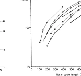
fibrillation than dogs with steep baroreflex slopes (resistant Fig. 1 shows the relation between the basic cycle length dogs). These results were interpreted as indicating that and the action potential duration or effective refractory strong vagal reflexes would protect an individual with an period in the ventricle (or in isolated trabeculae or isolated infarction against stress- and ischaemia-induced ventricular cells) in several species [157–161]. In contrast to all other fibrillation. Indeed, clinical studies [149,150] have con- species it may be appreciated that in the rat there is no firmed that baroreflex sensitivity is an important deter- shortening of refractory periods at the shorter cycle minant for sudden death and inducibility of ventricular lengths. Also, there are considerable differences in re-tachycardia in post-infarction patients. Still, both in ani- fractory periods between the dog and the pig, although
Fig. 1. Relation between (steady state) cycle length and (monophasic) Fig. 2. Relation between (steady state) cycle length and diastolic interval action potential duration or effective refractory period in several species (see inset for details). Data have been taken as indicated in the legend to (see inset for details). Data have been taken as follows: pig [159], dog Fig. 1.
[157], rat [158], man [161], rabbit [160], man / HF (heart failure) [160], man / HF (heart failure isolated cells (unpublished data, Veldkamp MW). ERP: effective refractory period; APD: action potential duration; MAPD:
monophasic action potential duration. Medline search for the number of studies performed on the
effects of anti-arrhythmic agents on action potential dura-tion in different species between 1966 and 1992, and between 1993 and 1996. The rabbit is often used for these species have comparable cardiac dimensions and in electrophysiological research, probably because it consti-vivo and in vitro cycle lengths. The discrepancy between tutes a reasonable compromise in terms of cardiac dimen-the monophasic action potential duration in dimen-the normal sions, basic electrophysiological characteristics and cost. human ventricle (Fig. 1: filled squares, solid line) and Therefore it has been chosen as the reference species for a action potential duration in ventricular trabeculae from comparison of the species chosen in this area of research in failing human hearts (Fig. 1: filled squares, dashed line) the past and in more recent years. Table 1 shows that the underscores the prolongation of action potential duration use of the pig and the guinea pig has increased during associated with heart failure. In the latter study [160] recent years and that the use of the rat is small compared patients were selected who were not on amiodarone, to other species which seems justified on the basis of data quinidine or sotalol, thereby excluding drug effects in in Figs. 1 and 2. Also, the use of human tissue has addition to the effect of heart failure itself. Action potential increased.
duration in single cells isolated from failing human hearts (Fig. 1: filled squares, fat line) is even further prolonged compared to trabeculae. It cannot be determined with
certainty whether this follows from the cell isolation Table 1
Studies on action potential duration and antiarrhythmic agents
procedure, but it certainly sets limits to the choice of a
relevant animal model. Fig. 2 shows the diastolic interval Species 1966–1992 Relative 1993–1996 Relative
along the ordinate (log scale used for better description of number number
the data at the more relevant shorter cycle lengths) versus Dog 123 1.9 21 1.2
the cycle length. Fig. 2 shows that rat ventricle is not the Pig 101 1.6 48 2.8
Cat 7 0.1 6 0.4
first choice if one aims at ‘filling up’ the diastolic interval
Rabbit 64 1.0 17 1.0
by means of a class I or class III or an experimental
Guinea pig 100 1.6 45 2.6
anti-arrhythmic agent. On the other hand, porcine
ventricu-Rat 19 0.3 6 0.4
lar myocardium appears to have a diastolic interval similar Man 22 0.3 22 1.3
to that in human ventricle. In line with these data it may be
Absolute and relative numbers of studies performed on the effect of
interesting to assess the number of studies performed in anti-arrhythmic agents on action potential duration in several species several species on the effects of anti-arrhythmic agents on between 1966 and 1992, and between 1993 and 1996, as included in
across an accessory atrioventricular connection in types A and B
4. Conclusions
preexcitation. Circulation 1970;41:375–397.
[11] Janse MJ, Anderson RH, McGuire MA, Ho SY. ‘AV nodal’ reentry:
It is clear that species differences do exist with respect part I. AV nodal reentry revisited. J Cardiovasc Electrophysiol to factors that determine arrhythmogenesis and it is also 1993;4:561–572.
[12] McGuire MA, Janse MJ, Ross DL. ‘AV nodal’ reentry: part II. AV
clear that no animal model will accurately mimic the
nodal, AV junctional, atrionodal reentry?. J Cardiovasc
Elec-human patient suffering from, or threatened by an
arrhyth-trophysiol 1993;4:573–586.
mia. [13] Coumel P, Cabrol C, Fabiato A, Gourgon R, Slama R. Tachycardie
Nevertheless, the knowledge gathered from animal permanente par rythme reciproque. Arch Mal Coeur 1967;60:1830–´ 1864.
studies undoubtedly has been instrumental in devising
[14] Ross DL, Johnson DC, Denniss R, Cooper MJ, Richards DA, Usher
diagnostic and therapeutic strategies both in
supraventricu-JB. Curative surgery for atrioventricular junctional (‘AV nodal’)
lar and ventricular arrhythmias. It is our conviction that in reentrant tachycardia. J Am Coll Cardiol 1985;6:1383–1392. the future, new knowledge will be obtained from experi- [15] Cox JL, Holman WL, Cain ME. Cryosurgical treatment of atrioven-ments performed at many levels: in systems expressing and tricular node reentrant tachycardia. Circulation 1987;76:1329–1376. [16] Sung RJ, Waxman HL, Saksena S, Juma Z. Sequence of retrograde
testing the functions of molecules involved in electrical
atrial activation in patients with dual atrioventricular nodal
path-excitation, in single cells, cell cultures, excised cardiac
ways. Circulation 1981;64:1059–1067.
preparations, isolated whole hearts, whole hearts in anaes- [17] Haissaguerre M, Warin JF, Lemetayer P, Saoudi N, Guillem JP, thetized animals, and in conscious animals. It will be the Blanchot P. Closed-chest ablation of retrograde conduction in patients with atrioventricular nodal reentrant tachycardia. N Engl J
combination of such investigations, rather than a single
Med 1989;320:426–433.
model or experimental technique, which will lead to novel
[18] Epstein LM, Scheinman MM, Langberg JL, Chilson D, Goldberg
strategies for diagnosis and treatment. Finally, electro- HR, Griffin JC. Percutaneous catheter modification of the atrioven-physiological studies should be encouraged in animals with tricular node. A potential cure for atrioventricular nodal reentrant
tachycardia. Circulation 1989;80:757–768.
‘naturally’ occurring cardiovascular disease [162,163].
[19] Mendez C, Moe GK. Demonstration of a dual AV nodal conduction system in the isolated rabbit heart. Circ Res 1966;19:378–393. [20] Janse MJ, van Capelle FJL, Freud GE, Durrer D. Circus movement
within the A–V node as a basis for supraventricular tachycardia as
Acknowledgements
shown by multiple microelectrode recording in the isolated rabbit heart. Circ Res 1971;28:403–414.
A.G.K. was supported by the Scientific Durrer Founda- [21] Wit AL, Goldreyer BN, Damato AN. An in vitro model of tion, Utrecht, Netherlands paroxysmal supraventricular tachycardia. Circulation 1971;43:862–
875.
[22] Janse MJ, Van Capelle FJL, Anderson RH, Touboul P, Billette J. Electrophysiology and structure of the atrioventricular node of the
References isolated rabbit heart. In: Wellens HJJ, Lie KI, Janse MJ editors. The
Conduction System of the Heart. Leiden: Stenfert Kroese, 1976, [1] Mines GR. On dynamic equilibrium in the heart. J Physiol 1988:296–315.
1913;46:349–383. [23] Jackman WM, Beckman KJ, McClelland JH, et al. Treatment of [2] Kent AFS. Observations on the auriculo-ventricular junction of the supraventricular tachycardia due to atrioventricular nodal reentry by mammalian heart. Quart J Exp Physiol 1913;7:193–195. radiofrequency catheter ablation of slow-pathway conduction. N [3] Mines GR. On circulation excitations in heart muscles and their Engl J Med 1992;327:313–318.
¨
possible relation to tachycardia and fibrillation. Trans Roy Soc of [24] Haissaguerre M, Gaıta F, Fisher B, et al. Elimination of atrioven-Canada 1914;section IV:42–53. tricular nodal reentrant tachycardia using discrete slow potentials to [4] Wolff L, Parkinson J, White PD. Bundle-branch block with short guide application of radiofrequency energy. Circulation
P-R interval in healthy young people prone to paroxysmal tachycar- 1992;85:2162–2175. ´
dia. Am Heart J 1930;5:685–704. [25] Sanjuan R, Morell S, Garcıa Civera R, et al. Transvenous ablation ¨
[5] Holzmann M, Scherf D. Ueber Elektrokardiogramme mit verkurzter with high frequency energy for atrioventricular junctional (AV Vorhof–Kammer-Distanz und positiven P-Zacken. Z Klin Med nodal) reentrant tachycardia. PACE 1989;12:1631–1639.
1932;121:404–423. [26] Iinuma HL, Dreifus LS, Mazgalev T, Price R, Michelson EL. Role [6] Durrer D, Roos JR. Epicardial excitation of the ventricles in a of the perinodal region in atrioventricular nodal reentry: evidence in patient with a Wolff–Parkinson–White syndrome (type B). Circula- an isolated rabbit heart preparation. J Am Coll Cardiol 1983;2:465–
tion 1967;35:15–21. 473.
[7] Burchell HB, Frye RB, Anderson MW, McGoon DC. Atrioventricu- [27] Mazgalev T, Dreifus LS, Bianchi J, Michelson EL. The mechanism lar and ventriculo-atrial excitation in Wolff–Parkinson–White of A–V junctional reentry: the role of the atrio-nodal junction. Anat syndrome (type B). Circulation 1967;36:663–672. Rec 1981;201:179–188.
[8] Durrer D, Schoo L, Schuilenburg RM, Wellens HJJ. The role of [28] Paes de Carvalho A. Cellular electrophysiology of the atrial premature beats in the initiation and termination of supraventricular specialized dissues. In: Peas de Carvalho A, de Mello WC, Hoffman tachycardia in the Wolff–Parkinson–White syndrome. Circulation BF, editors. The Specialized Tissues of the Heart. Amsterdam:
1967;36:644–662. Elsevier, 1961:113–115.
[9] Wellens HJJ. The electrophysiological properties of the accessory [29] Watanabe Y, Dreifus LS. Inhomogeneous conduction in the A–V pathway in the Wolff–Parkinson–White syndrome. In: Wellens HJJ, node. A model for reentry. Am Heart J 1965;70:505–514. Lie KI, Janse MJ, editors. The Conduction System of the Heart. [30] Moe GK, Cohen W, Vick RL. Experimentally induced paroxysmal Leiden: Stenfert Kroese, 1976, 1988:567–587. A–V nodal tachycardia in the dog. A ‘case report’. Am Heart J [10] Boineau JP, Moore EN. Evidence for propagation of activation 1963;65:87–92.
´
[31] Lin F-Y, Lo H-M, Cheng J-J. Experimentally created atrioventricular [54] Page P, Plumb VJ, Okumura K, Waldo AL. A new model of atrial node reentrant tachycardia in the dog: evidence of a brake system flutter. J Am Coll Cardiol 1986;8:872–879.
for nodal reentry in the anterior interatrial septum. J Am Coll [55] Allessie MA, Lammers WJEP, Bonke FIM, Hollen J. Intraatrial Cardiol 1993;22:1541–1547. reentry as a mechanism for atrial flutter induced by acetylcholine in [32] Denes P, Wu D, Dhingra RC, Chuquimia R, Rosen KM. Demonstra- rapid pacing in the dog. Circulation 1984;70:123–135.
tion of dual A–V nodal pathways in patients with paroxysmal [56] Mary-Rabine L, Mahaux V, Waleffe A, Kulbertus H. Atrial flutter: supraventricular tachycardia. Circulation 1973;48:549–555. historical background. J Cardiovasc Electrophysiol 1997;8:353–358. [33] Billette J. Atrioventricular nodal activation during periodic stimula- [57] Garrey WE. The nature of fibrillatory contraction of the heart: its tion of the atrium. Am J Physiol 1987;252:H163–H177. relation to tissue mass and form. Am J Physiol 1914;33:397–414. [34] Moe GK, Preston JB, Burlington H. Physiologic evidence for a dual [58] Garrey WE. Auricular fibrillation. Physiol Rev 1914;4:215–250.
A–V transmission system. Circ Res 1956;4:357–375. [59] Allessie MA, Lammers WJEP, Bonke FIM, Hollen J. Experimental [35] Simson MB, Spear J, Moore EN. The relationship between atrioven- evaluation of Moe’s multiple wavelet hypothesis of atrial fibrilla-tricular nodal refractoriness and the functional rerfactory period in tion. In: Zipes DP, Jalife J, editors. Cardiac Electrophysiology and the dog. Circ Res 1979;44:121–126. Arrhythmias. New York: Grune and Stratton, 1985:265–275. [36] Hoffman BF, Moore EN, Stuckey JH, Cranefield PF. Functional [60] Moe GK, Abildskov JA. Atrial fibrillation as a self-sustaining
properties of the atrioventricular conduction system. Circ Res mechanism independent of focal discharge. Am Heart J 1959;58:59–
1963;13:308–328. 70.
[37] Ferrier GR, Dresel PE. Relationship of the functional refractory [61] Moe GK. On the multiple wavelet hypothesis of atrial fibrillation. period to conduction in the atrioventricular node. Circ Res Arch Int Pharmacodyn Ther 1962;140:183–188.
1974;35:204–214. [62] Schuessler RB, Grayson TM, Bromberg BI, Cox JL, Boineau JP. [38] Loh P, de Bakker JMT. Unpublished observation (1997). Cholinergically mediated tachyarrhythmias induced by a single [39] Loh P, de Bakker JMT, Hocini M, Thibault B, Janse MJ. High extrastimulus in the isolated canine right atrium. Circ Res
resolution mapping and dissection of the triangle of Koch in canine 1992;71:1254–1276.
hearts: evidence for subatrial reentry during ventricular echoes. [63] Konings KTS, Kirchhof CJHJ, Smeets JRLM, Wellens HJJ, Penn PACE 1997;20:1080 (Abstract). OC, Allessie MA. High-density mapping of electrically induced [40] McGuire MA, Janse MJ. New insights on anatomical location of atrial fibrillation in humans. Circulation 1994;89:1665–1680.
components of the reentrant circuit and ablation therapy for at- [64] Cox JL, Canavan TE, Schuessler RB, Cain ME, Lindsay BD, Stone rioventricular junctional reentrant tachycardia. Curr Opin Cardiol C. The surgical treatment of atrial fibrillation II: Intraoperative
1995;10:3–8. electrophysiologic mapping and description of the
elec-[41] Josephson ME. Clinical Electrophysiology: Techniques and Inter- trophysiologic basis of atrial flutter and fibrillation. J Thorac pretation. Philadelphia: Lea and Febiger, 1993. Cardiovasc Surg 1991;101:406–426.
[42] Lewis T, Feil HS, Stroud WD. Observations upon flutter and [65] Wijffels MCEF, Kirchhof CJHJ, Dorland R, Allessie MA. Atrial fibrillation. Part II. The nature of auricular flutter. Heart fibrillation begets atrial fibrillation: a study in awake chronically
1920;7:191–245. instrumented goats. Circulation 1995;92:1954–1968.
´ ´
[43] Puech P. L’activite electrique circulaire normale et pathologique. [66] Attuel P, Leclercq JF, Coumel P. Atrial electrophysiological sub-Paris: Masson and Cie., 1956. strate remodeling after tachycardia in patients with and without [44] Cosio FG. Endocardial mapping of atrial flutter. In: Touboul P, atrial fibrillation. PACE 1995;18:804 pt. II.
Waldo AL, editors. Atrial Arrhythmias. St. Louis: Mosby Year [67] Murgatroyd FD. In: AJ Camm, editor. Nonpharmacological Man-Book, 1990:229–240. agement of Atrial Fibrillation. Amonk NY: Futura Publishing, 1997. [45] Hoffman BF. Experimental models of atrial flutter. in: Touboul P, [68] Daoud EG, Bogun F, Goyal R, et al. Effect of atrial fibrillation on
Waldo AL, editors. Atrial Arrhythmias. St. Louis: Mosby Year atrial refractoriness in humans. Circulation 1996;94:1600–1606. Book, 1990:183–189. [69] Daoud EG, Knight BP, Weiss R, et al. Effect of verapamil and
´
[46] Rosenblueth A, Garcıa Ramos J. Studies on flutter and fibrillation II. procainamide on atrial fibrillation-induced electrical remodeling in The influence of anatomical obstacles on experimental auricular humans. Circulation 1997;96:1542–1550.
flutter. Am Heart J 1947;33:677–684. [70] Goette A, Honeycut C, Langberg JJ. Electrical remodeling in atrial [47] Kimura E, Kato K, Murao S, Ajisaka H, Koyama S, Omiya Z. fibrillation. Time course and mechanisms. Circulation
Experimental studies on the mechanism of auricular flutter. Tohoku 1996;94:2968–2974. ´
J Exp Med 1954;60:197–207. [71] Coumel P, Attuel P, Lavallee JP, Flammang D, Leclercg JF, Slama [48] Boineau JP, Schuessler RB, Mooney CR, et al. Natural and evoked R. Syndrome d’arythmie auriculaire d’origine vagale. Arch Mal
atrial flutter due to circus movement in dogs. Am J Cardiol Coeur 1978;71:645–656.
1980;45:1167–1181. [72] Murgatroyd FD, Camm AL. Atrial Arrhythmia. Lancet
[49] Feld GF, Shahandeh-Rad F. Mechanisms of double potentials 1993;341:1317–1322.
recorded during sustained atrial flutter in the canine right atrial [73] Ravelli F, Allessie MA. Effects of atrial dilation on refractory period crush-injury model. Circulation 1992;86:628–641. and vulnerability to atrial fibrillation in the isolated Langendorff-[50] Inoue H, Matsuo H, Takayanagi K, Murao S. Clinical and ex- perfused rabbit heart. Circulation 1997;96:1689–1695.
perimental studies of the effects of extrastimulation and rapid pacing [74] Nazir SA, Lab MJ. Mechanoelectric feedback and atrial arrhythmias. on atrial flutter: evidence of macro reentry with an excitable gap. Cardiovasc Res 1996;32:52–61.
Am J Cardiol 1981;48:623–631. [75] Boyden PA, Tilley LP, Pham TD, Liu SK, Fenoglio Jr. JJ, Wit AL. [51] Frame LH, Page RL, Hoffman BF. Atrial reentry around an Effects of left atrial enlargement on atrial transmembrane potentials anatomic barrier with a partially refractory excitable gap. A canine and structure in dogs with mitral valve fibrosis. Am J Cardiol model of atrial flutter. Circ Res 1986;58:495–511. 1982;49:1896–1908.
[52] Waldo AL. Mechanisms of atrial fibrillation, atrial flutter, and [76] Hordof AJ, Edie R, Malm JR, Hoffman BF, Rosen MR. Elec-ectopic atrial tachycardia. A brief review. Circulation trophysiologic properties and response to pharmacologic agents of 1987;75:III37–III40. Suppl. III. fibers from diseased human atria. Circulation 1976;54:774–779. [53] Boyden PA, Hoffman BF. The effects on atrial electrophysiology [77] Ten Eick RA, Singer DH. Electrophysiologic properties of diseased
and structure of surgically induced right atrial enlargement in dogs. human atrium I. Low diastolic potential and altered cellular response Circ Res 1981;49:1319–1331. to potassium. Circ Res 1979;44:545–557.
[78] Le Heuzey JY, Boutjdir M, Gagey S, Lavergne T, Guize L. Cellular [97] Jenkins MG, Johnson TA, Engle C, Gettes LS. Metabolic protection aspects of atrial vulnerability. In: Attuel P, Coumel P, Janse MJ, by verapamil during graded coronary flow reduction independent of editors. The Atrium in Health and Disease. Mt Kisco NY: Futura effect on baseline systolic function. Separation of mechanical and Publishing, 1989:81–94. ionic markers of ischaemia. Circulation 1989;80:1870–1877. [79] Spach MS, Dolber PC. Relating extracellular potentials and their [98] Kaplinsky E, Ogawa S, Balke CW, Dreifus LS. Two periods of early
derivatives to anisotropic propagation at a microscopic level in ventricular arrhythmias in the canine acute myocardial infarction human cardiac muscle: evidence for electrical uncoupling of side-to- model. Circulation 1979;60:397–403.
side connections with increasing age. Circ Res 1986;58:356–371. [99] Hill JL, Gettes LS. Effects of acute coronary artery occlusion on 1
¨
[80] Moıse NS, Gilmour Jr. RF, Riccio ML. An animal model of local myocardial extracellular K activity in swine. Circulation spontaneous arrhythmic death. J Cardiovasc Electrophysiol 1980;61:768–778.
1997;8:98–103. [100] Wilde AAM, Asknes G. Myocardial potassium loss and cell ¨
[81] Gilmour Jr. RF, Moıse NS. Triggered activity as a mechanism for depolarisation in ischaemia and hypoxia. Cardiovasc Res inherited ventricular arrhythmias in German shepherd dogs. J Am 1995;29:1–15.
Coll Cardiol 1996;27:1526–1533. [101] Downar E, Janse MJ, Durrer D. The effect of acute coronary artery ¨
[82] Freeman LC, Pacioretty LM, Moıse NS, Kass RS, Gilmour Jr. RF. occlusion on subepicardial transmembrane potentials in the intact Decreased density of Ito in left ventricular myocytes from German porcine heart. Circulation 1977;56:217–224.
shepherd dogs with inherited arrhythmias. J Cardiovasc Elec- [102] Kleber AG, Janse MJ, van Capelle FJL, Durrer D. Mechanism and´ trophysiol 1997;8:872–883. time course of S–T and T–Q segment changes during acute
¨
[83] Doe M, Ursell P, Lee RJ, Stilson C, Chin M, Moıse NS. Heteroge- regional myocardial ischaemia in the pig heart determined by neous sympathetic innervation in German shepherd dogs with extracellular and intracellular recordings. Circ Res 1978;42:603– inherited ventricular arrhythmias and sudden death. J Am Coll 613.
Cardiol 1995;25:20A Abstract. [103] Shaw RM, Rudy Y. Electrophysiologic effects of acute myocardial [84] Schwartz PJ, Locati EH, Napolitano C, Priori, S. The long QT ischaemia. A mechanistic investigation of action potential
conduc-syndrome. In: Zipes DP, Jalife J, editors. Cardiac Electrophysiology: tion and conduction failure. Circ Res 1997;80:124–138. From Cell to Bedside. Philadelphia: Saunders, 1995:788–811. [104] Kleber AG, Janse MJ, Wilms-Schopman FJG, Wilde AAM,´ [85] Kass RS, Davies MP. The roles of ion channels in an inherited heart Coronel R. Changes in conduction velocity during acute ischaemia disease: molecular genetics of the long QT syndrome. Cardiovasc in ventricular myocardium of the isolated porcine heart. Circ Res
Res 1996;32:433–454. 1986;73:189–198.
[86] Leenhardt A, Glaser E, Burguera M, Nurnberg M, Maison-Blanch P, [105] Smith WT, Fleet WF, Johnson TA, Engle CL, Cascio WE. The Ib Coumel P. Short-coupled variant of torsade de pointes: a new phase of ventricular arrhythmias in ischaemic in situ porcine heart electrocardiographic entity in the spectrum of ventricular tach- is related to changes in cell-to-cell electrical coupling. Circulation yarrhythmias. Circulation 1994;89:206–215. 1995;92:3051–3060.
[87] Eisenberg SJ, Scheinman MM, Dullet N. Polymorphous ventricular [106] Rudy Y, Quan W. A model study of the effects of the discrete tachycardia in patients with normal cardiac function and QT cellular structure on electrical propagation in cardiac tissue. Circ interval. Am J Cardiol 1995;75:687–692. Res 1987;61:815–823.
´
[88] Janse MJ, Wit AL. Electrophysiological mechanisms of ventricular [107] Janse MJ, Kleber AG. Electrophysiological changes and ventricular arrhythmias resulting from myocardial ischaemia and infarction. arrhythmias in the early phase of regional myocardial ischaemia. Physiol Rev 1989;69:1049–1169. Circ Res 1981;49:1069–1081.
[89] Wit AL, Janse MJ. The ventricular arrhythmias of ischaemia and [108] Coronel R, Fiolet JWT, Wilms-Schopman FJG. Distribution of infarction. Electrophysiological mechanisms. Mount Kisco NY: extracellular potassium and its relation to electrophysiological Futura Publishing, 1993. changes during acute myocardial ischaemia in the isolated perfused [90] Janse MJ, Van Capelle FJL, Morsink H, et al. Flow of ‘injury’ porcine heart. Circulation 1988;77:1125–1138.
current and patterns of excitation during early ventricular arrhyth- [109] Coronel R, Fiolet JWT, Wilms-Schopman FJG, Opthof T, mias in acute regional myocardial ischaemia in isolated porcine and Schaapherder AFM, Janse MJ. Distribution of extracellular potas-canine hearts. Evidence for 2 different arrhythmogenic mechanisms. sium and electrophysiologic changes during two-stage coronary Circ Res 1980;47:151–165. ligation in the isolated, perfused canine heart. Circulation [91] Gray RA, Jalife J, Panfilov A, et al. Nonstationary vortexlike 1989;80:165–177.
reentrant activity as a mechanism of polymorphic ventricular [110] Coronel R. Distribution of extracellular potassium during myocar-tachycardia in the isolated rabbit heart. Circulation 1995;91:2454– dial ischaemia. Thesis, University of Amsterdam. Dordrecht: ICG
2459. Printing, 1988; p. 133.
´
[92] Myerburg RJ, Kessler KM, Castellanos A. Sudden cardiac death. [111] Cascio WE, Yan G-X, Kleber AG. Early changes in extracellular Structure, function, and time-dependence of risk. Circulation potassium in ischaemic rabbit myocardium. The role of extracellu-1992;85:2–10. Suppl. lar carbon dioxide accumulation and diffusion. Circ Res [93] Maseri A, Severi S, Marzullo P. Role of coronary arterial spasm in 1992;70:409–422.
sudden coronary ischaemic death. Ann NY Acad Sci 1982;382:204– [112] Case RB, Felix A, Castellana FS. Rate of rise of myocardial PCO2
217. during early myocardial ischemia in the dog. Circ Res
[94] Vermeulen JT, Tan HL, Rademaker H, et al. Electrophysiologic and 1979;45:324–330.
extracellular ionic changes during acute ischaemia in failing and [113] Wilde AAM, Escande D, Schumacher CA, et al. Potassium normal rabbit myocardium. J Mol Cell Cardiol 1996;28:123–131. accumulation in the globally ischaemic mammalian heart. Circ Res [95] Winterton SJ, Turner MA, O’Gorman DJ, Flores NA, Sheridan DJ. 1990;67:835–843.
´
Hypertrophy causes delayed conduction in human and guinea pig [114] Cascio WE, Yan G-X, Kleber AG. Passive electrical properties, myocardium: accentuation during ischaemic perfusion. Cardiovasc mechanical activity, and extracellular potasium in arterially per-Res 1994;28:47–54. fused and ischaemic rabbit ventricular muscle. Effect of calcium [96] Watanabe I, Johnson TA, Buchanan J, Engle CL, Gettes LS. Effect entry blockade or hypocalcemia. Circ Res 1990;66:1461–1473.
of graded coronary flow reduction on ionic, electrical, and me- [115] Shattock MJ, Bers DM. Rat vs. rabbit ventricle: Ca flux and chanical indexes of ischaemia in the pig. Circulation 1987;76:1127– intracellular Na assessed by ion-selective microelectrodes. Am J
[116] Baxter GF, Heads RJ, Yellon DM. Oxidative stress and the second tachycardia and fibrillation by drugs and antitachycardia pacing. In: Allessie MA, Fromer M, editors. Atrial and Ventricular Fibrilla-window of protection after preconditioning. Circulation
tion: Mechanisms and Device Therapy. Armonk NY: Futura 1996;94:2992–2993. letter.
Publishing, 1997:145–175. [117] Speechly DM, Mocanu MM, Yellon DM. Protein kinase C. Its role
[136] Borggrefe M, Chen X, Hindricks G, et al. Catheter ablation of in ischemic preconditioning in the rat. Circ Res 1994;75:586–590.
ventricular tachycardia in patients with coronary heart disease. In: [118] Boyden PA, Jeck CD. Ion channel function in disease. Cardiovasc
Zipes DP, Jalife J, editors. Cardiac Electrophysiology: From Cell to Res 1995;29:312–3188.
Bedside. Philadelphia: WB Saunders, 1995:1502–1517. [119] Severs NJ. Gap junction alterations in the failing heart. Eur Heart J
[137] Lawrie GM, Pacifico A. Surgery for ventricular tachycardia. In: 1994;15:53–57. Suppl.
Zipes DP, Jalife J, editors. Cardiac Electrophysiology: From Cell to [120] Peters NS. New insights into myocardial arrhythmogenesis:
dis-Bedside. Philadelphia: WB Saunders, 1995:1547–1552. tribution of gap-junctional coupling in normal, ischaemic and
[138] Wilber DJ, Lynch JJ, Lucchesi BR. Postinfarction sudden death: hypertrophied human hearts. Clin Sci (Colch) 1996;90:447–452.
significance at inducible ventricular tachycardia and infarct size in [121] Spach MS, Boineau JP. Microfibrosis produces electrical load
a conscious canine model. Am Heart J 1985;109:8–18. variations due to loss of side-to-side cell connections: a major
[139] Gardner PI, Ursell PC, Pham TD, Fenoglio Jr JJ, Wit AL. mechanism of structural heart disease arrhythmias. PACE
Experimental chronic ventricular tachycardia: anatomic and elec-1997;20:397–413.
trophysiologic substrates. In: Josephson ME, Wellens HJJ, editors. [122] Endo T, Ribeiro LGT, Cheung WM, Faria DB, Petranto M, Tachycardias: Mechanisms, Diagnosis, Treatment. Philadelphia:
Maroko PR. Relationship between the extent of the hypoperfused Lea and Febiger, 1984:29–60.
zone of the myocardium and the occurrence of ventricular fibrilla- [140] Euler DE, Prood CE, Spear JF, Moore EN. The interruption of tion. Am Heart J 1983;105:915–920. collateral blood flow to the ischaemic canine myocardium by [123] Austin M, Wenger TL, Harrell Jr. FE, Luzzi FA, Strauss HA. embolization of a coronary artery with latex. Effects on conduction
Effect of myocardium at risk on outcome after coronary artery delay and arrhythmias. Circ Res 1981;49:97–108.
occlusion and release. Am J Physiol 1982;243:H340–H345. [141] Wetstein L, Mark R, Kaplinsky E, et al. Histopathologic factors [124] Bolli R, Fisher DJ, Entman ML. Factors that determine the conducive to experimental ventricular tachycardia. Surgery
occurrence of arrhythmias during acute myocardial ischaemia. Am 1985;98:532–538.
Heart J 1986;111:261–270. [142] De Bakker JMT, Coronel R, Tasseron S, et al. Ventricular tachycar-[125] Rosen MR, Janse MJ, Myerburg RJ. Arrhythmias induced by dia in the infarcted, Langendorff-perfused human heart: role of the coronary artery occlusion: What are the electrophysiological arrangement of surviving cardiac fibers. J Am Coll Cardiol mechanisms? In: Hearse DJ, Manning AC, Janse MJ, editors. 1990;15:1594–1607.
Life-threatening Arrhythmias during Ischaemia and Infarction. [143] Zipes DP. Influence of myocardial ischaemia and infarction on New York: Raven Press, 1987:11–47. autonomic innervation of the heart. Circulation 1990;82:1095– [126] Meesmann W. Early arrhythmias and primary ventricular fibrilla- 1105.
tion after acute myocardial ischaemia in relation to preexisting [144] Gaide MS, Myerburg RJ, Kozlovskis PL, Bassett AL. Elevated coronary collaterals. In: Parratt JR, editor. Early Arrhythmias sympathetic response of epicardium proximal to healed myocardial Resulting from Myocardial Ischaemia. London: Macmillan Press, infarction. Am J Physiol 1983;245:H646–H652.
1982:93–112. [145] Herre JM, Wetstein L, Lin Y-L, Mills AS, Dae M, Thames MD. [127] Meesmann W, Schulz FW, Schley G, Adolphsen R. Ueberleben- Effect of transmural versus nontransmural myocardial infarction on squote nach akutem experimentellen Koronarverschlusz in inducibility of ventricular arrhythmias during sympathetic
stimula-¨
Abhangigkeit von Spontankollateralen des Herzens. Z ges exp Med tion in dogs. J Am Coll Cardiol 1988;11:414–421.
1970;153:246–264. [146] Billman GE, Schwartz PJ, Stone HL. Baroreceptor reflex control of [128] Schaper W. Experimental infarcts and the microcirculation. In: heart rate: a predictor of sudden death. Circulation 1982;66:874–
Hearse DJ, Yellon DM, editors. Therapeutic Approaches to 880.
Myocardial Infarct Size Limitation. New York: Raven Press, [147] Schwartz PJ, Billman GE, Stone HL. Autonomic mechanisms in 1984:79–90. ventricular fibrillation induced by myocardial ischaemia during [129] Trolese-Mongheal Y, Duchene-Marullaz P, Trolese J-F, Leinot M, exercise in dogs with healed myocardial infarction: An
experimen-Lamar J-C, Lacroix P. Sudden death and experimental acute tal model for sudden death. Circulation 1984;69:790–800. myocardial infarction. Am J Cardiol 1985;56:677–681. [148] Schwartz PJ, Vanoli E, Stramba-Badiale M, De Ferrari G, Billman [130] Hunt GB, Ross DL. Influence of infarct age on reproducibility of GE, Foreman RD. Autonomic mechanisms and sudden death. New ventricular tachycardia induction in a canine model. J Am Coll insights from analysis of baroreceptor reflexes in conscious dogs Cardiol 1989;14:765–773. with and without a myocardial infarction. Circulation [131] Karagueuzian HS, Fenoglio Jr. JJ, Weiss MB, Wit AL. Protracted 1988;78:969–979.
ventricular tachycardia induced by premature stimulation of the [149] La Rovere MT, Specchia G, Mortara A, Schwartz PJ. Baroreflex canine heart after coronary artery occlusion and reperfusion. Circ sensitivity, clinical correlates, and cardiovascular mortality among Res 1979;44:833–846. patients with a first myocardial infarction. A prospective study.
¨
[132] Kuck K-H, Costard A, Schluter M, Kunze K-P. Significance of Circulation 1988;78:816–824.
timing programmed electrical stimulation after acute myocardial [150] Farrell TG, Paul V, Cripps PV, et al. Baroreflex sensitivity and infarction. J Am Coll Cardiol 1986;8:1279–1288. electrophysiological correlates in patients after acute myocardial [133] Breithardt G, Borggrefe M, Haesten K. Role of programmed infarction. Circulation 1991;83:945–952.
ventricular stimulation and noninvasive recording of ventricular [151] Legato MJ. The anatomic matrix as a factor in susceptibility to late potentials for the identification of patients at risk of ventricular lethal arrhythmias in a canine model of sudden death. J Mol Cell tachyarrhythmias after acute myocardial infarction. In: Zipes DP, Cardiol 1993;25:501–508.
Jalife J, editors. Cardiac Electrophysiology and Arrhythmias. [152] Pressler ML. Passive electrical properties of cardiac tissue. In: Orlando: Grune and Stratton, 1985:553–561. Zipes DP, Jalife J, editors. Cardiac Electrophysiology: From Cell to
´
[134] Roy DE, Marchand E, Theroux P, Waters DD, Pelletier GB, Bedside. Philadelphia: WB Saunders, 1990:108–122.
Bourassa MG. Programmed ventricular stimulation in survivors of [153] Pressler ML. Membrane properties of the cardiac conduction an acute myocardial infarction. Circulation 1985;72:487–494. system: comparative aspects. Proc Kon Ned Akad v Wet [135] Shenassa M, Shenassa H. Prevention and termination of ventricular 1990;93:477–487.