Traumatic Hemipelvectomy
Ludwig Labler, Otmar Trentz, Marius Keel
1Ab stract
Purpose: Open or closed traumatic hemipelvectomy is defined as a uni- or bilateral avulsion of the bony hemi-pelvis in combination with rupture of the large pelvic nerves and vessels and is usually accompanied by inju-ries of the genitourinary tract and bowel. According to a literature review between 1960 and 2005, 96 cases of traumatic hemipelvectomy were documented.
Patients and Methods: Between 1998 and 2004, nine male patients fulfilled the criteria for a traumatic hemi-pelvectomy, out of 1.8% pelvic injuries (n = 507) and of 2.4% pelvic ring injuries (n = 373). Seven patients were admitted directly to the authors’ trauma center, one patient was admitted 3 h after the accident, and one patient was stabilized in another hospital and trans-ferred 5 days later.
Results: All seven patients admitted primarily after trauma and the patient transferred 3 h later were in shock class IV. The traumatic hemipelvectomy was uni-lateral in eight patients with one complete avulsion and bilateral in one patient. Injuries of the pelvic ves-sels occurred in all patients. Most of the patients had injuries of both the genitourinary tract and the intes-tine. Associated injuries were mostly those of extremi-ties, thorax and head. Neurologic deficits could be doc-umented clinically on admission in four patients. Laparotomy for damage control with packing of the ab-dominal cavity and the retroperitoneum was per-formed in all cases. Four patients died during stabiliza-tion attempts in hemorrhagic shock during the first 4 h of treatment and three patients died after 3, 5, and 7 days in the intensive care unit because of septic compli-cations. Two patients survived with a follow-up of 21 and 34 months.
Conclusion: Traumatic hemipelvectomy is a most se-vere pelvic ring injury. If the diagnosis of traumatic hemipelvectomy is clear, surgical hemipelvectomy should be performed. Limb-saving procedures
endan-ger patient’s life. Early and frequent second-look opera-tions and aggressive management of associated pelvic injuries minimize wound problems and septic compli-cations.
Key Words
Pelvic fractures · Polytrauma · Infection of bone and joints · General trauma · Fractures · Abdominal trauma
Eur J Trau ma 2005;31:543–50 DOI 10.1007/s00068-005-2135-4
Introduction
Traumatic hemipelvectomy is the most severe form of closed or open pelvic fractures caused by a high-energy impact on the hemipelvis and results in a uni- or bilat-eral wide bony separation of the pelvic ring at the sym-physis and the sacrum combined with a traumatic rup-ture of iliac vessels. In addition, a severe stretch injury or a disruption of the femoral and sciatic nerves takes place [1–3]. The traumatic hemipelvectomy results ei-ther in a complete avulsion or an incomplete disruption with closed or severely damaged soft tissue and is often associated with injuries of the genitourinary tract and intestine as well as severe soft-tissue disruption of the pelvic diaphragm. The injury occurs mostly after motor-cycle or bimotor-cycle traffic accidents and a rollover or high-velocity abduction movements are the cause of a disruption of the pelvic ring. Some cases of this severe trauma were due to accidents at work and one reported case of traumatic hemipelvectomy was a result of a blast injury [4]. The mortality rate is very high and the rare cases of surviving patients appeared sporadically in the literature, mostly as case reports, in the course of the recent 45 years. The aim of this retrospective study was to discuss the management and to analyze the course of 1 Division of Trauma Surgery, Department of Surgery, University
Hospital Zurich, Switzerland.
traumatic hemipelvectomy in patients treated at our in-stitution in the last 6 years.
Patients and Methods
Out of a total of 507 pelvic injuries treated at the De-partment of Trauma Surgery, University Hospital Zu-rich, Switzerland, between 1998 and 2004, 373 were pel-vic ring injuries and nine of them fulfilled the criteria of traumatic hemipelvectomy (Figures 1a and 1b) with an incidence of 2.4%. All patients were males of an
aver-age aver-age of 30 ± 6 years (range 13–75 years). The causes of the injury were six traffic accidents and three acci-dents at work. Seven patients were transferred primarily to our department by a helicopter (#1, 3, 4, 5, 6, 8, 9). The remaining two patients were transferred from other hospitals as soon as they were stable for further trans-port, one patient 3 h after the accident (#7) and the oth-er aftoth-er 5 days (#2). Except one complete avulsion in patient #4 (Figures 2a to 2d), the traumatic hemipelvec-tomy was incomplete in the remaining patients,
com-Figures 1a to 1d. Patient #8.
a) X-ray of open traumatic hemipelvectomy. The plane of the hemipelvectomy runs through the symphysis pubis and the sacroiliac joint on the right side. Notice the bony lesion of the acetabulum of the contralateral hemipelvis.
b) Patient on arrival in the operating room with incomplete traumatic hemipelvectomy. Complete transection of the vascular and neurogenic structures (clamp positioned on external iliac artery). Complete disrupture of the anorectum as an associated injury.
c) Temporary closure of the soft-tissue defect after surgical hemipelvectomy. Open abdomen situation after damage control procedure. d) Complete necrosis of gluteus muscle and the associated soft tissue 6 days after traumatic hemipelvectomy. Open abdomen closed by Ab-dominal V.A.C.™ Dressing. The patient died 1 day later due to septic complication in multiple organ failure.
a b
bined with a wide selection of soft-tissue injuries from closed skin situation up to deep perineal defects. Except one patient with a bilateral hemipelvectomy (#2), six patients named at last had a unilateral hemipelvectomy on the left side and one on the right side (#8). Injury Severity Score (ISS) [5], associated injuries to the pelvic ring injury, and associated extrapelvic lesions are sum-marized in Tables 1 and 2. The following parameters were evaluated: Revised Trauma Score (RTS), the probability of survival (TRISS), shock classification, time management, emergency operation procedures, transfusion analysis, intensive care unit (ICU) stay and the outcome.
Results
Seven patients (#1, 3, 4, 5, 6, 8, 9) admitted primarily after trauma and the patient transferred after 3 h (#7) were in shock class IV. The mean pre-hospital rescue time by a helicopter amounted to 70 ± 4 min (range
55–85 min). The mean ISS of the patients was 49 ± 3 points (range 41–66 points) and the mean RTS at admit-tance 3.269 ± 0.257 (range 2.628–4.094). On arrival, the TRISS for blunt trauma was 16.2 ± 5% (range 3–42.2%). After arrival, the patients were managed according to Advanced Trauma Life Support Guidelines [6]. Initial diagnostic steps on arrival included solely thoracic and pelvic X-ray and FAST (focused assessed sonography in trauma). Injuries of the genitourinary tract and colorec-tal injuries were found in most of the cases (Table 1). Neurologic deficits could be documented clinically on admittance in four patients only. Injuries of the pelvic vessels were present in all patients. Severe soft-tissue injuries such as Morel-Lavallée lesion [7] were docu-mented in four patients (#1, 7, 8, 9). Most of the associ-ated extrapelvic lesions included thorax, abdomen and head (Table 2). The mean time from admittance to emergency surgery was 33 ± 6 min (range 5–60 min) and the mean operating time 156 ± 27 min (range 55–240
Figures 2a to 2d. Patient #4.
a) X-ray on arrival. Com-plete avulsion of the left hemipelvis.
b) Avulsed extremity with hemipelvis on arrival. c) Soft-tissue injury of the complete traumatic hemi-pelvectomy. Clamps are positioned on iliac vessels in the operating room. d) X-ray of complete trau-matic hemipelvectomy on the left side with associ-ated multifragmented fe-mur fracture.
a
b
c
min). In all cases a laparotomy with packing of the abdominal cavity and the retroperitoneum was carried out. A pelvic clamp was used for bleeding control in five patients. Aortic clamping was performed dur-ing six damage control procedures. Damage control [8] of the colon and colostomies were performed in six and of small bowel injury in two pa-tients. Suture repair of the urinary bladder was carried out in five cases. On average, 45 ± 10 blood units, 29 ± 7 fresh frozen plasmas and 6,400 ±
2,500 ml Ringer’s infusion were necessary for transfu-sions during the emergency procedures.
Four of the patients admitted primarily died during attempts at stabilization as a result of hemorrhagic shock in the course of the first 4 h of treatment (#1, 3, 5, 6). At admittance we operated one surviving patient with complete traumatic hemipelvectomy (#4). In the group of the surviving patients with incomplete trau-matic hemipelvectomy, a surgical hemipelvectomy was performed initially (#9) and one 8 h after stabilization on the first day of admittance (#8). In two patients, bilat-eral surgical hemipelvectomies were performed on the 5th and 6th day after trauma, in one case (#2) because of completely missing perfusion of both lower limbs and in the other (#7) because of a septic complication and a severe soft-tissue defect (Morel-Lavallée). Frequent and repeated aggressive debridements of the hemipel-vectomy wounds were performed. Three patients (#2, 8, 9) had large septic wounds and died after 3, 5, and 7 days in the ICU because of septic-toxic shock and/or multiple organ failure. All three patients, in addition to the direct mechanical injury of the structures, also had an ischemic injury caused by a ligation of the internal or common artery and vein. A large necrosis of the gluteus maximus muscle, bladder and pelvic musculature was observed in these cases during second-look procedures (Figures 1c and 1d). Two patients (#4, 7) could be stabilized and un-derwent ten and 30 second-look operations, respective-ly, in order to achieve a complete debridement of the injured and necrotic tissue. In one case (#4), the soft-tis-sue defects were covered with a free flap. Both patients survived their catastrophic pelvic ring injury and were available for a follow-up after 21 and 34 months. The now 15-year-old boy (#7) is visiting school again and the 25-year-old man (#4) has a job and is working full time.
Both survivors refused prosthetic rehabilitation. The boy is mobilized in a wheelchair and the young man is ambulatory on crutches. Early psychological support of the patients and their families was necessary for social reintegration.
Discussion
Traumatic hemipelvectomy is caused by an extremely high kinetic energy force applied to a partially abducted and externally rotated lower extremity [1, 9]. The injury results predominantly in motor vehicle accidents and mostly the left side is entangled typically involving mo-torcycle or bicycle riders colliding with an oncoming car or an object in right-hand traffic [10]. When the impact occurs, the pubic symphysis of the involved extremity is disrupted and as the deformation continues, disruption of the remaining pelvic supporting structures continues posterior to and through the sacroiliac joint. In 71%, the plane of the hemipelvectomy runs through the
symphy-Table 1. Associated abdominal injuries after traumatic hemipelvectomy. ISS: Injury Severity Score [5].
Patient # Age (years) ISS Bladder Urethra Perineum Anus Rectum Colon
1 29 57 + + 2 22 50 + + 3 75 41 + + + 4 22 41 + + + 5 33 50 + + + + + + 6 24 41 + + 7 13 45 + + + + 8 18 66 + + + + 9 33 50 + + + +
Table 2. Injury severity of associated extrapelvic lesions. AIS: Abbrevi-ated Injury Scale [39].
Patient # AIS head AIS thorax AIS abdomena
1 2 4 4 2 5 3 3 4 5 3 4 6 4 7 8 5 9
a all injuries in the abdominal cavity except associated injuries of traumatic
sis pubis and the sacroiliac joint [11]. This tremendous shearing force and displacement of the affected hemi-pelvis result in an extensive soft-tissue destruction in-cluding avulsion of iliac vessels, genitourinary and colorectal injuries [10, 12–14]. Direct avulsion of the limb after entanglement in heavy machinery is the next most common mechanism of injury [3, 15]. Complete avulsion of the hindquarter amounts to about 50% of cases [10, 16]. Other author describes an incidence of 32%, whereas 68% of the limbs remained connected by means of a soft-tissue bridge [11].
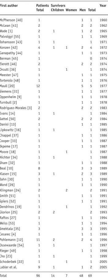
The incidence for traumatic hemipelvectomy is about 0.6% of all pelvic fractures and about 4.9% of complex pelvic fractures [12]. In our own patient group we found an incidence of 1.8% and 2.4%, respectively. No more than 96 documented traumatic hemipelvecto-mies were reported in the literature in the last 45 years (Table 3). The actual incidence, however, remains un-known because most of the victims, as a consequence of an uncontrollable hemorrhage, die before reaching the hospital [17–19]. Nevertheless, the number of patients arriving at the hospital alive has increased during the recent 20 years (Table 3), presumably as a result of im-proved pre-hospital aggressive resuscitation, a shorter transport time by helicopter to the trauma centers and an improved shock treatment. This correlates well with our own patient group where the time for the rescue and diagnostic procedures after arrival in the hospital was relatively short. Consequently, the surgeon is, and will be in the future, more often confronted with this kind of injury than in the past. However, Table 3 shows that the rate of successful management of these surviving pa-tients still remains unknown. Except few studies of groups of patients published [10, 12, 20], including our own study (Table 3), their number being too small to be statistically relevant, the majority of publications are case reports describing only successful management of selected cases of traumatic hemipelvectomies leaving the rate of survivors who died later in the course of treatment unnoticed.
Most of the cases described, as well as our own pa-tient group, showed a severe shock at arrival [10, 12, 15, 16, 21]. The survivors, almost < 30 years, were healthy individuals able to tolerate massive hemorrhage and soft-tissue destruction [16, 22, 23]. Patients with com-plete hemipelvectomy seem to have a better chance of survival than patients with partial hemipelvectomy. This supposedly is due to a retraction of the vessels, which takes place after complete transection and allows a
clo-Table 3. Traumatic hemipelvectomies in the literature.
First author Patients Survivors Year
Total Children Women Men Total
McPherson [40] 1 1 1 1960 McLean [41] 2 2 2 1962 Wade [1] 2 1 1 2 1965 Palvolgyi [55] 1 1 1 1969 Johansson [43] 1 1 1 1971 Konzen [42] 4 1 1 2 4 1972 Ganapathy [44] 1 1 1 1973 Berman [45] 1 0 1974 Zierott [46] 2 2 2 1974 Orcutt [36] 1 1 1 1974 Meester [47] 1 1 1 1975 Torterolo [48] 1 1 1 1976 Maull [20] 12 5 5 1977 Siemens [31] 1 1 1 1977 Oppenheim [9] 1 1 1 1978 Turnbull [2] 1 1 1 1978 Rodriguez-Morales [3] 2 2 2 1983 Evans [14] 1 1 1 1984 Sattel [50] 2 2 2 1984 Danisi [13] 1 1 1 1985 Lipkowitz [16] 1 1 1 1985 Chappel [37] 1 1 1 1986 Cooper [33] 1 1 1 1987 Ikpeme [17] 1 1 1 1987 Moore [18] 1 1 1 1987 Nichter [34] 1 1 1 1988 Sham [32] 1 1 1 1988 Beal [10] 8 3 3 1989 Klasen [15] 3 1 2 3 1989 Jahn [30] 1 1 1 1990 Wand [26] 1 1 1 1990 Klingman [24] 2 2 2 1991 Smith [51] 1 1 1 1991 Spiers [52] 1 1 1 1991 Dendrinos [19] 1 1 1 1992 Glorion [25] 2 2 2 1993 Raftos [27] 1 1 1 1994 Weiss [53] 1 1 1 1994 Smektala [35] 3 3 3 1995 Cesarec [4] 1 1 1 1996 Pohlemann [12] 11 2 2 4 1996 Ossewaarde [54] 1 1 1 1997 Rieger [49] 1 1 1 1998 Cho [23] 1 1 1 1999 Schoderbek [22] 1 1 1 2005 Labler et al. 9 1 1 2 2005 Total 96 14 7 48 69
sure by a muscular contraction. Partial vessel injuries usually do not allow this kind of mechanism [24].
Associated injuries of the pelvic region are common and 88% of them are genitourinary or anorectal injuries [11]. Associated extrapelvic regions, not regarding inju-ries of the ipsilateral lower extremity, were observed in 48% of the survivors [11]. The associated injuries might substantially reduce the survival rate and careful atten-tion should be paid to them. A survival of only 8% was reported for traumatic hemipelvectomies with more that two associated injuries [10, 13].
The aim of initial treatment is to save patient’s life by a shock therapy and a hemorrhage control. Immedi-ate application of direct pressure starting at the scene of the accident was described by many authors [14, 25–27]. Direct judicious clamping of large bleeding vessels as a control of hemorrhage at the scene was re-ported [27]. The role of the pneumatic anti-shock gar-ment (PASG), however, remains controversial [13, 15, 28]. Contralateral leg loss after PASG-associated com-partment syndrome has been reported [10, 29]. On ar-rival at the hospital, a vigorous resuscitation and expe-ditious evaluation of the injuries should take place simultaneously [12, 16] but an exploration of the wound before the operation should be avoided [12, 15, 16, 24, 27, 30]. An early laparotomy as a damage control pro-cedure is usually necessary for clamping the abdominal aorta to achieve a hemostasis before the particular in-jured vessels can be managed individually [18, 31]. Bleeding from the sacral and prosthetic venous plexus often cannot be adequately controlled by suture liga-tion and requires mechanical compression by local packing [32]. In patients with incomplete separation where the limb remained partially attached, bleeding may continue and is difficult to control. In such a case, as a life-saving intervention, the hemipelvectomy should be completed [10, 12, 15, 18, 24, 33] because at-tempted salvage of the lower extremity, out of the un-controllable bleeding, often leads to septic complica-tions or organ failure [11]. Salvage of hemipelvectomy bone fragments by means of internal fixation as an ini-tial procedure converts hemipelvectomy to a hip disar-ticulation level [34]. This procedure, however, remains controversial in view of a massive contamination [24]. Also attempted primary salvage of a part of the iliac bone failed because of extensive infection [12].
Problems regarding wound healing were reported in 75% of the survivors [11]. If the wound is closed pri-marily, it should be reexplored and debrided early in the
postoperative period [3, 16]. Other authors prefer an open amputation, which allows good drainage and makes reexploration easier [15, 17]. Reexploration of an amputation wound with about three repeated de-bridements on average (range one to ten redebride-ments) was carried out in 86% of the survivors [11]. De-layed debridement contributes directly to sepsis and organ failure [3, 10, 12, 15, 27, 34, 35]. The definitive closure of an amputation wound is fastidious and re-quires reconstructive surgery techniques. The use of a full-thickness gluteus myocutaneous flap was reported [18, 24]. The used musculocutaneous flaps included tho-racoabdominal, rectus abdominis, latissimus dorsi, con-tralateral gluteus flaps, or a composite island flap [10, 27, 33, 34, 36]. Split-thickness skin grafting able to with-stand the stress generated by prosthesis was also report-ed [10, 13]. Skin can be harvestreport-ed from the amputatreport-ed extremity [9, 14, 15]. The use of a tissue expander was also described. The latter was placed under the skin of the buttock and fully inflated over 6 weeks to finally completely close the amputation wound [27].
Prevention of a sepsis caused by associated injuries in the pelvic region is another goal of the management. Broad-spectrum antibiotic and tetanus prophylactic must not be forgotten. A local infection may lead to le-thal systemic sepsis [10, 34, 35]. Any injury of the perine-um should be treated by a diverting colostomy or ileos-tomy and by a thorough irrigation of the distal colon segment (rectal washout) to prevent continued fecal contamination of the pelvic wound [3, 12, 13, 18, 24]. A literature review showed that 80% of the survivors had a colostomy [11]. The position of the colostomy should take the eventual prosthesis into account and should be placed in a safe area of the abdominal wall [3, 15]. The main therapeutic aim in the management of injuries to the lower urinary system is prevention of a sepsis by means of a suprapubic urinary drainage to ensure sepa-ration of the urinary flow. A damaged urethra should be splinted with a catheter [15, 16]. The question of an im-mediate reconstruction of the urethra is still controver-sial. Some authors recommend a secondary repair be-cause of a breakdown of the initial repair resulting in an urinary fistula. Intraperitoneal bladder injuries have to be treated surgically. The treatment of extraperitoneal bladder ruptures is still under discussion. Some authors prefer catheter drainage, whereas others recommend surgical repair. Injuries of ipsilateral ureter require stenting and primary repair [10, 15, 16, 24, 32]. Genito-urinary complications such as infections, calculi of the
urinary tract, fistulae, urinary incontinence, neurogenic bladder, urethral stricture, sexual dysfunction or infec-tion of penile prosthesis are reported in 46% of the sur-vivors [11]. Another risk is the development of meningi-tis, probably secondary to an ascending infection along the course of the avulsed lumbar and sacral nerve roots [3]. As reported in about 28% of the survivors, the pa-tients may suffer from intractable phantom limb pain [10, 24, 37].
Attention must be paid to associated extrapelvic le-sions such as skeletal and neurologic injuries. As these injuries will significantly influence the patient’s poten-tial for rehabilitation [16, 18, 19, 36], they should be managed, provided the patient’s condition permits.
According to the literature a total of 71% of the sur-vivors received a prosthesis [11]. However, not all pa-tients were able or willing to wear it, some preferred mobilization on crutches, others a wheelchair. Only 55% of the patients accepted a prosthesis and were able to ambulate with it, often in combination with crutches [11]. Some patients refused rehabilitation or were un-motivated due to organic brain syndrome, psychic con-fusion, alcoholism, or drug dependency.
The last determinant of a successful outcome is the patient’s ability to psychologically adapt to the injury [19]. Early integration of the patient’s family in the psy-chological therapy has shown good results [12]. Only a multidisciplinary support by psychologically instructed staff and a psychiatrist may suppress the depressive mood and prolong the euphoric phases [13, 17]. Infor-mation concerning a long-term social reintegration is available in the literature [38]. Successful courses exist for survivors of a traumatic hemipelvectomy [9, 12, 13, 16, 18, 23, 24]. On the other hand, there are patients with severe psychological and social problems, drug or alco-hol dependency, and isolation [10, 15, 38].
Conclusion
Traumatic hemipelvectomy is a most severe and muti-lating but seldom survivable injury. Management of traumatic hemipelvectomy includes pre-hospital hemo-stasis by local pressure, shock therapy, and prompt transfer to a trauma center. In-hospital management consists of immediate surgical hemostasis. If the diagno-sis of traumatic hemipelvectomy is clear, surgical hemi-pelvectomy should be performed. Limb-saving proce-dures endanger patient’s life. Early and frequent second-look operations and aggressive management of associated pelvic injuries minimize wound healing
prob-lems and septic complications. Successful rehabilitation is possible in this patient group.
References
1. Wade FV, Macksood WA. Traumatic hemipelvectomy; a report of two cases with rectal involvment. J Trauma 1965;5:554–62. 2. Turnbull H, Wyndham NR. A case of traumatic hindquarter
ampu-tation. Br J Surg 1978;65:390–2.
3. Rodriguez-Morales G, Phillips T, Conn AK, et al. Traumatic hemi-pelvectomy: report of two survivors and review. J Trauma 1983;23:615–20.
4. Cesarec M, Majski-Cesarec S. Hemipelvectomy due to blast inju-ries: possibilities of occupational rehabilitation. Arh Hig Rada Tok-sikol 1996;47:289–93 (PMID 9012336).
5. Baker SP, O’Neill B, Haddon W Jr, et al. The Injury Severity Score: a method for describing patients with multiple injuries and evalu-ating emergency care. J Trauma 1974;14:187–96.
6. Collicott PE, Hughes I. Training in advanced trauma support. JAMA 1980;243:1156–9.
7. Hak DJ, Olson SA, Matta JM. Diagnosis and management of closed internal degloving injuries associated with pelvic and ace-tabular fractures: the Morel-Lavallée lesion. J Trauma 1997;42: 1046–51.
8. Shapiro MB, Jenkins DH, Schwab CW, et al. Damage control: col-lective review. J Trauma 2000;49:969–78.
9. Oppenheim WL, Tricker J, Smith RB. Traumatic hemipelvectomy – the tenth survivor: a case report and a review of the literature. In-jury 1978;9:307–12.
10. Beal SL, Blaisdell FW. Traumatic hemipelvectomy: a catastrophic injury. J Trauma 1989;29:1346–51.
11. Rieger H, Dietl KH. Traumatic hemipelvectomy: an update. J Trau-ma 1998;45:422–6.
12. Pohlemann T, Paul C, Gansslen A, et al. Die traumatische Hemipel-vectomie: Erfahrungen aus 11 Fällen. Unfallchirurg 1996;99:304–12. 13. Danisi FJ, Stromberg BV. Traumatic hemipelvectomy. Plast
Recon-str Surg 1985;76:945–7.
14. Evans RN Jr, Foss FE. Traumatic hemipelvectomy in combination with traumatic amputation of an upper extremity. J Trauma 1984;24:342–5.
15. Klasen HJ, ten Duis HJ. Traumatic hemipelvectomy. J Bone Joint Surg Br 1989;71:291–5.
16. Lipkowitz G, Phillips T, Coren C, et al. Hemipelvectomy, a lifesaving operation in severe open pelvic injury in childhood. J Trauma 1985;25:823–7.
17. Ikpeme JO, Craig RP. Traumatic hemipelvectomy: a further case re-port and comments on management. Injury 1987;18:206–10. 18. Moore WM, Brown JJ, Haynes JL, et al. Traumatic hemipelvectomy.
J Trauma 1987;27:570–2.
19. Dendrinos G, Koronias D, Papagiannopoulos G. Traumatic hemi-pelvectomy. A case report and comments on associated injuries. Arch Orthop Trauma Surg 1992;111:293–5.
20. Maull KI, Sachatello CR, Ernst CB. The deep perineal laceration – an injury frequently associated with open pelvic fractures: a need for aggressive surgical management. A report of 12 cases and re-view of the literature. J Trauma 1977;17:685–96.
21. Beard JD, Davidson CM, Scott DJ, et al. Pelvic injuries associated with traumatic abduction of the leg. Injury 1988;19:353–6. 22. Schoderbek RJ, Battaglia TC, Dorf ER, et al. Traumatic
hemipelvec-tomy: case report and literature review. Arch Orthop Trauma Surg 2005;125:358–62.
23. Cho KJ, Kang YJ, Ahn J, et al. Traumatic hemipelvectomy before body image has developed. Yonsei Med J 1999;40:80–3.
24. Klingman RR, Smith P, Stromberg B, et al. Traumatic hemipelvec-tomy. Ann Plast Surg 1991;27:156–63.
25. Glorion C, el Helou S, Lortat-Jacob S, et al. Traumatic amputation of the lower limb and traumatic hemipelvectomy. Report of 2 cas-es in children and review of the literature. Rev Chir Orthop Repa-ratrice Appar Mot 1993;79:670–6.
26. Wand JS. Traumatic hemipelvectomy without visceral injury. J Bone Joint Surg Br 1990;72:327–8.
27. Raftos JR, Ethell AT, Bye WD, et al. Traumatic hemipelvectomy as-sociated with contralateral hip dislocation: case report. J Trauma 1994;36:583–8.
28. Richardson JD, Harty J, Amin M, et al. Open pelvic fractures. J Trau-ma 1982;22:533–8.
29. Smejkal R, Izant T, Born C, et al. Pelvic crush injuries with occlusion of the iliac artery. J Trauma 1988;28:1479–82.
30. Jahn R, Heinrich P. Die traumatische Hemipelvektomie. Eine selten überlebte Unfallfolge. Zentralbl Chir 1990;115:631–4. 31. Siemens R, Flint LM Jr. Traumatic hemipelvectomy: a case report.
J Trauma 1977;17:245–7.
32. Sham AR, Spencer RF. Traumatic hemipelvectomy. A case report. S Afr J Surg 1988;26:79–81.
33. Cooper MA, Waterhouse N. Reconstruction of the pelvis using a composite island flap salvaged from the remaining leg. Ann Plast Surg 1987;19:276–83.
34. Nichter LS, Bolton LL, Rink D. Bony and soft tissue reconstruction and rehabilitation following traumatic hemipelvectomy, exsan-guination and cardiac arrest. Ann Plast Surg 1988;20:326–30. 35. Smektala R, Hahn MP, Henkel M, et al. Chirurgisches
Manage-ment der Komplikationen offener Beckenverletzungen. Zentralbl Chir 1995;120:893–8.
36. Orcutt TW, Emerson CW Jr, Rhamy RK, et al. Reconstruction and rehabilitation following traumatic hemipelvectomy and brachial plexus injury. J Trauma 1974;14:695–704.
37. Chappel R, Herregods P, Gevaert M, et al. Traumatic hemipelvec-tomy, case report. Acta Belg Med Phys 1986;9:79–83.
38. Losch A, Stankovic P, Sturmer KM. Traumatische Hemipelvectomie und die postoperative Lebensqualität. Bericht über 2 Fälle 18 Jahre nach dem Trauma. Unfallchirurg 2001;104:91–4.
39. Committee on Medical Aspects of Otomotive Safety. Rating the severity of tissue damage. I. The Abbreviated Scale. JAMA 1971; 215:277–80.
40. McPherson JH Jr. Traumatic hindquarter amputation. J Med Assoc Ga 1960;49:494–5.
41. McLean EM. Avulsion of the hindquarter. J Bone Joint Surg Br 1962;44:384–5.
42. Konzen CM, Koepke GH. Hemipelvectomy following trauma. Arch Phys Med Rehabil 1972;53:530–2.
43. Johansson H, Olerud S. Traumatic hemipelvectomy in a ten-year-old boy. J Bone Joint Surg Am 1971;53:170–2.
44. Ganapathy DH. A report of traumatic hindquarter amputation. Injury 1973;5:51–3.
45. Berman AT, Tom L. Traumatic separation of the pubic symphysis with associated fatal rectal tear: a case report and analysis of mechanism of injury. J Trauma 1974;14:1060–7.
46. Zierott G, Havemann DH. Hemipelvektomie nach Trauma und ihre prothetische Versorgung. Akt Chir 1974;9:29–36.
47. Meester GL, Myerley WH. Traumatic hemipelvectomy: case report and literature review. J Trauma 1975;15:541–5.
48. Torterolo E, Farcic A, Fernandez-Perdomo G, et al. Traumatische Hemipelvektomie. Akt Chir 1976;11:309–16.
49. Rieger H, Dietl KH, Wetterkamp D, et al. Ein mutilierendes Kom-plextrauma des Beckens. Chirurg 1998;69:1275–9.
50. Sattel W, Stankovic P. Traumatic hemipelvectomy – report of 2 cases. Unfallchirurgie 1984;10:213–5.
51. Smith RJ. Hemipelvectomy for trauma: case report. J Natl Med Assoc 1991;83:265–8.
52. Spiers JP, Croce MA, Fabian TC. Traumatic hemipelvectomy. J Tenn Med Assoc 1991;84:383–4.
53. Weiss WM, Egan MC, Amundson DE. Traumatic hemipelvectomy: a survivable injury. Mil Med 1994;159:164–6.
54. Ossewaarde S. Case study: traumatic hemipelvectomy. Int J Trau-ma Nurs 1997;3:13–7.
55. Palvolgyi L. Traumas hemipelvectomia. Orv Hetil 1969;110:970–3.
Address for Correspondence Ludwig Labler, MD
Division of Trauma Surgery Department of Surgery University Hospital Zurich Rämistraße 100
8091 Zürich Switzerland
Phone (+41/1) 255-1111, Fax -4406 e-mail: [email protected]
![Table 1. Associated abdominal injuries after traumatic hemipelvectomy. ISS: Injury Severity Score [5].](https://thumb-eu.123doks.com/thumbv2/123doknet/14848491.628568/4.892.346.824.177.377/table-associated-abdominal-injuries-traumatic-hemipelvectomy-injury-severity.webp)