S U R G I C A L T E C H N I Q U E
Extraarticular Knee Resection for Sarcomas with Preservation
of the Extensor Mechanism
Surgical Technique and Review of Cases
Pawel Zwolak MD, Stefanie P. Ku¨hnel MD, Bruno Fuchs MD, PhD
Received: 23 October 2009 / Accepted: 7 April 2010 / Published online: 24 April 2010 Ó The Association of Bone and Joint Surgeons1 2010
Abstract
Background Sarcomas in or contaminating the knee are rare but extremely challenging to treat. Complete resection of the joint is necessary, and often the entire extensor mechanism is removed as well. Reconstruction of the knee is challenging, and the resulting function may be compromised.
Description of technique We describe a surgical tech-nique of extraarticular resection of the knee while preserving the extensor mechanism combined with pros-thetic reconstruction. The medial and lateral retinaculum is prepared such that it allows extraarticular placement of K-wires that are driven through the patella and the proxi-mal tibia, serving as in situ guides for the osteotomies. Patients and Methods We retrospectively reviewed 11 patients with sarcomas contaminating the knee. The mini-mum followup was 14 months (mean, 38 months; range, 14–80 months).
Results At last followup patients had a mean flexion of 88° (range, 65°–120°). We observed no complications related to the extensor mechanism, and there was one local recurrence.
Conclusions We believe extraarticular resection of the knee with preservation of the extensor mechanism is a reasonable treatment option for intraarticular sarcomas with functional scores comparable to those for patients having intraarticular resections.
Level of Evidence Level IV, therapeutic study. See the Guidelines for Authors for a complete description of levels of evidence.
Introduction
Sarcomas around the knee most often are removed using a transarticular resection because the joint rarely is directly involved. Knee contamination occurs infrequently owing to inappropriate biopsy placement, extension of tumor along the intraarticular cruciate ligaments, pathologic fracture, or on rare occasions, direct involvement of the knee [9,14]. Wide resection of sarcomas with safe margins is the main goal of surgery. When a sarcoma is diagnosed in or is contaminating the knee, extraarticular (instead of intraar-ticular) resection of the entire knee en bloc should be done. Because such a scenario is infrequent, there is only sparse information in the literature regarding the usefulness of such an approach [1,5,6,8].
Historically, resection arthrodesis is the preferred treat-ment after complete resection of the knee [10]. However, because of functional limitations, patients prefer keeping the joint whenever possible and therefore prosthetic reconstruction may be preferred over amputation, arthro-desis, or rotationplasty [18]. After complete arthrectomy without amputation, the extensor mechanism must be reconstructed either using muscle transfers or allograft augmentation [4, 15,16]. Because this requires extensive surgery with considerable failure potential, there is interest Each author certifies that he or she has no commercial associations
(eg, consultancies, stock ownership, equity interest, patent/licensing arrangements, etc) that might pose a conflict of interest in connection with the submitted article.
Each author certifies that his or her institution has approved or waived approval for the reporting of these cases and that all investigations were conducted in conformity with ethical principles of research.
P. Zwolak, S. P. Ku¨hnel, B. Fuchs (&)
Department of Orthopaedics, Division of Orthopaedic Oncology, Balgrist University Hospital, University of Zurich, Forchstrasse 340, CH-8008 Zurich, Switzerland
e-mail: [email protected] DOI 10.1007/s11999-010-1359-8
in developing a surgical technique that allows performing extraarticular knee resection while preserving the extensor mechanism.
The surgical technique of extraarticular resection of the knee while preserving the extensor mechanism was first mentioned by Dubousset et al. [8] and later by Healey [13], however without describing surgical details. In addition to describing the technique, we specifically asked: (1) what ROM and what complications associated with the pre-served extensor mechanism occurred, and (2) whether there were any local recurrences in these 11 patients operated on using the same surgical technique.
Surgical Technique
Indications to perform the proposed procedure (1) include inappropriate biopsy placement, (2) prior curettage of a whoops lesion contaminating the knee, (3) extension of the tumor along the intraarticular cruciate ligaments, (4) a pathologic fracture in or contaminating the joint, (5) direct involvement of the knee, and (6) a sarcoma located at the dorsal aspect of the condyles involving the origin of the gastrocnemius heads. In this latter case, the joint capsule extends above the condyles, and therefore, the condyle covering the head of the gastrocnemius muscle, even when dissected distally during an intraarticular resection, does not provide protection in terms of tumor margin.
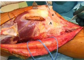
The patient is positioned supine. A longitudinal incision is performed, depending on the tumor location and pref-erence of the surgeon, on the lateral aspect of the thigh extending from the midportion of the thigh to the knee and then ventrally over the tibial tuberosity. The skin site with biopsy tract is kept with the tumor. The tensor fascia is split in line to Gerdy’s tubercle and retracted posteriorly to find access to the posterior compartment. The biceps femoris muscles are kept intact. The popliteal vessels and peroneal and tibial nerves are dissected and tagged with rubber loops, and both heads of the gastrocnemius muscles are dissected free. Anteriorly, the fascia is dissected off the retinaculum respecting the biopsy site but preserving as much tissue as possible for later soft tissue coverage. This is a critical step because the retinaculum is very thin and because at the level of the superior pole of the patella, there is no clear but rather a virtual anatomic plane between these two layers (Fig.1). A preoperative MRI allows assessment of the fat layers and the type of tendon arrangement at the distal end of the quadriceps muscles to plan the resection and enhances the chance of a safe dis-section while preserving the main tendon of the extensor mechanism [21]. The lateral facet of the patella is
prepared to preserve the retinaculum without violating the capsule and the knee. The patellar tendon is dissected by opening the prepatellar bursa. The Hoffa fat pad, which lies extraarticular but partly intracapsular, is left entirely with the specimen (Fig. 2) [2,3, 11, 17, 19]. The vastus lateralis then is dissected according to the tumor extent, now having access to the femur on all sides. At this step, two Kirschner wires are placed under C-arm fluoroscopy control through the patella in the frontal plane, allowing in situ vertical osteotomy of the patella using an oscillating saw (Fig.3). The patellar osteotomy is analogous to that used in standard knee replacement, leaving approximately 15 mm thickness. The extensor mechanism then is everted. The femoral osteotomy is performed at the level that was determined before surgery. The specimen is brought anteriorly and dissected free from the posterior compart-ment structures. The heads of the gastrocnemius muscles are dissected distally to cover the posterior joint capsule to obtain a safe margin. The knee (or specimen) then is extended while the extremity is in flexion, allowing dis-section of the fascia from the retinaculum on the medial side and the capsular structures around the tibia. Distal osteotomy of the tibia is made with an oscillating saw over the Kirschner wires (Fig.2). These Kirschner wires are placed approximately 12 mm below the tibial plateau thereby keeping the joint capsule intact but being proximal to the tibial tubercle to preserve the patellar tendon attachment (Fig.4). The resected specimen now can be removed after dissection of the retinaculum from the capsule on the medial side (Fig.5), and a tumor prosthesis including replacement of the patella retrosurface is implanted the usual way. Postoperative management and rehabilitation do not differ from protocols after transar-ticular resection.
Fig. 1 This lateral view of the knee shows the development of the
virtual plane between the retinaculum (*) and the joint capsule (^) at the superior lateral pole of the patella.
Patients and Methods
We retrospectively reviewed 11 patients (five female and six male patients) who underwent this procedure from 2000 to 2008. The technique was performed as a primary pro-cedure in nine patients and as a secondary propro-cedure in two; these two patients had metastasis to the distal femur, one from an osteosarcoma of the distal tibia and the other from a fibromyxoid sarcoma of the ipsilateral leg extensors. During this period, we treated 411 other patients with tumors about the knee with other approaches. From the medical records we recorded the age, gender, diagnosis, type of surgery, followup, occurrence of metastasis, local recurrence, ROM, and complications. All patients had tumors with intraarticular extension either through direct tumor growth or erosion or pathologic fracture, and two of
the tumors extended into the cruciate ligaments. The mean age of the patients was 39.8 years (range, 15–79 years). There were four osteosarcomas, two leiomyosarcomas, two high-grade pleomorphic sarcomas, one chondrosarcoma, one synovial sarcoma, and one low-grade fibromyxoid sarcoma, all located in the distal femur. Patients were fol-lowed for a minimum of 14 months (mean, 37.5 months; range, 14–80 months). Six of the 11 patients had metastases develop during the followup time and two died from
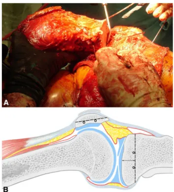
Fig. 2A–B (A) After dissection of the distal joint capsule around the
tibia, two Kirschner wires are placed laterally to guide the osteotomy at the correct level such that the tibial tubercle with its tendon is preserved. (B) A schematic shows the intended resection planes at the tibia and the extent of the synovial and membranous parts of the capsule in relation to the fat pad.
Fig. 3A–B (A) Two Kirschner wires were placed through the patella
in the frontal plane under fluoroscopy guidance at the level of the planned extracapsular osteotomy. (B) A schematic figure shows placement of the wires.
Fig. 4 The intact extensor mechanism with the frontal plane
osteotomy through the patella (*) is seen after removal of the tumor specimen.
widespread metastatic disease. No patients were lost to followup.
Postoperatively, the patients were partial weightbearing of 10 to 15 kg for 6 weeks and then gradually increased to full weightbearing by 3 months postoperatively. Patients had physiotherapy while in the hospital and were taught to climb stairs before they were discharged.
Patients usually were seen at 3-month intervals for 2 to 3 years, and thereafter every 6 months until 5 years. On these occasions, conventional radiographs, MRI of the knee, and CT of the chest usually were performed.
Results
The patients had a mean flexion of 88° (range, 65°–120°); all patients had full extension. There were no complica-tions associated with the extensor mechanism or patella, specifically no patellar fractures (Table 1).
One patient had local recurrence develop 9 months after surgery in the proximal dorsal leg distal to the incision. This patient (Patient 10, Table 1) had presented with a metastatic fibromyxoid sarcoma to the lung. Six months after histologically proven wide resection and reconstruc-tion with a MUTARS tumor endoprosthesis (Implantcast, Buxtehude, Germany) locally and the lung lesion via thoracotomy, metastatic disease again developed in the lung. A second thoracotomy was followed by chemother-apy. The local recurrence 9 months after initial surgery was removed surgically followed by postoperative radiotherapy (33 9 2 = 66 Gy). The patient had no evidence of disease at last followup 4 months after resection of the recurrence.
Discussion
On rare occasions, bone or soft tissue sarcomas may infiltrate or contaminate the knee necessitating resection of the entire joint in toto. Surprisingly, there is only sparse information in the literature regarding the surgical tech-nique of resection and reconstruction in such situations. We describe an extraarticular resection of the knee with pres-ervation of the extensor mechanism. We were interested specifically in the functional ROM of the newly recon-structed knee and the local recurrence rate in these patients. We recognize some limitations of this study. First, because the indication for extraarticular knee resection is
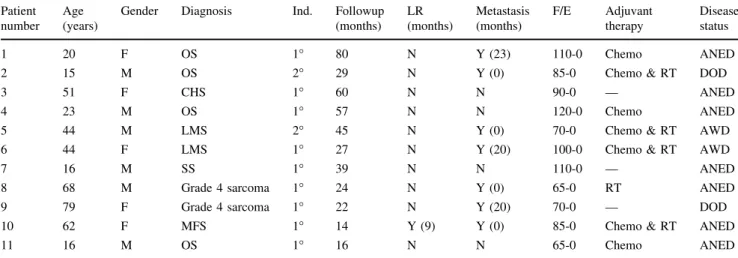
Table 1. Demographics of patients
Patient number
Age (years)
Gender Diagnosis Ind. Followup
(months) LR (months) Metastasis (months) F/E Adjuvant therapy Disease status 1 20 F OS 1° 80 N Y (23) 110-0 Chemo ANED
2 15 M OS 2° 29 N Y (0) 85-0 Chemo & RT DOD
3 51 F CHS 1° 60 N N 90-0 — ANED
4 23 M OS 1° 57 N N 120-0 Chemo ANED
5 44 M LMS 2° 45 N Y (0) 70-0 Chemo & RT AWD
6 44 F LMS 1° 27 N Y (20) 100-0 Chemo & RT AWD
7 16 M SS 1° 39 N N 110-0 — ANED
8 68 M Grade 4 sarcoma 1° 24 N Y (0) 65-0 RT ANED
9 79 F Grade 4 sarcoma 1° 22 N Y (20) 70-0 — DOD
10 62 F MFS 1° 14 Y (9) Y (0) 85-0 Chemo & RT ANED
11 16 M OS 1° 16 N N 65-0 Chemo ANED
Chemo = chemotherapy; RT = radiation therapy; LR = local recurrence; F/E = flexion/extension; F = female; M = male; OS =
osteosar-coma; CHS = chondrosarcoma; LMS = leiomyosarcoma; SS = synovial sarcoma; MFS = myxofibrosarcoma; N = no; Y = yes;
ANED = alive with no evidence of disease; DOD = dead of disease; AWD = alive with evidence of disease; Ind. = Indications.
Fig. 5 The entire tumor specimen with the osteotomy planes of the
relatively rare, we can report only on a small group of patients. Second, the sarcomas included in this study are biologically heterogeneous in terms of type, stage, and adjuvant treatment. Third, the indications for performing an extraarticular resection of the knee may vary; for example, when there are signal alterations on MRI in the proximal cruciate ligaments adjacent to a sarcoma in the distal femur, different surgeons may make different decisions. Therefore, we can make no conclusive statements regarding the ade-quacy of margins of this procedure, particularly with respect to local recurrence. Furthermore, this technique may be contraindicated when the main tumor mass is located in the proximal tibia because obtaining a safe margin while pre-serving the tibial tubercle may be compromised.
The technique of extraarticular knee resection was described more than 30 years ago by Enneking and Shirley [10]. To obtain a safe margin, they performed an osteotomy distal to the tibial tubercle thereby removing the entire extensor mechanism. Reconstruction was performed using an intramedullary rod with an allograft or vascularized or nonvascularized fibulae [10, 22]. Although resection arthrodesis of the knee still has its place in the armamen-tarium of orthopaedic oncologists, it is used less frequently because many patients do not want limited ROM of the knee [5,7,20]. Complications specifically include fatigue frac-tures and rod-associated problems, which do not occur when an extraarticular resection with prosthetic reconstruction is used [20,22] (Table2). Alternatively, if knee mobility is to be preserved and a tumor megaprosthesis is implanted, reconstruction of the extensor mechanism can be achieved either through muscle transfer or allograft reconstruction to compensate for the loss of the extensor mechanism. Capanna et al. reported an MSTS score of only 50% and loss of extensor strength in 16 patients when they transferred flexors to extensors after arthrectomy [4]. The decreased function was presumed to be the effect of extensive scarring, which led to decreased extensor strength and knee stabil-ization. They concluded prosthetic replacement with muscle transfer was an experimental alternative to the conventional arthrodesis [4]. In comparison, the patients in our series had normal extensor strength because the extensor mechanism was preserved. Another reconstruction option was reported by Anract et al., who used gastrocnemius and pes anserinus transfer for reconstruction of the extensor mechanism after total knee arthrectomy in nine patients [1]. The overall function in this group was satisfactory with a mean knee flexion of 62° and an extension lag of 12°. They concluded this technique provides excellent tissue coverage, a strong extensor mechanism, and therefore a viable alternative to arthrodesis [1]. Although stronger extension was noticed using this technique, the reconstruction described in our series provides full active extension without lag. Patients having allografts to reconstruct the knee after tumor Table
2. Overview of literature Study Reconstruction type Cases Localization MSTS % Flexion (mean) Extension lag (mean) Followup Rupture of the extensor mechanism Local recurrence Anract et al. [ 1 ] Soft tissue/ muscle transfer 9 6 distal femur, 2 proximal tibia, and Hoffa Average 61% 62 ° 12 ° 23 ± 10 months 1 None Capanna et al. [ 4 ] Soft tissue/ muscle transfer 16 Distal femur 50% [ 70 ° Poor 2 to 5 years No information None Dubousset et al. [ 8 ] 8 children Distal femur No information No information No information No information No information 1 Wunder et al. [ 23 ] Allograft and prosthesis 11 allograft reconstructions 6 distal femur, 5 proximal tibia 57% — — 55 months for both groups None None 64 modular tumor prostheses 50 distal femur, 14 proximal tibia 75% — — 2 (3%) extensor mechanism repair None Wolf et al. [ 22 ] Arthrodesis 40 29 femur; 11 tibia 77% — — 17 years — 2 Wada et al. [ 20 ] Arthrodesis 12 Distal femur 90% — — 95 months — — Gebhardt et al. [ 12 ] Allograft 25 distal femur, 10 proximal tibia ——— Current study Prosthesis 11 All distal femur — 8 8 ° None 37.5 months None 1
resection have a high complication rate compared with patients with prosthetic reconstruction in general [12,23], and although used for reconstruction after arthrectomy, to the best of our knowledge, there are no published studies.
When reporting a new surgical technique that accepts closer surgical margins to gain better postoperative func-tion, it is crucial to determine the rate of local recurrence. There are few reports on this issue [1, 8] which set the standard. Dubousset et al. [8] and Healey [13] mentioned the technique but did not report the surgical details. The local recurrence rate in our series compares with that reported in the literature (Table2). Obviously, larger series need to be analyzed to confirm whether preservation of the extensor mechanism through extraarticular resection offers the same safe margins. Anatomic details are of great importance, and care is taken that the resection be extra-capsular. For example, because the Hoffa fat pad lies partly intracapsularly, the patellar tendon does not need to be resected. Care also must be taken to avoid opening the joint around the superior pole of the patella where the retinac-ulum and the capsule are fused to one layer.
Our patients had no extensor lag, a mean flexion of 88°, and no obviously increased local recurrence rate; these findings may be superior to those for patients with tendon transfer or allograft reconstructions but comparable to those for patients having intraarticular resection and con-ventional tumor prosthetic reconstruction. When it is doubtful whether the tumor extends into the knee based on preoperative MRI, extraarticular resection is considered. Using the described technique with preservation of the extensor mechanism and reconstruction using a tumor prosthesis, ROM of the knee and local recurrence rate are comparable to those for patients who undergo intraarticular resection for tumors without extension into the knee.
Acknowledgments We thank Carol de Simio, University Hospital
of Zurich, for drawing and preparing the figures included in this manuscript.
References
1. Anract P, Missenard G, Jeanrot C, Dubois V, Tomeno B. Knee reconstruction with prosthesis and muscle flap after total arthrectomy. Clin Orthop Relat Res. 2001;384:208–216. 2. Aydingoz U, Oguz B, Aydingoz O, Bayramoglu A, Demiryurek
D, Akgun I, Uzun I. Recesses along the posterior margin of the infrapatellar (Hoffa’s) fat pad: prevalence and morphology on routine MR imaging of the knee. Eur Radiol. 2005;15:988–994. 3. Boles CA, Martin DF. Synovial plicae in the knee. AJR Am J
Roentgenol. 2001;177:221–227.
4. Capanna R, Ruggieri P, Biagini R, Ferraro A, DeCristofaro R, McDonald D, Campanacci M. The effect of quadriceps excision on functional results after distal femoral resection and prosthetic replacement of bone tumors. Clin Orthop Relat Res. 1991;267: 186–196.
5. Casadei R, Donati D, Ferraro A, Giacomini S, Gozzi E, Gigli M, Boni F, Mercuri M. Knee resection arthrodesis with allograft: a long-term follow-up study. Chir Organi Mov. 2003;88:123–135. 6. Cho Y, Kim JD, Chung SH. Osteosarcoma of the patella: biologic reconstruction with allograft. Orthopedics. 2009 Oct;32(10). pii:
orthosupersite.com/view.asp?rID = 43783. DOI:10.3928/014774
47-20090818-01477427.
7. Donati D, Giacomini S, Gozzi E, Sorin E, Borz S, Mercuri M, Bacci G. Knee arthrodesis with a temporary spacer performed in malignant tumor around the knee. Arch Orthop Trauma Surg. 2002;122:123–128.
8. Dubousset J, Missenard G, Kalifa C. Management of osteogenic sarcoma in children and adolescents. Clin Orthop Relat Res. 1991;270:52–59.
9. Eckardt JJ, Springfield D, Peabody TD. Distal femur. In: Simon MA, Springfield D, eds. Surgery for Bone and Soft Tissue Tumors. Philadelphia, PA: Lippincott-Raven; 1997:357–373. 10. Enneking WF, Shirley PD. Resection-arthrodesis for malignant
and potentially malignant lesions about the knee using an intra-medullary rod and local bone grafts. J Bone Joint Surg Am. 1977;59:223–236.
11. Garcia-Valtuille R, Abascal F, Cerezal L, Garcia-Valtuille A, Pereda T, Canga A, Cruz A. Anatomy and MR imaging appearances of synovial plicae of the knee. Radiographics. 2002;22:775–784. 12. Gebhardt MC, Flugstad DI, Springfield DS, Mankin HJ. The use
of bone allografts for limb salvage in high-grade extremity osteosarcoma. Clin Orthop Relat Res. 1991;270:181–196. 13. Healey JH. Bone and soft tissue tumors around the knee. In: Insall
J, Scott WN, eds. Surgery of the Knee. New York, NY: Churchill Livingstone; 2000:1997–1920.
14. Malawer MM. Distal femoral resection with endoprosthetic reconstruction. In: Malawer MM, Sugarbaker PH, eds. Muscu-loskeletal Cancer Surgery. Dordrecht, The Netherlands: Kluwer Academic Publishers; 2001:459–483.
15. Osanai T, Tsuchiya T, Ogino T. Gastrocnemius muscle flap including Achilles tendon after extensive patellectomy for soft tissue sarcoma. Scand J Plast Reconstr Surg Hand Surg. 2008;42:161–163.
16. Pritsch T, Malawer MM, Wu CC, Squires MH, Bickels J. Functional reconstruction of the extensor mechanism following massive tumor resections from the anterior compartment of the thigh. Plast Reconstr Surg. 2007;120:960–969.
17. Schweitzer ME, Falk A, Berthoty D, Mitchell M, Resnick D. Knee effusion: normal distribution of fluid. AJR Am J Roentgenol. 1992;159:361–363.
18. Simon MA, Aschliman MA, Thomas N, Mankin HJ. Limb-salvage treatment versus amputation for osteosarcoma of the distal end of the femur. J Bone Joint Surg Am. 1986;68:1331–1337.
19. Vahlensieck M, Linneborn G, Schild H, Schmidt HM. Hoffa’s recess: incidence, morphology and differential diagnosis of the globular-shaped cleft in the infrapatellar fat pad of the knee on MRI and cadaver dissections. Eur Radiol. 2002;12:90–93. 20. Wada T, Usui M, Nagoya S, Isu K, Yamawaki S, Ishii S.
Resection arthrodesis of the knee with a vascularised fibular graft: medium- to long-term results. J Bone Joint Surg Br. 2000;82: 489–493.
21. Waligora AC, Johanson NA, Hirsch BE. Clinical anatomy of the quadriceps femoris and extensor apparatus of the knee. Clin Orthop Relat Res. 2009;467:3297–3306.
22. Wolf RE, Scarborough MT, Enneking WF. Long-term followup of patients with autogenous resection arthrodesis of the knee. Clin Orthop Relat Res. 1999;358:36–40.
23. Wunder JS, Leitch K, Griffin AM, Davis AM, Bell RS. Com-parison of two methods of reconstruction for primary malignant tumors at the knee: a sequential cohort study. J Surg Oncol. 2001;77:89–99; discussion 100.