Phagocytosis and Phagosomal Fate of
Surface-Modified Microparticles in
Dendritic Cells and Macrophages
Lars Thiele,1Hans P. Merkle,1and Elke Walter1,2
Received October 7, 2002; accepted November 1, 2002
Purpose. We compared cationic, polyamine-coated microparticles (MPs) and anionic, protein-coated MPs with respect to their phago-cytosis and phagosomal fate in dendritic cells (DCs) and macro-phages (M⌽).
Methods. Polystyrene MPs were surface modified by covalent cou-pling with fluorescein isothiocyanate-labeled polyamines or proteins. Phagocytosis of MP and the pH of their intracellular microenviron-ment was assessed in human-derived DCs and M⌽ in a fluorescence plate reader. Visualization of MP phagocytosis in DCs was performed by transmission electron microscopy.
Results. Phagocytosis of bovine serum albumin-coated MPs was low with significant differences between DC and M⌽, whereas phagocy-tosis of IgG-coated MPs was significantly enhanced in both cell types. Phagocytosis of both particle types resulted in an acidified phago-somal microenvironment (pH 4.6–5.1). In contrast, cationic, poly-amine-coated MPs were equally phagocytosed by DCs and M⌽ to a high extent and showed lower degrees of acidification (pH 6.0–6.8) in the phagosomal microenvironment. Transmission electron micros-copy examination demonstrated all phagocytosed particles to be sur-rounded by a phagosomal membrane, which was more tightly ap-posed to the surface of cationic MPs and more loosely to bovine serum albumin-coated MPs.
Conclusion. Phagocytosis of cationic, polyamine-coated MPs is sug-gested to lead to diminished phagosomal acidification. Thus, cationic MP are potential carriers that may display beneficial features for the intracellular delivery of immunomodulating therapeutics and their protection against lysosomal degradation.
KEY WORDS: dendritic cells; macrophages; surface-modified
mi-croparticles; phagocytosis, intracellular fate.
INTRODUCTION
The internalization of particles in the micrometer range, i.e., microparticles (MP), is a special feature of phagocytic cells and has recently received high interest to enable targeted delivery of therapeutics into dendritic cells (DCs) and mac-rophages (M⌽; 1–3). DCs and M⌽ are professional antigen-presenting cells and, thus, represent targets for the delivery of vaccines (4,5). Particularly, DC were found to elicit T-cell responses that were up to 3000 times stronger than those observed with M⌽. In addition, DCs feature unique capabili-ties to modulate the immune system toward immunity or tol-erance (6,7). Upon phagocytosis, antigens associated to MPs have been shown to elicit strong cytotoxic T-cell responses (8,9), being a consequence of their access to the MHC class I pathway of presentation (10–13). In this context, the
advan-tage of MP-associated antigens over soluble antigens appears to result from at least the following two factors: 1) the op-portunity to internalize large quantities of antigen when em-bodied in MP, and 2) their prolonged release and thus more efficient presentation as compared with a short-lived bolus of antigen delivered in soluble form.
Phagocytosis of MPs as well as the presentation of MP-associated antigens have been demonstrated to be strongly influenced by the chemical and physical nature of the MPs (3,14–16). To initiate phagocytosis, the MP surface needs to stimulate binding to the cellular membrane, which subse-quently sets off internalization into the cell. Recent studies revealed that particularly MPs displaying a positive surface charge were subject to efficient phagocytosis by both M⌽ and DCs (14,17). Interestingly, although the phagocytosis activity of DCs is generally considered to be lower than that of more efficient phagocytes, such as M⌽, the phagocytosis of cationic MPs by DCs has been demonstrated to measure up with M⌽ (4,5,14,18,19).
Phagocytosis leads to the intracellular entrapment of MPs in phagosomes, which mature under the influence of acidification, resulting from their fusion with lysosomes by forming so-called phagolysosomes (20). The process of phagosome maturation has been shown to relate to the de-gree of apposition of the phagosomal membrane to the en-trapped MPs (20–22). There are two alternatives. The first is hydrophilic MPs, entrapped in spacious phagosomes with the phagosomal membrane and in loose contact to the MP sur-face, are subjected to rapid fusion with lysosomes. The second is mycobacteria or hydrophobic latex particles were observed to resist rapid fusion with lysosomes, which was suggested to be caused by their tight apposition to the phagosomal mem-brane (21).
MPs carrying positively charged surfaces may be ob-tained by coating them with cationic excipients owing to the protonation of their amino groups (23–25). Because of their buffer capacity, these compounds have been suggested to de-lay phagosomal acidification (26) and subsequently cause im-paired fusion with lysosomes or even deliver particle-associated antigens to the cytosol. Cytosolic delivery of anti-gens or DNA vaccines represents an important prerequisite for antigen presentation via the major histocompatibility complex class I to specifically stimulate cytotoxic T lympho-cyte response.
For the first time, this study aims to investigate the in-fluence of positively charged MPs on their phagocytosis and subsequent intracellular fate in DCs and M⌽. Moreover, for comparison with positively charged MPs, we also studied MPs with various other surface modifications that were expected to cause either unspecific or receptor-mediated phagocytosis. The various MP surfaces were characterized for charge and hydrophobicity, and the phagocytosis of the respective MPs was assessed with human blood-derived DCs and M⌽ by us-ing an automated fluorescence plate reader. By the same methodology, we determined the intracellular pH in the mi-croenvironment of phagocytosed MP using the pH-dependent intensity of a fluorescence signal to probe the phagosomal pH. Additionally, we used transmission electron microscopy (TEM) to study the mechanisms of phagocytosis and assess the residence of phagocytosed MP.
1Department of Applied BioSciences, Swiss Federal Institute of
Technology Zurich (ETH), Winterthurerstrasse 190, CH-8057 Zu-rich, Switzerland.
2To whom correspondence should be addressed. (e-mail: elke_walter@
gmx.net)
MATERIALS AND METHODS
Materials
Poly-L-lysine hydrobromide (PLL 29, mean molecular weight [MW] 29 kDa and PLL 99, mean MW 99 kDa), fluo-rescein isothiocyanate (FITC), bovine serum albumin (BSA), human IgG, and phosphate-buffered saline (PBS, pH 7.4) were purchased from Sigma, Buchs, Switzerland. Polyethyl-enimine (PEI, MW 600,000–1,000,000) was obtained from Fluka, Buchs, Switzerland. Trypan blue was obtained from Merck AG, Darmstadt, Germany. Materials for cell culture experiments were obtained from Life Technologies, Basel, Switzerland. All other chemicals used were of analytical grade unless otherwise specified and obtained from Fluka, Buchs, Switzerland.
Surface Modification of MPs
Surface modified MPs were obtained by covalent conju-gation of carboxylated polystyrene MPs (1.0m in diameter, PolybeadTM, Polysciences, Eppelheim, Germany) with IgG,
BSA, PLL, or PEI (50g/mg MP) using the carbodiimide method (data sheet #238 C, Polysciences, Eppelheim, Ger-many). To separate coated MPs from unbound coating ma-terial, the MPs were centrifuged (6 min, 21,000 × g, 20°C) and subsequently washed three times in PBS, pH 7.4, to remove unbound material. The amount of unbound coating material was determined in the supernatant upon conjugation with fluorescamine (0.3 mg/mL in acetone) according as described previously (27). Quantitative analysis was performed by using an automated plate reader (Fluoro Count, Canberra Packard, Frankfurt, Germany; filters: 360/460 nm).
FITC Labeling of Coating Material and MPs
Soluble IgG, BSA, PLL, and PEI or surface-modified MPs, respectively, were fluorescence-labeled with FITC in carbonate buffer (0.5 M, pH 9.5) according to Arvinte et al. (28). FITC-labeled soluble IgG, BSA, PLL, and PEI were purified by dialysis in PBS using a Spectra/Por® Membrane
(MWCO 3500, Spectrum Medical Industries, Inc., Hollister, CA, USA). FITC-labeled surface-modified MP were sepa-rated from excess FITC by repeated washings and subsequent centrifugation.
Determining the Surface Charge of MPs by
-Potential Measurement
The-potentials of various MPs were determined using a Zeta-Meter System 3+ (ZM3+ 331, Zeta-Meter, Inc., Staun-ton, VA, USA). Three samples of each MP type were mea-sured at 25°C in double distilled water containing 0.01 M NaCl.
Determining the Hydrophobicity of MP by the Rose Bengal Assay
Different amounts of MP (0.3–1.8 mg/mL, corresponding to 5.5*108–3.3*109particles per mL) were suspended in a
so-lution of 20g/mL Rose Bengal in 0.1 M phosphate buffer of pH 7.4. Samples were incubated at room temperature for 3 h in the dark, centrifuged at 21,000 × g, and the amount of non-adsorbed Rose Bengal in the supernatant was determined by
using an automated fluorescence plate reader (filters: 530/590 nm). The partition quotient was calculated by dividing the amount of Rose Bengal bound on the MP surface by the amount of Rose Bengal remaining in the dispersion medium. Plotting of partition quotient against the total surface area of the MP resulted in straight lines. The slope of such lines was taken as parameter for the hydrophobicity of the MP surface (29). Slopes were calculated by linear regression analysis.
Fluorescence Intensity of FITC-Labeled Coat Materials as a Function of pH
Purified FITC-labeled coating materials were dissolved at various pH (pH 2–5.5 in 0.5 M citrate buffer, pH 5.5–10 in PBS). The fluorescence intensity was measured using an au-tomated fluorescence plate reader (filters: 485/530) and ex-pressed as relative fluorescence intensity with 100% fluores-cence at pH 7.4.
M⌽ and DC Cell Cultures
DCs and M⌽ were obtained from human peripheral blood monocytes according to Sallusto et al. (30). Briefly, peripheral blood monocytes were isolated from buffy coats (Bloodbank Zurich, Switzerland) by density gradient cen-trifugation on Ficoll-Paque (Pharmacia Biotech). Peripheral blood monocytes were resuspended in RPMI 1640 supple-mented with 10% heat inactivated (pooled) human serum (Bloodbank Zurich, Switzerland) and then allowed to adhere for 2 h in culture flasks (25 cm2). Nonadherent cells were
removed and adherent cells were cultured in RPMI 1640 me-dium supplemented with 5% heat inactivated (pooled) hu-man serum in the presence of 1000 IU/mL interleukin-4 (Sigma) and 50 ng/mL GM-CSF (R+D Systems) for the gen-eration of DCs, whereas M⌽ were obtained without any ad-ditional supplements (31). For phagocytosis studies, cells were mechanically removed from the flasks during the first day of culture and reseeded in 96-well plates. Cultures were kept at 37°C in 5% CO2humidified atmosphere for 7–10 days before used for further experiments.
Characterization of DCs and M⌽
Surface antigen expression of DCs and M⌽ was analyzed by flow cytometry (32). For further identification of DCs, the cells were also challenged with lipopolysaccharide (LPS; 1 g/mL) for 48 h to induce DC maturation. The cells were incubated (45 min, 4°C) with each of the following primary anti-human antibodies: CD11b (Mac-1, ICRF44, Pharmingen; 44), CD14 (Clone UCHM-1, Sigma), CD83 (Clone HB15e, Pharmingen), CD86 (B70/B7-2, Clone IT2.2, Pharmingen), MHC I (HLA-ABC, Clone W6/32, Dako), and MHC II (HLA-DP, DQ, DR, Clone CR3/43, Dako). In a similar way controls were performed by using mouse isotype control IgG2a (UPC-10, Sigma). The cells were washed three times and subsequently incubated with the secondary antibody (anti-mouse IgG R-Phycoerythrin conjugate, Sigma) for 45 min at 4°C. Afterwards, the cells were washed again three times and then transferred to FACS tubes and analyzed on a FACScan type Becton Dickinson. The presence of CD14 and the absence of CD83 after LPS stimulation is regarded to be characteristic for M⌽, whereas the presence of CD11b, the absence of CD14 and the upregulation of CD83 and CD86
after LPS stimulation is characteristic for DC (6,32). Accord-ing to these criteria, more than 90% of the cells were identi-fied as DC or M⌽, respectively.
Cell Viability and Trypan Blue Quenching
Trypan blue quenching simultaneously allows the deter-mination of cell viability and phagocytosis activity (14). Cell viability was checked by trypan blue exclusion (1% in PBS pH 7.4) as described elsewhere (14,33). When used as a so-lution of 250g/mL in citrate buffer (pH 4.4), trypan blue has been demonstrated to completely quench extracellular fluo-rescence of FITC as described in a previous study in detail (14). In addition, penetration of trypan blue into necrotic cells typically leads to quenching of intracellular fluorescence. Thus, the recorded fluorescence strictly originates from phagocytosed MP in viable cells only .
Determination of MP Phagocytosis and Intracellular pH
DCs and M⌽ were cultured in 96-well plates at 104cells
per well for 10 days. For phagocytosis studies, the medium was replaced by 50L of MP dispersed in HEPES buffer (0.2 M, pH 7.4) containing 1 mM Ca2+(3.3*108 MP/mL). Cells
were incubated at pH 7.4, either 4 or 37°C, for 4 h. The subsequent measurement of fluorescence intensity was per-formed at room temperature by using an automated plate reader (filters: 485/530). The following parameters were used: Fall: Fluorescence intensity of all MP incubated in the pres-ence of cells at 4°C. At this temperature phagocytosis was blocked to allow determination of F for all MP in the extracellular milieu (pH 7.4).
F37: Fluorescence intensity of all MP incubated in the
pres-ence of cells at 37°C.
Fintra: Measured after quenching with trypan blue,
represent-ing the fluorescence intensity of all phagocytosed, in-tracellular MP.
Pa: Number of MP per well. C: Number of cells per well.
Calculation of MP Uptake
Because of the pH sensitivity of the fluorescence label, its reduced intracellular fluorescence in a low pH environment can be compensated as follows:
Fluorescence intensity of a single
particle: FMP⳱ Fall/Pa
Amount of nonphagocytosed
extracellular MP at 37°C: Fextra⳱ F37− Fintra Number of extracellular MP: Pe⳱ Fextra/FMP
Number of intracellular MP: Pi⳱ Pa− Pe Average number of MP phagocytosed per cell: Pi/C
Calculation of Intracellular pH in the Microenvironment of Phagocytosed MPs
The pH-dependent fluorescence intensity of phagocy-tosed MPs relates to Piand the difference between
extracel-lular and intracelextracel-lular pH as previously used by (34,36–38):
Change in fluorescence intensity
of Paafter phagocytosis: Fphago⳱ Fall− F37 Fluorescence intensity of Piat
pH 7.4: F7.4⳱ Fintra+ Fphago Remaining relative fluorescence
intensity of Pi: Fremain%⳱ Fintra/F7.4*100 By means of the fluorescence intensity of various FITC-labeled polymers (PLL, PEI, IgG, and BSA) as a function of pH, microenvironmental pH values can be derived from the experimentally obtained fluorescence intensities, i.e., Fremain%. The respective profiles are depicted in Fig. 1 and
are further described in the results section.
Microscopic Assessment of Phagocytosis
The intracellular fluorescence of very small numbers of MPs was below the detection limit of the fluorescence plate reader. Thus, phagocytosis of BSA-coated MPs by DCs was assessed by fluorescence microscopy (Axiovert 35, Zeiss, Oberkochen, Germany). Phagocytosis studies and subsequent quenching of extracellular MPs with trypan blue were per-formed as described above. The extent of phagocytosis was then monitored by using an inverted microscope (Axiovert 35, Zeiss, Oberkochen, Germany) equipped for fluorescence microscopy (excitation 450–490 nm, emission 520 nm). Data indicate the mean number of MPs counted in 150 represen-tative cells per sample.
Fig. 1. Fluorescence intensity of FITC-labeled coating materials, (a)
FITC-PLL and FITC-PEI, (b) FITC-BSA and FITC-IgG, as a func-tion of pH. The fluorescence intensity is expressed in % was set to 100% at pH 7.4.
Processing of Cells for TEM
Cells were washed three times in Hank’s balanced salt solution and resuspended in phosphate buffered 2.2% glutar-aldehyde solution at pH 6.8. The cells were postfixed in 1% osmium tetroxide in 0.1 M sodium cacodylate buffer and con-trasted in 0.5% uranyl acetate in 0.05 M maleate buffer. De-hydration was performed in a graded series of ethanol-water mixtures (v/v; 70, 80, 96, and 100%). Gradual replacement of ethanol was performed with propylene oxide before infiltra-tion and embedding in epoxy resin. Ultrathin secinfiltra-tions were cut using an ultramicrotome (Reichert, Vienna, Austria) and were collected on 200-mesh carbon-coated copper grids. Af-ter staining with uranyl acetate and counAf-terstaining with lead citrate, samples were observed by TEM at 60 kV using a Phillips 300 (Phillips Electronic Instruments, Inc., New Han-over, NJ, USA).
RESULTS
Properties of Various Surface-Modified MPs
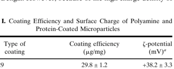
MPs displaying different surface properties were gener-ated by covalent coating with either proteins (BSA or IgG) or polyamines (PEI or PLL; Table I). All surface modifications resulted in high coating efficiencies ranging from 15–30g/mg MPs (equivalent to 2.6–5.2 ng/mm2). Surface charges were
determined by measuring the-potentials. Polyamine-modi-fied MPs displayed highly positively charged surfaces in the range of +45.7 to +38.2 mV, whereas protein-modified MP displayed negatively charged surfaces in the range of –16.9 to −21.1 mV (Table I). As a control, the surface charge of plain polystyrene MPs was practically neutral. Moreover, we deter-mined the hydrophobicity of the various surface-modified MPs by the previously established Rose Bengal partitioning method (29,36). The method was modified and performed as a microassay. In principle, MPs dispersed in an aqueous me-dium were regarded as a two-phase system with the surface layer of the MPs as one phase and the dispersion medium as the second phase. Plotting of the partition coefficients of the dye against the total surface area of the microspheres yielded straight lines. The slopes represent arbitrary hydrophobicity values for the surface of the microspheres. As expected, plain polystyrene MPs displayed the highest hydrophobicity fol-lowed by BSA- and IgG-coated MPs (Fig. 2). The Rose Ben-gal partitioning method was not suitable for cationic surfaces because of ionic interactions with the negatively charged Rose Bengal. However, because of the high charge density of
the polyamine coating, assumption of a rather hydrophilic surface is reasonable.
Assessment of Phagocytosis and Intracellular pH in the Microenvironment of Phagocytosed MP
We measured the phagocytosis of the various surface-modified MPs in human blood-derived M⌽ and DCs by using an automated fluorescence plate reader. To not to change the surface properties of the MP as the result of serum protein adsorption, a serum-free incubation medium was used. Fur-ther studies are currently performed to assess the influence of serum protein adsorption on the phagocytosis of M⌽ and DCs. The assessment of intracellularly located MPs requires surface- but not bulk-conjugated FITC to allow quenching of extracellular fluorescence by trypan blue (14). The pH-dependent fluorescence intensity of various FITC-derivatives is characterized as outlined in Fig. 1. Because the phagocy-tosed MPs may be exposed to an acidic intracellular environ-ment as it occurs in phagosomes or phagolysosomes, the change in fluorescence intensity must be compensated to as-sess phagocytosis capacity. Furthermore, we used the pH-dependent fluorescence intensity to calculate the pH in the microenvironment of phagocytosed MPs. The change in pH was most pronounced between pH 4 and 7 (Fig. 1a and b) and, thus, the phagosomal pH can be determined depending on the state of phagosomal maturation (34,35,37,38).
Phagocytosis of the various surface-modified MPs was determined in DCs and M⌽ (Fig. 3). BSA-coated MP with a negatively charged and comparatively hydrophilic surface were phagocytosed by DCs at a significantly lower degree than by M⌽. In contrast, phagocytosis of IgG-coated MP dis-playing analogous surface properties was much larger and at
Table I. Coating Efficiency and Surface Charge of Polyamine and
Protein-Coated Microparticles Type of coating Coating efficiency (g/mg) -potential (mV)a PLL 29 29.8 ± 1.2 +38.2 ± 3.3 PLL 99 27.4 ± 2.6 +42.0 ± 3.5 Polyethylenimine 26.3 ± 3.3 +45.7 ± 4.1 IgG 14.5 ± 1.9 −21.1 ± 1.6
Bovine serum albumin 18.4 ± 2.1 −16.9 ± 1.2
Non-coated MPs — −0.8 ± 1.5
aMeasured in 0.01 N NaCl; data are means of three samples ± SD.
Fig. 2. Relative hydrophobicity of various surface-modified
micropar-ticles determined by the Rose Bengal adsorption assay. Data show the mean ± SD (n⳱ 3). Non-coated polystyrene microparticles (PS).
Fig. 3. Phagocytosis of various surface-modified microparticles by
comparable efficiencies with both cell types. All cationic poly-amine-modified MPs induced phagocytosis on the highest level in both DCs and M⌽ (Fig. 3).
The pH in the microenvironment of phagocytosed MPs was probed using a pH-dependent fluorescent label conju-gated to the various tested polyamine (PLL and PEI) or pro-tein coatings (IgG and BSA). The relationships between the recorded fluorescence intensities and the pH of the tested polymers are depicted in Fig. 1, showing more or less sigmoi-dal relationships for PLL, IgG, and BSA.
An acidified microenvironment was found in the phago-somes of the IgG-coated MP in the range of pH 4.6 to 5.1, both in DC and M⌽ (Table II). The low incidence of phago-cytosis of BSA-coated MPs by DCs was below the detection limit of the fluorescence plate reader. Thus, information about the effect of BSA-coated MPs on the phagosomal pH in DCs is missing, but phagosomes of M⌽ were shown to be similarly acidified as upon IgG-coated MPs. In contrast, the microenvironment of phagocytosed PLL-coated MPs was less acidic for both DCs and M⌽, i.e., pH 6.0–6.3 for both PLL coatings (Table II). The peak-shaped profile, which was found in the case of PEI led to two potential pH values of 6.0 and 6.8, both indicating a significantly increased microenvi-ronmental pH compared with protein-coated MPs (Table II). In summary, our data may suggest diminished phagosomal acidification in the presence of polyamine-coated MPs vs. protein-coated MPs.
Mechanism of Phagocytosis and Intracellular Fate of the MP
We used TEM to compare the histology of the phagocy-tosis and the intracellular fate of cationic, polyamine-coated MPs with those of anionic, protein-coated MPs, both in DCs and M⌽. The objectives of these experiments were to distin-guish between two hypotheses for the diminished acidifica-tion. Firstly, intraphagosomal buffering and or delay of phagosomal maturation may be continued. Secondly, poly-amine-induced rupture of the endosomal membrane has been previously demonstrated to lead to the endosomal escape of PEI-associated material into the cytosol (26).
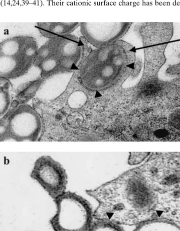
Previously, BSA-coated MPs have been demonstrated to be internalized into spacious phagosomes followed by a rapid fusion with lysosomes (21). In our study, PLL 99 and PEI-coated MPs were chosen as examples for cationic MP and compared with anionic, BSA-coated MP. As confirmed by TEM, the majority of the polyamine-coated MPs were
inti-mately engulfed by large pseudopods (Fig. 4a, arrows), result-ing in a tightly apposed membrane coverresult-ing the surface of the MPs (Fig. 4a, arrowheads). In some cases cationic MPs ap-peared to sink into the cells, especially for the sequential phagocytosis of several MPs in a row. Nonetheless, after phagocytosis cationic MPs were always enclosed by a tightly apposed membrane (Fig. 4b, arrowheads). In contrast, pseu-dopod formation was never observed upon phagocytosis of BSA-coated MPs, which appeared to sink into the cells as already observed in previous studies (21).
After 4 h, the vast majority of the polyamine and BSA-coated MPs were found to be enclosed by a phagosomal membrane (Fig. 5) with only a few exceptional polyamine-coated MPs, where such a membrane could not be clearly identified. BSA-coated MPs were mainly found in spacious phagosomes with large parts of the phagosomal membrane being distant from the surface of the MP (Fig. 5a). Only few MPs were found in phagosomes with a tightly apposed mem-brane (Fig. 5b). In contrast, the majority of PLL 99-coated MPs (Fig. 5c and d) and PEI-coated MPs (Fig. 5e and f) were engulfed in phagosomes with tightly apposed phagosomal membranes (Fig. 5c–f).
DISCUSSION
Cationic MPs have gained increasing interest as delivery systems to target phagocytic cells such as DC and M⌽ (14,24,39–41). Their cationic surface charge has been
demon-Fig. 4. Transmission electron microscopy of the phagocytosis of
poly-ethylenimine-coated microparticles (MPs) by dendritic cells. (a) The majority of MPs are internalized by the formation of pseudopods (arrows) or (b) in some cases by sinking into the cell. The cellular membrane is tightly adjoined to the MP surface (arrowheads).
Table II. Microenvironmental pH in the Phagosomes of
Phagocy-tosed Microparticles Coated with Various Polyamines and Proteins Type of coating pH in DCa pH in M⌽a PLL 29 6.0 ± 0.1 6.1 ± 0.2 PLL 99 6.3 ± 0.2 6.1 ± 0.3 Polyethylenimine 6.0 ± 0.2 (6.8 ± 0.2)b 6.0 ± 0.2 (6.7 ± 0.2)b IgG 5.1 ± 0.2 4.9 ± 0.1
Bovine serum albumin n.d. 4.6 ± 0.4
aData represent means of three samples ± SD.
bThe peak-shaped profile of fluorescence intensity chart led to two
potential pH values with the higher number given in parentheses (see Fig. 1).
strated to strongly enhance phagocytosis by such cells (14). In this study, we investigated the degree of phagocytosis of vari-ous polyamine-coated MPs in comparison to protein-coated MPs, including an IgG-coating that was previously shown to elicit receptor-specific phagocytosis (42,43). In addition, we evaluated the mechanism of phagocytosis and the intracellu-lar fate of internalized MPs by probing their microenviron-mental pH in combination with electron microscopy. Cationic MPs were efficiently phagocytosed by DCs through extensive pseudopod formation and ended up in phagosomes displaying a tightly apposed membrane. The expected acidification of the phagosomal microenvironment was only found with pro-tein-coated MPs, whereas the phagosomes containing poly-amine-coated MPs turned out to be less acidic. Typically, low phagosomal acidification was always concomitant with tight engulfment by the phagosomal membrane.
Consistent with other studies, hydrophilic BSA-coated MPs were internalized at a comparatively low frequency by M⌽ (41,44,45) and even more reduced by DC. The relatively low capacity of DC for nonspecific phagocytosis of foreign material has been demonstrated in previous studies (18,46, 47). Whereas particle scavenging by M⌽ was found to be highly efficient and led to the clearance of foreign material, DCs have been suggested to phagocytose only to an extent that is necessary to initiate the immune response (18).
Coating of MPs with IgG resulted in even more hydro-philic surfaces than BSA-coated MPs with comparable sur-face charge but led to more pronounced phagocytosis, which was comparably efficient in both DCs and M⌽. This may be explained by the interaction of the IgG-coated MPs with Fc receptors, which are abundantly expressed on the cell mem-brane of both DCs and M⌽, and mediate efficient receptor-mediated phagocytosis (15,48–50).
The strong phagocytosis of hydrophilic polyamine-coated MPs by DCs and M⌽ is suggested to be caused by their strongly positive surface charge, which was demon-strated to be independent of the molecular weight or the chemical structure of the polyamines used in this study. The ionic attraction between the positively charged MPs and the negatively charged cell surface of the cells is likely to repre-sent a strong stimulus that initiates binding and subsequent internalization (51).
Two principal mechanisms have been discussed to ex-plain the internalization of MPs by phagocytic cells (20). For cationic MPs, we observed active pseudopod advancement engulfing the MPs, which is in analogy to the zipper mecha-nism and resulted in the formation of tightly apposed phago-somes. After the zipper mechanism, ingestion occurs by se-quential engagement of a phagocyte’s membrane against the MPs surface. Pseudopod advancement proceeds no further than receptor-ligand interactions permit and is probably re-lated to a high affinity of the cell membrane to the particle surface. This could be induced by involvement of a receptor-ligand interaction, such as with IgG-opsonized or IgG-coated particles, which were actively engulfed by large protrusions, ending up in tightly apposed phagosomes (20). Alternatively, a high affinity of the particle surface to the cell membrane, such as for hydrophobic or positively charged particles, may result in a similar effect.
In contrast, hydrophilic BSA-coated MPs were phagocy-tosed without formation of pseudopods and ended up in loosely apposed phagosomes. This is in agreement with find-ings by others (21) and suggests phagocytosis in DCs and M⌽ to occur according to the trigger mechanism (20). In this case, the particle would be ingested by a sinking-in-to-the-cell-like manner in loose contact to the phagosomal membrane.
Phagocytosed material ends up in phagosomes where it is then subject to several “fusion and fission” processes between phagosomes and lysosomes in order to exchange membrane constituents and vesicular contents (52). Thereby, the phago-some matures and acquires enzymes and membrane bound proton pumps, which are typically associated with lysosomes leading to a significant acidification of the matured phago-some (34,36–39). However, phagosomal maturation has been demonstrated to be inhibited depending on the type of phago-cytosed MPs (21). Particularly, hydrophobic MPs were en-gulfed in tightly fitting phagosomes inhibiting phagosomal maturation. Protein-coated and hydrophilic MPs, however, were found to display loose interaction with the phagosomal membrane allowing rapid fusion with lysosomes (21). These observations are in accordance with our findings on protein-coated MPs carrying hydrophilic and negatively charged sur-faces. Moreover, phagocytosed BSA- and IgG-coated MPs were found to end up in more acidic microenvironments of pH 4.6 to 5.1, indicating matured phagosomes. In contrast, we found hydrophilic polyamine-coated cationic MPs to be en-gulfed in tightly fitting phagosomes with a significantly less
Fig. 5. Transmission electron microscopy of surface-modified MPs
phagocytosed by dendritic cells after 4 h for (a, b) BSA-coated mi-croparticles (MPs), (c, d) PLL 99-coated MPs, and (e, f) polyethyl-enimine-coated MPs. MPs are located in phagosomes with the phago-somal membrane tightly adjoined to the MP surface (b-f) or in spa-cious phagosomes with large parts of the phagosomal membrane distant from the MP surface (a).
acidic microenvironmental pH (6.0–6.8) as compared with protein-coated MPs. Phagosomal buffering, potentially fol-lowed by delayed phagosomal maturation, is one way to ex-plain the minor acidification through cationic, polyamine-coated MP vs. the pronounced acidification with anionic, pro-tein-coated MP (Table II). Another hypothesis is the polyamine-induced rupture of the endosomal membrane, which has been previously demonstrated to lead to the endo-somal escape of PEI-associated material into the cytosol (26). According to the proton sponge hypothesis introduced by Behr (26), extensive buffering of the phagosome may even provoke ongoing proton transport by membrane-bound pro-ton pumps and is followed by water influx. Finally, rupture of the phagosomal membrane and escape of the material en-trapped in the phagosomes into the cytosol could be the con-sequence (26).
Based on our findings, we suggest maturation of phago-somes containing cationic MPs to be inhibited because of ionic interactions with the negatively charged phagosomal membrane. Besides, the investigated polyamines are likely to delay phagosomal acidification because of their buffering ca-pacity (26). However, there was no visible evidence for phagosomal escape. The majority of the phagocytosed cat-ionic MP observed in our study were coated by a tight phago-somal membrane even after 4 h of incubation and displayed no significant membrane disruption.
In summary, although hydrophilic, cationic MPs were ef-ficiently phagocytosed by DCs, exceeding even the efficiency of receptor-mediated internalization of IgG-coated MPs. Electrostatic attraction between the positively charged MP surface and the negatively charged cell surface is likely to mediate binding and subsequent internalization. Phagocytosis was initiated by pseudopod advancement and led to the for-mation of phagosomes with a tightly apposed phagosomal membrane in a zipper-like manner. Furthermore, we found the intracellular pH in the microenvironment of cationic MPs to be significantly less acidic compared with the lower pH with other protein-coated MPs. This may be explained by the tight fit of the phagosomal membrane, which is suggested to lead to the inhibition of phagosomal maturation. Alterna-tively, intraphagosomal buffering by cationic coatings is likely to contribute to the reduced acidification and could also lead to delayed phagosomal maturation. In summary, the proper-ties of cationic MPs to strongly enhance phagocytosis by DCs in combination with a delayed phagosomal maturation may hold promise for the intracellular delivery of immunomodu-lating therapeutics, which must be protected against lysosom-al degradation to maintain their activity.
ACKNOWLEDGMENT
This work was supported by grants #4037-55144 from the Swiss National Research Foundation. Furthermore, we thank Dr. Ernst Wehrli and Dr. Markus Müller for their excellent help in electron microscopy.
REFERENCES
1. S. Raychaudhuri and K. L. Rock. Fully mobilizing host defense: building better vaccines. Nat. Biotech. 16:1025–1031 (1998). 2. E. Walter, D. Dreher, M. Kok, L. Thiele, S. G. Kiama, P. Gehr,
and H. P. Merkle. Hydrophilic poly(DL-lactide-co-glycolide)
mi-crospheres for the delivery of DNA to human-derived macro-phages and dendritic cells. J. Control. Release 76:149–168 (2001). 3. S. Prior, B. Gander, N. Blarer, H. P. Merkle, M. L. Subria, J. M. Irache, and C. Gamazo. In vitro phagocytosis and monocyte-macrophage activation with poly(lactide) and poly(lactide-co-glycolide) microspheres. Eur. J. Pharm. Sci. 15:197–207 (2002). 4. J. Banchereau, F. Briere, C. Caux, J. Davoust, S. Lebecque, Y. J.
Liu, B. Pulendran, and K. Palucka. Immunobiology of dendritic cells. Annu. Rev. Immunol. 18:767–811 (2000).
5. A. Aderem and D. M. Underhill. Mechanisms of phagocytosis in macrophages. Annu. Rev. Immunol. 17:593–623 (1999). 6. J. Banchereau and R. M. Steinman. Dendritic cells and the
con-trol of immunity. Nature 392:245–252 (1998).
7. F. D. Finkelman, A. Lees, R. Birnbaum, W. C. Gause, and S. C. Morris. Dendritic cells can present antigens in vivo in a tolero-genic or immunotolero-genic fashion. J. Immunol. 157:1406–1414 (1996).
8. Y. Men, H. Tamber, R. Audran, B. Gander, and G. Corradin. Induction of a cytotoxic T lymphocyte response by immunization with a malaria specific CTL peptide entrapped in biodegradable polymer microspheres. Vaccine 15:1405–1412 (1997).
9. K. Peter, Y. Men, G. Pantaleo, B. Gander, and G. Corradin. Induction of a cytotoxic T-cell response to HIV-1 proteins with short synthetic peptides and human compatible adjuvants. Vac-cine 19:4121–4129 (2001).
10. Z. Shen, G. Reznikoff, G. Dranoff, and K. L. Rock. Cloned den-dritic cells can present exogenous antigens on both MHC class I and class II molecules. J. Immunol. 158:2723–2730 (1997). 11. M. Svensson, B. Stockinger, and M. J. Wick. Bone
marrow-derived dendritic cells can process bacteria for I and MHC-II presentation to T cells. J. Immunol. 158:4229–4236 (1997). 12. C. Scheicher, M. Mehlig, H. P. Dienes, and K. Reske. Uptake of
microparticle-adsorbed protein antigen by bone marrow-derived dendritic cells results in up-regulation of interleukin-1 alpha and interleukin-12 p40/p35 and triggers prolonged, efficient antigen presentation. Eur. J. Immunol. 25:1566–1572 (1995).
13. M. Kovacsovics-Bankowski and K. L. Rock. A phagosome-to-cytosol pathway for exogenous antigens presented on MHC class I molecules. Science 267:243–246 (1995).
14. L. Thiele, B. Rothen-Rutishauser, S. Jilek, H. Wunderli-Allenspach, H. P. Merkle, and E. Walter. Evaluation of particle uptake in human blood monocyte-derived cells in vitro. Does phagocytosis activity of dendritic cells measure up with macro-phages? J. Control. Release 76:59–71 (2001).
15. Y. Tabata and Y. Ikada. Phagocytosis of polymer microspheres by macrophages. Adv. Polymer Sci. 94:107–141 (1990). 16. H. Ayhan, A. Tuncel, N. Bor, and E. Piskin. Phagocytosis of
monosize polystyrene-based microspheres having different size and surface properties. J. Biomat. Sci. 7:329–342 (1995). 17. M. Singh, M. Briones, G. Ott, and D. O’Hagan. Cationic
micro-particles: a potent delivery system for DNA vaccines. Proc. Natl. Acad. Sci. USA 97:811–816 (2000).
18. S. G. Kiama, L. Cochand, L. Karlsson, L. P. Nicod, and P. Gehr. Evaluation of phagocytic activity in human monocyte-derived dendritic cells. J. Aerosol Med. 14:289–299 (2001).
19. C. R. Sousa and J. M. Austyn. Phagocytosis of antigens by Lan-gerhans cells. Adv. Exp. Med. Biol. 329:199–204 (1993). 20. J. A. Swanson and S. C. Baer. Phagocytosis by zippers and
trig-gers. Trends Cell Biol. 5:89–93 (1995).
21. C. De Chastellier and L. Thilo. Phagosome maturation and fusion with lysosomes in relation to surface property and size of the phagocytic particle. Eur. J. Cell Biol. 74:49–62 (1997).
22. J. A. Swanson and C. Watts. Macropinocytosis. Trends Cell Biol.
5:424–428 (1995).
23. E. Walter and H. P. Merkle. Microparticle-mediated transfection of non-phagocytic cells in vitro. J. Drug Target. 10:11–21 (2002). 24. K. S. Denis-Mize, M. Dupuis, M. L. MacKichan, M. Singh, B. Doe, D. O’Hagan, J. B. Ulmer, J. J. Donnelly, D. M. McDonald, and G. Ott. Plasmid DNA adsorbed onto cationic microparticles mediates target gene expression and antigen presentation by den-dritic cells. Gene Ther. 7:2105–2112 (2000).
25. M. J. Mahoney and W. M. Saltzman. Transplantation of brain cells assembled around a programmable synthetic microenviron-ment. Nat. Biotech. 19:934–939 (2001).
26. J. P. Behr. Gene transfer with amino lipids and amino polymers. Compt. Rend. Seanc. Soc. Biol. 190:33–38 (1996).
27. A. Lorenzen and S. W. Kennedy. A fluorescence-based protein assay for use with a microplate reader. Anal. Biochem. 214:346– 348 (1993).
28. T. Arvinte, A. Cudd, and A. F. Drake. The structure and mecha-nism of formation of human calcitonin fibrils. J. Biol. Chem. 268: 6415–6422 (1993).
29. R. H. Mu¨ller, S. S. Davis, L. Illum, and E. Mak. Particle Charge and Surface Hydrophobicity of Colloidal Carriers. In G. Grego-riedes, J. Senior, and G. Poste (eds.), Targeting of drugs with synthetic system, Plenum, New York 1986, pp. 239–263. 30. F. Sallusto, M. Cella, C. Danieli, and A. Lanzavecchia. Dendritic
cells use macropinocytosis and the mannose receptor to concen-trate macromolecules in the major histocompatibility complex class II compartment: downregulation by cytokines and bacterial products. J. Exp. Med. 182:389–400 (1995).
31. F. Gantner, R. Kupferschmidt, C. Schudt, A. Wendel, and A. Hatzelmann. In vitro differentiation of human monocytes to mac-rophages: change of PDE profile and its relationship to suppres-sion of tumour necrosis factor-alpha release by PDE inhibitors. Br. J. Pharmacol. 121:221–231 (1997).
32. L. Cochand, P. Isler, F. Songeon, and L. P. Nicod. Human lung dendritic cells have an immature phenotype with efficient man-nose receptors. Am. J. Respir. Cell Mol. Biol. 21:547–554 (1999). 33. J. M. Coco-Martin, J. W. Oberink, T. A. van der Velden-de Groot, and E. C. Beuvery. Viability measurements of hybridoma cells in suspension cultures. Cytotechnology 8:57–64 (1992). 34. M. J. Geisow. Fluorescein conjugates as indicators of subcellular
pH. A critical evaluation. Exp. Cell Res. 150:29–35 (1984). 35. G. P. Downey, R. J. Botelho, J. R. Butler, Y. Moltyaner, P.
Chien, A. D. Scheiber, and S. Grinstein. Phagosomal maturation, acidification, and inhibition of bacterial growth in nonphagocytic cells transfected with Fc␥RIIA receptors. J. Biol. Chem. 274: 28436–28444 (1999).
36. R. H. Muller. Colloidal Carriers for Controlled Drug Delivery and Targeting. Wissenschaftliche Verlagsgesellschaft Stuttgart, 1990. 37. B. Poole and S. Ohkuma. Effect of weak bases on the intralyso-somal pH in mouse peritoneal macrophages. J. Cell Biol. 90:665– 669 (1981).
38. S. Ohkuma, J. Chudzik, and B. Poole. The effects of basic sub-stances and acidic ionophores on the digestion of exogenous and endogenous proteins in mouse peritoneal macrophages. J. Cell Biol. 102:959–966 (1986).
39. P. Erbacher, J. S. Remy, and J. P. Behr. Gene transfer with synthetic virus-like particles via the integrin-mediated endocyto-sis pathway. Gene Ther. 6:138–145 (1999).
40. S. E. Fong, P. Smanik, M. C. Smith, and S. R. Jaskunas. Cationic liposome-mediated uptake of human immunodeficiency virus type 1 Tat protein into cells. J. Virol. Methods 66:149–157 (1997).
41. Y. Tabata and Y. Ikada. Effect of the size and surface charge of polymer microspheres on their phagocytosis by macrophage. Bio-materials 9:356–362 (1988).
42. F. M. Griffin, J. A. Griffin, J. E. Leider, and S. C. Silverstein. Studies on the mechanism of phagocytosis. I. Requirements for circumferential attachment of particle-bound ligands to specific receptors on the macrophage plasma membrane. J. Exp. Med.
142:1263–1282 (1975).
43. F. M. Griffin, J. A. Griffin, and S. C. Silverstein. Studies on the mechanism of phagocytosis. II. The interaction of macrophages with anti-immunoglobulin IgG-coated bone marrow-derived lym-phocytes. J. Exp. Med. 144:788–809 (1976).
44. A. M. Torche, P. Le Corre, E. Albina, A. Jestin, and R. Le Verge. PLGA microspheres phagocytosis by pig alveolar macrophages: influence of poly(vinyl alcohol) concentration, nature of loaded-protein and copolymer nature. J. Drug Target. 7:343–354 (2000). 45. D. R. Absolom. Opsonins and dysopsonins: an overview.
Meth-ods Enzymol. 132:281–318 (1986).
46. K. Inaba, M. Inaba, M. Naito, and R. M. Steinman. Dendritic cell progenitors phagocytose particulates, including bacillus Calmette-Guerin organisms, and sensitize mice to mycobacterial antigens in vivo. J. Exp. Med. 178:479–488 (1993).
47. K. Matsuno, T. Ezaki, S. Kudo, and Y. Uehara. A life stage of particle-laden rat dendritic cells in vivo: their terminal division, active phagocytosis, and translocation from the liver to the drain-ing lymph. J. Exp. Med. 183:1865–1878 (1996).
48. A. Regnault, D. Lankar, V. Lacabanne, A. Rodriguez, C. Thery, M. Rescigno, T. Saito, S. Verbeek, C. Bonnerot, P. Ricciardi-Castagnoli, and S. Amigorena. Fcg receptor-mediated induction of dendritic cell maturation and major histocompatibility com-plex class I-restricted antigen presentation after immune comcom-plex internalization. J. Exp. Med. 189:371–380 (1999).
49. D. Maurer, E. Fiebiger, B. Reininger, C. Ebner, P. Petzelbauer, G. P. Shi, H. A. Chapman, and G. Stingl. Fc epsilon receptor I on dendritic cells delivers IgE-bound multivalent antigens into a ca-thepsin S-dependent pathway of MHC class II presentation. J. Immunol. 161:2731–2739 (1998).
50. N. A. Fanger, D. Voigtlaender, C. Liu, S. Swink, K. Wardwell, J. Fisher, R. F. Graziano, L. C. Pfefferkorn, and P. M. Guyre. Char-acterization of expression, cytokine regulation, and effector func-tion of the high affinity IgG receptor Fc gamma RI (CD64) ex-pressed on human blood dendritic cells. J. Immunol. 158:3090– 3098 (1997).
51. L. Josephson, C. H. Tung, A. Moore, and R. Weissleder. High-efficiency intracellular magnetic labeling with novel superpara-magnetic-Tat peptide conjugates. Bioconjug. Chem. 10:186–191 (1999).
52. M. Desjardins. Biogenesis of phagolysosomes: the ‘kiss and run’ hypothesis. Trends Cell Biol. 5:183–187 (1995).