Alexandra Platon Helmi Jlassi Olivier T. Rutschmann Christoph D. Becker Francis R. Verdun Pascal Gervaz Pierre-Alexandre Poletti Received: 7 April 2008 Revised: 7 July 2008 Accepted: 31 July 2008
Published online: 17 September 2008
# European Society of Radiology 2008
Evaluation of a low-dose CT protocol with oral
contrast for assessment of acute appendicitis
Abstract The aim of this study was to evaluate a low-dose CT with oral contrast medium (LDCT) for the diagnosis of acute appendicitis and compare its performance with stan-dard-dose i.v. contrast-enhanced CT (standard CT) according to patients’ BMIs. Eighty-six consecutive patients admitted with suspicion of acute appendicitis underwent LDCT (30 mAs), followed by standard CT (180 mAs). Both examinations were reviewed by two experienced radiol-ogists for direct and indirect signs of
appendicitis. Clinical and surgical follow-up was considered as the reference standard. Appendicitis was confirmed by surgery in 37 (43%) of the 86 patients. Twenty-nine (34%) patients eventually had an alternative discharge diagnosis to explain their abdominal pain. Clinical and biologi-cal follow-up was uneventful in 20 (23%) patients. LDCT and standard CT had the same sensitivity (100%, 33/33) and specificity (98%, 45/46) to diagnose appendicitis in patients with a body mass index (BMI)≥ 18.5. In slim patients (BMI < 18.5), sensitivity to diagnose appendicitis was 50% (2/4) for LDCT and 100% (4/4) for standard CT, while specificity was identical for both techniques (67%, 2/3). LDCT may play a role in the diagnostic workup of patients with a BMI≥ 18.5.
Keywords Computed tomography . Appendicitis . Radiation dose
Introduction
Abdominal CT is widely accepted as the most accurate imaging method to assess or rule out the diagnosis of acute appendicitis [1–9]. Because acute appendicitis is a common abdominal emergency affecting young adults, the high amount of radiation dose delivered by abdominal CT raises a major concern with regard to its systematic use as the first-line examination tool in this population [10,11]. To reduce the radiation dose, some authors suggest that
sonography should be the initial screening of patients with suspicion of appendicitis [12–15]. However, in spite of the fact that, in skilled hands, sonography has been reported to be highly specific in the depiction of appendicitis, it is limited by a high rate of false negative or indeterminate results [16–18]. Therefore, its role in a busy emergency centre is controversial [17,19]. Recently, reports suggested that CT protocols using very low tube current time products (such as 30 mAs instead of 180 mAs) might achieve similar diagnostic performances as unenhanced CT This paper was supported by the grant for
Research and Development of the Univer-sity Hospital of Geneva
A. Platon . H. Jlassi . C. D. Becker . P.-A. Poletti (*)
Department of Radiology, University Hospital of Geneva, 24, rue Micheli-du-Crest, 1211 Geneva 14, Switzerland e-mail: pierre-alexandre.poletti@ hcuge.ch Tel.: +41-22-3723311 Fax: +41-22-3727072 O. T. Rutschmann Emergency Center,
University Hospital of Geneva, Geneva, Switzerland
F. R. Verdun
University Institute for Radiation Physics,
Lausanne, Switzerland P. Gervaz
Clinic of Digestive Surgery, University Hospital of Geneva, Geneva, Switzerland
in patients presenting with acute abdominal conditions, such as appendicitis or renal colic, for a radiation dose close to that delivered by a standard abdominal radiograph [20–23]. Some authors reported that the performance of LDCT for the assessment of appendicitis may not be influenced by the patient’s BMI [22]. Other reports, in pediatric populations, suggested that an increased perito-neal fat significantly improves the rate of identification of a normal appendix [24].
The goal of the current study was to evaluate a low-dose CT protocol of the abdomen and pelvis (LDCT), with oral contrast medium only (no i.v. enhancement), for the diagnosis of acute appendicitis, and compare its perfor-mance with a standard-dose i.v. contrast-enhanced CT (standard CT), according to the patients’ BMIs.
Materials and methods
Patient population
Eighty-six consecutive adult patients (> 18 years old) admitted during daytime in our emergency department with a suspicion of acute appendicitis underwent LDCT in addition to standard-dose CT. Pregnant women were excluded. The study population consisted of 41 men and 45 women (age range 18–96 years; mean age 45.6 years, median 42.5 years). The study protocol was approved by the institutional review board of our institution (IRB 03– 049). Written informed consent was obtained from each patient. The patients’ BMIs were calculated by the radiologist and reported on the consent form as BMI < 18.5 (underweight), between 18.5 and 24.9 (normal range), between 25 and 29.9 (overweight) and≥ 30 (obese) [25]. Slim patients were defined as patients with a BMI <18.5; prior series suggested that absence of pericaecal fat in slim patients may explain false negative CT interpretation [26].
Technical imaging parameters
LDCTs and standard CT examinations were performed using a four-multi-row Philips MX 8000 CT system (Philips Medical Systems, Best, the Netherlands), from lung bases to symphysis pubis. A topogram was first obtained on the entire abdomen, using the default survey CT setting (120 kV, 50 mAs, 180° tube position). Only one topogram was used for both LDCT and standard CT examinations. A 400-mL dose of oral contrast material (4% ioxitalamate meglumine) was administered to every patient at least 50 min before scanning, according to the standard protocol in force in our institution, to achieve caecal opacification.
LDCTs were performed with the following parameters: 4×5-mm collimation, pitch 1.25, gantry rotation time 0.5 s, tube potential 120 kV, tube current time product per gantry
rotation 30 mAs (75 mA × 0.5 s/1.25=30 mAs leading to a volume computer tomography dose index (CTDIvol) of 2.1 mGy, reconstruction slice thickness 5.0 mm. The displayed CTDIvolwas checked by measuring the normal-ised weighted computer tomography dose index (nCTDIw) in a 32-cm-diameter CTDI test object with a 10-cm-long CT pencil ionization chamber connected to an electrometer (chamber 1035–10.3, and electrometer MDH 1015, Radcal, Monrovia, CA). The chamber and electrometer were calibrated in RQR9 and RQA9 beams according to IEC 61267 [27] and are traceable to international standards [27].
Standard CTs were obtained immediately after comple-tion of LDCT. A 120-mL aliquot (150 mL in patients with a BMI≥ 30) of 300 mg/mL of non-ionic intravenous contrast material was power injected at a rate of 3 mL/s, with a delay of 60 s before initiating CT data acquisition. Images were obtained with the following parameters: 4×5-mm collimation, pitch 1.0, gantry rotation time 1.0 s, tube potential 120 kV, tube current time product per gantry rotation 180 mAs (leading to a CTDIvol of 12.6 mGy), reconstruction slice thickness 5.0 mm. The average scanned length, for both standard CTs and LDCTs, was 40±5 cm for men and 35±5 cm for women.
Effective dose calculation
The effective dose calculation was performed by a federal expert in radioprotection (FV), independent from our institution.
1. LDCT
The dose delivered by LDCT was estimated using the ImPACT CT patient dosimetry calculator [28], using the defaultnCTDIwof 7.0 mGy/100 mAs proposed at 120 kV (value compatible with our measurements within 10%). The following results were provided by the program:
DLP (women)=84±10.5 mGy cm→ E (women)=1.7± 0.2 mSv
E (men)=84±10.5 mGy cm→ E (men)=1.2±0.1 mSv [28]
2. Standard CT
Using a CTDIvolof 12.6 mGy instead of 2.1 mGy, the same calculation as used for LDCT was performed to determine the effective dose delivered by standard CT (sixfold increase in effective dose):
E (women)=10.2±1.2 mSv and E (men)=7.2±0.6 mSv 3. Topogram
Based on a prior evaluation of the default survey CT setting and exposure value for 11 CT models [29], the effective dose of radiation delivered by our CT topogram was considered negligible and not subject to a specific calculation in the frame of this study.
Data collection and analysis
Standard CTs were immediately interpreted on the picture archiving and communication system (PACS) by the senior resident and attending radiologists on duty, and a written report was transmitted to the physician in charge of the patients (standard procedure in our institution). LDCTs were stored in the PACS, but not immediately interpreted by the radiologists. At the end of the case collection, LDCTs and standard CTs were interpreted independently, by two board certified experienced radiologists (PAP and AP), with respectively 11 and 7 years’ experience with abdominal CT imaging, blinded to patients’ demographics, clinical follow-up and to official CT interpretation.
LDCTs were analysed in a random order and indepen-dently by both radiologists, on the same workstation with the same visualization software (Cedara I-softview, 6.1, Cedara software Corp., Mississauga, Ontario, Canada), to allow inter-reader agreement evaluation, using Cohen kappa statistics. Disparities between readers were solved by consensus.
The following signs, commonly related to appendicitis or absence of appendicitis in standard CT [30,31], were recorded for each LDCT and reported on a standardized form: 1. appendicolith; 2. appendix diameter—an appendix was reported enlarged when its diameter was at least 7 mm because this value was considered an optimal balance between sensitivity and specificity of the CT [5]; 3. gas in the appendiceal lumen; 4. caecal wall thickening (localized thickening of the caecal wall at the origin of the appendix); 5. periappendiceal fat stranding, with or without conflu-ence (phlegmon); 6. pericaecal abscess; 7. arrowhead sign (arrowhead-shaped collection of contrast media localized to the upper portion of the caecum, near the root of the appendix) [31]; 8. contrast media in the appendiceal lumen; 9. free fluid in the lower-right paracolic gutter or Douglas’ pouch.
Radiologists had to make a diagnosis of appendicitis or absence of appendicitis, based on their own evaluation of the nine abovementioned signs. They were not allowed to report an LDCT result as uncertain for the presence of appendicitis. Appendicitis was excluded when gas or contrast media was depicted in the appendiceal lumen and/ or in the absence of any sign suggestive of appendicitis. In the absence of appendicitis, the presence of an alternative diagnosis that could explain the clinical presentation was also reported.
After completion of LDCT evaluations, standard CTs were interpreted by the same readers, in a random order, using the same criteria to define appendicitis as used for LDCT reading, except that an appendix wall enhancement was also considered a positive sign of appendicitis. Standard CTs were immediately interpreted by consensus by both readers, without preliminary separate analysis.
A definite diagnosis was obtained for every patient, based on the surgical findings and/or the final discharge
report, and was used as reference standard for LDCT and standard CT comparisons.
Statistical analysis
Statistical analyses were performed by using SPSS 11.5 for Windows (Chicago, IL). Two-tailed Fisher’s exact test was used for group comparison. The t test was used for comparison of means. Inter-observer agreement between the two radiologists for the depiction of appendicitis or alternate diagnosis was analysed using Cohen kappa statistics. An excellent inter-observer agreement was defined as a kappa value of 0.81 or more. A p value of less than 0.05 was indicative of a statistically significant difference between two different sample populations.
Consensual results for LDCT and CT were compared with definite diagnoses, to figure out the sensitivity and specificity for the assessment of appendicitis and of alternate diagnoses, in the following patient categories: 1. Slim patients (BMI < 18.5)
2. Patients with a BMI between 18.5 and 29.9 3. Obese patients (BMI≥ 30)
Univariate analyses of the nine abovementioned LDCT signs used to assess (or rule out) appendicitis were performed. Statistically significant parameters were then analysed in a stepwise multivariate logistic regression model, to adjust for potential confounding factors.
Results
Study population and clinical follow-up
Seven (8%) of the 86 patients included in our study were classified as slim, 63 (73%) had a BMI between 18.5 and 29.9, and 16 (19%) a BMI≥30.
A definitive diagnosis of appendicitis was confirmed by surgery in 37 of 86 patients (43%). An alternative diagnosis was reported in 29 (34%) patients. Twenty patients (23%) had a complete relief of their clinical symptoms and were discharged without specific diagnosis.
LDCT and standard CT diagnostic performances
Sensitivity and specificity of LDCT and standard CT for the diagnosis of appendicitis and for depiction of alternative diagnosis, for all patients, are reported in Table 1. Sensitivity of LDCT (95%) for the diagnosis of appendicitis (Fig. 1) was close to that of standard CT (100%), and specificities were identical (96%). There was an excellent inter-observer agreement for LDCT analysis for the presence or absence of appendicitis (Cohen kappa coefficient 0.93). The appendix (normal or abnormal) was
identified in 67 (78%) of 86 patients by LDCT and in 75 (87%) of 86 patients by standard CT. LDCT and standard CT achieved similar sensitivity and specificity for detection of appendicitis in patients with a BMI of 18.5–29.9 and in obese patients. LDCT was more limited than CT for detection of appendicitis in slim patients (sensitivity of 50% and 100%, respectively). The observed difference in sensitivities between slim and non-slim patients (including
obese patients) was statistically significant (p=0.009) (Table2).
An alternative diagnosis was considered in 29 (34%) of the 86 patients (Fig.2).
Sensitivity and specificity of LDCT and CT for detection of alternative diagnoses are reported in Table3. LDCT was 10% less sensitive and almost as specific as CT for detection of alternative diagnoses.
Table 1 Overall sensitivity and specificity of low-dose CT (LDCT) and standard-dose contrast-enhanced CT (standard CT) for detecting appendicitis and alternative diagnoses (n=86)
Detection of appendicitis Detection of alternative diagnosis
Sensitivity Specificity Sensitivity Specificity
LDCT 95 (35/37) 96 (47/49) 83 (24/29) 96 (55/57)
Standard CT 100 (37/37) 96 (47/49) 93 (27/29) 100 (57/57)
First number in each box corresponds to percentage found; patient numbers are enclosed in parentheses
Fig. 1 A 36-year-old man with true positive diagnosis of appen-dicitis by LDCT. a Axial LDCT image at the level of the appendix root shows arrowhead sign and caecal wall thickening (arrow). b Enlargement of the appendix (arrowhead) and stranding of the pericaecal fat (asterisk) are also demonstrated at a lower level. These
findings are consistent with appendicitis. c Same signs are shown on axial contrast-enhanced standard-dose CT images, which also display enhancement of appendiceal wall. d Same signs are shown on axial contrast-enhanced standard-dose CT images, which also display enhancement of appendiceal wall
There was no statistically significant influence of the patients’ BMIs on the diagnostic performance of LDCT and CT for depiction of alternative diagnoses.
Caecal opacification
Opacification of the caecum was obtained in 43 (50%) of 86 patients, in whom a 50-min delay (or more) between
oral contrast administration and CT was respected. This delay was shorter (<50 min) in the remaining patients. LDCT was 100% sensitive (22/22) and 95% (20/21) specific in detecting appendicitis in patients with caecal opacification and 87% (13/15) sensitive and 96% (27/28) specific in patients without opacification; the difference in sensitivities was not statistically significant (p=0.16). Univariate/multivariate analysis of LDCT signs to assess or rule out appendicitis
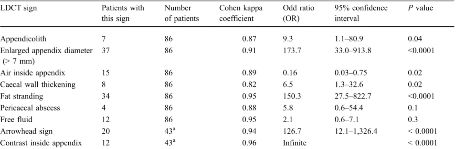
In univariate analyses, seven of the nine LDCT signs used in this study to assess (or rule out) appendicitis were significantly correlated with the final presence or absence of appendicitis (Table 4). Contrast medium within the appendix was never found in association with appendicitis and therefore displayed the higher (infinite) odds ratio (OR) among all evaluated LDCT criteria (Fig.3). Since the two signs associated with the administration of oral contrast medium (contrast medium within appendix and arrowhead sign) were only evaluated in a subset (n=43) of Table 2 Sensitivity and specificity of low-dose CT (LDCT) and standard-dose contrast-enhanced CT (standard CT) for detecting appendicitis, in slim and non-slim patients, as compared with clinical and surgical follow-up
Slim patients (n=7) Non-slim patients (n=79)
Sensitivity Specificity Sensitivity Specificity
LDCT 50 (2/4) 67 (2/3) 100 (33/33) 98 (45/46)
Standard CT 100 (4/4) 67 (2/3) 100 (33/33) 98 (45/46)
First number in each box corresponds to percentage found; patient numbers are enclosed in parentheses
Fig. 2 A 34-year-old woman with clinical suspicion of appendicitis. a Axial LDCT image at the level of the upper abdomen shows an enlarged right kidney and a discrete stranding of the perirenal fat (arrowheads), suggestive of acute pyelonephritis. b Axial contrast-enhanced standard-dose CT image, at the same level as (a), shows the stranding of perirenal fat (arrowheads), several peripheral hypodensities of the renal cortex (asterisks) and a ureteral wall enhancement (arrow), confirming the diagnosis of pyelonephritis
Table 3 Number of LDCT and CT suggestive of an alternative diagnosis with regard to the definite diagnoses at patient discharge Diagnoses at patient discharge LDCT Standard CT
Right ureteral stone (n=3) 3 3
Inflammation of the urinary tract (n=5) 4 5 Perforated duodenal ulcer (n=1) 1 1 Tubo-ovarian abscess or torsion (n=3) 2 2 Small bowel obstruction (n=3) 3 3
Acute cholecystitis (n=2) 2 2
Mesenteric panniculitis (n=1) 1 1
Non-Hodgkin lymphoma (n=1) 1 1
Mesenteric adenitis (n=1) 0 1
Terminal ileitis (n=2) 2 2
Colitis (including diverticulitis) (n=5) 4 5
Ovarian cyst (n=1) 1 1
Intermittent umbilical herniation (n=1) 0 0
patients, they were not included in the stepwise logistic regression model. Of the five non-contrast-dependent signs evaluated in 86 patients, only three remained statistically significant on multivariate analysis: enlarged appendix (OR 27.9, 95% CI [3.17–245.39]), periappendiceal fat stranding (OR 25.2, 95% CI [1.93–330.15]), gas within appendix (OR 0.014, 95% CI [0.002–0.893]).
Discussion
In the current study the specificity of LDCT to depict acute appendicitis was similar (96%) to standard enhanced CT, while its sensitivity was slightly lower (95% and 100%, respectively). In non-slim patients, both techniques had equivalent diagnostic performances, achieving sensitivities and specificities close to 100%. Therefore, our data suggest that, in patients with BMI > 18.5, LDCT may replace standard CT for the initial evaluation of patients with suspected appendicitis. Only one series to date has specifically compared unenhanced CT with enhanced CT in the same patient population [32]. The diagnostic accuracy of standard CT for acute appendicitis was improved significantly with the use of i.v. contrast media when“the borders of appendix are difficult to discern—on non i.v. enhanced CT—because of minimal retroperitoneal fat”.
The concern of using CT without i.v. contrast medium in the diagnostic workup of acute appendicitis in slim patients has also been addressed in a prior study [33] in which the authors reported that eight of ten patients in whom CT (without i.v. or oral contrast medium) failed to diagnose appendicitis were slender people. In spite of the fact that the number of slim patients was small (n=7), our data demonstrate that LDCT cannot replace conventional CT in these patients (50% false negative rate).
Only one series evaluated a low-dose CT protocol (< 50 mAs) in an adult patients population for the diagnosis of appendicitis [22]. In this study, LDCTs and CTs were Table 4 Low-dose CT (LDCT) signs predictive of appendicitis: univariate analysis
LDCT sign Patients with
this sign Number of patients Cohen kappa coefficient Odd ratio (OR) 95% confidence interval P value Appendicolith 7 86 0.87 9.3 1.1–80.9 0.04
Enlarged appendix diameter (> 7 mm)
37 86 0.91 173.7 33.0–913.8 <0.0001
Air inside appendix 15 86 0.89 0.16 0.03–0.75 0.02
Caecal wall thickening 8 86 0.82 6.5 1.3–32.6 0.02
Fat stranding 34 86 0.95 150.3 27.5–822.7 <0.0001
Pericaecal abscess 4 86 0.88 5.8 0.6–54.4 0.1
Free fluid 12 86 0.95 2.1 0.6–7.1 0.3
Arrowhead sign 20 43a 0.94 126.7 12.1–1,326.4 < 0.0001
Contrast inside appendix 12 43a 0.96 Infinite < 0.0001
a
Patients with caecal opacification only
Fig. 3 A 27-year-old woman admitted with right-lower quadrant pain with true negative LDCT and standard-dose CT for appendi-citis. a Axial LDCT. b Standard-dose enhanced CT images at the level of the pelvis. Appendix is of normal size and is filled with contrast (arrow). In the absence of signs suggestive of appendicitis or alternative diagnosis, both examinations were considered normal. Patient was discharged after complete relief of her clinical symptoms
performed without oral or i.v. contrast administration. Sensitivities of LDCT and CT were similar to our results (close to 100%); however, our protocols achieved higher specificities (96% in our study, 89% in Keyzer et al.’s study). These results are probably explained by the fact that, in our series, depiction of contrast media within the appendix excluded appendicitis with a high certainty. This observation, along with the finding that arrowhead sign was among the three most predictive signs of appendicitis by univariate analysis, highlights the importance of caecal opacification before performing LDCT. Many series also emphasized the importance of caecal opacification before scanning patients with suspicion of acute appendicitis [1,2,
7–9,31,34–36]. In a recent review of 60 patients with false negative diagnoses of appendicitis by standard CT, Levine et al. stressed the benefit of caecal opacification, especially when imaging lean patients [26]. Those authors observed caecal opacification in 34 (94%) of 36 patients with a correct diagnosis of appendicitis by CT and in only 12 (50%) of 24 patients with a false negative CT (p<0.001). Our results substantiate these findings, since LDCT was 100% and 86% sensitive in depicting appendicitis in patients with and without caecal opacification, respectively.
In the current study, an enlarged appendiceal diameter (≥ 7 mm) and the presence of periappendiceal fat stranding were the most significant non-contrast-dependent LDCT signs associated with appendicitis on multivariate analysis. These signs were also reported by Keyzer et al. to be highly predictive of appendicitis by LDCT [22]. A high inter-observer agreement was reported in both the latter series and ours. These findings suggest that the most predictive LDCT signs to assess or rule out appendicitis are reproducible and relatively observer-independent.
An alternative diagnosis was found by CT in 34% of patients in our study, which is consistent with other series [4,6,22]. Our LDCT protocol achieved 83% sensitivity to depict alternative diagnoses. This percentage is also in the range of previously reported studies using standard-dose CT, without i.v. enhancement, to assess patients with abdominal pain [7, 22]. These findings suggest that performance of CT to detect alternative diagnoses is independent of the radiation dose [22]. No influence of the BMI on the depiction of alternative diagnoses was observed in our study; however, the small number of patients with alternative diagnoses in each subset of BMI did not allow us to perform meaningful statistics.
Clinical suspicion of appendicitis (adult patients)
Sonography Male Female LDCT Standard CT Positive for appendicitis or gynecological pathology Negative or indeterminate Appendicitis Stop Alternative diagnosis No appendicitis No alternative diagnosis
Management according to imaging findings Discuss standard CT
(according to clinical presentation)
BMI >18.5 BMI <18.5
18.5 > BMI <30 BMI <18.5 or >30 BMI >30 BMI <30
Our data showed that standard CT with administration of i.v. contrast media allowed a 10% improvement in sensitivity with regard to LDCT for alternative diagnoses (93% versus 83%), while specificities were similar for both techniques (100% and 96%, respectively).
Since LDCT achieves similar performances as standard CT with i.v. enhancement for the diagnosis of acute appendicitis in patients with BMI >18.5, and similar sensitivity and specificity as unenhanced standard-dose CT for detection of alternative diagnoses [33, 35], it could constitute a useful tool for the triage of patients with suspicion of appendicitis towards standard i.v. enhanced CT. Indeed, injecting i.v. contrast media in patients with suspicion of acute appendicitis might only be required in a limited number of patients with no evidence of appendicitis or alternative diagnosis on LDCT. The decision to perform contrast-enhanced standard CT after a negative LDCT should therefore be tailored to the patients’ clinical presentation, as also suggested in prior studies [37–39].
Some limitations of the current study must still be addressed. First, in spite of the fact that LDCTs were collected in a prospective fashion, they were not immedi-ately interpreted and therefore not used in true acute clinical conditions. Second, our study design did not evaluate the influence of the radiologist’s training level on LDCT interpretation. Indeed, a radiologist not accustomed with this technique may require a learning experience before being confident in interpreting LDCT images.
Third, our study was not designed to assess the percentage of patients in whom LDCT could have replaced standard CT, and the benefit in terms of reduction of radiation dose. Nevertheless, our data suggest that LDCT would have been sufficient to assess the diagnosis of appendicitis in at least 41% of our patient population (33/79) with a BMI≥ 18.5, without need for further examination. The dose reduction among patients with a negative LDCT for appendicitis (with or without alternative diagnosis) cannot be inferred from our data. Finally, wide confidence intervals were found for the most important predictors of appendicitis diagnosis. This can be explained by the relatively small numbers of subjects included in our analyses.
In spite of these limitations, our results suggest that LDCT may play a significant role in the initial assessment of patients with suspected appendicitis. Therefore, we propose an algorithm to further evaluate our LDCT protocol (Chart1). Although sonography was not included in the current study, we have integrated this imaging method in our algorithm because many authors suggest that sonography should be used in women with right-lower quadrant pain [37–39]; indeed, a gynaecological cause can often be suggested by sonography.
We urge emergency clinicians and radiologists to perform prospective studies to validate this algorithm and to determine to what extent it will reduce the radiation dose delivered to patients admitted with a suspicion of acute appendicitis.
References
1. Naffaa LN, Ishak GE, Haddad MC (2005) The value of contrast-enhanced helical CT scan with rectal contrast enema in the diagnosis of acute ap-pendicitis. Clin Imaging 29:255–258 2. Funaki B, Grosskreutz SR, Funaki CN
(1998) Using unenhanced helical CT with enteric contrast material for sus-pected appendicitis in patients treated at a community hospital. AJR Am J Roentgenol 171:997–1001
3. Stroman DL, Bayouth CV, Kuhn JA et al (1999) The role of computed tomo-graphy in the diagnosis of acute ap-pendicitis. Am J Surg 178:485–489 4. Rhea JT, Halpern EF, Ptak T, Lawrason
JN, Sacknoff R, Novelline RA (2005) The status of appendiceal CT in an urban medical center 5 years after its introduction: experience with 753 pa-tients. AJR Am J Roentgenol 184:1802–1808
5. Blackmore CC, Terasawa T (2006) Optimizing the interpretation of CT for appendicitis: modeling health utilities for clinical practice. J Am Coll Radiol 3:115–121
6. Lane MJ, Liu DM, Huynh MD, Jeffrey RB Jr, Mindelzun RE, Katz DS (1999) Suspected acute appendicitis: none-nhanced helical CT in 300 consecutive patients. Radiology 213:341–346 7. Rao PM, Rhea JT, Novelline RA,
Mostafavi AA, Lawrason JN, McCabe CJ (1997) Helical CT combined with contrast material administered only through the colon for imaging of suspected appendicitis. AJR Am J Roentgenol 169:1275–1280
8. Walker S, Haun W, Clark J, McMillin K, Zeren F, Gilliland T (2000) The value of limited computed tomography with rectal contrast in the diagnosis of acute appendicitis. Am J Surg 180:450– 454 discussion 454–455
9. Wijetunga R, Tan BS, Rouse JC, Bigg-Wither GW, Doust BD (2001) Diag-nostic accuracy of focused appendiceal CT in clinically equivocal cases of acute appendicitis. Radiology 221:747– 753
10. Johnson PT, Horton KM, Mahesh M, Fishman EK (2006) Multidetector computed tomography for suspected appendicitis: multi-institutional survey of 16-MDCT data acquisition protocols and review of pertinent literature. J Comput Assist Tomogr 30:758–764 11. Dixon AK, Goldstone KE (2002)
Ab-dominal CT and the Euratom Directive. Eur Radiol 12:1567–1570
12. van Breda Vriesman AC, Kole BJ, Puylaert JB (2003) Effect of ultraso-nography and optional computed tomography on the outcome of appen-dectomy. Eur Radiol 13:2278–2282 13. Keyzer C, Zalcman M, De Maertelaer
V et al (2005) Comparison of US and unenhanced multi-detector row CT in patients suspected of having acute appendicitis. Radiology 236:527–534 14. Gracey D, McClure MJ (2007) The
impact of ultrasound in suspected acute appendicitis. Clin Radiol 62:573–578
15. Garcia-Aguayo FJ, Gil P (2000) Sonography in acute appendicitis: di-agnostic utility and influence upon management and outcome. Eur Radiol 10:1886–1893
16. Balthazar EJ, Birnbaum BA, Yee J, Megibow AJ, Roshkow J, Gray C (1994) Acute appendicitis: CT and US correlation in 100 patients. Radiology 190:31–35
17. Doria AS, Moineddin R, Kellenberger CJ et al (2006) US or CT for diagnosis of appendicitis in children and adults? A meta-analysis. Radiology 241:83–94 18. Wise SW, Labuski MR, Kasales CJ et
al (2001) Comparative assessment of CT and sonographic techniques for appendiceal imaging. AJR Am J Roentgenol 176:933–941
19. Marincek B (2002) Nontraumatic ab-dominal emergencies: acute abab-dominal pain: diagnostic strategies. Eur Radiol 12:2136–2150
20. Poletti PA, Platon A, Rutschmann OT, Schmidlin FR, Iselin CE, Becker CD (2007) Low-dose versus standard-dose CT protocol in patients with clinically suspected renal colic. AJR Am J Roentgenol 188:927–933
21. Poletti PA, Platon A, Rutschmann OT et al (2006) Abdominal plain film in patients admitted with clinical suspi-cion of renal colic: should it be replaced by low-dose computed tomography? Urology 67:64–68
22. Keyzer C, Tack D, de Maertelaer V, Bohy P, Gevenois PA, Van Gansbeke D (2004) Acute appendicitis: comparison of low-dose and standard-dose unen-hanced multi-detector row CT. Radiol-ogy 232:164–172
23. Kim BS, Hwang IK, Choi YW et al (2005) Low-dose and standard-dose unenhanced helical computed tomo-graphy for the assessment of acute renal colic: prospective comparative study. Acta Radiol 46:756–763
24. Grayson DE, Wettlaufer JR, Dalrymple NC, Keesling CA (2001) Appendiceal CT in pediatric patients: relationship of visualization to amount of peritoneal fat. AJR Am J Roentgenol 176:497– 500
25. Garrow JS, Webster J (1985) Quetelet’s index (W/H2) as a measure of fatness. Int J Obes 9:147–153
26. Levine CD, Aizenstein O, Lehavi O, Blachar A (2005) Why we miss the diagnosis of appendicitis on abdominal CT: evaluation of imaging features of appendicitis incorrectly diagnosed on CT. AJR Am J Roentgenol 184:855– 859
27. Malone JF, Bischoff N (2008) IEC standards for radiological equipment: issues for the industry and for end users. Radiat Prot Dosimetry 129 (1–3):132–134 Epub 2008 May 10 28. ImPACT (2006) Imaging performance
assessment of CT scanners: a medical devices agency evaluation group. CT scanner matching data, tables of CTDI values in air, CTDIw, and phantom factor values, version 0.99x London UK.http://www.impactscan. org/ctdosimetry.htm. Accessed 14 July 2008
29. O’Daniel JC, Stevens DM, Cody DD (2005) Reducing radiation exposure from survey CT scans. AJR Am J Roentgenol 185:509–515 30. Rao PM, Rhea JT, Novelline RA
(1997) Sensitivity and specificity of the individual CT signs of appendicitis: experience with 200 helical appendi-ceal CT examinations. J Comput Assist Tomogr 21:686–692
31. Rao PM, Wittenberg J, McDowell RK, Rhea JT, Novelline RA (1997) Appen-dicitis: use of arrowhead sign for diagnosis at CT. Radiology 202:363– 366
32. Jacobs JE, Birnbaum BA, Macari M et al (2001) Acute appendicitis: compari-son of helical CT diagnosis focused technique with oral contrast material versus nonfocused technique with oral and intravenous contrast material. Ra-diology 220:683–690
33. Malone AJ Jr., Wolf CR, Malmed AS, Melliere BF (1993) Diagnosis of acute appendicitis: value of unenhanced CT. AJR Am J Roentgenol 160:763–766 34. Lee SY, Coughlin B, Wolfe JM, Polino
J, Blank FS, Smithline HA (2006) Prospective comparison of helical CT of the abdomen and pelvis without and with oral contrast in assessing acute abdominal pain in adult emergency department patients. Emerg Radiol 12:150–157
35. Tamburrini S, Brunetti A, Brown M, Sirlin C, Casola G (2007) Acute ap-pendicitis: diagnostic value of none-nhanced CT with selective use of contrast in routine clinical settings. Eur Radiol 17:2055–2061
36. Hershko DD, Awad N, Fischer D et al (2007) Focused helical CT using rectal contrast material only as the preferred technique for the diagnosis of suspected acute appendicitis: a prospective, ran-domized, controlled study comparing three different techniques. Dis Colon Rectum 50(8):1223–1229
37. Rao PM, Feltmate CM, Rhea JT, Schulick AH, Novelline RA (1999) Helical computed tomography in dif-ferentiating appendicitis and acute gy-necologic conditions. Obstet Gynecol 93:417–421
38. Birnbaum BA, Wilson SR (2000) Ap-pendicitis at the millennium. Radiology 215:337–348
39. Leschka S, Alkadhi H, Wildermuth S, Marincek B (2005) Multi-detector computed tomography of acute abdo-men. Eur Radiol 15:2435–2447