Lipid and Fatty Acid Composition of the Marine Brown Alga Dictyopteris
membranacea
Markus Hofmann and Waldemar Eichenberger
Department of Chemistry and Biochemistry, University of Bern, Freiestr. 3, CH-3012 Bern, Switzerland
Glycerolipids and fatty acids of D. membranacea (Dic-tyotales) were analysed. The betaine lipid DGTA and the glycolipids MGOG, DGDG and SQDG were major com-ponents. The phospholipids PE, PG, PI and PHEG were present in minor amounts only. This lipid pattern, which is characterised by the presence of DGTA and the absence of PC, has been found exclusively in brown algae belonging to the orders Dictyotales, Durvillaeales and Fucales. Major fatty acids were 16:0, 18:1, 18:2, al8:3, 18:4 and 20:4 acids. MGDG was the most unsaturated lipid with high levels of 18:4 acid. SQDG showed the highest degree of sat-uration containing a considerable proportion of 16:0 acid. DGTA contained 14 : 0,18:1,18:2 and 20:4 as major fatty acids. Among phospholipids, PE and PHEG had a very similar pattern which was enriched in 20:4 acid. Analysis of the positional distribution of fatty acids revealed that DGTA and MGDG were almost exclusivly of the "euka-ryotic" type, whereas SQDG was predominantly of the "prokaryotic" type. For the first time, molecular species of selected lipids have been analysed in a brown alga. In DGTA, 14:0/18:1, 14:0/18:2 and 14:0/20:4 were the main molecular species. In MGDG the highly unsaturated erl8:3/18:4, 18:4/18:4 and 18:4/20:5 were predominant. Key words: DGTA — Dictyopteris membranacea — Fatty acids — Lipids — Molecular species — Phaeophyceae.
Marine brown algae represent a considerable part of the littoral biomass (South and Whittick 1987) and many of them are considered to be important for industrial uses. Their high contents of minerals, vitamines and dietary fibres are interesting for nutritional purposes (Mabeau and Fleurence 1993). In addition, many of their constituents Abbreviations: DAG, diacylglycerol; DGDG, digalactosyl-diacylglycerol; DGTA, diacylglycerylhydroxymethyl-yV,7V,yV-tri-methyl-/?-alanine; DGTS, diacylglyceryl-MMW-trimethylhomo-serine; DPG, diphosphatidylglycerol; FID, flame ionization detector; GLC, gas liquid chromatography; RP-HPLC, re-versed-phase high-performance liquid chromatography; MGDG, monogalactosyldiacylglycerol; PC, phosphatidylcholine; PE, phos-phatidylethanolamine; PG, phosphatidylglycerol; PHEG, phospha-tidyl-O-[Ar-(2-hydroxyethyl)glycine]; PI, phosphatidylinositol; sn, stereospecific numbering; PUFAs, polyunsaturated fatty acids; SQDG, sulfoquinovosyldiacylglycerol; TAG, triacylglycerol; TLC, thin-layer chromatography.
are of interest from a pharmaceutical point of view (Rad-wan 1991). Considering brown algal lipids, some of the generally observed trends have been summarised (Dembit-sky 1996, Harwood and Jones 1989). The distinct fatty acid and lipid pattern of brown algae in comparison to higher plants (Gurr and Harwood 1991), suggest the metabolic pathways in this group of organisms to be different from those of higher plants. Little is known, e.g., about the bio-synthesis of the mainly "eukairyotic" galactolipids (Arao and Yamada 1989, Jones and Harwood 1992) and PUFAs such as 18:4, 20:4 and 20:5 (Harwood and Jones 1989). Also the metabolic role of additional lipids like the phos-pholipid PHEG (Eichenberger et al. 1995) and the betaine lipid DGTA (Araki et al. 1991) is not known. DGTA was shown to be widely distributed among Phaeophyceae (Eichenberger et al. 1993) and has been suggested to sub-stitute for PC in some cases. Therefore, members of the orders Dictyotales, Durvillaeales and Fucales which all lack PC (Eichenberger et al. 1993) are supposed to be excellent organisms to study the metabolic role of this betain lipid. Experiments on the incorporation of [l-l4C]acetate into lipids of the Fucales Fucus serratus (Smith and Harwood 1984), Fucus vesiculosus and Ascophyllum nodosum (Jones and Harwood 1993) have already revealed a signifi-cant labelling of DGTA. For our investigations, we used for the first time a member of the Dictyotales, namely
Dic-tyopteris membranacea which can be cultivated under
labo-ratory conditions. The aim of this study was to establish an analytical background for the subsequent work on the lipid metabolism and the metabolic role of DGTA in D.
mem-branacea. Therefore, the lipid composition, the fatty acid
pattern of single and total lipids as well as the positional dis-tribution of fatty acids among the diglyceride moieties of glycolipids and DGTA were analysed. Furthermore, the molecular species of DGTA and MGDG have been separat-ed and quantifiseparat-ed for the first time in a brown alga.
Materials and Methods
Plant material—A unialgal clonal culture of a Dictyopteris membranacea (Stackhouse) Batters tetrasporophyte (collected in Villefranche-sur-mer, France, Mediterranean Sea) was obtained from Prof. D.G. Muller, Faculty of Biology, University of Konstanz, Germany. The alga was cultivated continuously in glass dishes with 50 ml of culture medium prepared from autoclaved natural seawater (North Sea, salinity 2%%,) supplemented with PES as specified by Starr and Zeikus (1993). The alga was grown 1046
in a light/dark cycle of 12/12h at 18°C under white fluorescent light (60/JE m~2 S~') and weekly provided with new culture medi-um.
Lipid isolation and analysis—Total lipids were extracted with hot methanol containing 0.05% butyl hydroxytoluene as an antiox-idant. The crude extract was evaporated or dried under a stream of N2 and further purified by phase partition between 1 volume
each of sodium bicarbonate (1%) and diethyl ether. Lipids were separated on precoated silica gel plates (Merck 5715) with a stand-ard solvent system containing chloroform/ methanol/ water (65 : 25 : 4 , v/v) in the 1st dimension and chloroform/ methanol/ isopropylamine/ cone, ammonia (65 : 35 : 0.5 : 5, v/v) in the 2nd dimension. Spots were detected under UV light (366 nm) after spraying with 2',7'-dichlorofluorescein. Single lipids were iden-tified by spraying with specific reagents (Vogel and Eichenberger 1990). Betaine lipids were stained with Dragendorff reagent, phos-pholipids with molybdenum blue and glycolipids with anthrone reagent. Ninhydrin was used for the staining of amino groups. TAG was separated from polar lipids and pigments by TLC with chloroform/ methanol (50: 1, v/v). Lipids were quantified by their constituent fatty acids by GLC using eicosanoic acid (20:0) methyl ester as an internal standard.
Fatty acid analysis—Fatty acid methyl esters from total lipids were obtained by alkaline hydrolysis with KOH/ water/ ethanol ( 1 : 2 : 20, w/v/v), followed by methylation with diazomethane (Vogel and Eichenberger 1992). Fatty acid methyl esters from single lipids were prepared by transesterification with sodium methoxide (Thies 1971).
For analytical separation, a Shimadzu GC-14A equiped with FID was used. The separations were performed on a fused silica capillary column (25 m length, 0.25 mm I.D.) coated with chemi-cally bound Carbowax 20 M at 185-210°C (2°C min"1) with H2 as
carrier gas. Processing of data and integration of peaks was done with a Shimadzu C-R4A integrator.
Analysis of the positional distribution of fatty acids—The positional distribution of fatty acids among the sn-l and sn-2 posi-tion of glycolipids and DGTA was determined by cleavage with a lipase from Rhizopus arrhizus (Fischer et al. 1973). The products were separated on precoated silica gel plates (Merck 5719) using chloroform/ methanol/ water (70: 15 : 2 , v/v) for MGDG, chloroform/ methanol/ water (65 : 35 : 8, v/v) for DGDG and DGTA and chloroform/ methanol/ acetic acid/ water (65 : 35 : 6 : 4, v/v) for SQDG. For GLC analysis, the fatty acids cleaved from the sn-l position were methylated with diazomethane. The fatty acid methyl esters of the sn-2 position were prepared by trans-esterification of the lyso compounds.
Analysis of molecular species—Pure lipids were obtained by TLC separation of total lipids. DGTA was first isolated using ace-tone/ benzene/ methanol/ water ( 8 : 3 : 2 : 1 , v/v) as a solvent. The lipid was then eluted with methanol, and for removing pigments and PHEG, chromatographed with chloroform/ metha-nol/ isopropylamine/ cone, ammonia (65 : 35 : 0.5 : 5, v/v). For the isolation of MGDG, chloroform/ methanol/ water (65 : 2 5 : 4 , v/v) and chloroform/ methanol/ isopropylamine/ cone, ammonia (65 : 35 :0.5 : 5, v/v) were used for the 1st and 2nd step, respectively.
Molecular species were separated by RP-HPLC on a Shimadzu LC-6A with a Nucleosil 100-5 C,8 (250x4 mm, Macherey Nagel)
column. For the separation of DGTA molecular species, metha-nol/ water/ acetonitril (80 : 12 : 8, v/v) (A) and methametha-nol/ water/ acetonitril (94 : 3.5 : 2.5, v/v) with 20 mM choline chlo-ride (B) (Vogel and Eichenberger 1992) were used as solvents. The gradient was from 70% to 100% B in 40 min with a flow rate of
1.5 ml min ' and detection was at 202 nm with a Shimadzu SPD-6A detector. Molecular species of MGDG were separated in a isocratic manner with methanol/ water (94: 6, v/v) as a sol-vent (Giroud et al. 1988). The flow rate was 1.1 ml min"1 and
de-tection was at 210 nm. Peaks were collected and single species of both lipids were quantified by determination of their constituent fatty acids by GLC with 20:0 methyl ester as an internal standard.
Results
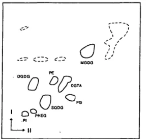
Lipid composition—A typical separation of the total lipids of D. membranacea by two-dimensional TLC is shown in Fig. 1 and the amounts of the different com-pounds are presented in Table 1.
Each lipid component was identified by its mobility and by its reaction with specific staining reagents. The gly-colipids MGDG, DGDG and SQDG were major com-ponents representing 83.1% of the polar lipids. MGDG was the most abundant component (44.1%), followed by SQDG (25.5%) and DGDG (13.5%). The ratio of MGDG to DGDG was 3.3 in this organism. Another prominent constituent was the betaine lipid DGTA accounting for 9.0% of the polar lipids. In contrast, the phospholipids PE, PG, PI and PHEG, which has recently been identified (Eichenberger et al. 1995), were present in minor amounts only. PC was not detectable with neither molybdenum blue nor Dragendorff reagent, although with the latter a de-tection limit of 0.5 fig mg~" total lipid was found under the-se conditions (Eichenberger et al. 1993). TAG was not the- sepa-rated from the main spot of unpolar lipids and pigments (Fig. 1) and was therefore isolated by a separate procedure. The amount of TAG was 105 nmol mg~" total lipid.
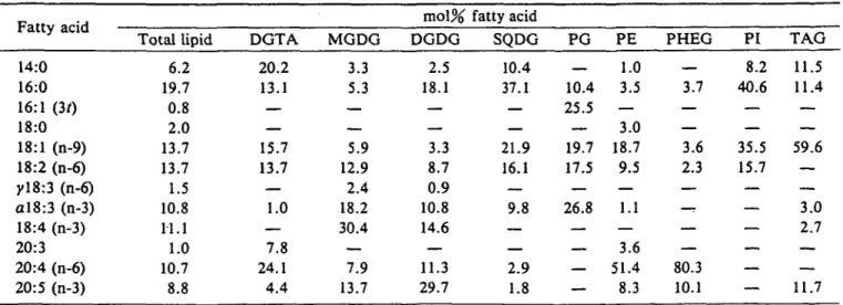
Fatty acid composition of total lipids and single lipid classes—The fatty acids of total lipids and their distribu-tion among different lipid classes are summarized in Table 2.
"0
V_/SQOQ (EGo
\ /
o V
MQOQ TA PGFig. 1 Two dimensional TLC of the total lipids from D. mem-branacea. Pigments are indicated by broken lines. Conditions were as described in Materials and Methods.
Table 1 Lipid composition of D. membranacea
Lipid nmol per mg
total lipid % polar lipid
DGTA MGDG DGDG SQDG PG PE PHEG PI TAG 37.0 181.9 55.6 105.3 11.1 12.9 4.2 4.8 105.2 9.0 44.1 13.5 25.5 2.7 3.1 1.0 1.2 — Values are means of 2 determinations.
The major fatty acids ( > 10% of the total) in D.
mem-branacea were 16:0, 18:1, 18:2, al8:3, 18:4 and 20:4 acids.
Minor components were 14:0 and 20:5, whereas 16:1(30, 18:0, yl8:3 and 20:3 acids were present in small amounts only. Considering the different chain lengths, Cu and CI6
acids accounted for almost 26.1%, the C18 acids for 52.8%
and C20 acids for 20.5% of the total.
Each lipid class was characterised by a distinct pattern of fatty acids. The highest variability of fatty acids was ob-served in the galactolipids. MGDG was the most unsaturat-ed lipid containing 18:2, al8:3, 18:4 and 20:5 as major fat-ty acids. DGDG was slightly more saturated than MGDG due to a higher proportion of 16:0. Furthermore 20:5 was the main fatty acid in DGDG followed by 16:0, 18:4, 20:4 and al8:3. It is remarkable that 18:4 acid is limited to the galactolipids. In contrast, SQDG was the most saturated lipid due to high levels of 16:0 and 14:0 acids and also con-tained 18:1, 18:2 and al8:3 acids as major components. Of
DGTA, high amounts of 14:0, 16:0, 18:1, 18:2 and 20:4 acids were characteristic. High levels of 20:4 acids were also observed in PE and PHEG, where this fatty acid ac-counted for 51% and 80%, respectively. PG which proba-bly contained traces of DPG, was occupied by 16:0, 18:1, 18:2, ctl8:3 and 16:1(30 acids as major components. The presence of 16:1(30 acid was a specific feature of PG. PI showed a rather simple fatty acid composition with mainly 14:0, 16:0, 18:1 and 18:2 acids. TAG contained large amounts of 18:1 (60%) and almost equal amounts (12%) each of 14:0, 16:0 and 20:5 acids.
Positional distribution of fatty acids in DGTA and the glycolipids—Table 3 shows the positional distribution of
fatty acids in the major lipid classes.
In DGTA, the sn-l position was mainly occupied by 14:0 and 16:0 acids, whereas the sn-2 position was occupied by 94% by C18 and CM acids indicating the predominantly
"eukaryotic" structure of the betaine lipid. Interestingly, 20:4 acid was distributed about equally among both posi-tions. Both galactolipids showed high levels of C13 acids at
the sn-2 position with 18:4 acid being almost exclusively located at this position. In contrast, 14:0, 20:4 and 20:5 acids were concentrated at the sn-l position in these lipids. Based on the total amount of Cig and CM fatty acids in the
sn-2 position, MGDG was by 97% of the "eukaryotic"
type. DGDG, due to the elevated amount of 16:0 acid in the sn-2 position, was by only 75% of the "eukaryotic" type. In SQDG, in contrast, the "prokaryotic" structure predominated (53%) because of the large amount of 16:0 acid in the sn-2 position of this lipid in which 14:0, 20:4 and 20:5 acids were restricted to the sn-l position.
Molecular species of DGTA and MGDG—For the first
time, the molecular species of DGTA and MGDG from a brown alga have been separated and quantified. A
RP-Table 2 Fatty acid composition in lipids of D. membranacea Fatty acid 14:0 16:0 16:1 (30 18:0 18:1 (n-9) 18:2 (n-6) yl8:3 (n-6) al8:3 (n-3) 18:4 (n-3) 20:3 20:4 (n-6) 20:5 (n-3) Total lipid 6.2 19.7 0.8 2.0 13.7 13.7 1.5 10.8 11.1 1.0 10.7 8.8 DGTA 20.2 13.1 — — 15.7 13.7 — 1.0 — 7.8 24.1 4.4 MGDG 3.3 5.3 — — 5.9 12.9 2.4 18.2 30.4 — 7.9 13.7 mol% DGDG 2.5 18.1 — — 3.3 8.7 0.9 10.8 14.6 — 11.3 29.7 fatty acid SQDG 10.4 37.1 — — 21.9 16.1 — 9.8 — — 2.9 1.8 PG _ 10.4 25.5 — 19.7 17.5 — 26.8 — — — — PE 1.0 3.5 — 3.0 18.7 9.5 — 1.1 — 3.6 51.4 8.3 PHEG __ 3.7 — — 3.6 2.3 — — 80.3 10.1 PI 8.2 40.6 — — 35.5 15.7 — — — — — — TAG 11.5 11.4 — — 59.6 — — 3.0 2.7 — — 11.7 Values are means of 2 determinations.
Table 3 Positional distribution of fatty acids in DGTA and the glycolipids of D. mem-branacea Fatty acid 14:0 16:0 18:0 18:1 (n-9) 18:2 (n-6) yl8:3 (n-6) al8:3 (n-3) 18:4 (n-3) 20:3 20:4 (n-6) 20:5 (n-3) 2C,4+C,4 DGTA sn-l 40.6 24.6 0.6 3.2 4.5 — 1,1 — — 21.7 3.7 65.2 34.8 sn-2 5.7 — 24.1 23.2 — — — 8.8 29.2 8.9 5.7 94.3 MGDG sn-l 2.8 7.4 — 3.0 11.5 — 22.0 8.1 — 14.2 31.1 10.2 89.8 sn-2 2.9 — 10.7 14.7 3.8 13.3 50.4 — 2.1 2.1 2.9 97.1 mol% DGDG sn-l 9.1 17.1 3.9 3.8 5.5 — 6.9 — — 15.1 38.6 26.2 73.8 sn-2 . 24.8 — 10.2 20.4 0.8 18.6 24.0 — 0.7 0.6 24.8 75.2 SQDG sn-l 13.9 19.0 1.6 35.8 10.8 — 8.3 — — 6.6 3.9 32.9 77.1 sn-2 53.3 — 11.4 24.3 — 11.0 — — — — 53.3 46.7 Values are means of 2 determinations.
HPLC separation of DGTA molecular species is shown in Fig. 2. The decreasing base line during the separation is probably due to the increasing choline chloride concentra-tion. The fractions corresponding to the different peaks have been further analysed and quantified by GLC and the data are presented in Table 4.
The most abundant species were 14:0/18:1, 14:0/18:2 and 14:0/20:4 combinations, accounting for almost 40% of the total. The bulk of acyl combinations contained
Time (ram)
Fig. 2 RP-HPLC separation of DGTA molecular species. Peak numbers refer to Table 4. Conditions were as described in Materials and Methods, u: unknown, no fatty acids detected.
either a 14:0 or 16:0 residue which could be attributed to the sn-l position of these molecules according to Table 3. Based on the fatty acid composition, fractions 1-4 and 8 contained two molecular species each. The molecular
spe-Table 4 Molecular species of DGTA from D.
mem-branacea
Acyl
combination peak number
m o l % molecular species 14:0/18:1 14:0/18:2 14:0/18:3 14:0/20:3 14:0/20:4 14:0/20:5 16:0/18:1 16:0/18:2 16:0/18:3 16:0/20:3 16:0/20:4 16:0/20:5 18:2/20:4 18:2/20:5 20:4/20:4 20:4/20:5 8 4 2 6 3 1 11 9 .7 10 8 5 4 2 3 1 12.7 13.1 5.4 2.2 14.0 8.0 3.2 3.2 1.9 1.0 9.6 2.2 8.3 1.6 9.6 4.1
Values are means of 2 determinations. Major species are underlin-ed. The given order of the acyl chains does not necessarily corre-spond with their positional distribution. Peak numbers refer to Fig. 2.
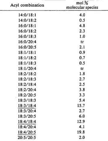
Table 5 Molecular species of MGDG from D. mem-branacea Acyl combination 14:0/18:1 14:0/18:2 16:0/18:1 16:0/18:2 16:0/18:3 16:0/20:4 16:0/20:5 18:1/18:1 18:1/18:2 18:1/18:3 18:1/20:4 18:2/18:2 18:2/18:3 18:2/18:4 18:2/20:4 18:2/20:5 18:3/18:3 18:3/18:4 18:3/20:4 18:3/20:5 18:4/18:4 18:4/20:4 18:4/20:5 20:5/20:5 m o l % molecular species 4.0 0.5 4.8 2.3 1.0 tr 2.1 0.9 0.7 0.5 tr 1.8 2.7 2.5 3.8 3.3 5.4 15.7 2.7 6.0 12.9 4.1 19.8 2.0
Values are means of 2 determinations. Major species are underlin-ed. The given order of the acyl chains does not necessarily corre-spond with their positional distribution.
cies composition of MGDG was even more complex con-taining a large number of acyl combinations, as expected from the complex fatty acid composition of this lipid and as shown in Table 5.
For example, almost all possible acyl combinations containing two C,8 residues, except 18:1/18:4, have been
identified. The major species were the highly unsaturated al8:3/18:4, 18:4/18:4 and 18:4/20:5 combinations accoun-ting for almost 50% of total MGDG and reflecaccoun-ting on the molecular level, the high degree of unsaturation of this lipid. It is interesting to note that the 14:0/18:1 and 16:0/ 18:1 combinations occurred in both MGDG and DGTA.
Discussion
The lipid and fatty acid composition of Dictyopteris
membranacea (Dictyotales) was analysed in detail in order
to obtain an analytical basis for a subsequent work on the lipid metabolism of this alga. The lipid pattern was dominated by the glycolipids MGDG, SQDG and DGDG
which accounted for as much as 83% of the polar lipids. MGDG was the most prominent lipid in D. membranacea, as found in many of the brown algae examined so far (Dem-bitsky et al. 1990, Dem(Dem-bitsky 1996) and as reported also for higher plants (Harwood and Jones 1989). The ratio of MGDG to DGDG was 3.3 and thus even higher than in higher plants (Gurr and Harwood 1991). In contrast, DGDG was found to be the most abundant lipid in some species of the order Ectocarpales (Dembitsky 1996), whereas SQDG predominated in Hizikia fusiformis (Araki et al. 1991), Fucus vesiculosus (Jones and Harwood 1992) and F.serratus (Smith and Harwood 1984) all of which belong to the order Fucales. SQDG was the most promi-nent lipid also in Padina pavonia and Taonia atomavia of the order Dictyotales (Dembitsky 1996) indicating the quan-titative importance of SQDG for marine chromophyte algae as reported earlier (Harwood and Jones 1989, Heinz 1993). These findings clearly show that the relative amounts of glycolipids considerably vary within the class of brown algae.
The phospholipids found in D. membranacea were PE, PG, PHEG and PI. PHEG has been exclusively found in brown algae so far and its structure has been reported recently (Eichenberger et al. 1995). This hpid was demon-strated to be distributed in all of the brown algal orders (Eichenberger et al. 1995, Khotimchenko and Titlyanova 1996). In contrast, PC was not detected in D.
mem-branacea. The absence of PC is a characteristic feature of
the brown algal orders Dictyotales, Durvillaeales and Fucales all of which, however, produce the zwitterionic be-taine lipid DGTA (Eichenberger et al. 1993). In D.
mem-branacea this betaine lipid accounted for 9% of the polar
lipids. An even higher amount (24%) has been reported for
F. vesiculosus (Jones and Harwood 1992). The occurrence
of DGTA in brown algae was shown to reflect their tax-onomy (Araki et al. 1991, Eichenberger et al. 1993). Within the genus Ectocarpus, DGTA is a taxonomical marker to distinguish single species (Miiller and Eichenberger 1994).
D. membranacea contained significant amounts of
TAG accounting for 20 mol% of the total lipids. This value might vary according to the physiological conditions, since in field-grown F. serratus, seasonal variations of the TAG content were observed (Kim et al. 1996).
The main fatty acids of D. membranacea were 16:0, 18:1, 18:2, 18:4, al8:3, 20:4, 20:5 and 14:0. This is in ac-cordance with the results obtained for other brown algae (Aknin et al. 1992, Arao and Yamada 1989, Dembitsky et al. 1990, Fleurence et al. 1994, Harwood and Jones 1989, Jones and Harwood 1992, Khotimchenko 1991, 1995, Vaskovsky et al. 1996). The 16:1 (n-5) acid, although reported for members of the genera Dictyota and
Dic-tyopteris (Aknin et al. 1992, Khotimchenko 1995), could
not be found in D. membranacea. The ratio of 20:4 to 20:5 acid (1.2) was in accordance with the predominance of 20:4
acid in brown algae (Harwood and Jones 1989), although a few exceptions have been reported (Dembitsky 1990). It should be noted, however, that the fatty acid composition is influenced by environmental factors such as light, heavy metals and temperature (Harwood and Jones 1989, Jones and Harwood 1993).
In the galactolipids of D. membranacea, PUFAs pre-dominated as in other brown algae (Araki et al. 1991, Arao and Yamada 1989, Jones and Harwood 1992, Khotim-chenko 1995, Smith and Harwood 1984). The 18:4 acid was almost restricted to the galactolipids in D. membranacea suggesting this fatty acid to be synthesized in the chloro-plast as reported for the haptophyte alga Isochrysis
gal-bana (Stern and Tietz 1993). The separation of the
mo-lecular species of the highly unsaturated MGDG which have been analysed for the first time in a brown alga, reveal-ed al8:3/18:4, 18:4/18:4 and 20:5/18:4 acyl combinations to be the main constituents. The presence of almost all of the possible C18/C18 combinations made a complete
separa-tion of species rather difficult. The posisepara-tional distribusepara-tion of fatty acids revealed "eukaryotic" MGDG to account for 97%. This value is in keeping with data from Padina
ascor-bens, Sargassum ringgoldianum and F. vesiculosus (Arao
and Yamada 1989, Jones and Harwood 1992).
DGDG was slightly more saturated than MGDG due to a higher level of 16:0 as also observed in other algae (wood and Jones 1989) and higher plants (Gurr and Har-wood 1991). In addition, the higher content of C^ acids in DGDG compared to MGDG is in accordance with data from several other brown algae (Araki et al. 1991, Arao and Yamada 1989, Khotimchenko 1995). Accordingly, the proportion of "eukaryotic" species in DGDG accounting for 75% is lower than in MGDG. The high proportions of "eukaryotic" galactolipids in the members of the Dic-tyotales and Fucales are surprising, because PC which generally acts as a DAG donor for the synthesis of "euka-ryotic" plastidial lipids (Roughan and Slack 1982) could not be detected in these algae (Eichenberger et al. 1993). This points towards the presence of an alternative pathway as already suggested for Cryptomonas CR-1 (Sato 1991b). SQDG was the most saturated lipid in D.
mem-branacea with relatively high proportions of 16:0 and 18:1
acids. The accumulation of 16:0 acid at the sn-2 position of the sulfolipid gives raise to a high proportion of "proka-ryotic" species (53%) in this lipid. It is interesting to note that in SQDG as well as in the galactolipids, C20 acids were restricted to the sn-l position as already observed in Ishige
okamurai, P. ascorbens, S. ringgoldianum and F. vesicu-losus (Arao and Yamada 1989, Jones and Harwood 1992).
DGTA of D. membranacea mainly contained 20:4, 14:0, 18:1, 18:2 and 16:0 acids. This is in agreement with data obtained for other members of the orders Dictyotales (Araki et al. 1991, Khotimchenko 1995, Dembitsky 1996) and Fucales (Jones and Harwood 1992). In DGTA of
D. membranacea, the 14:0 and 16:0 acids were
concen-trated in the sn-l position, whereas 18:1 and 18:2 acids were almost limited to the sn-2 position leading to the almost exclusively "eukaryotic" structure which has been also observed in F. vesiculosus (Jones and Harwood 1992) and in the unicellular algae Cryptomonas CR-1 (Sato 1991a) and Pavlova lutheri (Kato et al. 1995). The main mo-lecular species of DGTA in D. membranacea were 14:0/ 18:1, 14:0/18:2 and 14:0/20:4 combinations. Interestingly, a predominance of 14:0/18:1 and 14:0/18:2 acyl combina-tions has also been found in the betaine lipid DGTS of
Ochromonas danica where this structural isofner of DGTA
was shown to act as the primary acceptor of exogenous oleate and to be involved in the desaturation and redistribu-tion of fatty acids (Vogel and Eichenberger 1992). Also, rapid labelling of DGTA has been observed on incubation with [l-l4C]acetate of F.serratus (Smith and Harwood 1984), F. vesiculosus and A. nodosum (Jones and Harwood 1993) suggesting an active role of the betaine lipid in these organisms.
Among the phospholipids of D. membranacea, both PE and PHEG were enriched in 20:4 acid accounting for 51% in PE and for as much as 80% in PHEG. The fatty acid pattern of these lipids was very similar suggesting a biosynthetic relationship between the two compounds (Eichenberger et al. 1995). PG mainly contained a 18:3, 16:0 and 16:1(3/) acid which is specific for this lipid in algae (Harwood and Jones 1989) and higher plants (Gurr and Harwood 1991). For PI, 16:0 and 13:1 acids were typical in D. membranacea as well as in F. vesiculosus and
A. nodosum (Jones and Harwood 1992). In TAG, 14:0,
16:0 and 18:1 acids accounted for 83% of total fatty acids demonstrating the predominance of medium-chain fatty acids in this unpolar lipid in which the fatty acid composi-tion has been reported to undergo seasonal variacomposi-tions in
F. serratus (Kim et al. 1996). The role of the different polar
lipids, especially that of DGTA, in the metabolism of
D. membranacea is the subject of further investigations.
This work has been supported by the Swiss National Science Foundation (grant 31-45901.95). We are indebted to Prof. D.G. Miiller, Faculty of Biology, University of Konstanz, Germany, for the gift of the algal material and for helpful discussions.
References
Aknin, M.t Dogbevi, K., Samb, A., Kornprobsl, J-M., Gaydou, E.M. and
Miralles, J. (1992) Fatty acid and sterol compositions of eight brown algae from the senegalese coast. Comp. Biochem. Physiol. 102: 841-843. Araki, S., Eichenberger, W., Sakurai, T. and Sato, N. (1991) Distribution of diacylglycerylhydroxymethyltrimethyl-/?-alanine (DGTA) and phos-phatidylcholine in brown algae. Plant Cell Physiol. 32: 623-628. Arao, T. and Yamada, M. (1989) Positional distribution of fatty acids in
galactolipids of algae. Phytochemistry 28: 805-810.
Dembitsky, V.M. (1996) Betaine ether-linked glycerolipids: chemistry and biology. Prog. Lipid Res. 35: 1-51.
Glyco-lipids, phospholipids and fatty acids of brown algae species.
Phytochem-istry 29: 3417-3421.
Eichenberger, W., Araki, S. and Muller, D.G. (1993) Betaine lipids and phospholipids in brown algae. Phytochemistry 34: 1323-1333. Eichenberger, W., Bigler, P.. Gfeller, H., Gribi. C. and Schmid, C.E.
(1995) Phosphatidyl-O-[W-(2-hydroxyethyl) glycine] (PHEG), a new gly-cerophospholipid from brown algae (Phaeophyceae). /. Plant Physiol. 146: 398-404.
Fischer, W., Heinz, E. and Zeus, M. (1973) The suitability of lipase/rom
Rhizopus arrhizus delemar for analysis of fatty acid distribution in
dihex-osyl diglycerides, phospholipids and plant sulfolipids. Hoppe-Seyler's Z.
Physiol. Chem. 354: 1115-1123.
Fleurence, J., Gutbier, G., Mabeau, S. and Ceray, C. (1994) Fatty acids from 11 marine macro-algae of the French Brittany coast. J. Appl.
Phycol. 6: 527-532.
Giroud, C , Gerber, A. and Eichenberger, W. (1988) Lipids of
Chlamydo-monas reinhardtii. Analysis of molecular species and intracellular site(s)
of biosynthesis. Plant Cell Physiol. 29: 587-595.
Gurr, M.I. and Harwood, J.L. (1991) Lipid biochemistry. Chapman & Hall, London, pp. 246-297.
Harwood, J.L. and Jones, A.L. (1989) Lipid metabolism in algae. Adv.
Bot. Res. 16: 1-53.
Heinz, E. (1993) Recent investigations on the biosynthesis of the plant sulfolipid. In Sulfur Nutrition and Assimilation in Higher Plants. Edited by De Kok, L.J. et al. pp. 163-178. Academic Publishing, The Hague. Jones, A.L. and Harwood, J.L. (1992) Lipid composition of the brown algae Fucus vesiculosus and Ascophyllum nodosum. Phytochemistry 31: 3397-3403.
Jones, A.L. and Harwood, J.L. (1993) Lipid metabolism in the brown marine algae Fucus vesiculosus and Ascophyllum nodosum. J. Exp. Bot. 44: 1203-1210.
Kato, M., Hajiro-Nakanishi, K. and Miyachi, S. (1995) Polyunsaturated fatty acids and betaine lipids from Pavlova lutheri. Plant Cell Physiol. 36: 1607-1611.
Khotimchenko, S.V. (1991) Fatty acid composition of seven Sargassum spe-cies. Phytochemistry 30: 2639-2641.
Khotimchenko, S.V. (1995) Uncommon 16:1 (n-5) acid from Dictyota
dichotoma and fatty acids from some brown algae of Dictyotaceae. Phytochemistry 38: 1411-1415.
Khotimchenko, S.V. and Titlyanova, T.V. (1996) Distribution of an amino
acid-containing phospholipid in brown algae. Phytochemistry 41: 1535-1537.
Kim, M.K., Dubacq, J.-P., Thomas, J.-C. and Giraud, G. (1996) Seasonal variations of triacylglycerols and fatty acids in Fucus serratus.
Phyto-chemistry 43: 49-55.
Mabeau, S. and Fleurence, J. (1993) Seaweed in food products: biochemi-cal and nutritional aspects. Trends Food Sci. Technol. 4: 103-107. Muller, D.G. and Eichenberger, W. (1994) Betaine lipid content and
spe-cies delimitation in Ectocarpus, Feldmannia and Hincksia (Ectocarpales, Phaeophyceae). Eur. J. Phycol. 29: 219-225.
Radwan, S.S. (1991) Sources of Carpolyunsaturated fatty acids for biotech-nological use. Appl. Microbiol. Biotechnol. 35: 421-430.
Roughan, P.G. and Slack, C.R. (1982) Cellular organisation of glycer-olipid metabolism. Annu. Rev. Plant Physiol. 33: 97-132.
Sato, N. (1991a) Lipids in Cryptomonas CR-1. I. Occurrence of betaine lipids. Plant Cell Physiol. 32: 819-825.
Sato, N. (1991b) Lipids in Cryptomonas CR-1. II. Biosynthesis of betaine lipids and galactolipids. Plant Cell Physiol. 32: 845-851.
Smith, K.L. and Harwood, J.L. (1984) Lipids and lipid metabolism in the brown alga Fucus serratus. Phytochemistry 23: 2469-2473.
South, G.R. and Whittick, A. (1987) Introduction to Phycology. pp. 191-236. Blackwell Scientific Publications, Oxford.
Starr, R.C. and Zeikus, J.A. (1993) UTEX: the culture collection of algae at the University of Texas at Austin. J. Phycol. 29: Suppl: 1-106. Stern, N. and Tietz, A. (1993) Octadecatetraenoate synthesis in the
unicellu-lar alga Isochrysis galbana: studies with intact and broken chloroplasts.
Biochim. Biophys. Ada 1167: 248-256.
Thies, W. (1971) Schnelle und einfache Analyse der Fettsaurezusammenset-zung in einzelnen Raps-Kotyledonen. Z. Pflanzenzuchtung 65: 181-202. Vaskovsky, V.E., Khotimchenko, V., Xia, B. and Hefang, L. (1996) Polar lipids and fatty acids of some marine macrophytes from the yellow sea.
Phytochemistry 42: 1347-1356.
Vogel, G. and Eichenberger, W. (1990) Biosynthesis and metabolism of be-taine lipids in Ochromonas danica (Chrysophyceae). In Plant Lipid Bio-chemistry, Structure and Utilization. Edited by Quinn, P.J. and Har-wood, J.L. pp. 235-237. Portland Press Ltd., London.
Vogel, G. and Eichenberger, W. (1992) Betaine lipids in lower plants. Bio-synthesis of DGTS and DGTA in Ochromonas danica (Chrysophyceae) and the possible role of DGTS in lipid metabolism. Plant Cell Physiol. 33: 427-436.