Photomedicine of the endometrium: experimental
concepts
Pius Wyss1-3, Lars O.Svaasand4, Yona Tadir1>2S, Urs Haller3, Michael W.Berns1, Marie T.Wyss1-3 and Bruce J.Tromberg1'5
'Beckman Laser Institute and Medical Clinic and departments of Obstetrics and Gynecology, University of California, Irvine, CA, USA; 'University of Zurich, Switzerland; and 4Norwegian Institute of Technology, University of Trondheim, Norway
5To whom correspondence should be addressed at: Beckman Laser Institute and Medical Clinic, University of California, Irvine, 1002 Health Science Road East, Irvine, CA 92715, USA
Gynaecological photomedicine offers new diagnostic and therapeutic methods based on the interaction of light with the reproductive organs. One example is photodynamic therapy (PDT) in which photosensitizers are applied systemically or topically for selective endometrial ablation. Several studies describing the potential use of PDT for this application are reviewed. Basic experimental and clinical aspects of PDT, such as photosensitizer types, application modes, irradiation parameters, optical properties of tissues and photodegradation of photosensitizers are discussed.
Key words: endometrium/photodegradation/photodynamic therapy/photomedicine/photosensitizer
Introduction
Photodynamic therapy (PDT) is an experimental technique for selective tissue destruction. The process typically involves i.v. or topical administration of a photosensitizing drug. Although the photosensitizer is generally retained longer in malignant tissue (Dougherty, 1984), an accumulation of drug in normal tissue may also occur. When light of sufficient energy and appropriate wavelength interacts with the sensitizer, highly reactive oxygen intermediates are generated. These intermediates, primarily singlet molecular oxygen, irreversibly oxidize essential cellular components. The resultant photodestruction of cells and microvasculature ultimately causes tissue necrosis (Dougherty,
1987).
PDT has been used extensively in the treatment of tumours (Dougherty, 1984, 1987; DeLaney et al, 1993). Recently, several groups have studied the potential use of PDT for endometrial tissue destruction (Schneider etal., 1988a; Petrucco etal, 1990; Bhatta, 1992; Yang etal., 1993; Chapman et al., 1993). In view of the growing interest in this new method, this article summarizes the concepts that are important to the development of PDT for the study of endometrial physiology and the treatment of dysfunctional uterine bleeding, endometriosis
and endometrial cancer. The authors explore both in-situ and ectopic endometrial ablation using a variety of photosensitizers and light doses.
Characteristics of photosensitizers
The biophysical characteristics of various photosensitizers are currently an area of intense study (Bhatta et al., 1992; Yang etal., 1993; Chapman et al., 1993). Perhaps most common among these are porphyrins, a class of compounds recognized since the beginning of this century to include a number of effective photosensitizers (Von Trappenheimer and Jesionek, 1903). The clinical effect of these photo-active substances in porphyria cutanea tarda, a cutaneous form of endogenous porphyrin-opathies, is well known. These observations led to the development of exogenously administered porphyrins such as haematoporphyrin derivative (HPD). HPD is a mixture of porphyrins which tend to be selectively retained in tumours (Lipson et al., 1961). The purified fraction of HPD is thought to be primarily composed of a mixture of di-haematoporphyrin esters and ethers (DHE), and its use has allowed the therapeutic dose of HPD to be reduced by almost one-half (Kessel, 1990; Richter et al., 1990a). Additional modification to this compound has resulted in the development of a more potent photosensitizer, benzoporphyrin-derivative-monoacid-ring A (BPD-MA), which is 10—70 times more phototoxic to various cell lines than HPD (Richter et al., 1987).
Another approach to PDT utilizes 5-aminolevulinic acid (ALA), a precursor of protoporphyrin IX (Pp IX) in the haem biosynthetic pathway (Kennedy and Pottier, 1992). Haem biosynthesis is essential to life and occurs in all aerobic cells. The slowest step in haem synthesis is the conversion of Pp IX to haem. Therefore, the exogenous administration of photodynamically inactive ALA induces the production and accumulation of Pp IX, a potent photosensitizer. Since only certain types of cells have a capacity to sensitize substantial amounts of Pp IX, the use of ALA can provide an additional element of selectivity to PDT.
Uterine PDT
The human endometrium is particularly well suited to PDT for a variety of reasons. First, from a practical standpoint, both drug and light can be easily administered using topical sensitizers and transvaginal optical fibre illumination. Second, from a clinical perspective, PDT affects tumour microvasculature (Star et al., 1986; Chaudhuri et al., 1987; Nelson et al., 1988), and it seems likely that endometrial microvasculature can respond in a
Fig. 1. Distribution of di-haematoporphyrin esters and ethers (DHE) after topical administration in rat uterus: (a) bright-field image,
(b) fluorescence image. The fluorescence of DHE is almost identical in glands and stroma, but less in the myometrium. comparable manner. Third, the requirements for optimizing
endometrial PDT are much less stringent than those for conventional tumour PDT. This is due to the fact that light interacts with the endometrium for several millimetres before it encounters untargeted myometrial, serosal, and peritoneal structures. As a result, tissue destruction is primarily limited by our ability to deliver an effective photodynamic light dose throughout the 2 - 1 0 mm thick endometrial layer.
These factors suggest that endometrial PDT may have several important medical applications, including treatment of dysfunctional uterine bleeding and endometriosis, and studying endometrial regeneration mechanisms. In general, the clinical management of localized diseases like menorrhagia can be effectively accomplished using intra-uterine drug administration (vide infra). The practical benefit of this approach is 2-fold. First, endometrial drug concentration can be easily and selectively raised to the highest possible values. Since endometrial thickness and vascularity depend on hormonal status, uterine light and drug penetration can be modulated by manipulating hormone concentrations. Second, intra-uterine drug delivery does not result in cutaneous photosensitization, an undesirable side-effect of many i.v. administered sensitizers. This latter feature is a particularly important consideration in the management of patients with non-life-threatening conditions.
Application modes
In order to obtain a better understanding of drug distribution in uterine tissue after various application routes, the relative merits of i.v., i.p. and intra-uterine administration of the drug Photofrin (DHE) were evaluated in medically or surgically castrated rats (Chapman et al., 1993). Tissues were examined using both microscopy and extraction methods. Extraction of Photofrin from uterine tissue was conducted according to a modified porphyrin fecal extraction technique (Rossi and Curnow, 1986). Frozen sections were analysed by fluorescence microscopy in order to characterize Photofrin fluorescence and drug distribution in different uterine layers.
Regardless of delivery route, Photofrin fluorescence gradually increases in the endometrium over time (Chapman et al., 1993). In addition, myometrial and stromal drug uptake is minimized by intra-uterine application. However, it is important to note that intra-uterine application results in extremely high drug concentrations in the columnar epithelium (Figure la and b). This 'barrier' effect of the columnar epithelium can be minimized to some extent by administering Photofrin with a penetration-enhancing agent such as Azone. Although Photofrin/Azone can facilitate an increase in endometrial gland and stroma drug concentrations, the contrast between endometrial and myometrial concentrations decreases with time (see Steiner et al., 1995). Finally, despite a 10-fold reduction in dose, intra-uterine application yielded a significant increase in extracted Photofrin. This supports the hypothesis that site-specific delivery of photosensitizer can result in selective retention of the drug at a much reduced dose. Since systemic application of photosensitizers inherently involves a higher risk of adverse reactions (primarily skin photosensitivity), topical drug delivery may be preferable for endometrial PDT (Chapman et al, 1993).
Kennedy and Pottier (1992) studied Pp IX fluorescence following the systemic administration of ALA in the mouse uterus. The endometrium became strongly fluorescent, the myometrium did not. In a recent article, Judd et al. (1992) studied fluorescence of rabbit uterine layers following an i.v. injection of ALA. Endometrial fluorescence peaked 2 - 3 h after injection and the endometrial layer showed fluorescence levels five times higher than the myometrium. Similar levels were observed in our preliminary studies following topical (intra-uterine) application of ALA. Moreover, a 4:1 fluorescence ratio between endometrial glands and stroma was observed (Figure 2a and b). No skin photosensitivity was observed for intra-uterine ALA delivery.
Basic physical considerations for light apph'cation (PDT)
Porphyrin photosensitizers (e.g. HPD, DHE) are most efficiently activated by blue light since these wavelengths are generally
Fig. 2. Distribution of protoporphyrin IX after topical application of 5-aminolevulinic acid (ALA) in rabbit uterus: (a) bright-field image, (b) fluorescence image. The fluorescence of protoporphyrin IX is mainly localized in glands, less in stroma, minimal in myometrium.
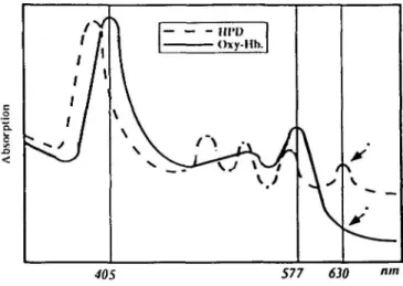
absorbed most readily by this class of compounds. However, blue light tissue penetrance is severely limited by haemoglobin absorption to a fraction of a millimeter (Figure 3), thus preventing efficient photodynamic treatment in deeper tissue layers. Since haemoglobin absorption diminishes at wavelengths >600 nm, red light can be used to photosensitize deep tissue structures. Unfortunately, photosensitizers typically absorb red light less efficiently than blue. For example, Pp EX and DHE red absorption maxima at 630 nm are nearly 35 times less intense than their corresponding 405 nm blue peaks (Kimel et al., 1989). Newer compounds, such as BPD, are designed to absorb light more readily farther into the red spectral region; the 692 nm BPD molar extinction coefficient is approximately eight times greater than that of the 630 nm DHE absortion peak (Richter et al., 1990b). This is important since the attenuation of light in tissue typically decreases with increasing wavelength. Consequently, the use of BPD and compounds with similar photophysical characteristics can facilitate more efficient treatment at greater tissue depths, provided, of course, that drug localization occurs in critical cellular structures. In order to achieve therapeutic effects comparable to BPD for 630 nm absorbing photosensitizers, increased light intensities (mW/cm2) or doses (J/cm2) would
have to be used. Of course, several interrelated factors, including light dose, drug dose, drug localization and photosensitizer photophysical properties are involved in determining the overall clinical effectiveness of PDT.
Light propagation through tissue
When tissues are irradiated with 600-800 nm light, - 3 0 % of the incident dose will be reflected in the surface layers (Svaasand et al., 1989 and Cilesiz and Welch, 1993). In other words, only 70% of the applied light will propagate into the tissue. Photons that propagate into the endometrium will be multiply scattered before they are absorbed. Scattering is caused by optical inhomogeneities, or, more precisely, by refractive index discontinuities in cells and tissue. In the near-infrared spectral regions, scattering is the dominant form of light—tissue
c o "5. SO T J3 <
'Y
• r
' 1
I 1 § ^> ^ / — — - nro Oxy-Hh. \ \ \ \ / . \ \ 40S 577 630Fig. 3. Oxyhaemoglobin (Oxy-hb) and haematoporphyrin derivative (HPD) spectra: HPD exhibits an absorption peak at 630 nm. The absorption of light by oxyhaemoglobin is small at this wavelength.
interaction. Exceptions include ocular media, such as the cornea, the lens and the vitreous humour.
Although tissue scattering is predominantly forward-directed, after a distance of about 1-2 mm, multiple scattering events produce nearly isotropic light distributions in most tissues. Thus, the actual photon path length at a given tissue depth is much longer than the linear distance. This increased path length enhances the probability for absorption. As a result, scattering will increase the attenuation of light in the tissue even though the scattering process itself occurs without loss (Figure 4).
Penetration depth and fluence rate
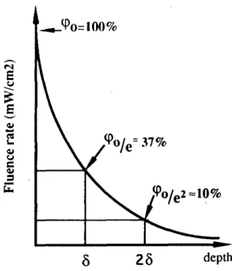
The intensity of light propagation through tissue decreases exponentially with distance due to scattering and absorption. The penetration depth (mm) is defined as the distance corresponding to a decrease in the optical fluence rate by a factor of \le = 0.37 (where e = 2.718) (Svaasand, 1984). The fluence rate measures the quantity of photons per second passing through a defined area
A I R T I S S U F.
Fig. 4. Diagrammatic presentation of light propagation through
tissue: photons are reflected, scattered and absorbed.
(mW/cm2); the fluence rate is, by definition, equal to the optical
power falling onto an infinitesimally small sphere, divided by the cross-sectional area of that sphere. If, for example, the optical penetration depth for a given tissue is 3 mm, the fluence rate of photons at this depth is 37% of its rate at the surface. If the distance is doubled to 6 mm, the fluence rate is decreased by a factor lie2, i.e. to ~ 10% (Figure 5). Since the optical penetration depth is determined by absorption and scattering properties, it is a unique parameter which can be used to characterize a particular tissue.
Back-scattered light in tissues enhances the optical fluence rate close to the surface. For example, in a non-melanotic skin tumour, the fluence rate just below the surface is typically 2 - 3 times higher than the incident irradiation. In a hollow organ such as the uterus, the surface fluence rate may be increased by a factor of 5 - 6 due to the additional reflection of light from the surrounding walls (Marijnissen, 1993) (Figure 6). Thus, in PDT of the endometrium, the fluence rate of the endometrial surface will generally be much higher than the incident unscattered irradiation.
The fluence rate in the endometrium is thus dependent on the reflection conditions at the surface, the incident irradiation, and the tissue scattering and absorption properties. Since the absorption and scattering coefficients typically decrease with increasing wavelength, longer wavelengths have greater tissue penetrance (Figure 3).
Endometrial optical properties (i.e. absorption and scattering coefficients) should be determined in order to optimize light dosimetry for uterine PDT. We have recently conducted optical property measurements on fresh uterine samples immediately after hysterectomy (Madsen et al., 1994). Our preliminary results indicate that bulk tissue absorption and scattering coefficients are, respectively, in the ranges 0.19-0.52 cm1 and 7.3-9.1 cm1
at 630 run. The corresponding values for the optical penetration depth are in the region of 2.59—5.11 mm. Although we have not observed substantial differences in light penetrance for human endometrium and myometrium, we are exploring this possibility with additional measurements.
0>
c
3
(
( p
°/e
2 = 1 0 %
depth
Fig. 5. Penetration depth: the optical penetration depth (5) of a given tissue is equal to the distance over which the optical fluence rate is reduced by a factor Me = 0.37 (where e = 2.718).incidenl (luf nee rale \
fluence rule in livs 5-6 timcN higher
dcpih
Fig. 6. The fluence rate in tissues of a hollow organ: in a hollow
organ the light level builds up through multiple reflections. The fluence rate at the tissue surface is 5—6 times higher than the direct unscattered light from the source.
Optical dose
The optical dose (i.e. the optical fluence) which is deposited in the tissue is proportional to the incident light power per unit area (irradiance) and the exposure time. The irradiant optical dose (J/cm2) is defined by the irradiation (W/cm2) multiplied by the
exposure time in seconds. The tissue optical dose (J/cm2) is
defined as the product of the in-situ fluence rate and the exposure time. The tissue optical dose decreases exponentially with depth in the same manner as the fluence rate. As indicated above, the tissue optical dose depends on the tissue type and geometry and, just below the surface, usually exceeds the incident fluence rate by a factor of 2 - 6 . Of course, the optical dose at a given depth can be enhanced by increasing the incident optical power and/or prolonging the exposure time.
J/cm2
200
50 tnW /cm2
no [henna] damage
1000 2000 4000 time (s)
Fig. 7. Thermal damage in relation to light intensity: high optical dose (J/cm2) may be achieved without causing thermal changes in the tissue by increasing the exposure time.
OPTICAL rx>sr. will induce phntoblcjching
KII1XTIVH OPTICAL IX)SI:
10 inni (depttn
Fig. 8. The 'effective' optical dose: because of photobleaching, the effective optical dose is always smaller than the applied optical dose. The maximum amount of singlet oxygen generated is limited to the amount that would have been produced by an optical dose of 75 J/cm2 if the drug had been completely resistant to bleaching.
Thermal damage
PDT is intended to induce photochemical changes at irradiation levels which do not cause thermal damage. In order to avoid hyperthermal effects, tissue temperature should be kept below ~43—45°C. A temperature rise of 6—8°C above normal body temperature corresponds to an incident optical power density in the range of 100-200 mW/cm2 (Svaasand and Potter, 1989).
In order to achieve deep photodynamic effects with a power density limited to 100 mW/cm2, the exposure time should be
prolonged (Figure 7).
Photodegradation (bleaching) of the photosensitizer
In response to optical irradiation, photosensitizers tend to decompose. The concentration of the non-degraded sensitizer which can participate in the production of singlet oxygen decreases continuously during irradiation. (Moan, 1986; Svaasand and Potter, 1992). The efficiency of the singlet oxygen generation process is, therefore, maximal at the beginning of irradiation,
and it decreases to zero when the sensitizer has been completely degraded.
This phenomenon can be remedied by introducing the concept of an 'effective' optical dose. The effective optical dose is proportional to the amount of in-situ generated singlet oxygen rather than to the optical fluence. For example, the amount of active Photofrin is reduced by photobleaching to Me = 0.37 after application of an optical dose of 75 J/cm2. An optical dose
several times larger than this value will 'bleach' the photosensitizer almost completely; an infinitely large optical dose will only produce a finite amount of singlet oxygen. The total amount of singlet oxygen generated by an infinitely large optical dose will, in fact, be equal to the amount of singlet oxygen that would have been produced by an optical dose of 75 J/cm2, if
Photofrin had been completely resistant to bleaching. Bleaching is, on the other hand, insignificant if the optical dose is much smaller than 75 J/cm2; in this case, the effective optical dose
will be approximately equal to the real optical dose (Figure 8). The effective optical dose will, therefore, be close to the upper limit of 75 J/cm2 in the region close to the irradiated surface
where the optical fluence is high. Bleaching will play an insignificant role in the deeper layers where the fluence has fallen off. In the case of endometrial destruction, bleaching will limit the 'over-kill' in the upper layer of the endometrium; at the same time, it has only an insignificant effect on the depth of necrosis.
Conclusion
In the past 5 years, several studies in rats and rabbits have suggested that PDT may offer a new approach for selective endometrial ablation. Different elements of PDT, such as the type and concentration of photosensitizer, the interval between drug application and light treatment ( 3 - 7 2 h), drug delivery mode (systemic versus topical), light sources (gold vapour laser, argon pumped dye laser, incoherent filtered red light), light doses and light delivery modes, have been studied. As one might expect, these are just a few of the parameters which affect the development of viable treatment protocols in humans. Based on our preliminary results, PDT appears to be a promising, minimally invasive alternative to surgery for treatment of dysfunctional uterine bleeding. However, additional studies must be undertaken to evaluate carefully factors associated with functional loss, long-term tissue regeneration, and selective binding of photosensitizers to endometrial structures. Ultimately, these efforts may lead to the development of a novel clinical technique as well as to contributions to our understanding of endometrial physiology and the implantation process.
Acknowledgements
This study was supported by grants from the National Institutes of Health (grant nos. 2RO1 CA32248, 5P41 RR01192, and R29GM50958), Department of Energy (grant no. DE-FG03-91ER61227) and Office of Naval Research (grant no. N00014-91-C-0134) and Academ. Nachwuchsfoerderung, University of Zurich, Switzerland and Schweizerische Krebsliga, Switzerland.
References
Bhatta.N., Anderson,R., Flotte,T., Schiff.L, Hasan.T. and Nishioka,N.S. (1992) Endometrial ablation by means of photodynamic
therapy with Photofrin n. Am. J. Obstet. Gynecol., 167, 1856-1863. Chapman.J.A., Tadir.Y., Tromberg,B.J., Yu,K., Manetta,A., Sun,C.H. and Berns.M.W. (1993) Effect of administration route and estrogen manipulation on endometrial uptake of Photofrin. Am. J. Obstet.
Gynecol, 168, 685-692.
Chaudhuri,K., Keck.R.W. and Selman,H. (1987) Morphologic changes of tumor microvasculature following hematoporphyrin derivative sensitized photodynamic therapy. Photochem. Photobiol., 46, 823-827.
Cilesiz.I.F. and Welch,A.J. (1993) Light dosimetry: effects of dehydration and thermal damage on the optical properties of the human aorta. Appl. Optics, 32, 477-487.
DeLaney,T.F., Sindelar.W.F., Tochner,Z., Smith.P.D., Friauf,W.S., Thomas,G., Dachowski,L., Cole.J.W., Steinberg,S.M. and Glatstein,E. (1993) Phase I study of debulking surgery and photodynamic therapy for disseminated intraperitoneal tumors. Int.
J. Radiat. Oncol. Biol. Phys., 25, 445-457.
Dougherty,T.J. (1984) Photodynamic therapy (PDT) of malignant tumors. CRC Crit. Rev. Oncol./Hematol., 2, 8 3 - 1 1 6 .
Dougherty,T.J. (1987) Photosensitizers: therapy and detection of malignant tumors. Photochem. Photobiol, 45, 879—889. Judd.M.D., Bedwell.J. and MacRobert.A.J. (1992) Comparison of the
distribution of phthalocyanine and ALA-induced porphyrin sensitizers within the rabbit uterus. Lasers Med. 5c/., 7, 203-211.
Kennedy,J.C. and Pottier,R.H. (1992) Endogenous protoporphyrin IX, a clinically useful photosensitizer for photodynamic therapy. J.
Photochem. Photobiol B. Biol, 14, 275-292.
Kessel,D. (1990) Proposed structure of the tumor-localizing fraction of
HPD J. Photochem. Photobiol, 44, 193-196.
Kimel,S., Tromberg.B.J., Roberts,W.G. andBerns,M.W. (1989) Singlet oxygen generation of porphyrins, chlorins and phthalocyanines.
Photochem. Photobiol, 50, 175-183.
Lipson,R.L., Baldes,E.J. and Olsen,A.M. (1961) The use of derivative of hematoporphyrin in tumor detection. J. Natl Cancer Inst., 26,
1 - 1 1 .
Madsen.S.J., Tromberg,B.J., Wyss.P., Svaasand,L.O., Haskell,R.C. and Tadir,Y. (1994) The optical properties of human uterus at 630 run. Advances in Optical Imaging and Photon Migration. Proceedings of the Optical Society of America, Topical meeting, March 21—23, Orlando, Florida, USA.
Marijnissen,J.A.P. (1993) In Situ Light Dosimetry during Whole Bladder
Photodynamic Therapy, Clinical Results and Experimental Verification. Ph.D. thesis, University of Amsterdam, pp. 75—95.
Moan.J. (1986) Effects of bleaching of porphyrin sensitizers during photodynamic therapy, Cancer Lett., 33, 45—53.
Nelson,J.S, Liaw,L.H., Orenstein,A., Roberts,W.G. and Berns,M.W. (1988) Mechanism of tumor destruction following photodynamic therapy with hematoporphyrin derivative, chlorin, and phthalocyanine.
J. Natl. Cancer Inst., 80, 1599-1605.
Petrucco.O.M., Sathananden.M., Petrucco,M.F., Knowles.S., McKenzie.L., Forbes.I.J., Cowled,P.A. and Keye,W.E. (1990) Ablation of endometriotic implants in rabbits by hematoporphyrin derivative photoradiation therapy using the gold vapor laser. Lasers
Surg. Med., 10, 344-348.
Richter.A.M., Kelly,B., Chow.J., Liu.J., Towers.G.H.N., Dolphin,D. and LevyJ.G. (1987) Preliminary studies on a more effective phototoxic agent than hematoporphyrin. J. Natl. Cancer Inst., 79,
1327-1332.
Richter.A.M., Ceerruti-Sola,S., Sternberg.E.D., Dolphin.D. and Levy.J.G. (1990a) Biodistribution of tritiated benzoporphyrin derivative (3H-BPD-MA), a new potent photosensitizer, in normal and tumor-bearing mice. J. Photochem. Photobiol. B. Biol, 52, 231-244.
Richter.A.M., Waterfield.E., Jain.A.K, Sternberg.E.D., Dolphin.D., and Levy.J.G. (1990b) Evaluation of phototoxic properties of four structurally related benzoprophyrin derivatives. J. Photochem.
Photobiol. B. Biol, 52, 495-500.
Rossi,E., and Curnow,D. (1986) Porphyrins. In Lim.C.L. (ed.), HPLC
of Small Molecules. A Practical Approach. IRL Press, Oxford, pp.
266-267.
Schneider.D.F., Schellhas,H.F., Wesseler.T.A. and Moulton.B.C. (1988a) Endometrial ablation by DHE photoradiation therapy in estrogen-treated ovariectomized rats. Colposcopy Gynecol Laser
Surg., 4, 73-77.
Schneider.D.F., Schellhas.H.F., Wesseler.T.A. and Moulton.B.C. (1988b) Hematoporphyrin derivative uptake in uteri of estrogen treated ovariectomized rats. Colposcopy Gynecol. Laser Surg., 4, 67—71. Star.W.M., Marijnissen.H.P.A., van den Berg-Blok,A.E., Versteeg,J.A.C, Franken,K.A.P. and Reinhold,H.S. (1986) Destruction of rat mammary tumor and normal tissue microcirculation by hematoporphyrin derivative photoradiation observed in vivo in sandwich observation chambers. Cancer Res., 46, 2532-2540. Steiner.R., Tromberg,B.J., Wyss.P., Krasieva.T., Chandanani.N.,
McCullough,J, Berns.M.S. and Tadir.Y. (1995) Rat reproductive performance following photodynamic therapy with optically-administered photofrin. Hum. Reprod., 10, 227-233.
Svaasand.L.O. (1984) Optical dosimetry for direct and interstitial photoradiation therapy of malignant tumors. In Doiron.D. and C. Gomer.C. (eds), Porphyrin Localization and Treatment of Tumors. Alan R. Liss, New York, pp. 91-114.
Svaasand.L.O. and Potter.W.R. (1992) The Implications of Photobleaching for Photodynamic Therapy. In Henderson,B. and Dougherty,T.J. (eds), Photodynamic Therapy: Basic Principles and
Clinical Aspects. Marcel Dekker, New York, pp. 369-385.
Svaasand.L.O., Gomer.C.J. and Profio.A.E. (1989) Laser induced hyperthermia of ocular tumors. Appl. Optics, 28, 2280-2287. Von Trappenheimer,H. and Jesionek.A. (1903) Therapeutische Versuche
mit fluoreszierenden Stoffen. Muench. Med. Wochenschr. 50, 2041-2051.
Yang.J.Z., van Vugt,D.A., Kennedy,J.C. and Reid.R.L. (1993) Evidence of lasting functional destruction of rat endometrium after 5-aminolevulinic acid-induced photodynamic ablation: prevention of implantation. Am. J. Obstet. Gynecol, 168, 995-1001.