The role of the human acetylation polymorphism hi the
metabolic activation of the food carcinogen
2-amino-3-methyhinidazo[4,5-/lquhioline (IQ)
Markus R.Probst
1, Martin Blum, Ingrid Fasshauer
2,
Daniel D'Orazio, Urs A.Meyer and Dieter Wild
2"
3 Department of Pharmacology, Biocenter of University of Basel, Klingelbergstrasse 70, CH^4O56 Basel, Switzerland, and 2Institute ofPharmacology and Toxicology, University of Wurzburg, Versbacher Strasse 9, D-8700 Wurzburg, Germany
^To whom reprint requests should be sent
'Present address: University of California Los Angeles, Laboratory of Biomedical and Environmental Sciences, 900 Veteran Ave, Los Angeles, CA 90024, USA
Hie metabolic activation of the heterocyclk food carcinogen
2-amino-3-methylimidazo[4,5-/|quinolin€ (IQ) by two human
cytochrome P450 monooxygenases (P4501A1 and P4501A2)
and two human N-acetyltransferases (NAT1 and NAT2) was
investigated. Various combinations of these enzymes were
functionally expressed in COS-1 cells. DNA adducts resulting
from the activation of IQ were assayed quantitatively by the
32
P-postlabeling procedure. The highest adduct frequency
was observed in cells expressing both CYP1A2 and NAT2.
CYP1A2 in combination with NAT1 was 3—6 times less
active. When expressed alone these enzymes gave rise to low
adduct frequencies. Experiments with iV-acetyl-IQ as
substrate suggest that NAT1 and NAT2 in addition to their
known role in A
r-acetyiation display arylhydroxamic acid
N,O-acetyttransferase (AHAT) activity. Quantitative differences
in adduct formation between IQ and A'-acetyl-IQ indicated
that metabolic activation of these arylamines preferentially
occurs by P4501A2-catalyzed W-hydroxylation followed by
O-acetylation mediated through NAT1 and/or NAT2. These
data, in combination with the known genetic polymorphism
of NAT2, may explain the clinical observation that the
acetylation polymorphism constitutes a risk factor in the
carcinogenic activation of environmental mutagens.
Introduction
Frying of meat and fish results in the generation of heterocyclic
arylamines (1), which present a major source of dietary
carcinogens. Among a number of food-derived heterocyclic
amines, 2-aiTuno-3-memylimidazo[4,5-/]quinoline (IQ*) was
found to be highly mutagenic in Salmonella typhimurium, and
its carcinogenic potential was demonstrated in various rodent
species and a primate (2,3).
The effects of heterocyclic arylamines, like those of other
arylamines, require metabolic activation by drug-metabolizing
enzymes. It is generally accepted that the ultimate
mutagens/car-cinogens are electrophilic arylnitrenium ions, which can react
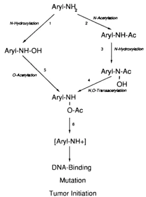
with DNA and form adducts ( 4 - 6 ) . Reactions involved in the
metabolic activation include N-hydroxylation and N- and
O-acetylation (Figure 1), catalyzed by microsomal monooxygenase
enzymes of the cytochrome P4501A family (for review see 7,8),
•Abbreviations: IQ, 2-amino-3-methylimidazo{4,5-/|quinoline; NAT,
N-acetyltransferase; RFLP, restriction fragment length polymorphism; AHAT, arylhydroxamic acid A'.O-acetyltransferase; OAT, O-acetyltransferase.
and acetyl coenzyme A-dependent arylamine A'-acetyltransferases
(NATs) in liver cytosol (9). In the case of IQ and related
heterocyclic arylamines, N-acetylation appears to be a minor
metabolic pathway (10,11). Considerable interindividual
varia-tions in the activities of these enzymes has been observed caused
either by environmental conditions or genetic polymorphisms.
P4501A1 is induced by cigarette smoking, and individuals with
genetically controlled high inducibility are at higher risk of
developing cigarette-induced bronchogenic carcinoma (12). A
restriction fragment length polymorphism (RFLP) has been
reported in the human CYP1 Al gene downstream of the coding
region (13 - 1 5 ) . The association of this RFLP with an increased
incidence of bronchogenic carcinoma has been observed in some
but not all populations tested (12). Point mutations in the NAT2
gene, resulting in decreased enzyme protein levels (16), are
responsible for the acetylation polymorphism, which divides the
population into so-called 'rapid' and 'slow' acetylators. Between
40 and 70% of individuals in Caucasian populations are slow
acetylators (17). Slow acetylators display impaired metabolism
of a variety of arylamine and hydrazine drugs and chemicals.
These include many therapeutically useful drugs (for review see
17,18), as well as potential arylamine carcinogens present in dyes,
antioxidants, pesticides and explosives (19).
There is circumstantial evidence for a link between rapid
acetylator status and certain diseases, e.g. colorectal cancer
(20-22). A second human NAT gene, NAT1, is not affected
by the acetylation polymorphism, and does not contribute to the
Aryl-NK,
N-AcMytmtionAryl-NH-Ac
Aryl-NH-OH
N-HydmxylatJon O-ActytmtkmAryl-NH
iO-Ac
Aryl-N-Ac
OH
' N.QTnniacatytMSion[Aryl-NH+]
DNA-Binding
Mutation
Tumor Initiation
M.R.Probst et al.
acetylator status (23,24). The availability of the cloned
NAT1/NAT2 genes allows us to test in a cell culture situation,
which one of these genes is involved, if at all, in the metabolic
activation of IQ. Upon transfection of different combinations of
NAT and CYP1A genes, P4501A2 and NAT2 resulted in the
highest IQ-DNA adduct frequencies. This suggests that the human
acetylation polymorphism may affect the carcinogenic action of
the food mutagen IQ.
Materials and methods
ChemicalsIQ was purchased from WAKO Pure Chem. Industries, Ltd (Neuss, Germany). N-Acetyl-IQ was prepared from IQ and acetic anhydride, and was further purified by chromatography on silica gel. Phenoxazone ethers and resorufin were purchased from Pierce Chemicals. All other chemicals were of HPLC or analytical grade quality and obtained from local suppliers.
Functional expression ofNATl, NAT2 CYPJA1 and CYPIA2 in monkey kidney COS-1 cells
COS-1 cells were cultured and transfected by the DEAE-dextran/chloroquine method as described (25). The human NAT1 and NAT2 expression constructs in the vector p91023(B) were as described (23). cDNA clones encoding human CYP1A1 (26) and CYP1A2 (27) were kindly provided by D.W.Nebert and F.J.Gonzales (NCI, NIH Bethesda, MD), and were subcloned into p91023(B). Cultures were treated with IQ or W-acetyl-IQ (dissolved in ethanol) 50 h after transfection, and cells were harvested 9—12 h later.
Enzyme assays and detection of expressed proteins on Western blots Enzyme activity of expressed NAT proteins was determined using sulfamethazine as a substrate and quantitated using a HPLC assay as described (28).
P4501A1 and P4501A2 proteins in transfected COS-1 cultures and in microsomes prepared from kidney donor liver (KDL35) were identified by immunoreaction on Western blots with a rabbit antiserum raised against the corresponding rat P4501A1 (P45Oc) enzyme. This antiserum recognizes both human P4501A1 and P4501A2 proteins. Their enzymatic activities were determined by dealkylation of phenoxazone ethers 7-methoxy- and 7-ethoxy-resorufin as described by Burke et al. (29). Deacetylation of JV-acetyl-IQ was measured by reversed-phase HPLC (MN 5-C18 Macherey-Nagel, Germany) by isocratic elution using 50 mM sodium acetate/methanol (40:60) pH 6.3 as mobile phase and UV detection at X = 254 nm.
DNA isolation and 3iP-postlabeling
Cell pellets were homogenized by means of an Ultra-Turrax, and treated with RNase A, RNase T, and proteinase K. DNA was extracted with phenol/chlorofbrm
A B C
and precipitated with ethanol. After digestion of the DNA to 3'-nucleotides with mjcrococcus endonuclease and spleen exonuclease (30), an aliquot of the digest equivalent to 4 /ig was labelled with 100-120 /iCi [ T -3 2P ] A T P (sp. act. 3000
Ci/mmol; Amersham, Braunschweig, Germany) and by use of T4 polynucleotide kinase (Pharmacia). The intensification procedure was applied according to Randerath et aL (31). Four-directional TLC separation of the adducts was performed on PEI-cellulose sheets (Macherey-Nagel) with the following solvents: Dl, 1 M sodium phosphate, pH 6.8; D3, 3.8 M lithium formate, 6.8 M urea, pH 3.4; D4, 0.6 M sodium phosphate, 0.38 M Tris-HCl, 6.5 M urea, pH 8.2; D5, 1.7 M sodium phosphate, pH 6.0. After detection of adducts by autoradiography, spots were excised and quantitated by Cerenkov counting; intensification factors required for the quantitative analysis were determined in each experiment by use of a highly IQ-modified DNA (32).
Results
Metabolic activation of IQ
Figure 1 shows the proposed enzyme reactions involved in the
metabolic activation of arylamines such as IQ. The first step of
metabolism of arylamines can either be an N-hydroxylation
(reaction 1 in Figure 1) or an N-acetylation (reaction 2 in
Figure 1). Reaction 1 may be catalyzed by either P4501A1 or
P4501A2 (7,8), and it has been shown that liver cytosolic NAT
accounts for the N-acetylation reaction (24). The A'-arylacetamide
resulting from reaction 2 serves as substrate for P4501A1 or
P4501A2, and N-hydroxylation yields the respective
aryl-hydroxamic acid (reaction 3, Figure 1). The two pathways
converge through reactions 4 (N.Otransacetylation) and 5
(O-A CYP1(O-A1
2OO 1OO 10 20 30 40 50 60 Time (min) -54kDa -52kDaFig. 2. Immunoreaction on Western blots of CYP1A1 and CYP1A2 in human liver microsomes and upon expression of the respective cDNAS in COS-1 cells. Homogenates of COS-1 cells transfected with cDNAS encoding CYP1A1 in sense (lane A) and antisense orientation (lane B), and CYP1A2 in antisense (lane C) and sense (lane D) orientation, as well as human liver microsomes (lane E) were subjected to SDS-PAGE (150 jig of COS-1 homogenates and 50 fig microsomal protein), transferred to nitrocellulose and immunoreacted with a polyclonal rabbit antiserum raised against purified rat P4501A1.
B
CYP1A2
O 5 10 15 20 25 30 40 50 60
Time (min)
Fig. 3. Expressed cytochrome P4501A1 and cytochrome P4501A2 are functionally active in COS-1 cells. Homogenates of COS-1 cells transfected with CYP1A1 (A) and CYP1A2 (B) were incubated with methoxyresorufin ( • ) and ethoxyresorufin ( • ) , and formation of the dealkylated products was determined at the time points indicated. Note: P4501A1 preferentially metabolized ethoxyresorufin, whereas P45O1A2 displayed higher activity with methoxyresorufin.
acetylation) to yield the product, N-acetoxy-A'-arylamine. Both
of these reactions have been attributed to the activity of cytosolic
NAT in hamster and rabbit (33,34), whereas no such correlation
could as yet be established for the human NAT enzymes.
Spontaneous degradation of A
/-acetoxy-A
r-arylamines (reaction 6
in Figure 1) leads, presumably via unstable arylnitrenium ions
(4—6), to the formation of DNA adducts.
COS-] cell cultures provide an in vitro system to analyze
IQ-adduct formation
COS-1 cells were used to analyze the involvement of the proposed
enzymes in the above-mentioned reactions in an in vitro system,
Table I. Formation of IQ-nucleotide adducts in COS-1 cells transfected with cytochrome P450 monooxygenase and W-acetyltransferase genes Gene combinations Adducts/108 nucleotides (±SD)
CYP1A1 CYP1A2 NAT1 NAT2 CYP1A2 CYP1A2 + NAT1 + NAT2 IQ 100,1 7.3 13.6 1.7 1.6 81.6 250.3 ± ± ± ± ± ± M 0.2 2.3 0.4 0.2 5.9 28.8 IQ \0 n ND 0 ND ND 9.3 ± 57.2 ± M 0 5 .7 .8 JV-Acetyl-IQ 10 /JM ND 0 0 0 1.0 ± 0.2 14.4 ± 1.3 COS-1 cells were transfected with CYP1A1, CYP1A2, NAT1 and NAT2, either alone or in the combination indicated. Cultures were incubated in the presence of 10 or 100 yM IQ or W-acetyl-IQ for 12 h, followed by isolation of the DNA and analysis of IQ-adducts.
Note: the combination of CYP1A2 and NAT2 resulted in the highest adduct frequencies in all three experiments. Adduct formation with the acetylated IQ substrate still required the NAT enzyme(s), indicating their inherent N,0-transacetylase activities.
ND, not done; 0, no adducts detectable.
I
r—r
M
I
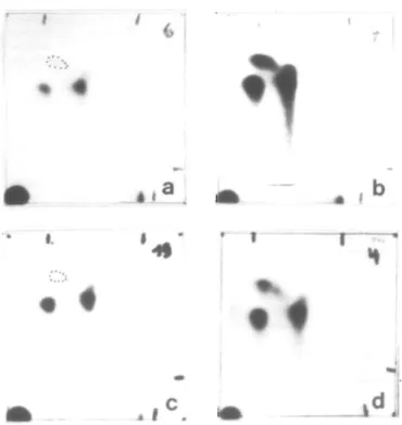
Fig. 4. Identical patterns of IQ and W-acetyl-IQ adducts in COS-1 cells expressing CYP1A2 + NAT1 and CYP1A2 + NAT2. Autoradiographs of IQ-DNA adducts separated by TLC. (a) COS-1 cells transfected with CYP1A2 + NAT1 (substrate: IQ, 10 /iM); (b) COS-1 cells transfected with CYP1A2 + NAT2 (IQ. 10 nM); (c) COS-1 cells transfected with CYP1A2 + NAT2 (substrate: JV-acetyl-IQ, 10 /iM); (d) IQ adducts formed in the liver of a F344 rat 24 h after a 30 mg/kg oral dose of IQ; adduct frequency 123/10" nucleotides. The origin of the chromatograms is in the lower left-hand comer.
both individually and in combination. We have previously
demonstrated that NAT1 and NAT2 can be functionally expressed
in COS-1 cells, and that COS-1 cells have no detectable
endogenous N-acerylation activity (25). In this study, we have
additionally analyzed the expression of CYP1A1 and CYP1A2
cDNAs in COS-1 cells. Immunoreaction on a Western blot of
proteins expressed from the cDNAs using a polyclonal rabbit
antiserum raised against the rat P4501A1 is shown in Figure 2.
Protein bands of the expected mol. wts of 52 and 54 kDa in
cultures transfected with CYP1A1 (lane A, Figure 2) and
CYP1A2 (lane D, Figure 2) respectively were detected. These
were indistinguishable in electrophoretic mobility from
microsomal proteins isolated from human liver (lane E, Figure 2),
suggesting that in this particular liver both P4501A proteins were
present. No immunoreactive proteins were detected in cultures
transfected with the constructs in antisense orientation (lanes B
and C, Figure 2). Cell homogenates of cultures transfected with
CYP1A1 or CYP1A2 both displayed enzyme activity with the
substrates 7-methoxy- and 7-ethoxyresorufin (Figure 3).
Homogenates of untransfected cells and cells transfected with
cDNAs in antisense orientation did not display any detectable
activities with either substrate (not shown).
The highest DNA adduct frequency occurs in COS-1 cells
transfected with CYP1A2 and NAT2
COS-1 cells were transfected with NAT and CYP1A genes
individually and in the combinations shown in Table I.
Transfected cultures were treated with IQ (at a concentration of
100 ftM) and the resulting adduct frequencies determined
(Table I, left-hand column). Transfections with NAT1 or NAT2
alone resulted in low levels of IQ-DNA adducts ( 1 - 2 adducts/
10
8nucleotides; see Table I). Slightly more DNA adducts were
found after transfection of COS-1 cells with either CYP1A1 or
CYP1A2 ( 7 - 1 4 adducts/10
8nucleotides; Table I). From these
experiments we conclude that COS-1 cells have virtually no
endogenous enzyme activity for the acetylation and
hydroxyla-tion of arylamines. Because expressed P4501A2 was twice as
active as P4501A1, combinations of CYP1A2 and NAT genes
were used in all further experiments. As shown in Table I, these
combinations resulted in adduct frequencies 5—20 times higher
than those obtained with single genes, and NAT2 was three times
more effective than NAT1.
A lower IQ concentration (10 /tM) was applied in a second
series of transfection experiments (Table I). Qualitatively, the
same results were obtained, namely highest adduct frequencies
following transfection of CYP1A2 in combination with NAT2.
The chromatographic patterns of IQ—nucleotide adducts obtained
after activation by P4501A2 and NAT1 and by P4501A2 and
NAT2 were qualitatively identical (Figure 4a and b); the
difference was merely quantitative. The same pattern had been
found previously in liver DNA of rats that had received an oral
dose of IQ (Figure 4d), and with that obtained from the IQ-related
Table H. COS-i cells have no endogenous JV-acetyl-TQ deacetylase activity A/-Acetyl-IQ concentration COS-1 cell homogenate Human liver
microsomes 100 200 400 0.002* 0.002 0.017* 0.085 0.232 Untransfected COS-1 cells were homogenized and assayed for endogenous deacetylase enzyme activity using chemically synthesized JV-acetyl-IQ as substrate. Microsomes isolated from human liver were similarly analyzed. 'Numbers represent nmol IQ/mg protein/min.
M.R.Probst et al.
compounds nitro-IQ in Salmonella typhimurium and azido-IQ in
calf thymus DNA (32,35). The quantitatively most prominent,
slow migrating adduct has been identified as
A^-(deoxyguano-sin-8-yl)-IQ (36).
NAT1 and NAT2 both display N,O-transacetylase (AHAT) enzyme
activity
Kinetic characterization of purified NAT enzyme activities from
rabbit and mouse liver have suggested that the same enzyme has
NAT, arylhydroxamic acid yV.Oacetyltransferase (AHAT) and
O-acetyltransferase (OAT) activities (33,34).
We used the COS-1 expression system to address the question
of whether human NATs have AHAT activity. The adduct
formation by N-acetyl-IQ was studied, which, provided that this
substrate is not deacetylated by COS-1 cells, can be converted
to an arylnitrenium ion only in the presence of AHAT activity
(Figure 1). Therefore the TV-acetyl-IQ deacetylase activity of
COS-1 cells was tested and compared with that of human liver
microsomes. As is evident from Table n , COS-1 cells display
only minute endogenous deacetylase activities at all substrate
concentrations tested, whereas significant activities were found
in human liver microsomes.
In another series of transfection experiments N-acetyl-IQ was
added to cultures and the resulting adducts were determined
(Table I). Adducts were detected with combinations of CYP1A2
and either NAT1 or NAT2. NAT2 was 14 times as effective as
NAT1, and overall frequencies were considerably lower
(1/4—1/9) than those observed with IQ at the same concentration
(10 /iM). The chromatographic pattern of the N-acetyl-IQ—DNA
adducts (Figure 4c) was the same as that of the IQ—DNA
adducts. Thus, the same arylnitrenium ion was formed from IQ
and N-acetyl-IQ and it can be concluded that both NAT1 and
NAT2 display AHAT enzyme activity. The lower adduct
frequency observed with A'-acetyl-IQ compared to that observed
with IQ is probably due to an efficient direct activation of IQ
(via N-hydroxylation and O-acetylation) and a less efficient
activation of N-acetyl-IQ (via N-hydroxylation and
N,O-transacetylation).
Discussion
In the present report we used transient expression in COS-1 cells
of each of four drug-metabolizing enzymes to study their role
in the activation of the food mutagen and carcinogen IQ. Our
experiments suggest that (i) human P4501A2 is more efficient
than P4501A1 for N-hydroxylation of this heterocyclic amine;
(ii) N-hydroxy-IQ is only weakly reactive towards DNA; (iii)
the most efficient metabolic activation and DNA adduct formation
is achieved with P4501A2 in combination with NAT2. Finally,
we propose that NAT1 and NAT2 in addition to their known
role in N-acetylation display N.O-transacetylase activity.
Our first finding is in keeping with published results obtained
in rat and rabbit. When purified rat (37) and rabbit (38) P4501A1
and P4501A2 were used in the Salmonella mutagenicity assay
with IQ, the rat P4501A2 enzyme was found to be 12 times,
and the corresponding rabbit enzyme 8 times more efficient than
P4501A1. Similarly, when mouse P4501A1 and P4501A2
enzymes were expressed in Hep G2 human hepatoma cells and
used in the Salmonella assay with IQ, P4501A2 was nine times
as active as P4501A1 (39). Thus, the available evidence points
to P4501A2 as the predominant physiological activator of IQ.
N-Hydroxylation of IQ, obtained by expression of CYP1A2 alone
(Table I), results in only minor formation of IQ-nitrenium ions
and DNA binding: an acetylation step is required as well. This
is in agreement with mutagenicity data obtained in Salmonella
strains with low, normal and high acetyltransferase activity,
where the mutagenic activity of IQ (in the presence of rat liver
S9) strongly depends on the acetylator status of the strain used
(40).
The data presented indicate that the human acetylation
poly-morphism has to be considered an important factor in the
metabolic activation of arylamine carcinogens. This becomes
evident because (i) the polymorphic NAT2 in our analysis was
about five times as effective as NAT1 in activating IQ into the
arylnitrenium ion; and (ii) NAT2 possesses ten times as much
Af.O-transacetylase (AHAT) activity as NAT1, thus broadening
the spectrum of reactions affected by the polymorphism.
In addition we suggest that NAT1 and NAT2 can catalyze the
hydroxylamine O-acetylation. This was concluded from the very
low rate of IQ A/-acetylation by rodent NATs (10,41), and from
the relatively high adduct frequencies found in our IQ
experi-ments, which can only be explained by the presence of OAT
activity (see Figure 1).
In conclusion, this study provides a mechanistic basis for the
clinical observation that the acetylation polymorphism constitutes
a risk factor in the metabolic activation of environmental and
dietary mutagens/carcinogens.
Acknowledgements
We thank A.Demierrc, M.Beer and D.Beer for expert technical assistance. This work was supported by the Swiss National Research Foundation (grant 32-31266.91). Work at the University of WQrzburg was supported by the Deutsche Forschungsgemeinschaft (SFB 172 'Molekulare Mechanismen kanzerogener PrimarverSnderungen').
References
1. FehonJ.S. and Knize.M.G. (1990) Heterocyclic amine mutagens/carcinogens in foods. In Cooper.C.S. and Grover.P.L. (eds), Handbook of Experimental Pharmacology. Vol. 94, Springer, Berlin, pp. 471-502.
2. Sugimura.T., Sato.S. and Wakabayashi,K. (1988) Mutagens/carcinogens in pyrolysates of amino acids and proteins in cooked foods: heterocyclic aromatic amines. In Woo.Y., Lai.D.Y., ArcosJ.C. and Argus.M.F. (eds), Chemical Induction of Cancer. Academic Press, San Diego, Vol. iiiC, pp. 681-710. 3. Adamson.R.H., Thorgeirsson.U.P., Snyderwine.E.G., Thorgeirsson.S.S., Reeves,J., Dalgard.D.W., Takayama.S. and Sugimura.T. (1990) Carcinogenicity of 2-arnino-3-methylimidazo(4,5-/]quuK>line in nonhuman primates: induction of tumors in three macaques. Jpn. J. Cancer Res., 81,
10-14.
4. Kadlubar.F.F. and Beland.F.A. (1985) Chemica] properties of ultimate carcinogenic metabolites of arylamines and arylamides. In Harvey,R.G. (ed.), PolycycUc Hydrocarbons and Carcinogenesis, ACS Symposium series no. 283. American Chemical Society, Washington, DC, pp. 341-370. 5. Kriek,E. (1965) On the interaction of Af-2-fluorenylhydroxylamine with nucleic
acids in vitro. Biochem. Biophys. Res. Common., 20, 793—799. 6. LutgerinkJ.T., StavenuiterJ.F.C, Zomer.G., Hamzink.M., van Dijk,P.,
WestraJ.G. and Kriek,E. (1989) Synthesis of heterocyclic /V-acetoxy-arylamines and their reactivity with DNA. Carcinogenesis, 10, 1957-1960. 7. Kadlubar.F.F. and Hammons,GJ. (1987) The role of cytochrome P-450 in the metabolism of chemical carcinogens. In Guengerich.F.P. (ed.), Mammalian Cytochromes P-450- CRC Press, Boca Raton, FL, Vol. II, pp. 81-130. 8. Butler.M.A., Iwasalri.M., Guengerich.F.P. and Kadlubar.F.F. (1989) Human cvtochrome P450pA (P4501A2) the phenacetin-0-deethylase is primarily
responsible for the hepatic 3-demethylation of caffeine and N-oxidation of carcinogenic arylamines. Proc. Nail. Acad Sci. USA, 86, 7696-7700. 9. Weber.W.W. (1987) The Acetylator Genes and Drug Response. Oxford
University Press, New York.
10. Kato,R. (1986) Metabolic activation of mutagenic heterocyclic aromatic amines from protein pyrolysates. CRC Crit. Rev. Toxicol.. 16, 307-348. 11. Turesky.RJ., Bracco-Hammer.I., Markovic,J., Richli.U., Kappeler,A.-M.
and Wdti.D.H. (1990) The contribution of N-oxidanon to the metabolism of the food-borne carcinogen 2-amino-3,8-dimethylimidazo[4,5-/lquinoxaline in rat hepatocytes. Chem. Res. ToxicoL, 3, 524—535.
12. Nebert.D.W., Petersen.D.D. and Puga,A. (1991) Human AH locus polymorphism and cancer inducibility of CYP1A1 and other genes by combustion products and dioxin. Pharmacogenetics, 1, 6 8 - 7 8 . 13. Kawajiri.K., Nakachi.K., Kazue,!., Yoshii.A., Shinoda.N. and WatanabeJ.
(1990) Identification of genetically high risk individuals to lung cancer by DNA polymorphisms of the cytochrome P4501A1 gene. FEBS Lett., 263, 131-133.
14. Hayashi,S., WatanabeJ., Nakachi.K. and KawajiriJC. (1991) Genetic linkage of lung-cancer associated Mspl polymorphisms with amino acid replacement in the heme binding region of the human cytochrome P4501A1 gene. /. Biochem, 110, 407-411.
15. Tefre.T., Ryberg.D., Haufen.A., Nebert.D.W., Skaug.V., Brogger.A. and Borrensen,A.-L. (1991) Human CYP1A1 (cytochrome P,450) gene: lack of association between the Mspl restriction fragment length polymorphism and the incidence of lung cancer in a Norwegian population. Pharmacogenetics, 1, 2 0 - 2 5 .
16. Blum.M., Demierre.A., Grant.D.M., Heim.M. and Meyer.U.A. (1991) Molecular mechanism of slow acetylation of drugs and carcinogens in humans. Proc. Natl. Acad. Sci. USA, 88, 5237-5241.
17. Evans.D.A.P. (1989) A'-Acetyltransferase. Pharmac. Ther., 42, 157-234. 18. Meyer.U.A. (1990) Genetic polymorphisms of drug metabolism. Fundam.
din. Pharmacol, 4, 595-615.
19. National Academy of Sciences (1981) Aromatic Amines: an Assessment of the Biological and Environmental Effects. National Academy Press, Washington, DC.
20. WohllebJ.C., Hunter.C.F., Blass.B., Kadlubar.F.F., Chu.D.Z.J. and Lang.N.P. (1990) Aromatic aminc acetyltransferase as a marker for colorectal cancer environmental and demographic associations. Int. J. of Cancer, 46, 2 2 - 3 0 .
21. Lang.N.P., Chu.Z.J., Hunter.C.F., Kendall.D.C, Flammang.T.J. and Kadlubar.F.F. (1986) Role of aromatic amine acetyltransferase in human colorectal cancer. Arch. Surg., 121, 1259-1261.
22.Ilett,K.F., David.B.M., Detchon.P., Castleden.W.M. and Kwa.R. (1987) Acetylation phenotype of colorectal carcinoma. Cancer Res., 47, 1466-1469. 23.Blum,M., Grant.D.M., McBride.W., Heim.M. and Meyer.U.A. (1990) Human arylamine A'-acetyltransferase genes: isolation, chromosomal localization and functional expression. DNA Cell Biol, 9, 193-203. 24. Grant.D.M., Blum.M., Beer.M. and Meyer.U.A. (1991) Monomorphic and
polymorphic human arylamine A'-acetyltransferases: a comparison of liver isozymes and expressed products of two cloned genes. MoL Pharmacol., 39, 184-191.
25. Blum.M., Grant,D.M., Demierre.A. and Meyer.U.A. (1989) A'-Acetylation pharmacogenetics: a gene deletion causes absence of arylamine A'-acetyl-transferase in liver of slow acetylator rabbits. Proc. Natl. Acad. Sci. USA, 86, 9554-9557.
26. Jaiswal.A.K., Nebert.D.W. and Gonzakz.F.J. (1987) Human P3-450: cDNA
and complete amino acid sequence. Nucleic Acids Res., 14, 6773-6774. 27. Jaiswal.A.K., Gonzalez,F.J. and Nebert.D.W. (1985) Human dioxin-inducible cytochrome P,-450: complementary DNA sequence and amino acid sequence. Science, 228, 8 0 - 8 3 .
28. Grant.D.M., Moerike.K., Eichelbaum.M. and Meyer.U.A. (1990) Acetylation pharmacogenetics: the slow acetylator phenotype is caused by decreased or absent arylamine A'-acetyltransferase in human liver. J. Clin. Invest., 85, 968-972.
29. Burke.M.D., Tompson.S., Elcombe.C.R., HalpertJ., Haaparanta.T. and Mayer.R.T. (1985) Ethoxy-, pentoxy-, and benzyloxyphenoxazones and homologues: a series of substrates to distinguish between different induced cytochrome P-450. Biochem Pharmacol., 34, 3337-3345.
30. Gupta,R.C, Reddy.M.V. and Randerath.K. (1982) 32P-Postlabelling analysis
of non-radioactive aromatic carcinogen-DNA adducts. Carcinogenesis, 3, 1081-1092.
31. Randerath.E., Agrarwal.H.P., WeaverJ.A., Bordelon.C.B. and Randerath.K. (1985) 32P-Postlabelling analysis of DNA-adducts persisting for up to 42
weeks in the skin, epidermis and dermis of mice treated topically with 7,12-dimethylbenzManthracene. Carcinogenesis, 6, 1117 — 1126. 32.Wild,D., Dirr.A., Fasshauer.I. and Henschler.D. (1989) Photolysis of
arylazides and generation of highly electrophilic DNA-binding and mutagenic intermediates. Carcinogenesis, 10, 335—341.
33. Glowinski.I.B., Weber.W.W., FyshJ.M., VaughU.B. and King.C.M. (1980) Evidence that arylhydroxamic acid A'.O-acyitransferase and the genetically polymorphic A'-acetyltransferase are properties of the same enzyme in rabbit liver. J. Biol. Chem, 255, 7883-7890.
34. Mattano.S.S., Land.S., King.C.M. and Weber.W.W. (1989) Purification and biochemical characterization of hepatic arylamine A'-acetyltransferase from rapid and slow acetylator mice: identity with arylhydroxamic N,O-aceyltransferase and A'-hydroxylamine Oacetyltransferase. MoL Pharmacol., 35, 599-609.
35. Dirr.A., Fasshauer.I., Wild.D. and Henschler.D. (1989) The DNA-adducts of the food mutagen and carcinogen IQ (2-amino-3-mentylimidazo(4,5-/lquino-line). Arch. Toacol., Suppl.. 13, 224-226.
36. Synderwine.E.G., Roller.P.P., Adamson.R.H., Sato.S. and Thorgeirsson.S.S. (1988) Reaction of A'-hydroxylamine and A'-acetoxy derivatives of
2-amino-3-methylimidazo[4,5-/]quinoline with DNA. Synthesis and identification of A/-(deoxyguanosin-8-yl)-IQ. Carcinogenesis, 9, 1061 — 1065. 37. Yamazoe.Y., Shimada.M., Kamataki.T. and Kato.R. (1983) Mkrosomal
activation of 2-amino-3-methylimidazo[4,5-/]quinoline. Cancer Res., 43, 5768-5774.
38. McManus,M.E., Burgess.W., Snyderwine.E. and Stupans.I. (1988) Specificity of rabbit cytochrome P-450 isozymes involved in the metabolic activation of the food derived mutagen 2-amino-3-memylimidazo{4,5-/)quinoline. Cancer Res., 48, 4513-4519.
39. Ayoama.T., Gonzalez.F.J. and Gelboin.H.V. (1989) Mutagen activation by cDNA-expressed P,450, P345O and P450a. Mol. Carcinogenesis, 1, 253-259.
40. Wild.D., Watkins.B.E. and Vanderlaan.M. (1991) Azido- and nitro-PhIP, relatives of the heterocyclic arylamine and food mutagen PhIP—mechanism of their mutagenicity in Salmonella, Carcinogenesis, 12, 1091-1096. 41. Turesky.RJ., Lang.N.P., Buner.M.A., Teitel.C.H. and Kadlubar.F.F. (1991)
Metabolic activation of carcinogenic heterocyclic aromatic amines by human liver and colon. Carcinogenesis, 12, 1839 — 1845.