Determination of fetal chromosome
aberrations from fetal DNA in
maternal blood: has the challenge
finally been met?
Sinuhe Hahn
1,*, Olav Lapaire
2, Sevgi Tercanli
2, Varaprasad Kolla
1and Irene Hösli
2The analysis of cell-free fetal nucleic acids in maternal blood for prenatal
diagnosis has been transformed by several recent profound technology
developments. The most noteworthy of these are
‘digital PCR’ and
‘next-generation sequencing
’ (NGS), which might finally deliver the long-sought goal
of noninvasive detection of fetal aneuploidy. Recent data, however, indicate
that NGS might even be able to offer a much more detailed appraisal of the
fetal genome, including paternal and maternal inheritance of point mutations
for
mendelian
disorders
such
as
β-thalassaemia. Although these
developments are very exciting, in their current form they are still too complex
and costly, and will need to be simplified considerably for their optimal
translation to the clinic. In this regard, targeted NGS does appear to be a step
in the right direction, although this should be seen in the context of ongoing
progress with the isolation of fetal cells and with proteomic screening markers.
In the past few years it has become clear thatthe demographic shift towards increasing
maternal age for pregnancies in developed nations, with its associated risks of fetal
chromosomal anomalies, will necessitate a
change in current strategies for prenatal
screening and detection of fetal aneuploidies (Refs 1, 2, 3, 4). This is illustrated by a recent analysis of the English and Welsh National
Down Syndrome (DS) Cytogenetic Register over the period 1989–2008 (Ref. 5), which reported a 71% increase in the number of diagnosed cases with DS, with no comparable increase in birth rate. The increase in the number of cases
with DS was largely attributed to the
concomitant increase in maternal age, as more than 20% of pregnancies now occur in mothers older than 35 years (Ref. 5).
1
Laboratory for Prenatal Medicine, Department of Biomedicine / Department of Obstetrics and Gynecology, University Hospital Basel, Switzerland.
2Department of Obstetrics and Gynecology, University Hospital Basel, Switzerland.
*Corresponding author: Sinuhe Hahn, Laboratory for Prenatal Medicine, Department of Biomedicine/Department of Obstetrics and Gynecology, University Hospital Basel, Hebelstrasse 20, CH-4031 Basel, Switzerland. E-mail: shahn@uhbs.ch
1
Determina
tion
of
fetal
chr
omosome
aberr
a
tions
fr
om
fetal
DNA
in
ma
ter
nal
blood:
has
the
challenge
finally
been
met?
This development is likely to add additional costs to already strained healthcare budgets, because positive cases from initial noninvasive screening based on ultrascans and maternal serum biochemical analysis need to be verified by invasive analysis such as amniocentesis or chorionic villous sampling, which are labour-intensive examinations requiring highly skilled personnel (Ref. 6). To break this spiralling cost, it would be advantageous for any future noninvasive method to be so accurate that it could function as a ‘stand-alone’ test and not require further invasive verification. Naturally, it will also need to be considerably cheaper than
current invasive practices. An alternative
scenario might be to use such a noninvasive test to reduce the cost and risk associated with the invasive analysis of the high number of false-positive cases resulting from current screening practice (Refs 7, 8). In either case, strenuous efforts will need to be undertaken in order to ensure that cost-effective noninvasive prenatal diagnosis of DS cases finally becomes a reality (Ref. 8).
Has research in the field of noninvasive prenatal diagnosis become ‘mature’?
Since the discovery of fetal cell-free DNA (cf-DNA) in maternal plasma or serum in 1997 (Ref. 9), more than 1000 papers have been published on this topic. A cursory review of publications listed in public depositories such as PubMed (http:// www.ncbi.nlm.nih.gov/pubmed) suggests that the most prolific period was probably the latter half of the previous decade. In this period, several large-scale studies were initiated to test the efficacy of this new-found tool for the noninvasive determination of fetal genetic traits such as the fetal RhD (Rhesus D) gene or gender (Refs 10, 11, 12, 13). These data indicated that this approach was indeed sufficiently robust that widespread clinical application could be safely implemented, and as a result concerted efforts were undertaken by multicentre consortia, such as the European Union (EU)-funded SAFE Network, to standardise these assays (Refs 11, 14). These data also indicated that the analysis of cf-fetal DNA could form the secure and sound basis for subsequent developments, such
as the noninvasive detection of fetal
aneuploidies (Ref. 15).
In recent years this prolific output in
publications seems to have largely tapered off,
although the number of reviews dealing with the field has increased tremendously. Hence the scenario appears very similar to economic
models of the ‘product life cycle’, where a
particular item has progressed through
development, introduction and growth, and is
settling into a pattern of maturity and
subsequent decline (Ref. 16). Although this simplistic view might lead to the impression that research in this field has waned, this is far from the truth, for this punctuated modicum in published reports indicates that the field has finally entered a phase where quality, using a quantum leap in technological development, rather than quantity is the prevailing trend. This facet will become very evident in this review.
The question of fetal cell-free DNA fragment size and concentration
It is necessary to reiterate that the major problem still hampering the use of fetal cf-DNA for the noninvasive detection of fetal genetic loci that are not distinct from maternal loci is that fetal sequences constitute only 5–10% of the total cf-DNA in maternal plasma, and even less in maternal serum (Refs 17, 18, 19).
Groundbreaking research performed in the middle of the past decade on the biophysical properties of DNA indicated that fetal cf-DNA was more fragmented and had a shorter
size than comparable maternal cf-DNA
fragments (Refs 20, 21). Interestingly, this topic still continues to be the focus of intense research efforts and considerable debate (Refs 22, 23, 24).
The fragmentation was exploited for the
selective enrichment of fetal cf-DNA sequences (to up to 50% of total cf-DNA), thereby permitting the detection of otherwise masked fetal genetic loci such as short tandem repeats, single-nucleotide polymorphisms (SNPs) and paternally inherited point mutations, such as
those involved in β-thalassaemia or
achondroplasia (Refs 21, 25, 26, 27, 28). These
experiments, however, used conventional
agarose gel electrophoresis for the physical-size-based separation of fetal and maternal cf-DNA
species – a cumbersome, labour-intensive,
inefficient procedure that is prone to
contamination. For the widespread applicability of this approach in the clinic, new approaches such as microfluidics will be required.
Another key development during this period was verification of the long-standing suspicion
2
Determina
tion
of
fetal
chr
omosome
aberr
a
tions
fr
om
fetal
DNA
in
ma
ter
nal
blood:
has
the
challenge
finally
been
met?
that fetal cf-DNA was of placental origin, and was not the result of the demise of trafficking fetal cells. This was confirmed by a number of different approaches, such as analysis of (1) placental mosaicism, where key fetal loci such as the Y chromosome were missing both in the placenta and in fetal cf-DNA, but not in the male fetal karyotype (Ref. 29), (2) molar pregnancies, where cf-DNA specific for the molar karyotype (46 XY) could be detected (Ref. 30), and (3) epigenetic markers specific for placental tissues, such as the hypomethylated maspin (SERPINB5)
or the hypermethylated RASSF1 genetic loci
(Refs 31, 32, 33).
The latter markers could prove to be useful as gender-independent tools to verify the presence of fetal cf-DNA in ambiguous diagnostic cases, such as when determining fetal gender or RhD status (Ref. 10). It might also be possible to use these as tools to quantify fetal cf-DNA levels (Ref. 34). Reliable, accurate assessment of fetal cf-DNA levels, however, appears to require the use of high-copy sequences, such as DYS14 on the Y chromosome (Ref. 18).
Indirect methods for the noninvasive detection of fetal aneuploidy using
cell-free nucleic acids
Allelic transcript ratios
The first indication that cell-free fetal nucleic acids could be used for the noninvasive prenatal detection of DS, in 2007, came from an indirect approach in which chromosomal dosage was
inferred from allelic gene transcript copy
numbers (Ref. 35). The study focused on the
gene PLAC4 (placenta-specific 4), located on
chromosome 21, which was specifically
transcribed in the placenta but not in any maternal tissues. In this manner, the analysis of this gene product would be similar to that of the Y chromosome, in that it would not be hindered by maternal background. To assess the dosage of each allelic transcript, heterozygous SNP loci were used. These could then be quantitatively
assessed using mass spectrometry. In the
analysis of samples from 10 DS cases and 56 healthy controls, DS cases could be detected with a sensitivity of 90% and a specificity of 96.5%. However, a serious caveat of this approach is
that the SNP in PLAC4 needs to be
heterozygous in order to derive a conclusion concerning chromosomal dosage. Hence, a very large number of cases had to be excluded from
the study, because they failed to meet this important criterion. Furthermore, the approach makes a fundamental assumption concerning
the underlying biology for it to work
effectively– namely, that the interrogated alleles are transcribed at exactly the same rate. This might not be the case in many instances (Refs 36, 37), which would make subsequent analysis unreliable.
Although this RNA-based approach was subsequently explored by Sequenom, Inc., USA,
it appears not to have been successful.
Unfortunately, details of this study have not been divulged; it would have been interesting to determine the cause for its apparent failure, which perhaps could have been rectified in subsequent studies.
Epigenetic allelic ratios
A second indirect approach, which was explored around the same time, involved epigenetic
differences of methylation to distinguish
placental (fetal) genetic loci from maternal loci (Refs 31, 32, 38, 39, 40). The first report, on the maspin gene on chromosome 18 (Ref. 38), again used heterozygous SNP alleles to determine chromosomal dosage; in this study, however, no clear distinction could be discerned between cases with trisomy 18 and unaffected healthy
controls, even when using ‘pure’ fetal and
maternal genetic material, such as fetal material
obtained by amniocentesis. Subsequently,
however, the epigenetic approach has been explored successfully for cases with trisomy 21
and trisomy 18 (Edwards syndrome; ES)
(Refs 39, 40). In these recent studies much more encouraging results were obtained, in that all five DS cases and eight out of nine cases with ES were correctly identified by the analysis of epigenetic markers in plasma DNA. The efficacy of these assays might be improved by the
inclusion of further candidate epigenetic
biomarkers (Refs 39, 40).
In a very recent report, it has been demonstrated that the enrichment of fetal cf-DNA fragments by methylated DNA immunoprecipitation can be
used for the successful determination of
chromosome 21 ploidy (Ref. 41). In this study,
hypermethylated fetal cf-DNA fragments,
identified in a previous study (Ref. 42), were enriched by immunoprecipitation and then examined by conventional real-time quantitative polymerase chain reaction (PCR). The
fetal-3
Determina
tion
of
fetal
chr
omosome
aberr
a
tions
fr
om
fetal
DNA
in
ma
ter
nal
blood:
has
the
challenge
finally
been
met?
specific DNA methylation ratio for each sample was then calculated by comparing the sample CT (cycle threshold) value with the median CT value of a pool of normal control cases. In those cases where the fetus had a normal karyotype this ratio was determined to be of the order of 1, whereas in cases with trisomy 21 this ratio was larger than 1. To increase the accuracy of this system, eight methylated genetic regions on chromosome 21 were examined in parallel. In a blinded analysis, 14 cases with trisomy 21
could be correctly distinguished from 26
normal cases. The advantage of this system is that it does not require any specialised equipment, and can readily be performed by the instruments currently present in most routine diagnostic laboratories.
Digital PCR: first hint at direct noninvasive aneuploidy detection
Most of the studies examining fetal cf-DNA to date have used a form of real-time PCR and Y-chromosome-specific sequences (Refs 17, 43). These studies indicated that the amount of fetal cf-DNA increases during gestation, and disappears rapidly from the maternal circulation following delivery. Increases were also observed
in several pregnancy-related disorders or
conditions such as preeclampsia, preterm
delivery and trisomy 21, suggesting that this phenomenon could serve as the basis for a new generation of screening tests.
During this period, studies had indicated that real-time PCR could be used for the rapid determination of chromosomal ploidy on pure fetal genetic material obtained by invasive means (Ref. 44). Because real-time PCR is not well suited for the detection of less than twofold differences in template copy numbers, special conditions had to be introduced to detect the 1.5-fold difference in template concentration occurring in trisomy 21. It was, however, very clear that this approach would not be suited for the noninvasive determination of DS, because of the overwhelming presence of maternal cf-DNA fragments.
Consequently, a different tack had to be taken, which was provided by ‘digital PCR’ (Ref. 45) and the advent of microfluidic devices (Ref. 46).
Unlike real-time PCR, where an ‘analogue’
signal of the entire PCR reaction containing the entire input template is obtained, in ‘digital PCR’ the PCR reaction is split into thousands of
minute individual reactions, with each
individual reaction containing at most a single template copy. A quantitative assessment of the concentration of input template in the sample examined is then made by counting the number of individual positive PCR reactions (Fig. 1).
This procedure has been demonstrated to permit a much more accurate quantification than more conventional approaches such as real-time PCR, but, more importantly, it permits the detection of very small changes in input DNA.
Thus, ‘digital PCR’ could perhaps indicate
whether a fetus was affected by DS, simply by counting the number of chromosome-21-specific target sequences in comparison to a similar locus on an unaffected chromosome (Refs 46, 47, 48, 49) (Fig. 1).
A strategy in which the amyloid gene locus on
chromosome 21 and theGAPDH (glyceraldehyde
3-phosphate dehydrogenase) gene locus on chromosome 12 were co-examined by digital PCR was shown to be able to detect DS when examining pure fetal genetic material obtained by invasive means (Ref. 47) (Fig. 1). The report indicated that the method might also be suitable for the analysis of cf-DNA, in that ‘DS cases’ could be detected using artificial mixtures involving only 10% DS material, which is very similar to the concentration of fetal cf-DNA in maternal plasma. A crucial limitation of these observations, however, was that they would hold true only when 10 000 or more individual PCR reactions were monitored.
Next-generation sequencing and the possible advent of noninvasive DS
detection
Although reports from two research groups indicated that digital PCR might offer success for the noninvasive detection of DS by the analysis of cf-DNA (Refs 47, 48, 49), these studies also made it clear that these analyses would be conducted at the limits of current digital PCR platforms, which at the time offered some 12 000 individual reaction events. Hence, if this strategy were to be pursued, then its successful transition would require an even greater level of ‘individual event analysis’ to permit the necessary degree of discrimination between normal and DS cases.
This was offered by the advent of
‘next-generation sequencing (NGS)’, also termed
‘deep sequencing’, whereby the entire genomic
4
Determina
tion
of
fetal
chr
omosome
aberr
a
tions
fr
om
fetal
DNA
in
ma
ter
nal
blood:
has
the
challenge
finally
been
met?
template is fragmented, sequenced in short reads
and then reassembled through complex
bioinformatic comparison with a genomic
database (Ref. 50). In this manner, subtle mutations having key roles in tumour initiation and progression could be ascertained (Refs 51, 52). Apart from offering unprecedented detail with regard to genomic alterations, the NGS approach also offered a unique opportunity to overcome the current limitation of digital PCR, by providing information concerning tens of thousands of sequence reads per chromosome; more than 60 000 reads might be recorded for chromosome 21, and many millions over the entire genome.
In the case of a DS fetus, there would be a small increase in the number of chromosome 21 reads, whereas there should not be any comparable alteration across the other chomosomes. As such, in a manner akin to digital PCR, by simply counting the number of chromosome 21 reads, and then comparing these to a much larger number of sequence reads over the entire genome, it should be possible to determine the ploidy of chromosome 21 (Fig. 2). This indeed was the case, and it was clearly demonstrated that even minute quantitative alterations in the
number of chromosome 21 reads could lead
to an unparalleled discrimination between
pregnancies bearing a fetus with trisomy 21 or those with a normal karyotype (Refs 53, 55). Furthermore, because this analysis was not restricted to chromosome 21, it was also possible to detect two cases with ES (trisomy 18) and one case with Patau syndrome (trisomy 13), thereby indicating the possible widespread applicability of this technology (Ref. 55).
The downside of this approach is the
prohibitively high cost per sample and the length of time taken for sample preparation,
sequencing and subsequent bioinformatic
analysis, which occupies the better part of several days per sample (Ref. 56).
Is NGS ready for clinical application?
Neverthless, two very recent publications have suggested that the NGS approach might be ready to make the transition from the research laboratory to clinical routine (Refs 7, 8, 34). In the first of these studies, 753 pregnant women at risk of having a fetus affected by DS, and who were therefore about to undergo an invasive prenatal diagnostic procedure, were recruited
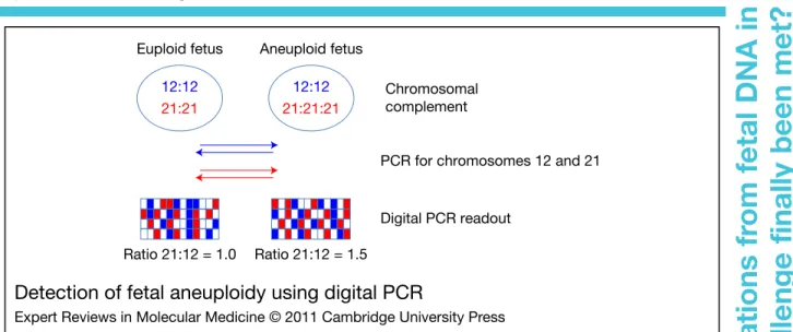
Ratio 21:12 = 1.0 Ratio 21:12 = 1.5 Euploid fetus Aneuploid fetus
PCR for chromosomes 12 and 21
Digital PCR readout Chromosomal complement 12:12 21:21 12:12 21:21:21
Detection of fetal aneuploidy using digital PCR
Expert Reviews in Molecular Medicine © 2011 Cambridge University Press
Figure 1. Detection of fetal aneuploidy using digital PCR. In this procedure, fluorescent PCR specific for sequences on chromosomes 12 and 21 is carried out in individual microreaction chambers. The amount of input template is titrated in such a fashion that each microreaction vessel contains >1 copy. After the plateau phase has been reached (approximately 40 cycles), the PCR reaction is terminated, and the number of positive reactions for each locus is counted. In euploid cases the ratio of blue (chromosome 12) to red (chromosome 21) signals should be 1, whereas in cases with Down syndrome the ratio of blue (chromosome 12) to red (chromosome 21) signals should be 1.5 (illustrated in more detail in Ref. 46). This method has to date not been successfully used for the detection of fetal aneuploidy using cell-free DNA in maternal plasma. 5
Determina
tion
of
fetal
chr
omosome
aberr
a
tions
fr
om
fetal
DNA
in
ma
ter
nal
blood:
has
the
challenge
finally
been
met?
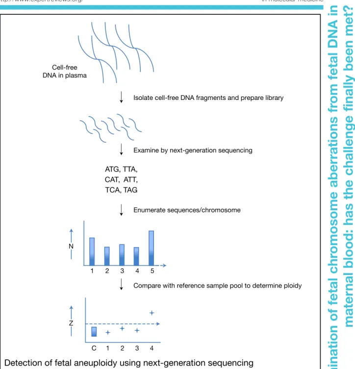
Cell-free DNA in plasma
Isolate cell-free DNA fragments and prepare library
Examine by next-generation sequencing
ATG, TTA, CAT, ATT, TCA, TAG
Enumerate sequences/chromosome
Compare with reference sample pool to determine ploidy
1 2 3 4 5
1
C 2 3 4
N
Z
Detection of fetal aneuploidy using next-generation sequencing
Expert Reviews in Molecular Medicine © 2011 Cambridge University Press
Figure 2. Detection of fetal aneuploidy using next-generation sequencing. In this procedure the cell-free DNA fragments in maternal plasma are isolated, and a library with special sequence tags is then made. These tags permit subsequent multiplex analysis. The library is examined by next-generation sequencing, which determines the sequence of each and every fragment. By bioinformatic analysis these sequences are ascribed to chromosomal locations. Following this, the number of sequence reads for each chromosome is counted. For chromosome 21 this is typically of the order of several thousand reads, which can then be compared with several million reads spread across the genome. If the fetus is affected by Down syndrome, then slightly more reads will be recorded for chromosome 21 compared with those from a euploid fetus. By comparing these data with a bank of reference samples, and by the use of predetermined cut-off values (Z score), the ploidy of the sample being examined can be determined (described in more detail in Refs 53, 54). 6
Determina
tion
of
fetal
chr
omosome
aberr
a
tions
fr
om
fetal
DNA
in
ma
ter
nal
blood:
has
the
challenge
finally
been
met?
(Ref. 7); of these, full karyotyping determined that 86 had a fetus affected by DS. To reduce the complexity and cost of the NGS analysis, a multiplex approach was tested, whereby eight samples were examined in parallel (8-plex). This was compared with a more costly duplex analysis on 314 samples (two samples examined simultaneously). The 8-plex analysis yielded a sensitivity of 79.1% and a specificity of 98.9%,
whereas the duplex assay yielded 100%
sensitivity and 97.9% specificity (Ref. 7). To make the assay more efficient, a cohort of normal samples was used as a standard, permitting a threshold cut-off value (termed Z) to be determined: values above this were deemed to show DS cases (Fig. 2).
Although it is unfortunate that the duplex analysis was determined to be superior to the less complex 8-plex analysis, it is not entirely surprising, in that for the 8-plex assay only approximately 300 000 total reads were recorded per sample. This translates to a small fraction of the billions of reads required to access the entire genome, and furthermore implies that perhaps less than 10 000 chromosome-21-specific reads were recorded. Because the NGS assay, like its digital PCR predecessor, relies simply on the counting of individual events, it would seem that here simply too few single events were being interrogated.
In the second, independent study, 449 samples were examined by a more detailed and robust
pair-end 4-plex NGS procedure in which
approximately 5 million reads per sample were obtained (Ref. 34). In this examination all 39
cases with DS were correctly identified,
although one normal case was incorrectly
classified as DS, thereby yielding 100%
sensitivity and 99.7% specificity. An important quality control in this study is that the concentration of fetal cf-DNA was monitored, and samples with less than 3.9% were excluded (Ref. 34).
Even if the NGS assays are considered not to be 100% effective, and could not be used as a stand-alone diagnostic test for DS, it has been suggested that their use as a secondary-tier screen of high-risk cases could lead to a 98% reduction in the number of invasive procedures (Ref. 7). As such, this avenue certainly does appear to be an attractive route to pursue (Ref. 8). Accordingly, large-scale clinical studies are being launched in Europe and elsewhere.
Can NGS be simplified?
The above results have indicated that the use of complex cutting-edge genomic tools such as NGS can finally offer the long-sought dream of
noninvasive detection of fetal aneuploidy
(Refs 1, 56). However, this comes at a very high cost and unacceptably long analysis. A possible strategy to overcome these disadvantages is the targeting of only those chromosomes or regions of interest such as those on chromosome 21 for the detection of DS cases. Although several different strategies exist that permit some form of target enrichment prior to the subsequent sequencing step, they have to date been limited to examination of intact genomic DNA samples, and not fragmented DNA species such as those found in cf-DNA (Ref. 57). Furthermore, these procedures, such as on-array capture, require rather large concentrations of input template
DNA (up to 7.5μg per sample). Hence, an
approach that permitted targeted sequencing of the small quantities of fragmented DNA in maternal plasma had to be sought.
Very recently, a solution hybrid selection
method using ultralong oligonucleotides,
commercially marketed by Agilent, USA, has been used for targeted enrichment (Ref. 57) (Fig. 3). A capture library specific for the X chromosome was hybridised with 500 ng of an amplified plasma DNA library, and the selected capture targets were pulled down using a
combination of biotinylated oligonucleotide
probes and streptavidin-coated magnetic beads. The enriched target DNA was then subjected to a further 12 rounds of PCR with specially tagged primers necessary for the subsequent sequence analysis. In the examination of 12 maternal plasma samples, this targeted capture procedure led to a mean enrichment of 213-fold. This enrichment was also reflected in the increased ability to detect fetal-specific loci on the X chromosome, which changed from 3.5% in the un-enriched samples to 95.9% in the samples subjected to a targeted enrichment step.
A concern with any enrichment approach is that it might lead to bias, by preferential enhancement of select sequences, including the preferential accumulation of maternal cf-DNA sequences over fetal ones. However, in this study the proportion of fetal to maternal sequences was
similar in un-enriched loci (∼16–30% from
first to third trimester) and in enriched X-chromosome regions (∼15–32%) (Ref. 57). These
7
Determina
tion
of
fetal
chr
omosome
aberr
a
tions
fr
om
fetal
DNA
in
ma
ter
nal
blood:
has
the
challenge
finally
been
met?
results are encouraging, because they could facilitate targeted enrichment of more crucial fetal loci, such as those on chromosome 21, thereby paving the way for a less complex noninvasive test for DS.
How much information is available from NGS?
The first series of experiments with NGS focused on select chromosomes and compared the number of reads obtained for these to those obtained for the entire genomic complement. Because close to
65 000 individual loci are counted on
chromosome 21 alone, it was open to debate whether this approach would be sufficiently sensitive to detect more subtle chromosomal
aberrations such as the Robertsonian
translocation between chromosomes 21 and 15 (or 14), which has a role in 2–3% of cases with DS (Ref. 1). As the Down Syndrome Critical Region associated with this translocation is a lot smaller than the entire chromosome 21 commonly involved in DS, it was unclear whether the NGS approach would be able to detect this alteration (Ref. 1).
However, this view has been altered by a new landmark publication, in which the entire cf-DNA present in a maternal plasma sample was sequenced (Ref. 24). The data indicated that the entire fetal genome complement is present in cf-DNA in maternal plasma, and that this can be mined to show minute details such as mutations
Cell-free DNA
Isolate cell-free DNA fragments and prepare library
Hybridise to SureSelectTM Oligo Capture Library
Select bound DNA fragments
Examine by next-generation sequencing ATG, TTA, CAT, ATT, TCA, TAG
Schematic representation of a
targeted sequencing approach using
the SureSelect
TMTarget Enrichment
System
Expert Reviews in Molecular Medicine © 2011 Cambridge University Press
Figure 3. Schematic representation of a targeted sequencing approach using the SureSelectTM Target Enrichment System.
Figure 3. Schematic representation of a targeted sequencing approach using the SureSelectTM Target Enrichment System. In this procedure the cell-free DNA fragments are isolated and a library is generated as per the standard next-generation sequencing protocol. Prior to sequencing, however, this library is hybridised to the SureSelectTM Oligo Capture Library, which is manufactured in such a manner that it will recognise a specific chromosome, such as chromosome 21. These oligo sequences contain magnetic particles to permit their retrieval in a magnetic field. Hence, following hybridisation (65°C, 24 h), captured sequences are selected by magnetic selection, and unselected sequences are washed away. The bound fragments are then purified and prepared for sequencing and examined as described for Figure 2. This procedure was recently shown to permit a 213-fold enrichment of the targeted X chromosome (Ref. 57).
8
Determina
tion
of
fetal
chr
omosome
aberr
a
tions
fr
om
fetal
DNA
in
ma
ter
nal
blood:
has
the
challenge
finally
been
met?
involved inβ-thalassaemia; surprisingly, the study correctly discerned that the fetus had inherited the paternal codon 41/42 mutation, involving a CTTT deletion, but not the maternal−28A→G mutation. Although this analysis was very complex, involving almost 4 billion reads and 900 000 SNPs, it is a striking indicator of what might lie ahead– namely, the ability to obtain a full fetal karyotype down to miniscule single-nucleotide detail from a single maternal blood sample (Ref. 24).
The question of intellectual property and how to optimise procedures
An important concern that needs to be
addressed is how intellectual property and commercialisation will affect future research and applications (Ref. 58). For the best possible service to reach the patient, clarity is essential; otherwise, conflicting reports and views might
prevent superior products from being
developed. In the case of fetal cf-DNA, such an issue was raised by the commercialisation of a noninvasive test for fetal RhD determination in the EU, and subsequently in the USA (Ref. 58). Although it was clear to leading researchers in the field that the tests initially marketed might be flawed or were not state of the art, it was felt that it would be a weary process to convince the commercial parties involved to change their standard of practice and to adopt more modern effective approaches. This issue was further highlighted by the near-simultaneous reports concerning the application of digital PCR or NGS, leading to conflicting reports in the lay media as to who the main patent claimants were (Refs 1, 59). It is currently also unclear what business model or strategies should be used in translating this research from the bench to the clinic. Hence, it might be worthwhile to echo previous concerns that such uncertainty might hinder or restrict further research in the field (Ref. 60).
Are fetal cells in maternal blood still worth pursuing?
Despite the enormous strides that have been made with the determination of fetal aneuploidies through the use of cf-DNA, the latest reports have indicated that these will be very labour
intensive, and require extremely high-tech
devices and bioinformatic services. Hence it is
unclear how quickly these developments will be translatable into the clinic.
For this reason there has been a resurgent interest in the isolation of fetal cells from maternal blood. This have been fostered by the development of (1) sophisticated high-throughput automated scanning devices, which permit the rapid identification of putative target fetal cells among a large pool of maternal cells (Refs 61, 62), and (2) efficient microfluidic
systems and electronic micromanipulation
systems, which permit the effective retrieval of individual trafficking fetal cells (Refs 59, 63, 64).
Even though this process will most likely also be laborious, it has several advantages over cf-DNA analysis, in that fetal cells offer a pure source of fetal genomic material. Furthermore, widely standardised procedures commonly used in many diagnostic laboratories, such as FISH (fluorescence in situ hybridisation), can be
applied for the very rapid detection of
chromosomal anomalies (Ref. 65). In addition, use can now be made of a wide body of experience in the analysis of single cells, a routine practice in many in vitro fertilisation
clinics offering preimplantation genetic
diagnosis (PGD) (Ref. 66).
A possible further advantage is that the intellectual property situation in this area is not as complex as that of cf-DNA, in that many of the original patents are close to expiry, and no commercial entity holds a wide portfolio of key elements. As such, it will be of interest to monitor progress in this sphere of research, because it might yet have a few surprises in store.
The challenge of proteomics
The development of the first-trimester DS screening test using a combination of ultrasound measurement of nuchal translucency, serum analyte analysis and maternal age has led to a dramatic increase in the ability to detect pregnancies at-risk of bearing an aneuploid fetus (85% sensitivity) when compared with the previous generation of second-trimester screening tests (65% sensitivity), which relied solely on serum analyte analysis in combination with maternal age (Ref. 67). This facet is reflected in the report on the English and Welsh National Down Syndrome Cytogenetic Register analysis, which indicated that the vast majority of DS cases were detected by current screening practice (Ref. 5). 9
Determina
tion
of
fetal
chr
omosome
aberr
a
tions
fr
om
fetal
DNA
in
ma
ter
nal
blood:
has
the
challenge
finally
been
met?
The downside of this startling development is that the first-trimester screen is still hampered by a rather high false-positive rate of 5–8% (Refs 68, 69). This implies that a large number of healthy pregnancies are being subjected to unnecessary invasive procedures – a healthcare risk for mother and unborn child, as well as a considerable financial burden for healthcare services. Furthermore, almost 15% of cases remain undetected and, because these occur in the group of pregnant women not judged to be at a higher risk (<35 years old), could result in undesired live births.
Therefore, increasing the efficiency of this test has been proposed, perhaps by the addition
of further biochemical analytes, such as
members of the activin family (Refs 70, 71). The inclusion of further highly specific blood-based analytes might be possible as a result of the
development of quantitative proteomic
technologies as part of the Human Proteome Association, especially the affiliated plasma proteome project (Ref. 72). The placenta in DS cases exhibits structural alterations, which are most likely the result of alterations in protein expression, and so it is possible that these changes will be reflected in the maternal plasma proteome (Ref. 73). In this regard, the use of quantitative isobaric labelling has been shown to permit the detection of significant alterations in the levels of several placenta-derived peptides in DS cases when compared with controls (Ref. 74). Analogous observations have been made by other research groups using a variety of different proteomic approaches (Refs 75, 76, 77, 78). It now remains to explore how useful these potential biomarkers will be in increasing the detection rate of the current first-trimester screen.
Summary and conclusion
Tremendous strides have been made with the analysis of fetal cf-DNA in maternal blood, in that we now stand on the threshold of a new generation of noninvasive diagnostic tools, which might even permit a detailed analysis of the entire fetal genome or a full karyotype. However, it remains to be seen how rapidly these can be translated into the clinic, in a manner that is cost effective and accessible to all. In this context it will be interesting to see
which system, namely NGS, digital PCR
or enrichment of methylated fetal cf-DNA fragments, passes the final hurdle to enter
clinical service. Although the isolation and analysis of trafficking fetal cells might remain a peripheral test, it could be useful in those centres already offering FISH or PGD services. Should multiparameter mass spectroscopy and quantitative proteomics finally come of age, it
might yield a set of biomarkers with
unparalleled specificity, not only for DS
screening but also for other pregnancy-related disorders such as preeclampsia.
Acknowledgements and funding
This report was funded by the clinical research fund of the University Women’s Hospital Basel. The authors thank the referees for their critical and insightful comments.
References
1 Hahn, S., Jackson, L.G. and Zimmermann, B.G. (2010) Prenatal diagnosis of fetal aneuploidies: post-genomic developments. Genome Medicine 2, 50
2 Bianchi, D.W. (2010) From Michael to microarrays: 30 years of studying fetal cells and nucleic acids in maternal blood. Prenatal Diagnosis 30, 622-623 3 Ferguson-Smith, M.A. and Bianchi, D.W. (2010)
Prenatal diagnosis: past, present, and future. Prenatal Diagnosis 30, 601-604
4 Lo, Y.M. (2010) Noninvasive prenatal diagnosis in 2020. Prenatal Diagnosis 30, 702-703
5 Morris, J.K. and Alberman, E. (2009) Trends in Down’s syndrome live births and antenatal diagnoses in England and Wales from 1989 to 2008: analysis of data from the National Down Syndrome Cytogenetic Register. British Medical Journal 339, b3794
6 Philip, J. et al. (2004) Late first-trimester invasive prenatal diagnosis: results of an international randomized trial. Obstetrics and Gynecology 103, 1164-1173
7 Chiu, R.W. et al. (2011) Non-invasive prenatal assessment of trisomy 21 by multiplexed maternal plasma DNA sequencing: large scale validity study. British Medical Journal 342, c7401
8 Jackson, L. and Pyeritz, R.E. (2011) Molecular technologies open new clinical genetic vistas. Science Translational Medicine 3, 65–62
9 Lo, Y.M. et al. (1997) Presence of fetal DNA in maternal plasma and serum. Lancet 350, 485-487 10 van der Schoot, C.E., Hahn, S. and Chitty, L.S. (2008)
Non-invasive prenatal diagnosis and determination of fetal Rh status. Seminars in Fetal and Neonatal Medicine 13, 63-68 10
Determina
tion
of
fetal
chr
omosome
aberr
a
tions
fr
om
fetal
DNA
in
ma
ter
nal
blood:
has
the
challenge
finally
been
met?
11 Chitty, L.S. et al. (2008) SAFE–the special non-invasive advances in fetal and neonatal evaluation network: aims and achievements. Prenatal Diagnosis 28, 83-88
12 Finning, K. et al. (2008) Effect of high throughput RHD typing of fetal DNA in maternal plasma on use of anti-RhD immunoglobulin in RhD negative pregnant women: prospective feasibility study. British Medical Journal 336, 816-818
13 Gautier, E. et al. (2005) Fetal RhD genotyping by maternal serum analysis: a two-year experience. American Journal of Obstetrics and Gynecology 192, 666-669
14 Maddocks, D.G. et al. (2009) The SAFE project: towards non-invasive prenatal diagnosis. Biochemical Society Transactions 37, 460-465 15 Lo, Y.M. (2009) Noninvasive prenatal detection of
fetal chromosomal aneuploidies by maternal plasma nucleic acid analysis: a review of the current state of the art. British Journal of Obstetrics and Gynecology 116, 152-157
16 Krentz, S.E. and Pilskaln, S.M. (1988) Product life cycle: still a valid framework for business planning. Top Health Care Finance 15, 40-48
17 Lo, Y.M. et al. (1998) Quantitative analysis of fetal DNA in maternal plasma and serum: implications for noninvasive prenatal diagnosis. American Journal of Human Genetics 62, 768-775
18 Zimmermann, B. et al. (2005) Optimized real-time quantitative PCR measurement of male fetal DNA in maternal plasma. Clinical Chemistry 51, 1598-1604 19 Sikora, A. et al. (2010) Detection of increased
amounts of cell-free fetal DNA with short PCR amplicons. Clinical Chemistry 56, 136-138
20 Chan, K.C. et al. (2004) Size distributions of maternal and fetal DNA in maternal plasma. Clinical Chemistry 50, 88-92
21 Li, Y. et al. (2004) Size separation of circulatory DNA in maternal plasma permits ready detection of fetal DNA polymorphisms. Clinical Chemistry 50, 1002-1011
22 Hahn, S. and Zimmermann, B.G. (2010) Cell-free DNA in maternal plasma: has the size-distribution puzzle been solved? Clinical Chemistry 56, 1210-1211
23 Fan, H.C. et al. (2010) Analysis of the size distributions of fetal and maternal cell-free DNA by paired-end sequencing. Clinical Chemistry 56, 1279-1286
24 Lo, Y.M. et al. (2010) Maternal plasma DNA sequencing reveals the genome-wide genetic and mutational profile of the fetus. Science Translational Medicine 2, 61ra91
25 Li, Y. et al. (2005) Detection of paternally inherited fetal point mutations for beta-thalassemia using size-fractionated cell-free DNA in maternal plasma. Journal of the American Medical Association 293, 843-849
26 Li, Y. et al. (2004) Improved prenatal detection of a fetal point mutation for achondroplasia by the use of size-fractionated circulatory DNA in maternal plasma– case report. Prenatal Diagnosis 24, 896-898 27 Li, Y. et al. (2007) Non-invasive prenatal detection of achondroplasia in size-fractionated cell-free DNA by MALDI-TOF MS assay. Prenatal Diagnosis 27, 11-17 28 Li, Y. et al. (2006) Genotyping fetal paternally
inherited SNPs by MALDI-TOF MS using cell-free fetal DNA in maternal plasma: influence of size fractionation. Electrophoresis 27, 3889-3896 29 Flori, E. et al. (2004) Circulating cell-free fetal DNA in
maternal serum appears to originate from cyto- and syncytio-trophoblastic cells. Case report. Human Reproduction 19, 723-724
30 Alberry, M. et al. (2007) Free fetal DNA in maternal plasma in anembryonic pregnancies: confirmation that the origin is the trophoblast. Prenatal Diagnosis 27, 415-418
31 Chim, S.S. et al. (2008) Systematic search for placental DNA-methylation markers on
chromosome 21: toward a maternal plasma-based epigenetic test for fetal trisomy 21. Clinical Chemistry 54, 500-511
32 Chim, S.S. et al. (2005) Detection of the placental epigenetic signature of the maspin gene in maternal plasma. Proceedings of the National Academy of Sciences of the United States of America 102, 14753-14758
33 Chiu, R.W. et al. (2007) Hypermethylation of RASSF1A in human and rhesus placentas. American Journal of Pathology 170, 941-950
34 Ehrich, M. et al. (2011) Noninvasive detection of fetal trisomy 21 by sequencing of DNA in maternal blood: a study in a clinical setting. American Journal of Obstetrics and Gynecology 204, 205.e1-205.e11 35 Lo, Y.M. et al. (2007) Plasma placental RNA
allelic ratio permits noninvasive prenatal chromosomal aneuploidy detection. Nature Medicine 13, 218-223
36 Gandhi, S.J. et al. (2010) Transcription of functionally related constitutive genes is not coordinated. Nature Structural and Molecular Biology 18, 27-34 37 Lionnet, T. and Singer, R.H. (2010) Transcription, one
allele at a time. Genome Biology 11, 129 38 Tong, Y.K. et al. (2006) Noninvasive prenatal
detection of fetal trisomy 18 by epigenetic allelic ratio analysis in maternal plasma: theoretical
11
Determina
tion
of
fetal
chr
omosome
aberr
a
tions
fr
om
fetal
DNA
in
ma
ter
nal
blood:
has
the
challenge
finally
been
met?
and empirical considerations. Clinical Chemistry 52, 2194-2202
39 Tong, Y.K. et al. (2010) Noninvasive prenatal detection of trisomy 21 by an epigenetic-genetic chromosome-dosage approach. Clinical Chemistry 56, 90-98
40 Tsui, D.W. et al. (2010) Systematic identification of placental epigenetic signatures for the noninvasive prenatal detection of Edwards Syndrome. PLoS One 5, e15069
41 Papageorgiou, E.A. (2011) Fetal-specific DNA methylation ratio permits noninvasive prenatal diagnosis of trisomy 21. Nature Medicine, Mar 6; [Epub ahead of print].
42 Papageorgiou, E.A. et al. (2009) Sites of differential DNA methylation between placenta and peripheral blood: molecular markers for noninvasive prenatal diagnosis of aneuploidies. American Journal of Pathology 174, 1609-1618
43 Zimmermann, B. et al. (2007) Real-time quantitative polymerase chain reaction measurement of male fetal DNA in maternal plasma. Methods in Molecular Medicine 132, 43-49
44 Zimmermann, B. et al. (2006) Use of real-time polymerase chain reaction for the detection of fetal aneuploidies. Methods in Molecular Biology 336, 83-100
45 Vogelstein, B. and Kinzler, K.W. (1999) Digital PCR. Proceedings of the National Academy of Sciences of the United States of America 96, 9236-9241 46 Zimmermann, B.G. et al. (2008) Digital PCR: a
powerful new tool for noninvasive prenatal diagnosis? Prenatal Diagnosis 28, 1087-1093 47 Fan, H.C. and Quake, S.R. (2007) Detection of
aneuploidy with digital polymerase chain reaction. Analytical Chemistry 79, 7576-7579
48 Fan, H.C. et al. (2009) Microfluidic digital PCR enables rapid prenatal diagnosis of fetal aneuploidy. American Journal of Obstetrics and Gynecology 200, 543, e541–547
49 Lo, Y.M. et al. (2007) Digital PCR for the molecular detection of fetal chromosomal aneuploidy. Proceedings of the National Academy of Sciences of the United States of America 104, 13116-13121 50 Ansorge, W.J. (2009) Next-generation DNA
sequencing techniques. New Biotechnology 25, 195-203
51 Jones, S. et al. (2008) Comparative lesion sequencing provides insights into tumor evolution. Proceedings of the National Academy of Sciences of the United States of America 105, 4283-4288
52 Leary, R.J. et al. (2010) Development of personalized tumor biomarkers using massively parallel
sequencing. Science Translational Medicine 2, 20ra14
53 Chiu, R.W. et al. (2008) Noninvasive prenatal diagnosis of fetal chromosomal aneuploidy by massively parallel genomic sequencing of DNA in maternal plasma. Proceedings of the National Academy of Sciences of the United States of America 105, 20458-20463
54 Chiu, R.W., Cantor, C.R. and Lo, Y.M. (2009) Non-invasive prenatal diagnosis by single molecule counting technologies. Trends in Genetics 25, 324-331
55 Fan, H.C. et al. (2008) Noninvasive diagnosis of fetal aneuploidy by shotgun sequencing DNA from maternal blood. Proceedings of the National Academy of Sciences of the United States of America 105, 16266-16271
56 Lo, Y.M. and Chiu, R.W. (2009) Next-generation sequencing of plasma/serum DNA: an emerging research and molecular diagnostic tool. Clinical Chemistry 55, 607-608
57 Liao, G.J. et al. (2010) Targeted massively parallel sequencing of maternal plasma DNA permits efficient and unbiased detection of fetal alleles. Clinical Chemistry 57, 92-101
58 Szczepura, A., Osipenko, L. and Freeman, K. (2011) A new fetal RHD genotyping test: costs and benefits of mass testing to target antenatal anti-D prophylaxis in England and Wales. BMC Pregnancy and Childbirth 11, 5
59 Hahn, S. et al. (2009) Noninvasive prenatal diagnosis of fetal aneuploidies and Mendelian disorders: new innovative strategies. Expert Review of Molecular Diagnostics 9, 613-621
60 Daniels, G., Finning, K. and Martin, P. (2010) Noninvasive fetal blood grouping: present and future. Clinics in Laboratory Medicine 30, 431-442 61 Ntouroupi, T.G. et al. (2008) Detection of circulating
tumour cells in peripheral blood with an automated scanning fluorescence microscope. British Journal of Cancer 99, 789-795
62 Seppo, A. et al. (2008) Detection of circulating fetal cells utilizing automated microscopy: potential for noninvasive prenatal diagnosis of chromosomal aneuploidies. Prenatal Diagnosis 28, 815-821 63 Lee, D. et al. (2010) Separation of model mixtures of
epsilon-globin positive fetal nucleated red blood cells and anucleate erythrocytes using a microfluidic device. Journal of Chromatography A 1217, 1862-1866 64 Huang, R. et al. (2008) A microfluidics approach for the isolation of nucleated red blood cells (NRBCs) from the peripheral blood of pregnant women. Prenatal Diagnosis 28, 892-899 12
Determina
tion
of
fetal
chr
omosome
aberr
a
tions
fr
om
fetal
DNA
in
ma
ter
nal
blood:
has
the
challenge
finally
been
met?
65 Evans, M.I. et al. (2006) Automated microscopy of amniotic fluid cells: detection of FISH signals using the FastFISH imaging system. Fetal Diagnosis and Therapy 21, 523-527
66 Wells, D. (2010) Embryo aneuploidy and the role of morphological and genetic screening. Reproductive Biomedicine Online 21, 274-277
67 Avgidou, K. et al. (2005) Prospective first-trimester screening for trisomy 21 in 30,564 pregnancies. American Journal of Obstetrics and Gynecology 192, 1761-1767
68 Falcon, O. et al. (2006) Screening for trisomy 21 by fetal tricuspid regurgitation, nuchal translucency and maternal serum free beta-hCG and PAPP-A at 11+ 0 to 13 + 6 weeks. Ultrasound in Obstetrics and Gynecology 27, 151-155
69 Cowans, N.J. et al. (2009) The impact of fetal gender on first trimester nuchal translucency and maternal serum free beta-hCG and PAPP-A MoM in normal and trisomy 21 pregnancies. Prenatal Diagnosis 29, 578-581
70 Spencer, K. et al. (2001) Maternal serum levels of dimeric inhibin A in pregnancies affected by trisomy 21 in the first trimester. Prenatal Diagnosis 21, 441-444 71 Spencer, K. et al. (2001) Maternal serum levels of total activin-A in first-trimester trisomy 21 pregnancies. Prenatal Diagnosis 21, 270-273
72 Schmidt, A. and Aebersold, R. (2006) High-accuracy proteome maps of human body fluids. Genome Biology 7, 242
73 Choolani, M. et al. (2009) Proteomic technologies for prenatal diagnostics: advances and challenges ahead. Expert Review of Proteomics 6, 87-101 74 Kolla, V. et al. (2010) Quantitative proteomics
analysis of maternal plasma in Down syndrome pregnancies using isobaric tagging reagent (iTRAQ). Journal of Biomedicine and Biotechnology 2010, 952047
75 Kolialexi, A. et al. (2008) Application of proteomics for the identification of differentially expressed protein markers for Down syndrome in maternal plasma. Prenatal Diagnosis 28, 691-698
76 Tsangaris, G.T. et al. (2006) Proteomic analysis of amniotic fluid in pregnancies with Down syndrome. Proteomics 6, 4410-4419
77 Lopez, M.F. et al. (2010) Mass spectrometric discovery and selective reaction monitoring (SRM) of putative protein biomarker candidates in first trimester trisomy 21 maternal serum. Journal of Proteome Research 10, 133-142
78 Nagalla, S.R. et al. (2007) Proteomic analysis of maternal serum in down syndrome: identification of novel protein biomarkers. Journal of Proteome Research 6, 1245-1257
Further reading, resources and contacts
Publications
Chiu, R.W., Cantor, C.R. and Lo, Y.M. (2009) Non-invasive prenatal diagnosis by single molecule counting technologies. Trends in Genetics 7, 324-331
Chitty, L.S. and Lau, T.K. (2011) First trimester screening– new directions for antenatal care. Prenatal Diagnosis 31, 1-2
Ferguson-Smith, M.A. and Bianchi, D.W. (2010) Prenatal diagnosis: past, present, future. Prenatal Diagnosis 30, 601-604
Websites
A study funded by the UK National Institute for Health Research to assist with the transfer of noninvasive prenatal diagnostic techniques into the clinic is described at:
http://www.rapid.nhs.uk/
Features associated with this article
Figures
Figure 1. Detection of fetal aneuploidy using digital PCR.
Figure 2. Detection of fetal aneuploidy using next-generation sequencing.
Figure 3. Schematic representation of a targeted sequencing approach using the SureSelectTMTarget Enrichment System. 13
Determina
tion
of
fetal
chr
omosome
aberr
a
tions
fr
om
fetal
DNA
in
ma
ter
nal
blood:
has
the
challenge
finally
been
met?
Citation details for this article
Sinuhe Hahn, Olav Lapaire, Sevgi Tercanli, Varaprasad Kolla and Irene Hösli (2011) Determination of fetal chromosome aberrations from fetal DNA in maternal blood: has the challenge finally been met? Expert Rev. Mol. Med. Vol. 13, e16, May 2011, doi:10.1017/S1462399411001852
14