557
Immunologic and Molecular Characteristics of
Encephalitozoon-Like
Microsporidia Isolated from Humans and Rabbits Indicate That
Encephalitozoon
cuniculi
Is a Zoonotic Parasite
Peter Deplazes, Alexander Mathis, Ruth Baumgartner,
Isabelle Tanner, and Rainer Weber
From the Institute of Parasitology and the Clinic of Exotic Pets and Zoo Animals, University of Zurich; and the Division of Infectious Diseases, University Hospital, Zurich, Switzerland To assess the zoonotic potential ofEncephalitozoon-likemicrosporidia, we isolated and cultivated
spores from specimens of urine, respiratory secretions, and stool from six patients infected with human immunodeficiency virus and from nine rabbits. Because spores ofEncephalitozoon-like species are indistinguishable by microscopy, we characterized the isolates by western blot analysis and by restriction enzyme analysis of the small subunit (SSU) rDNA after amplification by the polymerase chain reaction. We identifiedSeptata intestinalisin one patient and Encephalitozoon hellemin two symptomatic patients.Encephalitozoon cuniculiwas found in all rabbits and in three patients. One of these patients had clinical manifestations of infection with this parasite (severe interstitial pneumonitis). We observed abatement of symptoms and cessation of parasite excretion when these patients were treated with albendazole. Our findings suggest that E.cuniculimay be pathogenic in humans and that it is a zoonotic parasite.
In recent years, microsporidia have emerged as important opportunistic parasites in patients with AIDS. Three Encephali-tozoon-like microsporidia of animal origin that share most of the morphological features ofEncephalitozoon cuniculi have been found in humans [1].E. cuniculiis parasitic in birds and mammals including rabbits, rodents, carnivores, and primates
[2]. E. cuniculiwas detectedinthe CSF and/or urine (in 1959 and 1984) of two children with seizures and, subsequently, in HIV-infected patients with hepatitis, peritonitis, and keratocon-junctivitis [1]. Whether these infections indeed were caused byE. cuniculior by anotherEncephalitozoon-like microspori-dian is not known because only results of light microscopical studies and/or electron microscopical studies of the parasites are available.
Results of a western blot analysis performed in 1992 implied that humanEncephalitozoonisolates belong to a separate spe-cies, which was named E. hellem. Another
Encephalitozoon-like microsporidian that caused chronic diarrhea and systemic disease in HIV-infected patients was subsequently described; this organism was named Septata intestinalis on the basis of its specific ultrastructural appearance in small intestinal entero-cytes (reviewed in [1]). Recently, molecular characterization
Received 6 June 1995; revised 6 September 1995.
Informed consent was obtained from all patients to perform the examinations described in this paper. Compassionate use of a1bendazole was reviewed by the ethical committee of the University Hospital of Zurich.
Grant support: This work was supported by the Swiss National Science Foundation (grant no. 32 37399.93) and the Swiss National Program for AIDS Research (grant no. 91-7066).
Reprints or correspondence: Dr. Peter Deplazes, Institute of Parasitology, Winterthurerstrasse 266a, CH-8057 ZUrich, Switzerland.
Clinical Infectious Diseases 1996;22:557-9
© 1996 by The University of Chicago. All rights reserved. 1058-4838/96/2203 -0024$02 .00
of an Encephalitozoon-like organism isolated from the urine and respiratory specimens of a single HIV-infected patient has suggested that E. cuniculimay also be parasitic in humans [3].
Patients and Methods
Encephalitozoon-like microorganisms were detected in dif-ferent specimens from six HIV-infected patients (one female and five males) by light microscopical examination of chro-motrope-stained specimens [1]. Rabbits with antibodies to spores ofE. cuniculidetected with use of a routine immuno-fluorescence assay were selected for the isolation of microspo-ridia. Urine samples (0.1-2 L) and bronchoalveolar aspirates were centrifuged(l,OOOg for 20 minutes) and washed twice with distilled water. Unfixed stool specimens were homoge-nized in water, pressed through sieves (widths of mesh: 200
j..lm, 50 j..lm, and 20 j..lm), and washed five times in distilled water. The sediment was overlaid with a PBS-50% Percoll solution (Pharmacia, Silver Spring, MD) and centrifuged(600g
for 30 minutes) at room temperature. Kidney and brain tissue from rabbits were mechanically homogenized, washed, and centrifuged in PBS-30% Percoll(700gfor 40 minutes at room temperature).
The following microsporidia reference isolates were avail-able: an E.cuniculiisolate of animal origin (courtesy of Profes-sor F. Derouin, H6pital Saint-Louis, Paris), twoE. hellem iso-lates (CDC [Centers for Disease Control and Prevention] 0291:V213 [4] and CH-HI BG [5]), and anS.intestinalisisolate (CDC:V297 [6]). In vitro cultivation of spores was done as described [4] with human embryonic lung fibroblast (MRC-5) cells. Antibodies to spores of E. cuniculi (isolate CH-R2169 from a rabbit),E. hellem(isolate CH-HIBG), andS. intestinalis
558 Deplazes et al.
em
1996;22 (March)A
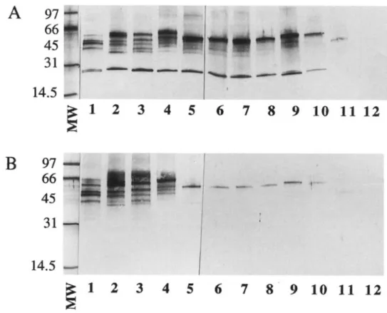
97
66
45
Figurepurified spores of1. Western blot analysis ofEncephalitozoon-31
like microsporidia isolated fromhumans and rabbits.Lanes1 and 2, Encephalitozoon hellem reference
14.5
strains CDC:029I :V213 andCH-~
1
2
3
4
5
6
7
8
9
10 11 12
laneHIBG.4, strain CH-H3WR (both iso-Lane3, strain CH-H2SF, and lated from HIV-infected patients). Lanes5 and 6,E. cuniculistrain CH-R2169 from a rabbit.Lane7,E.cun-B
97
iculireference isolate.Lane8, strain66
CH-H4BJ;and lane 10,lanestrain CH-H6FN (all9, strain CH-H5RB;45
isolatedtients).Lanefrom11,Septata intestinalisHIV-infectedpa-31
reference strain CDC:V297.12, strain CH-H70R isolated fromLanethe stool of an HIV-infected patient. Murine antibodies to inactivated and
14.5
E.purified spores ofhellem (B)were used. MWE. cuniculi (A)=mo-or~
1
2
3
4
5
6
7
8
9
10 11 12
lecular weights (in thousands).(non-inbred) mice that were immunized by subcutaneous injec-tion of 2X 10⁷spores (inactivated at +90°C for 5 minutes) in Freund's complete adjuvant. The procedure was repeated 4 weeks later with use of Freund's incomplete adjuvant, and serum samples were collected 2 weeks later.
Western blot analysis was performed as described previously [4]. Antigens from I X 106
purified spores per lane were sepa-rated by electrophoresis (4%-20% gradient gels). Specific anti-body reactions were detected with goat antianti-body to mouse IgG (H and L chains) (Cappel, Turnhout, Belgium). For riboprint-ing, DNA was released from the spores through a combination of mechanical disruption (vortexing with glass beads) and pro-teinase K digestion. The SSU rDNA was amplified under stan-dard PCR conditions with use of primers that corresponded to positions 1-19 and 1277-1299, respectively, of the SSU rRNA gene sequence ofE. cuniculi [7]. PCR products were cleaved
with restriction enzymes and resolved by electrophoresis in 2% agarose gels.
cytopathic BK virus was detected by electron microscopy. Spores directly isolated from stool from a sixth patient were used for further characterization of the S. intestinalis isolate
CH-H70R, as the MRC-5 cells were destroyed by adenovirus type 9 that was identified by seroneutralization with polyclonal type-specific antibodies (American Type Culture Collection, Rockville, MD). The isolates were characterized by western blot analysis with use of antibodies to spores ofE. cuniculi
(figure IA), E. hellem (figure IB), orS. intestinalis (data not
400bp - -....
M 1 2 3 4 5 6 7 8 9 10 11
Results
Encephalitozoon-like spores were isolated from five urine
sediments and from one bronchoalveolar lavage specimen from five patients and from nine rabbits (three isolates each were from urine, kidney, and brain). All of these isolates were estab-lished in culture for further characterization. The human E. cuniculi isolate CH-H4BJ was cultured, although strongly
Figure 2. Restriction patterns of the amplified SSU rRNA gene of Encephalitozoon-likeisolates after cleavage withMbo II HpaII.Lane M,DNA size marker (lOO-bp ladder).Lane1 and 2,Encephalitozoon hellem reference strains CDC:0291:V213 and CH-HlBG, respec-tively.Lane3, strain CH-H2SF, andlane4, strain CH-H3WR (both isolated from mY-infected patients).Lane5,E. cuniculistrain CH-R2169 from a rabbit.Lane 6,E. cuniculireference isolate. Lane 7, strain CH-H4BJ;lane 8, strain CH-H5RB; and lane 9, strain CH-H6FN (all isolated from HlV-infected patients). Lane 10, Septata intestinalisreference isolate CDC:V297.Lane11, strain CH-H70R.
eID
1996;22 (March) Encephalitozoon cuniculi 559shown) and by riboprinting (figure 2), with congruent results: all nine isolates from rabbits (not shown) as well as three of six isolates from humans showed banding patterns that were identical to those of theE. cuniculireference isolate (figure 1, lanes 5-10 and figure 2, lanes 5-9). Accordingly, two other isolates were indistinguishable from the two E. hellem refer-ence isolates (figures 1 and 2, lanes 1-4), and one was identi-fied asS. intestinalis(figure 2, lanes 10 and 11).
Discussion
Diagnosis of human microsporidial infections is presently based on morphological demonstration of the organisms by examination with light microscopy or electron microscopy. These techniques are sufficient to distinguish the species
Enter-ocytozoon bieneusifrom the Encephalitozoon-like microspo-ridia but are not appropriate for differentiating
Encephalito-zoon-like microsporidia, of which at least three morpholog-ically similar species are parasitic in humans [1]. So far, micro-sporidial isolates from humans and animals have rarely been compared by means of immunologic and molecular biological techniques. A single HIV-infected patient with confirmed
E. cuniculiinfection and seven patients with systemic E. hellem infection have been described [1, 3]. From the epidemiological standpoint, characterization of Encephalitozoon-like organisms that are parasitic in animals is of importance, as E. cuniculi is known to be prevalent in animals, but animal reservoirs of E. hellem andS. intestinalishave not been identified so far.
The fact that three of the six Encephalitozoon-like isolates from patients and all nine rabbit isolates were characterized as E. cuniculi supports earlier suggestions that E. cuniculi might be of importance as a zoonotic parasite. In an ongoing epidemi-ological survey conducted by our laboratory, we found that ,...., 16% of necropsied rabbits proved positive for the organism on serology. Rabbits infected with E. cuniculi excrete high numbers of spores in the urine [2]. However, no obvious source of infection was recognized for our six patients, as none owned a pet or reported exposure to animals. As E. cuniculi spores are resistant in the environment, infections caused by contaminated water, food, or other sources have to be considered.
All six HIV-infected patients were severely immunodefi-cient, with CD4 cell counts that ranged from 0 to 0.05 X 10⁹/L. Two patients withE.cuniculiinfection had no associated clinical manifestations. The third patient, who had E. cuniculi isolated from urine and bronchoalveolar lavage fluid, presented with severe interstitial pneumonitis. Of patients with E. hellem
infection, one presented with acute urinary tract infection, and one presented with systemic disease including conjunctivitis, sinusitis, bronchitis, and assumed cystitis. Although organisms were detected in urine specimens of all patients infected with
E. cuniculior E. hellem, only one patient had significant leuko-cyturia and erythroleuko-cyturia, and none had evidence of renal failure. In contrast to previous reports that described no patients with E. hellem and E. cuniculi identified in stool specimens, we found that one patient with systemic E. hellem infection and one with systemic E. cuniculi infection were excreting a few Encephalitozoon-like spores in stool. Albendazole (400 mg twice daily for 2-3 weeks) was found to cure infections due to encephalitozoon andS. intestinalisinfections in all six patients. No relapse of encephalitozoon infection was diag-nosed during the follow-up observation period of3-14 months, although no secondary prophylaxis with albendazole was given. Our results strongly suggest that E. cuniculi is pathogenic in severely immunodeficient HIV-infected individuals and that the organism is a zoonotic parasite. Modes of transmission have yet to be elucidated.
Acknowledgments
The authors thank Dr. Govinda Visvesvara for providing the CDC reference isolates and Herbert Kuster, Barbel Sauer, and Janet Skaggs for technical assistance.
References
1. Weber R, Bryan RT, Schwartz DA, Owen RL. Human microsporidial infec-tions. Clin Microbiol Rev 1994; 7:426-61.
2. Canning ED, LornJ.The microsporidia of vertebrates. London: Academic Press, 1986:189-241.
3. De Groote MA, Visvesvara GS, Wilson ML, et al. Polymerase chain reaction and culture confirmation of disseminated Encephalitozoon cuniculi in a patient with AIDS: successful therapy with a1bendazole. J Infect Dis 1995; 171:1375-8.
4. Visvesvara GS, LeitchGJ, Maura H, Wallace S, Weber R, Bryan RT. Culture, electron microscopy, and immunoblot studies on a microspori-dian parasite isolated from the urine of a patient with AIDS. J Protozool 1991; 38: lOSS-II S.
5. Weber R, Kuster H, Visvesvara GS, Bryan RT, Schwartz DA, LUthy R. Disseminated microsporidiosis due to Encephalitozoon hellem: pulmo-nary colonization, microhematuria, and mild conjunctivitis in a patient with AIDS. Clin Infect Dis 1993; 17:415-9.
6. Visvesvara GS, Da Silva AJ, Croppo GP, et al. In vitro culture and serologic and molecular identification of Septata intestinalis isolated from urine of a patient with AIDS. J C1in MicrobiolI995;33:930-6.
7. Hartskeerl RA, Schuitema ARJ, deWachter R. Secondary structure of the small subunit ribosomal RNA sequence of the microsporidium Encepha-litozoon cuniculi. Nucleic Acids Res 1993; 21 :1489.