Brigitte Wirth
Hubertus J.A. van Hedel Armin Curt
Ankle dexterity is less impaired
than muscle strength in incomplete
spinal cord lesion
Introduction
Patients with an incomplete spinal cord injury (iSCI) of-ten show considerable motor recovery [37] and the ma-jority of patients who initially had some preserved mo-tor function below the level of lesion become pedestrians (about 90 % in traumatic SCI) [19]. The
clin-ical assessment of motor deficits after iSCI currently consists of a measurement for muscle strength (manual muscle testing according to the American Spinal Injury Association (ASIA) [2]), gait tests [16, 17, 33, 34] and an assessment of independence in activities of daily living [9].
Muscle strength, however, is only one component of motor function that can be impaired after a lesion of the
JON 2724
Received: 21 April 2007
Received in revised form: 21 July 2007 Accepted: 21 August 2007
Published online: 22 January 2008
B. Wirth (쾷) · H. J. A. van Hedel Spinal Cord Injury Center Balgrist University Hospital Forchstrasse 340 8008 Zurich, Switzerland Tel.: +41-44/386-3729 Fax: +41-44/386-3731 E-Mail: bwirth@paralab.balgrist.ch B. Wirth
Institute of Human Movement Sciences and Sport
Swiss Federal Institute of Technology 8092 Zurich, Switzerland
A. Curt
ICORD and Division of Neurology University of British Columbia 2469-6270 University Blvd Vancouver, BC V6T 1Z4, Canada
■ Abstract Background Motor
assessment after incomplete spinal cord injury (iSCI) currently con-sists of tests for muscle strength (manual muscle testing) and gait. The ability to adequately time a movement, an aspect of dexterity, is not tested. Thus, this study assessed the timing of ankle dorsiflexion in iSCI patients in the supine position and during gait and examined its relation to measures for muscle strength, corticospinal conductivity and gait speed. Methods In 12 subjects with iSCI and 12 matched controls, tim-ing of ankle dorsiflexion was tested by means of auditory-paced dorsi-and plantar-flexion movements at three frequencies in the supine position and by determining initi-ation and termininiti-ation of dorsiflex-ion in swing during gait. In addi-tion, maximal movement velocity (MMV) in the ankle task, maximal voluntary contraction (MVC), cor-ticospinal conductivity (motor evoked potentials (MEP)) and gait speed (10 Meter Walk Test) were
assessed. Results The groups did not significantly differ in timing of ankle dorsiflexion, neither in the supine position nor in gait. How-ever, they significantly differed in MMV at all frequencies, MEP latency, MEP amplitude and gait speed. In contrast to ankle timing in the supine position, the onset of dorsiflexion in swing during gait significantly correlated to the dynamic MEP parameters.
Conclu-sions Although MMV and gait
speed were significantly reduced, timing of ankle dorsiflexion, both in the supine position and during gait, was less impaired in iSCI patients. This indicates that the loss of strength, particularly of dy-namic strength, is the major motor impairment in iSCI, which might be considered when assessing treatment interventions.
■ Key words dexterity ·
incomplete spinal cord injury · motor evoked potential · muscle strength
central nervous system (CNS). In upper limb studies with stroke patients, dexterity, defined as adroitness and competency in use of the limbs [7], was shown to be a separate aspect of motor control, which is not restricted to manual tasks [1, 8]. In iSCI, a recent study showed that dexterity in the supine position, defined as the adequate timing of ankle dorsi- and plantar-flexion movements, was only slightly affected, while muscle strength was substantially reduced [38].
In the assessment of gait in iSCI patients, only gait speed and the usage of walking aids are currently being assessed [16, 17, 33, 34], while studies focusing on kine-matic gait characteristics in iSCI patients have been rare [27]. Nevertheless, the swing phase of gait is particularly susceptible to corticospinal influence on the motoneu-ron pool [31, 32]. Thus, the control of ankle dorsiflexion during swing might be altered in iSCI patients, which could lead to impaired walking ability and enhanced risk for falls [26].
For these reasons, the aims of this study were to com-pare timing of ankle dorsiflexion as an aspect of dexter-ity in both the swing phase during gait and in the supine position between iSCI patients and control subjects and to relate it to corticospinal conductivity (motor evoked potentials (MEP) [12, 14, 36]) and to measures for mus-cle strength and gait speed.
Methods
■Subjects
All procedures were in accordance with the standards of the local ethics committee and with the Declaration of Helsinki. All subjects gave informed written consent to participate in the study. The 12 pa-tients with iSCI (9 males; age = 58.3 years ± standard deviation 10.7 years) were recruited from the Spinal Cord Injury Center, Balgrist, Zurich, Switzerland. All of them had preserved motor function below the neurological level (ASIA C or D) and the spinal lesion occurred on average 2.65 years (± 3.53 years) ago, ranging from 1 to 117 months (Table 1). The elderly control subjects (matched for gender and age = 59.2 years ± 11.3 years) were recruited via the local university department for senior citizens. Data of the more affected limb of the iSCI patients were compared to those of the weaker limb of the con-trols, which was defined by the muscle strength of the dorsiflexor muscles, since these muscles were the focus of this study.
Experimental procedure
■ Assessments during gaitTiming of ankle dorsiflexion in swing
The subjects walked on a treadmill at 2.5 km/h. All pa-tients (and the control subjects on request) wore a safety harness that was attached to the ceiling and all
partici-Table 1 Characteristics of the iSCI patients
Age Cause of lesion Level of ASIA Time ASIA motor ASIA motor WISCI II Maximal Preferred
(years) lesion category interval score score gait speed gait speed
since SCI dorsiflexor plantarflexor (m/s) (m/s) (months) muscles muscles
37 Epidural haematoma T6 D 1 3 4 16 0.69 0.50 44 Stenosis C5 D 15 5 5 20 1.83 1.44 53 Trauma C3 D 117 5 5 20 2.19 1.44 53 Trauma T10 D 112 5 5 20 1.14 1.00 57 Epidural phlegmon T11 D 7 4 4 19 0.94 0.69 59 Meningeom T9 C 5 3 4 16 0.97 0.92 60 Intramedullar ependymom T12 D 1 4 4 16 1.00 0.67 61 Intramedullar neurinom C2 D 57 5 4 20 1.83 1.00 63 Trauma C5 C 49 3 4 16 0.86 0.78 66 Trauma C6 D 22 5 5 20 2.08 1.14 70 Ischemia T7 D 1 4 4 16 0.86 0.53 76 Trauma C4 D 3 5 4 20 1.53 1.19
Level of lesion: C cervical; T thoracic; ASIA American Spinal Injury Association ASIA motor score:
3 active movement, full range of motion against gravity; 4 active movement, full range of motion against moderate resistance; 5 active movement, full range of motion against full resistance
ASIA category:
C more than half of the key muscles have a muscle grade less than 3; D at least half of the key muscles have a muscle grade greater than or equal to 3 WISCI II: Walking index for Spinal Cord Injury II:
16 Ambulates with two crutches, no braces and no physical assistance, 10 meters; 19 Ambulates with one cane/crutch, no braces and no physical assistance, 10 meters; 20 Ambulates with no devices, no braces and no physical assistance, 10 meters
pants were instructed to hold the hand railings that were parallel to the treadmill. Four force sensors underneath the treadmill recorded the phases of gait cycle, two elec-trogoniometers (Biometrics Ltd, Gwent, UK) the ankle movements. All subjects underwent a period of famil-iarization with treadmill walking under test conditions and subsequently, 20 consecutive complete step cycles (in order to avoid alterations of gait parameters due to fatigue) were collected for analysis. For analysis, the raw data were cut into single steps at the beginning of stance phase, averaged and normalized to 1000 samples. Initia-tion of dorsiflexion was determined by the time of the minimum in the ankle goniometer curve at the begin-ning of the swing phase [26], termination of dorsiflexion in swing by the maximum of the goniometer curve dur-ing swdur-ing. All data were analyzed usdur-ing SOLEASY soft-ware (ALEA solutions GmbH, Zurich Switzerland) and Matlab 6.5 (The MathWorks, Natick, Massachusetts, United States).
Gait speed
Gait speed was assessed by a 10 Meter Walk Test [33, 34]. The subjects walked on a flat stretch of about 14 meters length at their preferred and maximal gait speed. The time taken to walk the 10 meter distance in the middle (to avoid effects of acceleration and deceleration) were manually measured by means of a stopwatch. Gait speed data were normalized by dividing speed by body height
[s–1].
■ Assessments in the supine position
Timing of ankle dorsiflexion
A detailed description of the test protocol was published previously [38]. In short, computer-generated tones were presented to the subjects in blocks of different fre-quencies (0.8, 1.6 and 2.4 Hz). The subjects (in the supine position) were instructed to follow the tones (1) with their foot as accurately to the tones as possible and (2) with the largest range of motion (ROM) possible. For each frequency, the subjects had to perform 20 dorsi-and plantar-flexion repetitions. They were able to visu-ally control foot movements to compensate for impaired proprioception, but this was not explicitly instructed. Data from the first 5 movement cycles were not included in the analysis, since a minimum of 3 to 5 signals are re-quired for picking up the beat [3]. From the remaining 15 ankle dorsiflexions and 15 plantarflexions, accuracy of timing was determined for each frequency by averag-ing the duration of movement cycles, convertaverag-ing the re-sult to frequency and comparing it to the target fre-quency [38].
Maximal movement velocity and muscle strength
Maximal movement velocity (MMV) in dorsiflexion was calculated by deriving the goniometer data and then av-eraging the maximal movement speed per cycle. Maxi-mal voluntary contraction (MVC) was measured using a custom-built torque measuring device that prevented any movement at the ankle and any influence of the weight of the lower limb on the torque measurement [14]. The subjects were asked to pull their foot as force-fully as possible. The measurement was taken when they had been holding the torque constant for about two sec-onds. Finally, the torque data were normalized by divid-ing torque by body weight [Nm/kg] [24].
■ Transcranial magnetic stimulation
Transcranial magnetic stimulation (TMS) and EMG measurement were performed analogous to previous studies [14, 36]. Single pulses of 200 μs were delivered by means of a magnetic stimulator (MagPro, Denmark). For all measurements, a figure eight-shaped coil was used. Individual coil position and stimulation threshold were determined at the beginning of the recording. Threshold intensity was defined as the percentage of stimulator output that evoked a MEP amplitude of at least 50 μV in approximately 50 % of 10 consecutive stimuli [36]. Stimulation intensity was set at 1.2 x thresh-old intensity. TMS was performed in all patients at 20 % MVC [14] using the above described torque measuring device, while visual feedback about the contraction level was provided. Excitability and facilitation of MEP was studied during a static and a dynamic contraction con-dition of the tibialis anterior muscle (TA). The average of five measurements per condition was analysed [14, 36].
The EMG electrodes were placed on the middle of the TA muscle belly (inter-electrode distance 2 cm). The level of background muscle activity was calculated by the root mean square (RMS) of TA during 200 ms before the stimulus [14]. MEP amplitude was determined by calculating the RMS over a time window of 20 ms from the onset of the MEP and by subtracting background ac-tivity from the total MEP. MEP latency was defined as the time between TMS trigger and the MEP response using the cumulative sum method, which allows for a reliable determination of MEP latency and amplitude. Lastly, the MEP latency values were normalized by dividing MEP latency by body height [ms/m] [36].
■ Statistical analysis
With a view to the small sample size of the groups, dif-ferences in performance between the groups were
ana-lyzed using the non-parametric Wilcoxon rank sum tests. Spearman correlation was used to examine
corre-lations between the parameters. The significance level α
was set at 0.05 for all tests.
Results
■ Group differences in timing of ankle dorsiflexion
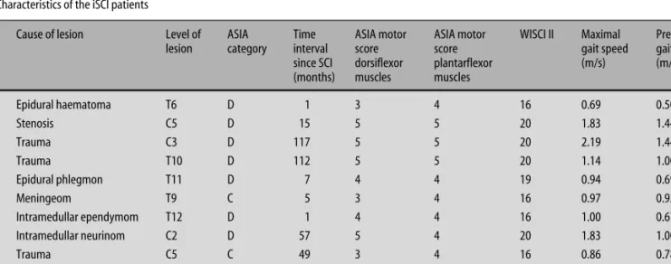
The groups did not significantly differ in the timing of ankle dorsiflexion, neither in gait (Fig. 1A) nor in the supine position (Fig. 1B). While walking, the iSCI pa-tients initiated dorsiflexion during swing on average at 67 % (± standard deviation 1.4 %) and terminated it at 83.3 % (± 3.3 %) of the gait cycle. The swing phase in the control group started on average at 66.5 % (± 1.4 %) and finished at 87.5 % (± 8.8 %) of the gait cycle. In the supine position, the deviation between performance and target frequency was larger in the iSCI group than in the control group at all frequencies, but the differ-ences between the groups were not significant (devia-tion from target frequency in the iSCI group: 0.8 Hz: av-erage = 0.009 Hz (± 0.015 Hz); 1.6 Hz: avav-erage = 0.067 Hz (± 0.127 Hz); 2.4 Hz: average = 0.280 Hz (± 0.358 Hz); de-viation from target frequency in the control group: 0.8 Hz: average = 0.004 Hz (± 0.003 Hz); 1.6 Hz: aver-age = 0.026 (± 0.040 Hz); 2.4 Hz: averaver-age = 0.142 Hz (± 0.157 Hz). Timing of ankle movements in the supine position at all frequencies and initiation or termination of dorsiflexion in swing did not correlate.
■ Timing of ankle dorsiflexion versus MEP
Static MEP amplitude was 0.065 mV (± 0.046 mV) in the iSCI group and 0.195 mV (± 0.176 mV) in the control group. Static MEP latency was 23.05 ms/m (± 4.3 ms/m) in the iSCI group and 20.33 ms/m (± 1.6 ms/m) in the control group. In the dynamic condition, the MEP am-plitude was 0.089 mV (± 0.040 mV) in the iSCI group and 0.226 mV (± 0.173 mV) in the control group. Dynamic MEP latency was 23.64 ms/m (± 5.0 ms/m) in the iSCI group and 18.99 ms/m (± 2.0 ms/m) in the control group. The groups significantly differed in static (p = 0.006) and dynamic (p = 0.006) MEP amplitude as well as in static (p = 0.050) and dynamic (p = 0.019) MEP latency. In the iSCI group, the time of dorsiflexion initiation in swing correlated significantly to static and dynamic
MEP latency (Spearman correlation coefficient rS= 0.79
(p = 0.006) and rS= 0.68 (p = 0.02), respectively). With a
view to the supine position, the MEP parameters did not correlate to the deviation from target frequency.
■ Timing of ankle dorsiflexion versus MMV and MVC
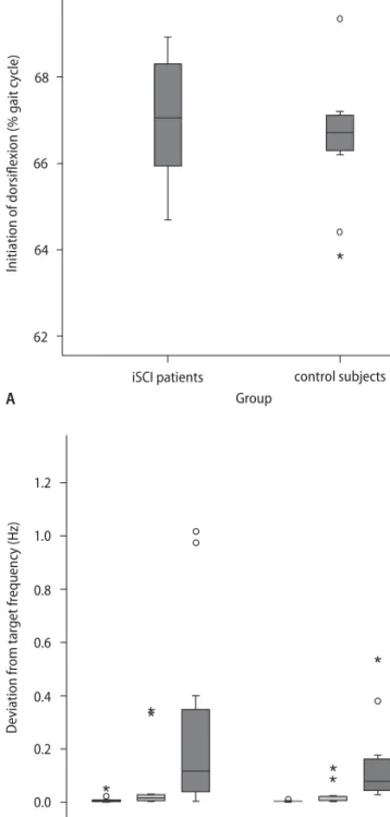
MMV in the foot task was significantly higher in the control group at all frequencies (0.8 Hz: p = 0.002; 1.6 Hz: p = 0.001; 2.4 Hz: p = 0.028) (Fig. 2A). At 0.8 Hz, MMV in dorsiflexion was 145.9 °/s (± 50.0 °/s) in the iSCI group
Fig. 1 Timing of ankle dorsiflexion. The timing of ankle dorsiflexion in the swing phase of gait (A) and in the supine position (B), as assessed at three different fre-quencies (0.8, 1.6, 2.4 Hz) of audio-paced movements, is not significantly reduced in the iSCI group. Circles in the boxplot indicate outlier values that are between the 1.5 and 3 interquartile range from the end of the box. Stars indicate extreme values that are more than 3 times the interquartile range from the end of the box.
70 A B 68 66 64 62
iSCI patients control subjects Group
Initiation of dorsiflexion (% gait cycle)
1.2
1.0
0.8
0.6
iSCI patients control subjects Group
Deviation from target frequency (
Hz)
0.4
0.2
0.0
and 222.4 °/s (± 57.2 °/s) in the control group. At 1.6 Hz, MMV of the iSCI patients and the controls was on aver-age 176.9 °/s (± 56.6 °/s) and 259.5 °/s (± 59.2 °/s), respec-tively. At 2.4 Hz, MMV was 180.4 °/s (± 54.8 °/s) in the iSCI group and 251.7 °/s (± 76.8 °/s) in the control group. However, the groups did not significantly differ in MVC in dorsiflexion (p = 0.456) (Fig. 2B). Isometric torque (normalized for body weight) was 0.35 Nm/kg (± 0.12 Nm/kg) in the iSCI group and 0.38 Nm/kg (± 0.07 Nm/kg) in the control group. Nevertheless, ankle timing in the supine position as well as the initiation and termination of dorsiflexion in swing were independent of MMV and MVC.
■ Timing of ankle dorsiflexion versus gait speed
In the iSCI group, preferred and maximal gait speed
were 0.55 s–1(± 0.18 s–1) and 0.77 s–1(± 0.29 s–1),
respec-tively. In the control group, preferred gait speed was
0.88 s–1 (± 0.09 s–1), maximal gait speed was 1.39 s–1
(± 0.18 s–1). Both, maximal and preferred gait speed
sig-nificantly differed between the groups (p < 0.001). Nei-ther accuracy in timing in the supine position nor the time of initiation of dorsiflexion in swing correlated with maximal or preferred gait speed. However, within the iSCI group, MMV in dorsiflexion at 2.4 Hz correlated
to gait speed (rS= 0.66 and p = 0.02 for maximal and
pre-ferred gait speed) as did MVC in dorsiflexion (rS= 0.80,
p = 0.006 for maximal and rS= 0.83, p = 0.003 for
pre-ferred gait speed).
Discussion
The purpose of this study was to investigate timing of ankle dorsiflexion in iSCI patients and to study the im-pact of spinal cord damage on this aspect of motor con-trol. Timing of ankle dorsiflexion was compared be-tween iSCI subjects and healthy controls and related to measures for CST conductivity (assessed by MEP), MMV, MVC and gait speed. Although gait speed, MEP parameters and MMV were significantly impaired in the iSCI subjects, there was no difference in timing of ankle dorsiflexion between iSCI patients and controls, neither during gait nor in the supine position. In addition, tim-ing of ankle dorsiflexion was not related to muscle strength and gait speed.
■ Cortical control of ankle dorsiflexion
Ankle dorsiflexion was shown to be under large cortical control, both during gait and in the supine position. En-hanced CST activity in the swing phase of gait was re-ported in animals [4, 11, 20, 28] as well as in man [31, 32].
Nevertheless, spinal networks that are involved in the generation of reciprocal rhythmic movement pattern for simple locomotion substantially enhance cortical control of locomotion [5, 15, 22]. In the supine position, a functional magnetic resonance imaging (fMRI) study in stroke patients using a paced dorsiflexion paradigm,
Fig. 2 MMV and MVC in dorsiflexion. A Maximal movement velocity (MMV) was significantly reduced in the iSCI group compared to healthy controls at all frequen-cies. B Maximal voluntary contraction (MVC), however, did not significantly differ between the groups. The star indicates extreme values that are more than 3 times the interquartile range from the end of the box.
400 p=0.028 p=0.001 p=0.002 350 300 250 200 150 100 50 iSCI patients Maximal mo vement v elocit y in dorsiflexion (degr e es/s) control subjects Group 0 A Normalised t o rque in dorsiflexion (N m/Kg) control subjects Group iSCI patients 0.6 0.5 0.4 0.3 0.2 0.1 0.0 B * 0.8 Hz 1.6 Hz 2.4 Hz
which was very similar to the task presented in this study, showed strong cortical control of the ankle move-ment and reported an increase in fMRI activation in parallel to progress in gait speed and lower extremity motor control (Fugl-Meyer assessment [21]) [18]. Therefore, timing of ankle dorsiflexion in the supine po-sition and during gait can be regarded as an aspect of dexterity, which is, apart from muscle strength, a sepa-rate aspect of motor control [1].
■ Dexterity in gait and in the supine position
Since initiation and termination of dorsiflexion in swing are dependent on gait speed [35], the same walking speed was chosen for both the iSCI patients and the con-trol subjects. Nevertheless, apart from a slight delay, none of these measures was significantly altered in the iSCI group, which indicates that gait cycle control was not considerably impaired. This is in contrast to other groups of patients with CNS lesions, where alterations in the duration of swing were reported [10, 13, 23]. In ad-dition, in the elderly, a delay in ankle dorsiflexion in swing was shown to be predictive of falls [26]. Although over-ground and treadmill walking were shown to be very similar in terms of kinematics and kinetic parame-ters [30], the sensory input provided by the driven walk-ing belts might help to improve the timwalk-ing of gait cycle. However, the time of initiation of dorsiflexion signifi-cantly correlated to MEP latency, which confirms the findings of a strong supraspinal (cortical) influence on the swing phase during gait [31, 32].
In the supine position, dexterity was only slightly re-duced in the iSCI patients, but not significantly im-paired.Although dexterity tests might be confounded by muscle strength, since a well controlled movement re-quires a certain amount of strength to be performed [7], the present motor paradigm in the supine position demonstrated that accuracy in timing did not depend on either MMV or on MVC. Thus, the iSCI patients and the controls were comparably able to switch from dorsal- to plantar-flexion and vice versa, although the MMV of the iSCI patients was significantly reduced. Furthermore, dexterity in the supine position did not correlate to the MEP parameters in the present study, despite previous evidence for a cortical involvement in ankle dorsiflexion tasks [18]. This result shows that the ability to generate
dynamic muscle strength is more responsive to an im-pairment of corticospinal pathways than dexterity (at least as dexterity was assessed in the present study). Al-though the iSCI patients showed considerable recovery of static muscle strength with preserved ankle dexterity, gait speed and dynamic strength were significantly re-duced. This indicates that impaired ankle dexterity is not the main factor that leads to impaired limb move-ments after iSCI.
■ Maximal movement velocity and maximal voluntary contraction
The sample in the present study included iSCI patients with good recovery of static strength (no significant dif-ference in MVC compared to controls) and walking ca-pacity. Nevertheless, their MMV remained significantly reduced, which confirms slowing of movement to be a common feature after CNS lesions [6, 29]. The dynamic measure MMV strongly correlates to muscle strength [38]. Thus, the interesting result of similar static, but sig-nificantly different dynamic muscle strength in the two groups is in line with a recent finding that the rate of torque development was dramatically reduced in iSCI patients, while electrically elicited contractile properties did not differ compared to control subjects [25]. In ad-dition, this finding emphasizes the need for a dynamic assessment tool to detect and follow motor deficits after iSCI [29].
Conclusions
The separate assessment of dexterity and paresis in the ankle showed that timing of ankle dorsiflexion was sig-nificantly less impaired than muscle strength in iSCI pa-tients. This supports the assumption that the loss of strength, particularly of dynamic strength, is a major component leading to motor impairment of the lower limb in iSCI, which might be considered in the assess-ment of treatassess-ment interventions.
■Acknowledgements We are much obliged to all patients for their participation in this study.We thank Rachel Jurd for editorial support. This work was funded by the International Spinal Research Trust (ISRT), Clinical Initiative Stage II.
1. Ada L, O’Dwyer N, Green J, Yeo W, Neilson P (1996) The nature of the loss of strength and dexterity in the upper limb following stroke. Hum Mov Sci 15:671–687
2. American Spinal Injury Association (2002) International Standards for Neurological and Functional Classifi-cation of Spinal Cord Injury, Chicago 3. Aschersleben G, Prinz G (1995)
Syn-chronizing actions with events: The role of sensory information. Percept Psychophys 57:305–317
4. Beloozerova, IN, Sirota MG, Swadlow HA (2003) Activity of different classes of neurons of the motor cortex during locomotion. J Neurosci 23:1087–1097 5. Butt S, Kiehn O (2003) Functional
identification of interneurons respon-sible for left-right coordination of hindlimbs in mammals. Neuron 38: 953–963
6. Canning C, Ada L, O’Dwyer N (1999) Slowness to develop force contributes to weakness after stroke. Arch Phys Med Rehabil 80:66–70
7. Canning CG, Ada L, O’Dwyer N (2000) Abnormal muscle activation charac-teristics associated with loss of dexter-ity after stroke. J Neurol Sci 176:45–56 8. Canning CG, Ada L, Adams R, O’Dwyer
N (2004) Loss of strength contributes more to physical disability after stroke than loss of dexterity. Clin Rehabil 18:300–308
9. Catz A, Itzkovich M, Agranov E, Ring H, Tamir A (2001) The spinal cord in-dependence measure (SCIM): Sensitiv-ity to functional changes in subgroups of spinal cord lesion patients. Spinal Cord 39:97–100
10. Chen G, Patten C, Kothari D, Zajac F (2005) Gait differences between indi-viduals with post-stroke hemiparesis and non-disabled controls at matched speeds. Gait Posture 22:51–56 11. Courtine G, Roy R, Raven J, Hodgson J,
Mckay H, Yang H, Zhong H, Tuszynski M, Edgerton (2005) Performance of locomotion and foot grasping follow-ing a unilateral thoracic corticospinal tract lesion in monkeys. Brain 128: 2338–2358
12. Curt A, Keck ME, Dietz V (1998) Func-tional outcome following spinal cord injury: significance of motor-evoked potentials and ASIA scores. Arch Phys Med Rehab 79:81–86
13. Den Otter A, Geurts A, Mulder Th, Duysens J (2006) Gait recovery is not associated with changes in the tempo-ral patterning of muscle activity dur-ing treadmill walkdur-ing in patients with post-stroke hemiparesis. Clin Neuro-physiol 117:4–15
14. Diehl P, Kliesch U, Dietz V, Curt A (2006) Impaired facilitation of motor evoked potentials in incomplete spinal cord injury. J Neurol 253:51–57 15. Dietz V (1992) Human neuronal
control of automatic functional move-ments: Interaction between central programs and afferent input. Physiol Rev 72:33–69
16. Ditunno JF, Ditunno PL, Graziani V, Bernardi M, Castellano V, Marchetti M, Barbeau H, Frankel HL, D’Andrea JM, Ko H-Y, Marshall R, Nance P (2000) Walking index for spinal cord injury (WISCI): an international multicenter validity and reliability study. Spinal Cord 38:234–243
17. Ditunno PL and Ditunno JF (2001) Walking index for spinal cord injury (WISCI II): scale revision. Spinal Cord 39:654–656
18. Dobkin B, Firestine A, West M, Saremi K, Woods R (2004) Ankle dorsiflexion as an fMRI paradigm to assay motor control of walking during rehabilita-tion. Neuroimage 23:370–381 19. Dobkin B, Apple D, Barbeau H, Basso
M, Behrmann A, Deforge D, Ditunno J, Dudley G, Elashoff R, Fugate L, Harkema S, Saulino M, Scott M (2006) Weight-supported treadmill vs over-ground training for walking after acute incomplete SCI. Neurology 66:484–493 20. Drew T, Jiang W, Widajewicz W (2002)
Contributions of the motor cortex to the control of the hindlimbs during locomotion in the cat. Brain Res Brain Res Rev 40:178–191
21. Gladstone D, Danells C, Black S (2002) The Fugl-Meyer Assessment of motor recovery after stroke: a critical review of its measurement properties. Neuro-rehabil Neural Repair 16:232–240 22. Grillner S, Ekeberg O, El Manira A,
Lansner A, Parker D, Tegner J, Wallen P (1998) Intrinsic function of a neuronal network – a vertebrate central pattern generator. Brain Res Rev 26:184–197 23. Gutierrez G, Chow J, Tillman M,
McCoy S, Castellano V, White L (2005) Resistance training improves gait kinematics in persons with multiple sclerosis. Arch Phys Med Rehabil 86:1824–1829
24. Hsu A, Tang P, Jan M (2002) Test-retest reliability of isokinetic muscle strength of the lower extremities in patients with stroke. Arch Phys Med Rehabil 83:1130–1137
25. Jayaraman A, Gregory CM, Bowden M, Stevens JE, Shah P, Behrmann AL, Van-denborne K (2006) Lower extremity skeletal muscle function in persons with incomplete spinal cord injury. Spinal Cord 44:680–687
26. Kemoun G, Thoumie P, Boisson D, Guieu D (2002) Ankle dorsiflexion delay can predict falls in the elderly. J Rehabil Med 34:278–283
27. Krawetz P, Nance P (1996) Gait analy-sis of spinal cord injured subjects: effects of injury level and spasticity. Arch Phys Med Rehabil 77:635–638 28. Lavoie S, Drew T (2002) Discharge
characteristics of neurons in the red nucleus during voluntary gait modifi-cations: a comparison with the motor cortex. J Neurophysiol 88:1791–1814 29. Miller T, Claiborne Johnston S (2005)
Should the Babinski sign be part of the routine neurologic examination? Neu-rology 65:1165–1168
30. Riley PO, Paolini G, Croce UD, Paylo KW, Kerrigan DC (2007) A kinematic and kinetic comparison of overground and treadmill walking in healthy sub-jects. Gait Posture 26:17–24
31. Schubert M, Curt A, Jensen L, Dietz V (1997) Corticospinal input in human gait: modulation of magnetically evoked motor responses. Exp Brain Res 115:234–246
32. Schubert M, Curt A, Colombo G, Berger W, Dietz V (1999) Voluntary control of human gait: conditioning of magnetically evoked motor responses in a precision stepping task. Exp Brain Res 126:583–588
33. Van Hedel H, Wirz M, Dietz V (2005) Assessing walking ability in subjects with spinal cord injury: validity and reliability of 3 walking tests. Arch Phys Med Rehabil 86:190–196
34. Van Hedel H, Wirz M, Curt A (2006) Improving walking assessment in subjects with a spinal cord injury: re-sponsiveness. Spinal Cord 44:352–356 35. Van Hedel HJ, Tomatis L, Müller R
(2006) Modulation of leg muscle activ-ity and gait kinematics by walking speed and bodyweight unloading. Gait and Posture 24:35–45
36. Van Hedel H, Murer C, Dietz V, Curt A (2007) The amplitude of lower leg motor evoked potentials is a reliable measure when controlled for torque and motor task. J Neurol 254: 1089–1098
37. Waters RL, Adkins RH, Yakura JS, Sie I (1994) Motor and sensory recovery following incomplete paraplegia. Arch Phys Med Rehabil 75:67–72
38. Wirth B, van Hedel H, Curt A (2007) Foot control in incomplete SCI: dis-tinction between paresis and dexterity. Still in press