Abstract The polymicrobial sepsis induced by cecal
ligation and puncture (CLP) in the rat is widely used in
shock research. For ethical reasons, narcotic analgesics
are often administered in this model, with the potential
risk of confounding effects. In conscious non-septic rats,
we investigated the cardiovascular effects of a continuous
i.v. infusion of fentanyl (20 µg/kg per h) administered
with fluid loading (10 ml/kg per h) for 24 h, a regimen
commonly applied in rat CLP. Animals were randomly
allocated to receive analgesia with fluid loading (Fentanyl
group), or fluid loading alone (Control). All endpoints
were assessed after 24 h of infusion. At that time,
Control animals had mild respiratory alkalosis, which
was essentially abolished by fentanyl. Analgesia mildly
elevated the plasma norepinephrine levels [median
(interquartile range): Control 232 pg/ml (0–292), Fentanyl
302 pg/ml (234–676), P=0.045] but was devoid of any
effect on blood pressure, heart rate, cardiac output (mean
±SD: Control 388±61
ml/kg per min, Fentanyl
382±62 ml/kg per min, P=0.87) and indices of left
ventricular function derived from high-fidelity recordings
of left ventricular pressure (dP/dt
max: Control 11782±
2324 mmHg/s, Fentanyl 12107±2816 mmHg/s, P=0.77).
In ex vivo experiments carried out immediately after
animal sacrifice, no differences were noted between the
Control and Fentanyl groups in the sensitivity of
endothelium-intact aortic rings to norepinephrine-induced
vasoconstriction (–logEC
50: Control 8.78±0.28, Fentanyl
8.83±0.26, P=0.52) or acetylcholine-induced
vasodilata-tion (–logEC
50: Control 7.00±0.37, Fentanyl 7.06±0.26±
0.53, P=0.75). In conclusion, the present data provide no
contraindication, and even some support for the ethical
use of a high dose i.v. infusion of fentanyl in
cardio-vascular studies of conscious catheterized rats undergoing
CLP or other painful procedures.
Keywords Analgesia · Hemodynamics ·
Left ventricular function · Opioids · Rats ·
Vascular endothelium · Vasoconstriction · Vasodilatation
Introduction
The polymicrobial sepsis induced by cecal ligation and
puncture (CLP) in the rat has gained wide popularity in
shock research. Its main advantage compared to models
based on the acute administration of endotoxin consists
in more closely mimicking key events observed in septic
patients, in particular the hyperdynamic circulation [13,
28, 48]. In CLP, sepsis develops subacutely, so that the
required observation period is too prolonged to allow the
full experiment to run under general anesthesia. This in
itself is an advantage, since confounding effects related
to the use of anesthetic agents [17] are avoided. However,
in a large number of studies with the CLP model,
continuous i.v. analgesia with opioid analogs in relatively
large doses has been administered for ethical reasons
[12, 13, 15, 25, 26, 27, 28, 30, 40, 41, 44, 47, 48]. The
specific physiological effects of these regimens have not
been documented, despite the fact that key endpoints
examined in several of these studies could theoretically
be influenced. In particular, it is well known that opioid
agonists may induce both venous and arterial
vasodilata-tion, and so alter the loading conditions of the heart [3,
43]. Furthermore, there are opioid receptors in the
myocardium [4], the stimulation of which may either
increase [31, 34] or decrease contractility [34, 46]. In
addition, narcotic analgesics have the ability to stimulate
F. Baechtold · D. Gasser · B. Waeber · F. Feihl (✉
)Division of Clinical Pathophysiology, University Hospital, 1011 Lausanne, Switzerland
e-mail: [email protected] Tel.: +41-21-3141423, Fax: +41-21-3141432 C. Cavadas · E. Grouzmann
Division of Hypertension, University Hospital, 1011 Lausanne, Switzerland
M. Markert
Laboratory of Clinical Chemistry, University Hospital, 1011 Lausanne, Switzerland
K.L. Peterson
Division of Cardiology, Department of Medicine,
University of California at San Diego, La Jolla, CA 92093, USA DOI 10.1007/s004240100693
I N S T R U M E N T S A N D T E C H N I Q U E S
François Baechtold · Claudia Cavadas · Didier Gasser
Michèle Markert · Eric Grouzmann · Kirk L. Peterson
Bernard Waeber · François Feihl
Cardiovascular effects of fentanyl in conscious rats
Received: 21 May 2001 / Revised: 26 June 2001 / Accepted: 18 June 2001 / Published online: 31 August 2001 © Springer-Verlag 2001
the release of nitric oxide by the vascular endothelium
[8, 42], a consideration of interest in view of the
importance of endothelial dysfunction in the
patho-genesis of sepsis.
The present work was designed to characterize in the
rat the cardiovascular effects of continuous intravenous
analgesia, with special regard to in vivo left ventricular
function and to vascular reactivity assessed ex vivo.
The agent tested was fentanyl, which is most commonly
used in rat CLP [12, 13, 15, 27, 28, 30, 40, 41, 44,
47, 48]. The rate of administration was the highest
one reported in these studies. For ethical reasons,
the investigation was restricted to nonseptic
condi-tions.
Materials and methods
The study was in accordance with Swiss laws and approved by the local review board on animal experimentation. It was divided into two separate experiments, each conforming to the same design, with essential differences relating to endpoints only.
Experiment 1
Twenty-five male adult Wistar rats (IFFA CREDO, Lyon, France) weighing between 300 and 400 g were randomly allocated to an experimental group (n=13) receiving a continuous i.v. infusion of fentanyl or to a control group (n=12) receiving an equivalent volume of vehicle alone.
Surgery
The rats were anesthetized with pentobarbital 50 mg/kg i.p. Poly-ethylene (PE) catheters were implanted into the femoral artery (PE50, with PE10 tip pushed into the abdominal aorta), the right jugular vein (PE50 with silicone tip), and the left ventricle (LV) through the right carotid artery. The LV catheter was specially designed to allow the subsequent passage of a miniature transducer-tipped probe for the high-fidelity recording of pressure, as shown in Fig. 1A and described in detail below. At the time of insertion, the catheter did not contain the probe, but was connected to an external pressure transducer in order to guarantee tip placement inside the LV. All catheters were tunneled subcutaneously, exteriorized at the neck, and ran through a protective spring affixed to the skin by means of a Teflon anchoring flange on one end and to a swivel adapter on the other. For our application, it was critical to avoid a sharp bend of the left ventricular catheter at the exit site. For this reason, we used a special technique to affix the anchoring flange at some distance from the skin (Fig. 1B).
The crural and cervical incisions were closed in one layer, and the animals were left to awaken, in an individual cage.
Protocol
On termination of anesthesia, the jugular venous catheter was connected to a syringe pump (Perfusor-Secura, Braun, Switzerland) containing isotonic saline with or without 2 µg/ml fentanyl. The i.v. infusion was started and run continuously for 24 h at a rate of 10 ml/kg per h, making up for a fentanyl dose of 20 µg/kg per h, in accordance with the analgesic scheme followed in several studies of CLP [15, 40]. Starting the fentanyl administration immediately after surgery, rather than waiting for post-operative stabilization, was part of the effort to imitate conditions prevailing in CLP studies, where catheter insertion and cecal perforation are usually grouped in a single operation [12, 13, 15, 27, 28, 30, 40, 41, 44, 47, 48].
Food and tap water were provided ad libitum during the entire observation period.
Twenty-four hours after the onset of infusion, taking care to chose a moment when the animal was resting quietly and to avoid the making of any noise, respiratory rate was noted by counting the breaths during 30 s. The femoral catheter was connected to a pressure transducer for the measurement of blood pressure (BP) and heart rate (HR). High-fidelity recording of LV pressure was carried out next, as described below. Finally, arterial blood was drawn through the left ventricular catheter for the measurement of blood gases, lactate, and plasma nitrate. The latter is an index of whole-body production of nitric oxide. Nitrate was determined by a spectrophotometric assay based on the oxidation of NADPH during the enzymatic conversion of nitrate to nitrite by a nitrate reducatase, as previously described [24].
High-fidelity recording of left ventricular pressure in the conscious rat
Following calibration with a mercury manometer and zeroing in a water bath, a miniature transducer-tipped pressure probe (Mikro.Tip SPR 671, Millar, external diameter of the distal part 1.4F=0.46 mm) was introduced into the LV from the proximal end of the LV catheter, using an O-ring valve to avoid the reflux of blood. In so doing, the operator did not touch the rat, nor did his hands enter the cage. Thus, there was no need for a restraining tube. The animal did not even appear aware of probe passage. The tip transducer could always be advanced to <5 cm (usually <5 mm) of the LV catheter distal end, as determined from distance marks made on the Millar probe.
The LV pressure signal was digitized at 2 kHz and stored on computer disk for the subsequent calculation of the maximum systolic LV pressure (LVPmax), the LV end-diastolic pressure (LVEDP), the maximum positive rate of change in LV pressure during isovolumic contraction (dP/dtmax), the minimum negative
Fig. 1A, B Technical details concerning left ventricular
catheter-ization in the conscious rat. A Construction of left ventricular catheter designed to allow the temporary introduction of a Millar transducer-tipped probe. The section of PE90 tubing is required to allow passage of the probe. Only the PE50 is intravascular. (OD Outer diameter.) B Attachment of the protective spring to the rat’s neck. To minimize catheter curvature at this site, the Teflon flange used to affix the protective spring to the rat’s neck is held in place at some distance from the skin, by means of a four-pronged wire frame embedded in cement
rate of change in left ventricular pressure during isovolumic relax-ation (dP/dtmin), and the time constant of isovolumic left ventricular relaxation (tau). The latter was obtained from the least-square fit of an exponential function to the isovolumic descent of LV pressure. For all these calculations, we used the Heart Beat Program (San Diego, CA) [33].
Studies in isolated aorta
Animals were sacrificed with 100 mg/kg pentobarbital i.v. The thoracic aorta was excised and placed in phosphate-buffered saline at 4°C. Two rings of length between 2 and 3 mm were cut. The endothelium was removed with a thin wire in one of them, and left untouched in the other. The two rings were mounted in parallel in organ chambers for the measurement of isometric force (Multi Myograph System, model 610M, JP Trading, Denmark). The chambers were filled with Earle’s Balanced Salt Solution (EBSS, Sigma) gassed at 37°C with 6% CO2+ 94% O2, pH 7.4. EBSS had the following composition (in mM): NaCl 116.3, KCl 5.3, NaHCO3 26.1, CaCl2 1.8, NaH2PO4 1.0, MgSO40.8, D-glucose 5.5, phenol red Na 0.03 (as pH indicator).
The rings were stretched and left to rest for 30 min. The applied preload was 90% of the circumference which, according to Laplace’s law, would correspond to an equivalent transmural pressure of 100 mmHg. The maximal active tension (Tmax) was determined by exposing the vessels to 80 mM KCl for a duration of 15 min, sufficient for stabilization. The solution used for that purpose was identical in composition to EBSS, except for the isosmolar substitution of KCl for NaCl. Following the washout of KCl (30 min), the rings were exposed to norepinephrine (NE), starting at a concentration of 10–9 M, with sequential 3.16-fold increases until the developed active tension was between 60% and 90% of Tmax. The relaxation response to the endothelium-dependent vasodilator acetylcholine (ACh) was then recorded. To that effect, the rings were exposed sequentially to ACh concentrations of 10–8, 10–7, 10–6and 10–5M.
From the recorded data, the following quantities were computed: the NE concentration required for a contractile response equal to 50% of Tmax (EC50[NE]); the maximum relaxation induced by ACh, expressed as a percentage of the precontraction tension (Rmax[ACh]), and the concentration of ACh required to halve the precontraction tension (EC50[ACh]).
Experiment 2
This experiment was carried out to complement the previous one by the determination of further endpoints, especially cardiac output and the plasma levels of NE. In the following description, unnecessary repetitions of features that were kept identical between experiments will be avoided. A total of 19 rats were randomly allocated to the control (n=10) and the experimental groups (n=9). Surgery
Anesthesia for surgery was with inhalation of 2% halothane in oxygen, which is more convenient than i.v. administration of pentobarbital. At the time of Experiment 1, our laboratory lacked a facility for the disposal of volatile anesthetics. In view of the 24-h interval from operation to actual measurements, we assumed that the use of different types of anesthesia in the two experiments would be of no consequence.
Only two vascular catheters were placed, one into the right jugular vein and one into the ascending aorta through the right carotid artery. The arterial catheter was a PE90 (outer diameter 1.26 mm) with a tip thinned to allow insertion. A thermistor wire dismounted from a used Swan-Ganz catheter (7F, Edwards Laboratories) ran through the lumen, all the way to the tip. The lumen was filled with saline containing 20 U/ml heparin. The catheter was advanced 3 cm from the vascular incision, a distance shown by experience to guarantee thermistor location within the ascending aorta.
Protocol
The endpoints, collected 24 h after the start of the fentanyl or saline-only infusion, were: respiratory rate, arterial BP, central temperature (°C), thermodilution cardiac output, plasma levels of catecholamines, arterial blood gases and lactate. Care was taken to obtain physiologic data and blood samples when the animal was resting quietly.
Thermodilution cardiac output in the conscious rat
The measurement of cardiac output conformed to the technique previously applied to the anesthetized rat in our laboratory [37]. Thermistor probe placement is described above. At the time of measurement, the thermistor wire was connected to a cardiac out-put comout-puter (COM-1, Edwards), 0.15 ml of room temperature isotonic saline was injected into the right jugular vein, and the thermodilution curve was recorded on paper. Cardiac output in an individual animal was the mean of four to six determinations made at least 90 s apart. The values corresponding to irregular thermodilution waveforms were excluded. The coefficient of variation of these multiple determinations was always <20% (mean ±SD: 8.6±3.6%).
Plasma levels of catecholamines
Special precautions were taken at the time of blood sampling, in order to avoid potential artifacts related to the triggering of baroreflexes or the rapid degradation of catecholamines ex vivo. When the measurement of cardiac output was completed, the thermistor wire was quickly withdrawn, allowing for the easy efflux of arterial blood. Two 300-µl samples were collected in heparinized microtubes and centrifuged at 4000 rpm for 15 min at 4°C, to provide two 125-µl plasma aliquots which were stored at –80°C. The time interval from the sampling of blood to the freezing of plasma did not exceed 20 min. The concentration of NE was subsequently determined in the plasma aliquot obtained from the first sample, the second one being kept as a back up. Samples for the determination of arterial blood gases were collected after those for NE.
Catecholamines were determined by liquid chromatography with amperometric detection as previously described [16]. 0.125 ml of serum or standard, added with dihydroxybenzylamine (Sigma, Buchs, Switzerland) as internal standard, was extracted on activated alumina (Al2O3) at pH 8.6. The alumina was allowed to settle and the supernatant aspirated followed by three washes with water. The catecholamines were then eluted by 120 µl of a mixture of acetic acid (0.2 M) and phosphoric acid (0.04 M, 8/2, v/v) and 100 µl was injected into the chromatography system (Alliance, Waters 2690 System). The separation was achieved on a reversed phase column (Nucleosil 5 µm C-18, 25 cm ×4.6 mm, Macherey-Nagel, Oesingen, Switzerland) using a mobile phase containing a phosphate buffer at pH 3.5 with sodium octyl sulfonate, as an ion-pairing agent, and acetonitrile. The electrochemical detector (Decade, from Antec, Basel) was set at +0.8 V. Recovery of catecholamines from alumina was 75%. The limit of quantification was set at 6 pg NE and epinephrine per injection, corresponding to 80 pg/ml of serum.
Statistical analysis
In each experiment, endpoints were compared between groups by means of the Student’s t-test for unpaired samples, with Welch adjustment for unequal variances when appropriate. Since the distribution of plasma NE data was highly skewed, a Mann–Whitney test was used in this instance. The alpha level of all tests was set at 0.05.
All actual P values are reported. Data are summarized as mean ±SD.
Results
As shown in Table 1, the control rats had a moderate
respiratory alkalosis, which appeared less marked in
Experiment 2, compared with Experiment 1. In contrast,
animals treated with fentanyl had a practically normal
acid–base status of the arterial blood, along with a
significantly lower respiratory rate. These effects of
analgesia were consistently observed in both experiments.
Blood lactate was within the normal range in all conditions,
although significantly lower with fentanyl treatment in
Experiment 1. The P
aO
2was significantly lower in the
treated, compared with control groups, but all animals
remained in the normoxemic range.
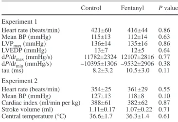
Hemodynamic data are shown in Table 2. Animals in
Experiment 1 were somewhat tachycardic, while those in
Experiment 2 had heart rates in complete agreement with
usually reported values [9, 19]. The mean BP was within
the normal range in all conditions. In Experiment 1, the
indices of LV function derived from the high-fidelity
recording of LV pressure were very consistent with data
published by others who used similar instrumentation in
conscious, unrestrained resting rats [9, 19]. The same
statement applies to the cardiac output measured in
Experiment 2 [12, 30]. Analgesia had no detectable
hemodynamic effect in any of these experiments. As
provided by the thermistor catheter in Experiment 2,
central temperature was in the lower normal range for
conscious rats [7] (Table 2, last line), and was not affected
by analgesia. The differences in heart rate and respiratory
rate between Experiment 1 and Experiment 2 are probably
related to the fact that animals were not comparably
instrumented. A less likely explanation in view of the
precautions taken (see Materials and methods) would be
Table 1 Pulmonary gas exchange and blood lactateControl Fentanyl P value Experiment 1
Respiratory rate (c/min) 101±13 75±6 <0.00001
PaO2(mmHg) 95±8 82±10 0.0016
PaCO2(mmHg) 33±4 39±3 0.0006
Arterial pH 7.53±0.06 7.45±0.03 0.0003
Blood lactate (mM) 0.92±0.41 0.62±0.12 0.02 Experiment 2
Respiratory rate (c/min) 88±13 73±11 0.015
PaO2(mmHg) 94.7±5.0 81.4±9.2 0.002
PaCO2(mmHg) 34.8±5.0 41.0±4.7 0.012
Arterial pH 7.49±0.03 7.44±0.03 0.002
Blood lactate (mM) 0.66±0.30 0.79±0.33 0.38
Table 2 Hemodynamic data. (BP Arterial blood pressure, LVPmax maximum left ventricular pressure in systole, LVEDP left ventricular end-diastolic pressure, dP/dtmaxmaximum positive rate of change in left ventricular pressure during isovolumic contraction, dP/dtmin minimum negative rate of change in left ventricular pressure during isovolumic relaxation, tau time constant of isovolumic left ventricular relaxation)
Control Fentanyl P value Experiment 1
Heart rate (beats/min) 421±60 416±44 0.86
Mean BP (mmHg) 115±13 112±14 0.63 LVPmax(mmHg) 136±14 135±16 0.86 LVEDP (mmHg) 13±7 12±5 0.64 dP/dtmax(mmHg/s) 11782±2324 12107±2816 0.77 dP/dtmin(mmHg/s) –10395±1306 –9532±2906 0.38 tau (ms) 8.2±3.2 10.5±3.0 0.11 Experiment 2
Heart rate (beats/min) 354±25 361±29 0.55
Mean BP (mmHg) 127±13 118±8 0.10
Cardiac index (ml/min per kg) 388±61 382±62 0.87 Stroke volume (ml) 1.11±0.17 1.07±0.22 0.71 Central temperature (°C) 36.6±1.7 36.3±1.4 0.61
Table 3 Vascular reactivity (Experiment 1). Rings of abdominal aorta mounted in the myograph in Experiment 1. Two rings per rat, with the endothelium intact and removed, respectively. Tmax Maximum tension developed in response to 80 mM KCl, –log EC50[NE] negative logarithm of the concentration of norepinephrine which induced a contractile response equal to 50% of Tmax, Rmax [ACh] maximum relaxation induced by acetylcholine in rings precontracted with NE (expressed as a percent of precontraction tension), –log EC50[ACh] negative logarithm of the concentration of ACh required to reduce the precontraction tension by 50%
Control Fentanyl P value Rings with endothelium
Tmax(mN/mm) 6.82±0.84 6.81±1.32 0.43
–log EC50[NE] 8.01±0.22** 8.03±0.50** 0.52
Rmax[ACh] (%) 96.9±3.9** 93.8±7.2** 0.43 –log EC50[ACh] 7.00±0.37 7.06±0.53 0.75 Rings without endothelium
Tmax(mN/mm 6.08±1.23 6.05±1.02 0.43
–log EC50[NE] 8.78±0.28 8.83±0.26 0.52
Rmax[ACh] (%) 7±7 6±6 0.43
–log EC50[ACh] – – –
P values in the last column refer to comparisons between the Control and Fentanyl groups. **P<0.001 compared with rings without endothelium
Fig. 2 In vitro relaxation of endothelium-intact aortas in response
the potential influence of the environment, in particular
ambient noise, on these variables [39], since the two
experiments were not performed by the same investigator.
If at all operative, this factor would have no bearing on
the comparisons of interest in our study, namely between
conditions with and without fentanyl, because each of
the two experiments was carried out entirely by a
single investigator (F.B. in Experiment 1, and D.G. in
Experiment 2).
Considering the potential influence of environmental
factors, in particular ambient noise, on these variables
[39], one might also note that the two experiments were
not carried out by the same operator.
The contractile behavior of isolated aortas
(Experi-ment 1) was entirely consistent with published
observa-tions [14, 20, 29, 45, 47] and was not influenced by
treatment with fentanyl (Table 3). In particular, analgesia
had no effect on the endothelium-dependent relaxation
induced by ACh (Table 3, Fig. 2).
The plasma levels of nitrate obtained in Experiment 1
were 16.8±12.6 µM in the Control and 10.8±4.7 µM in
the Fentanyl group (P=0.20).
As determined in Experiment 2, the median plasma
level of NE was 232 pg/ml (lower quartile below
the detection limit of 80
µg/ml, upper quartile
292 pg/ml) in the Control group, in agreement with
values previously found in this laboratory in a study of
conscious unrestrained rats [49]. As shown in Fig. 3,
plasma NE was mildly augmented by treatment with
fentanyl (median 302 pg/ml, lower quartile 234 pg/ml,
upper quartile 676 pg/ml, P=0.045). Plasma
epi-nephrine was below the detection limit of 80 pg/ml
in all animals.
Discussion
This investigation sought to identify potential confounding
cardiovascular effects of fentanyl, an analgesic that is
widely applied in studies of sepsis employing CLP in the
rat [12, 13, 15, 27, 28, 30, 40, 41, 44, 47, 48]. Accordingly,
the dosage rate and route of administration of fentanyl,
the duration of observation for its effect, and the fluid
load (10 ml/kg/h) administered were dictated by these
previous studies. Based on previous observations in
humans [35, 43], we expected that the cardiovascular
effects of analgesia under these conditions would be
minimal. Indeed, no impact was found on systemic
hemodynamics, left ventricular function, or vascular
reactivity of aortas examined ex vivo.
Mild hyperventilation was present in the control
animals, as shown by the moderate hypocapnia and
slightly higher P
aO
2observed in this group in both
experiments (Table 1). Hyperventilation could have been
induced by the physical discomfort and/or the stress
related to various aspects of the experimental setup, in
particular the implantation of vascular catheters.
Further-more, respiratory stimulation could have come from
some degree of pulmonary vascular congestion, induced
by the fluid load received, combined in Experiment 1
with alteration in left ventricular function by the LV
catheter. Nevertheless, in the absence of hypoxemia
(Table 1) and clear elevation of LVEDP (Table 2),
pulmonary congestion due to left heart failure is unlikely.
Whatever the cause of hyperventilation, it was markedly
blunted by treatment with fentanyl, indicating that the
dosing regimen used here did have central effects. The
lack of complete tolerance to the respiratory depressant
action of a mu-receptor agonist after 24 h of continuous
i.v. administration is consistent with previous observations
in the rat [1, 5].
Opiates, notably fentanyl, are known for an ability to
induce bradycardia through vagal stimulation [3]. The
lack of bradycardia in the fentanyl-treated rats (Table 2)
might be related to the continuous infusion, as opposed
to bolus administration, because the negative chronotropic
action of opiates is inversely related to speed of
adminis-tration [3].
To rule out any effect of fentanyl on cardiac
contrac-tility in the chosen experimental conditions, we carried
out an assessment of the dP/dt
max. The required
intraven-tricular placement of a high-fidelity pressure sensor has
rarely been achieved in the conscious rat [9, 38]. These
authors inserted the Millar probe under anesthesia and
left it in place while consciousness and motility were
regained. In this method, the necessity to tape the bulky
connector to the back of the rat adds to the restraint
imposed on the animal. A further potential problem is
the risk of damage inflicted on the expensive device,
especially if left in situ for a prolonged period. Probe
introduction through the proximal part of a LV catheter
obviates these disadvantages. The latter principle was
originally described in conscious rats by Flaim and
coworkers [10, 11], but the transducers available at that
Fig. 3 Plasma concentration of norepinephrine (Experiment 2).
Three animals in the Control group had values below the detection limit of 80 pg/ml (plotted as zeros)
time were too bulky to be placed inside PE50 tubing and
thus could not be brought closer than a few centimeters
to the LV. We removed this limitation by using the newly
developed miniature Millar probe. The values of
dP/dt
maxthus measured in Experiment 1 agreed with
those obtained in conscious rats with operatively inserted
high-fidelity transducers [9, 38]. Treatment with fentanyl
had no effect on myocardial contractility as reflected by
these values (Table
2). Isovolumic (pre-ejection)
dP/dt
maxis independent of afterload but depends on
ventricular preload, or end-diastolic volume. However,
the identity of LVEDP in Experiment 1 supports the
equivalency of preload in the control and treated rats. We
acknowledge that a constant LVEDP does not in itself
guarantee an identical LV preload, or myocardial fiber
stretch, due to potential variations in the diastolic
proper-ties (relaxation kinetics, active and passive stiffness) of
the myocardium. The lack of significant difference in
dP/dt
minand tau (control versus fentanyl, Table 2) would
not favor an effect of this analgesic on LV relaxation
kinetics. Taken together, we believe our data strongly
support the lack of effect of fentanyl on LV contractility.
This interpretation is consistent with recent observations
in isolated canine hearts [22], showing that
concentra-tions of fentanyl up to 240 ng/ml fail to modify LV
end-systolic elastance in this preparation. Furthermore, while
fentanyl is essentially a mu agonist [36], kappa but not
mu opioid receptors have been identified in the rat
myocardium [23, 34].
The fact that systemic hemodynamics were not
detectably affected by the continuous infusion of fentanyl
at 24 h (Table 2) could reflect the development of
tolerance [6]. Alternatively, a compensatory activation of
the sympatho-adrenergic system might have come into
play, a possibility suggested by the higher plasma level
of NE in the treated compared with the control group
(Fig. 3). In addition, the sizable amount of i.v. fluid
received in the course of the experiment might have been
protective. Thus, one should caution against the
extrapolation of the present results to conditions of less
intensive fluid loading.
Opioids may induce vasodilatation by a variety of
mechanisms, including histamine release [36], stimulation
of nitric oxide production by the endothelium [42], and
interference with alpha-adrenergic receptor signal
trans-duction [21]. In vitro, fentanyl has no effect on cultured
endothelial cells [42], but is able to relax
endothelium-denuded rat aortas precontracted with an alpha-adrenergic
agonist [21] or to increase the basal tone of dog coronary
arteries [18]. To our knowledge, the effects of prolonged
opioid administration in vivo on vascular reactivity
examined in vitro have not been documented. In the
present study, the reactivity of isolated aortas was not
affected by treatment with fentanyl (Table 3, Fig. 2).
Whether explained by washout of the opioid after
excision or by other factors, this observation indicates
that the present regimen of analgesia may be given to
rats without undue concern for subsequent interference
with aortic reactivity, when examined ex vivo. Further
experiments are required before extending the present
finding to other vessels. In this context, it is noteworthy
that plasma nitrate did not differ between groups,
suggesting that fentanyl analgesia did not stimulate the
expression of inducible nitric oxide synthase. This would
be in contrast to pentobarbital anesthesia, which has
been shown in the rat to depress vascular contractility by
this very mechanism [17].
In conclusion, the present data provide no
contraindi-cation to the ethical use of a high dose i.v. infusion of
fentanyl in conscious catheterized rats undergoing CLP.
This statement is valid for experiments conducted under
substantial fluid loading and lasting 24 h as is the case in
most sepsis studies with this model. The reasons are as
follows. First, there is no gross encroachment of this
regimen of analgesia per se on the cardiovascular
physiol-ogy of the animal. One might argue that this apparent
innocuousness is obtained at the cost of utilizing part of
the adaptation reserve, as possibly reflected by the
observed increase in plasma NE (Fig. 3). However, any
reduction in cardiovascular reserve due to fentanyl
would probably be of minor significance in the 24 h CLP
model, because cardiovascular collapse does not occur in
this setting, as reported by many studies, including one
by our group [2, 28, 48]. Finally, if only because of its
potential effects on organ blood flow [32], the respiratory
alkalosis found in conscious catheterized rats not receiving
fentanyl could in itself influence the time course of
sepsis, such that its blunting might in fact be a
methodologically useful side-effect of analgesia.
As a final note, two limitations of the present study
must be underscored. First, we have not assessed the
time course of potential cardiovascular effects related to
analgesia. It remains possible that observations made
especially earlier (see previous remark on tolerance)
might have differed from those presented. Our emphasis
on the 24 h time point is justified by its common use in
studies of sepsis with the rat CLP model. Second, fentanyl
was only evaluated in nonseptic animals. In view of the
emerging role of opioid receptors in the modulation of
the immune response, our results do not imply equivalent
behaviors of the CLP model in presence or absence of
analgesia. Inclusion of analgesia in the experimental
conditions is clinically relevant in view of the frequent
use of opioids in critically ill septic patients.
Acknowledgements The authors warmly thank Antoinette Ney for carrying out the assay of plasma nitrate, and Camille Anglada for technical assistance. The study was funded by a grant from the Swiss National Science Foundation (3200–055946.98/1).
References
1. Ayesta FJ, Florez J (1989) Tolerance to respiratory actions of sufentanil: functional tolerance and route-dependent differential tolerance. J Pharmacol Exp Ther 250:371–378
2. Baechtold F, Scott JA, Markert M, Mehta S, McCormack DG, Anglada A, Galaud D, Vaglio, Waeber B, Feihl F (2001) Does the activation of poly (ADP-ribose) synthetase mediate tissue injury in the sepsis induced by cecal ligation and puncture? Shock 16:137–142
3. Bailey PL, Stanley TH (1994) Intravenous opioid anesthetics. In: Miller RD (ed) Anesthesia, 4th edn. Churchill Livingstone, New York, pp 291–387
4. Barron BA (1999) Opioid peptides and the heart [editorial comment]. Cardiovasc Res 43:13–16
5. Burton CK, Ho IK, Hoskins B (1990) Evidence for involvement of cyclic GMP phosphodiesterase in morphine tolerance. J Pharmacol Exp Ther 252:104–111
6. Doorley BM, Knight VV, Spaulding TC (1988) Development of acute tolerance to the cardiorespiratory effects of alfentanil after subsequent bolus injection in conscious rats. Life Sci 43: 365–372
7. Farris EJ, Griffith JQ (1967) The rat in laboratory investigation. Hafner, New York
8. Fimiani C, Mattocks D, Cavani F, Salzet M, Deutsch DG, Pryor S, Bilfinger TV, Stefano GB (1999) Morphine and anandamide stimulate intracellular calcium transients in human arterial endothelial cells: coupling to nitric oxide release. Cell Signal 11:189–193
9. Fitzsimons DP, Bodell PW, Herrick RE, Baldwin KM (1990) Left ventricular functional capacity in the endurance-trained rodent. J Appl Physiol 69:305–312
10. Flaim SF, Nellis SH, Toggart EJ, Drexler H, Kanda K, Newman ED (1984) Multiple simultaneous determinations of hemodynamics and flow distribution in conscious rat. J Pharmacol Methods 11:1–39
11. Flaim SF, Stranieri MT, Gill A, Carson JR, Brannan MD (1988) Effects of the novel calcium channel blocker, McN-5691, on cardiocirculatory dynamics and cardiac output distribution in conscious spontaneously hypertensive rat. J Cardiovasc Pharmacol 11:489–500
12. Fox GA, Paterson NAM, McCormack DG (1994) Effect of inhibition of NO synthase on vascular reactivity in a rat model of hyperdynamic sepsis. Am J Physiol 267:H1377–H1382 13. Fox GA, Paterson NA, McCormack DG (1996) Cyclooxygenase
inhibition and vascular reactivity in a rat model of hyperdy-namic sepsis. J Cardiovasc Pharmacol 28:30–35
14. Furchgott RF, Zawadzki JV (1980) The obligatory role of endothelial cells in the relaxation of arterial smooth muscle by acetylcholine. Nature 288:373–376
15. Gocan NC, Scott JA, Tyml K (2000) Nitric oxide produced via neuronal NOS may impair vasodilatation in septic rat skeletal muscle. Am J Physiol 278:H1480–H1489
16. Grouzman E, Borgeat A, Fathi M, Gaillard RC, Ravussin P (2000) Plasma and cerebrospinal fluid concentration of neuro-peptide Y, serotonin, and catecholamines in patients under propofol or isoflurane anesthesia. Can. J. Physiol. Pharmacol. 78:100–107
17. Hoque AM, Marczin N, Catravas JD, Fuchs LC (1996) Anesthesia with sodium pentobarbital enhances lipopoly-saccharide-induced cardiovascular dysfunction in rats. Shock 6:365–370
18. Introna RP, Bridges MT Jr., Yodlowski EH, Grover TE, Pruett JK (1995) Direct effects of fentanyl on canine coronary artery rings. Life Sci 56:1265–1273
19. Irlbeck M, Muhling O, Iwai T, Zimmer HG (1996) Different response of the rat left and right heart to norepinephrine. Cardiovasc Res 31:157–162
20. Julou-Schaeffer G, Gray GA, Fleming I, Schott C, Parratt JR, Stoclet JC (1990) Loss of vascular responsiveness induced by endotoxin involves L-arginine pathway. Am J Physiol 259: H1038–H1043
21. Karasawa F, Iwanov V, Moulds RF (1993) Effects of fentanyl on the rat aorta are mediated by alpha-adrenoceptors rather than by the endothelium. Br J Anaesthiol 71:877–880
22. Kohno K, Takaki M, Ishioka K, Nakayama Y, Suzuki S, Araki J, Namba T, Suga H (1997) Effects of intracoronary fentanyl on left ventricular mechanoenergetics in the excised cross-circulated canine heart (revised publication). Anesthesiology 87:658–666 23. Krumins SA, Faden AI, Feuerstein G (1985) Opiate binding in
rat hearts: modulation of binding after hemorrhagic shock. Biochem Biophys Res Commun 127:120–128
24. Liaudet L, Fishman D, Markert M, Perret C, Feihl F (1997) L-Canavanine improves organ function and tissue adenosine triphosphate levels in rodent endotoxemia. Am J Respir Crit Care Med 155:1643–1648
25. Lundblad R, Giercksky KE (1995) Effect of volume support, antibiotic therapy, and monoclonal antiendotoxin antibodies on mortality rate and blood concentrations of endothelin and other mediators in fulminant intra-abdominal sepsis in rats. Crit Care Med 23:1382–1390
26. Lundblad R, Sandven P, Giercksky KE (1995) The physical nature of a large bowel perforation predicts severity of the subsequent inflammatory response. Shock 3:455–461
27. Martin CM, Yaghi A, Sibbald WJ, McCormack D, Paterson AM (1993) Differential impairment of vascular reactivity of small pulmonary and systemic arteries in hyperdynamic sepsis. Am Rev Respir Dis 148:164–172
28. Martin M, Sibbald WJ (1994) Modulation of hemodynamics and organ blood flow by nitric oxide synthase inhibition is not altered in normotensive, septic rats. Am J Respir Crit Care Med 150:1539–1544
29. McKenna TM (1990) Prolonged exposure of rat aorta to low levels of endotoxin in vitro results in impaired contractility. J Clin Invest 86:160–168
30. Morisaki H, Sibbald W, Martin C, Doig G, Inman K (1996) Hyperdynamic sepsis depresses circulatory compensation to normovolemic anemia in conscious rats. J Appl Physiol 80: 656–664
31. Novakova M, Ela C, Barg J, Vogel Z, Hasin Y, Eilam Y (1995) Inotropic action of sigma receptor ligands in isolated cardiac myocytes from adult rats. Eur J Pharmacol 286:19–30 32. Nunn JF (1993) The effects of changes in carbon dioxide
tension. In: Applied respiratory physiology, 4th edn. Butter-worth, London
33. Palakodeti V, Oh S, Oh BH, Mao L, Hongo M, Peterson KL, Ross J Jr. (1997) Force-frequency effect is a powerful determinant of myocardial contractility in the mouse. Am J Physiol 273:H1283–H1290
34. Pei JM, Yu XC, Bian JS, Wong TM (1999) Acidosis antagonizes intracellular calcium response to kappa-opioid receptor stimulation in the rat heart. Am J Physiol 277: C492–C500
35. Peng PW, Sandler AN (1999) A review of the use of fentanyl analgesia in the management of acute pain in adults [Review]. Anesthesiology 90:576–599
36. Reisine T, Pasternak K (1996) Opioid analgesics and antagonists. In: Hardman JG, Limbird LE (eds) Goodman and Gilman’s the pharmacological basis of therapeutics. McGraw-Hill, New York, pp 521–555
37. Rosselet A, Feihl F, Markert M, Gnaegi A, Perret C, Liaudet L (1998) Selective iNOS inhibition is superior to norepinephrine in the treatment of rat endotoxic shock. Am J Respir Crit Care Med 157:162–170
38. Salyers AK, Rozek LF, Bittner SE, Walsh GM (1988) Simulta-neous determination of ventricular function and systemic hemodynamics in the conscious rat. J Pharmacol Methods 19: 267–274
39. Schlatter J, Zbinden G (1982) Heart rate- and ECG-recording in the rat by biotelemetry. Arch Toxicol [Suppl.] 5:179–183 40. Scott JA, McCormack DG (1999) Selective in vivo inhibition
of inducible nitric oxide synthase in a rat model of sepsis. J Appl Physiol 86:1739–1744
41. Sielenkamper AW, Chin-Yee IH, Martin CM, Sibbald WJ (1997) Diaspirin crosslinked hemoglobin improves systemic oxygen uptake in oxygen supply-dependent septic rats. Am J Respir Crit Care Med 156:1066–1072
42. Stefano GB, Hartman A, Bilfinger TV, Magazine HI, Liu Y, Casares F, Goligorsky MS (1995) Presence of the mu3 opiate receptor in endothelial cells. Coupling to nitric oxide production and vasodilation. J Biol Chem 270: 30290–30293
43. Teeple E Jr. (1990) Pharmacology and physiology of narcotics. [Review]. Crit Care Clin 6:255–282
44. Tyml K, Yu J, McCormack DG (1998) Capillary and arteriolar responses to local vasodilators are impaired in a rat model of sepsis. J Appl Physiol 84:837
45. Umans JG, Wylam ME, Samsel RW, Edwards J, Schumacker PT (1993) Effects of endotoxin in vivo on endothelial and smooth-muscle function in rabbit and rat aorta. Am Rev Respir Dis 148:1638–1645
46. Xiao RP, Pepe S, Spurgeon HA, Capogrossi MC, Lakatta EG (1997) Opioid peptide receptor stimulation reverses beta-adrenergic effects in rat heart cells. Am J Physiol 272: H797–H805
47. Yaghi A, Paterson NAM, McCormack DG (1995) Vascular reactivity in sepsis: importance of controls and role of nitric oxide. Am J Respir Crit Care Med 151:706–712
48. Yu P, Boughner DR, Sibbald WJ, Keys J, Dunmore J, Martin CM (1997) Myocardial collagen changes and edema in rats with hyperdynamic sepsis. Crit Care Med 25:657–662 49. Zanchi A, Schaad NC, Osterheld MC, Grouzmann E,
Nussberger J, Brunner HR, Waeber B (1995) Effects of chronic NO synthase inhibition in rats on renin-angiotensin system and sympathetic nervous system. Am J Physiol 268: H2267–H2273