0099-2240/05/$08.00
⫹0 doi:10.1128/AEM.71.6.2880–2887.2005
Copyright © 2005, American Society for Microbiology. All Rights Reserved.
Lactobacillus casei DN-114 001 Inhibits the Ability of
Adherent-Invasive Escherichia coli Isolated from
Crohn’s Disease Patients To Adhere to and To
Invade Intestinal Epithelial Cells
Isabelle Ingrassia,
1Antony Leplingard,
2and Arlette Darfeuille-Michaud
1*
Pathoge´nie Bacte´rienne Intestinale, CBRV, Universite´ d’Auvergne, Clermont-Ferrand, France,
1and Danone Vitapole, Nutrivaleur, Palaiseau, France
2Received 14 September 2004/Accepted 21 December 2004
Ileal lesions in 36.4% of patients with Crohn’s disease are colonized by pathogenic adherent-invasive
Escherichia coli. The aim of this study was to determine the in vitro inhibitory effects of the probiotic strain,
Lactobacillus casei DN-114 001, on adhesion to and invasion of human intestinal epithelial cells by
adherent-invasive E. coli isolated from Crohn’s disease patients. The experiments were performed with undifferentiated
Intestine-407 cells and with undifferentiated or differentiated Caco-2 intestinal epithelial cells. Bacterial
adhesion to and invasion of intestinal epithelial cells were assessed by counting CFU. The inhibitory effects of
L. casei were determined after coincubation with adherent-invasive E. coli or after preincubation of intestinal
cells with L. casei prior to infection with invasive E. coli. Inhibitory effects of L. casei on
adherent-invasive E. coli adhesion to differentiated and undifferentiated intestinal epithelial cells reached 75% to 84% in
coincubation and 43% to 62% in preincubation experiments, according to the cell lines used. Addition of L. casei
culture supernatant to the incubation medium increased L. casei adhesion to intestinal epithelial cells and
enhanced the inhibitory effects of L. casei. The inhibitory effects on E. coli invasion paralleled those on
adhesion. This effect was not due to a bactericidal effect on adherent-invasive E. coli or to a cytotoxic effect on
epithelial intestinal cells. As Lactobacillus casei DN-114 001 strongly inhibits interaction of adherent-invasive
E. coli with intestinal epithelial cells, this finding suggests that the probiotic strain could be of therapeutic
value in Crohn’s disease.
Crohn’s disease (CD) is a chronic inflammatory bowel
dis-ease (IBD) for which the etiology is still unknown, but several
factors, including genetic, environmental, immunological, and
other endogenous host factors, have been incriminated (40).
Among the environmental triggers, luminal bacteria seem to
play a substantial role. Indeed, the onset of inflammation in
IBD may be associated with an imbalance in the intestinal
microflora, with a relative predominance of aggressive bacteria
and an insufficient amount of protective bacteria (46).
More-over, the efficacy of antibiotic therapy suggests a role of
bac-terial flora in CD. However, antibiotic treatments are often
associated with gastrointestinal side effects and bacterial
resis-tance, which may contribute to treatment failure (14, 21, 41, 43,
48).
In early and chronic ileal lesions of CD, an abnormal
pre-dominance of Escherichia coli has been observed (between 50
and 100% of the total number of aerobes and anaerobes).
Most of these strains are able to adhere to and invade
intes-tinal epithelial cells and to replicate within macrophages (9, 17,
19). These E. coli strains belong to a pathogenic group of E.
coli designated AIEC (for adherent-invasive E. coli) (9). In
neoterminal ileal specimens, AIEC strains were found in
36.4% of CD patients (16). Treatments aimed at eradicating
these pathogenic strains and replacing them by nonpathogenic
bacteria such as probiotic strains may be beneficial for the
course of CD and may provide an innovative approach to
treatment.
Probiotics are living microorganisms that upon ingestion in
sufficient numbers exert benefits on human health. By
modu-lating enteric flora, probiotic strains are effective in the
pre-vention and treatment of antibiotic-associated, rotavirus,
Clos-tridium difficile-associated, or traveler’s diarrhea (for a review,
see reference 47). The efficacy of several probiotics for IBD
has been investigated in clinical trials. E. coli Nissle 1917,
Saccharomyces boulardii, and a formula consisting of species of
Bifidobacterium, Lactobacillus, and Streptococcus salivarius
subsp. Thermophilus (VSL #3) have been reported as being as
effective as standard treatment in preventing relapse in
ulcer-ative colitis and chronic pouchitis (24, 42, 51). Probiotics are
used in these pathologies basically to restore the unbalanced
indigenous microflora, to inhibit the adverse effects of enteric
pathogens, and to counteract the inflammatory process (28,
45).
Lactobacillus casei DN-114 001 is a probiotic strain that
survives intestinal transit (35) and exerts beneficial effects in
vivo. It is able to modify the digestive microflora and enhance
the immune system during its transit in the digestive tract (23,
39). It was shown to reduce the incidence and duration of
diarrhea in children (37, 38). Moreover, a recent study has
provided evidence that this probiotic interacts with human
intestinal mucosa and can markedly reduced the mucosal
re-* Corresponding author. Mailing address: Pathoge
´nie Bacte
´rienne
Intestinale, Laboratoire de Bacte
´riologie, CBRV, 28 Place Henri
Du-nant, 63000 Clermont-Ferrand, France. Phone: 33 4 73 17 79 97. Fax:
33 4 73 17 83 71. E-mail: arlette.darfeuille-michaud@u-clermont1.fr.
lease of tumor necrosis factor alpha and interleukin-8 in active
Crohn’s disease (5, 6).
The aim of the present study was to investigate whether L.
casei DN-114 001 could inhibit the ability of pathogenic
ad-herent-invasive-E. coli strains isolated from patients with
Crohn’s disease to adhere to and invade intestinal epithelial
cells in vitro.
MATERIALS AND METHODS
Bacterial strains and culture conditions.L. casei DN-114 001 was provided by
Danone Vitapole (Paris, France). L. casei DN-114 001 was grown in De Man, Rogosa, and Sharpe (MRS) broth (Difco, Becton Dickinson, Meylan, France) at 37°C for 18 h. The culture was centrifuged (10,000⫻ g for 5 min at 4°C), and bacteria were suspended in cell culture medium. The final suspension was ad-justed to obtain the appropriate concentration. The number of CFU was deter-mined by plating serial 10-fold dilutions from bacterial suspensions on MRS agar plates. Plates were incubated at 37°C in a CO2atmosphere for 48 h.
Seven AIEC strains were assessed: the AIEC reference strain LF82 (9) and strains LF9, LF15, LF31, LF65, LF110, and LF134 (16). All these strains isolated from patients with Crohn’s disease were characterized by using the adhesion and invasion assays described below. All strains were highly sensitive to gentamicin.
E. coli LF32 was used as a positive control for cytotoxicity assay, since this strain
produces␣-hemolysin (17). E. coli strain K-12 C600 was used as a negative control. All E. coli strains were grown either in Luria-Bertani broth without shaking or on Mueller-Hinton agar plates (Institut Pasteur Production, Marnes-la-Coquette, France) overnight at 37°C.
Intestinal cell lines and cell cultures.The Intestine-407 cells (ATCC CCL6; Flow Laboratories, Inc., McLean, VA) derived from human embryonic jejunum and ileum were used as an intestinal model for undifferentiated intestinal epi-thelial cells mimicking the cells found in the crypts of the intestinal villi. They were cultured for 20 h and in an atmosphere containing 5% CO2at 37°C in Eagle
minimum essential medium (Eagle MEM; BioWhittaker-Cambrex, Emerain-ville, France) supplemented with 10% (vol/vol) fetal bovine serum (BioWhit-taker-Cambrex, Emerainville, France), 1% (vol/vol) nonessential amino acids (BioWhittaker), 1% (vol/vol) L-glutamine (Gibco BRL-Life Technologies, Cergy-Pontoise, France), 200 U of penicillin, 50 mg of streptomycin, 0.25 mg/liter of amphotericin B (Gibco BRL-Life Technologies, Cergy-Pontoise, France), and 1% (vol/vol) MEM vitamin solution X-100 (BioWhittaker). Caco-2 cells estab-lished from human colonic adenocarcinoma were kindly provided by Alain Zweibaum (INSERM U178, Villejuif, France). These cells were used as undif-ferentiated cells to mimic cells of the crypts and as difundif-ferentiated cells to mimic mature enterocytes of the small intestine (30). Undifferentiated and differenti-ated Caco-2 cells were grown for 2 days and 15 days, respectively. Cells were cultured in Dulbecco’s modified Eagle medium (DMEM) with 4.5 g/liter of glucose (BioWhittaker) supplemented with 20% (vol/vol) fetal bovine serum (BioWhittaker), 1% (vol/vol) nonessential amino acids (BioWhittaker), 1% (vol/ vol)L-glutamine (Gibco BRL-Life Technologies, Cergy-Pontoise, France), 200 U of penicillin, 50 mg of streptomycin, 0.25 mg/liter of amphotericin B (Gibco BRL-Life Technologies, Cergy-Pontoise, France), and 1% (vol/vol) MEM vita-min solution X-100 (BioWhittaker). The cells were grown at 37°C in 5% CO2.
Adhesion and invasion assays.Intestine-407 cells were seeded in 24-well tissue culture plates (Polylabo, Strasbourg, France) at 4⫻ 105cells per well and grown
for 20 h. Caco-2 cells were seeded in 24-well tissue culture plates (Polylabo, Strasbourg, France) at 2⫻ 105cells per well and grown for 2 days for
undiffer-entiated cells and 15 days for differundiffer-entiated cells. The culture medium was changed every 2 days. The cells were washed twice with phosphate-buffered saline (BioWhittaker).
To study the adhesion of L. casei DN-114 001, each cell line was infected in 1 ml of the cell culture medium supplemented with heat-inactivated (30 min; 56°C) fetal bovine serum, at a multiplicity of infection (MOI) of either 10, 100, or 500 bacteria per epithelial cell. After a 1- to 6-h incubation period at 37°C with 10% CO2, the infected cells were washed three times with phosphate-buffered saline.
To determine the total number of cell-associated bacteria, the cells were lysed with 1% (vol/vol) Triton X-100 (Sigma) in deionized water. This concentration of Triton X-100 did not affect bacterial viability for at least 30 min (data not shown). Samples were diluted and plated onto MRS agar plates to determine the number of CFU recovered from the lysed cells. To study the effect of spent L. casei culture supernatant (SN) on the adhesion of the strain, 10% (vol/vol) of its spent culture SN or neutralized spent culture SN were added to the cell culture medium. The spent culture supernatant of L. casei DN-114 001 was centrifuged
and sterilized by filtration through a sterile 0.22-m-pore-size filter unit (Milli-pore Molsheim, France). To check for potential interference of pH reduction linked to organic acid production, the spent culture supernatant was also ad-justed to a neutral value (pH 7.0) using 4 M NaOH.
AIEC adhesion was measured utilizing the same protocol. MOIs of 10 and 100 were used. To determine the number of CFU recovered from the lysed cells, samples were diluted and plated onto Mueller-Hinton agar plates. For invasion assays, fresh cell culture medium containing 100g/ml of gentamicin was added after the infection period to kill extracellular bacteria. After incubation for an additional hour, cultured cells were treated as described above. Each assay was performed three times with successive passages of intestinal cells.
Adhesion and invasion inhibition assays.Two different procedures were used to assess exclusion of AIEC strain by L. casei DN-114 001 and competition between the two strains. Exclusion was assessed by performing preinfection experiments in which cultured intestinal epithelial cells were first incubated with
L. casei DN-114 001 (MOI, 500) alone or in the presence of 10% of its spent
culture supernatant for 6 h at 37°C. AIEC strain LF82 (MOI, 100) was added and incubation was continued for a further 3 h. Competition was assessed by per-forming coinfection experiments in which L. casei DN-114 001 (MOI, 500) bacteria alone or in the presence of 10% of its spent culture supernatant (or neutralized spent culture supernatant), and each of the AIEC strains tested were added to the cultured cells at an MOI of 10 for 6 h. The numbers of strains adhering to or invading the intestinal cells were determined as described above. For each assay, a minimum of three experiments was performed with successive passage of intestinal cells. To evaluate the number of adherent or intracellular bacteria per intestinal epithelial cells, two additional wells were prepared when the cells were seeded. At the end of the culture period, the cells were trypsinized and enumerated microscopically.
Epithelial cell viability: lactate dehydrogenase (LDH) measurement.At the same time as each study of L. casei DN-114 001 adherence, a duplicate 24-well plate of cultured epithelial cells was inoculated with bacteria and assayed as described above. At the end of the incubation period, supernatants of the in-fected cells containing released LDH were collected, centrifuged at 2,500⫻ g for 3 min at 4°C, and assayed for lactate dehydrogenase activity. Enzymatic activity was determined in the supernatants by using NADH as the substrate. Release of LDH was expressed as units per liter of supernatant. The percentage of cyto-toxicity was calculated as follows: [(experimental release⫺ spontaneous release)/ (total release⫺ spontaneous release)] ⫻ 100, where spontaneous release is the amount of LDH activity in supernatants of cells incubated in medium alone and total release is the LDH activity measured in cell lysates. E. coli LF32 producing an␣-hemolysin was used as a positive control (17). E. coli strain K-12 C600 was used as a negative control.
Statistical analysis.The data were analyzed by Student’s t test. P values of ⱕ0.05 were considered to be statistically significant.
RESULTS
Ability of L. casei DN-114 001 to adhere to intestinal
epi-thelial cells.
The ability of L. casei DN-114 001 to adhere to
intestinal epithelial cells was determined using
undifferenti-ated Intestine-407 and Caco-2 cells and differentiundifferenti-ated Caco-2
cells. The adhesion of L. casei DN-114 001 increased with the
MOI as shown in Fig. 1A. It also increased with the incubation
period (Fig. 1B). The observed levels of adhesion varied
ac-cording to the cell lines tested. With Intestine-407 cells,
adhe-sion was maximal after 3 h of incubation, reaching an adheadhe-sion
level of 3 bacteria/cell. With undifferentiated and differentiated
Caco-2 cells, marked increases in adhesion levels were
ob-served between 3 and 6 h of incubation. After 6 h of
incuba-tion, the adhesion levels were 2.7 and 3.9 bacteria/cell with
undifferentiated and differentiated Caco-2 cells, respectively.
Thus, L. casei DN-114 001 exhibited a dose-dependent and
incubation time-dependent ability to adhere to
undifferenti-ated and differentiundifferenti-ated intestinal epithelial cells.
Increased ability of L. casei DN-114 001 to adhere to
intes-tinal epithelial cells in the presence of its spent culture
super-natant.
Since Lactobacilli can produce secreted compounds
able to interact with their adhesive abilities (10, 22), the effects
of putative secreted products on ability to adhere were tested.
L. casei DN-114 001 adhesion assays were performed at an
MOI of 500 for 6 h in the presence of 10% of L. casei DN-114
001 spent culture supernatant. The addition of 10% of L. casei
DN-114 001 spent culture supernatant to the incubation
me-dium induced 6.8-, 7.7-, and 7.1-fold increases in levels of
adhesion of L. casei DN-114 001 to Intestine-407,
undifferen-tiated Caco-2, and differenundifferen-tiated Caco-2 cells, respectively
(Fig. 2). Since the observed effect could be related to a drop in
pH due to acid lactic production, we performed similar
exper-iments in the presence of 10% spent culture supernatant
ad-justed to a neutral pH value. It continued to induce a marked
increase in L. casei DN-114 001 adhesion levels, indicating that
an acidic pH was not the main factor involved in the increased
ability of L. casei DN-114 001 to adhere to undifferentiated and
differentiated intestinal epithelial cells.
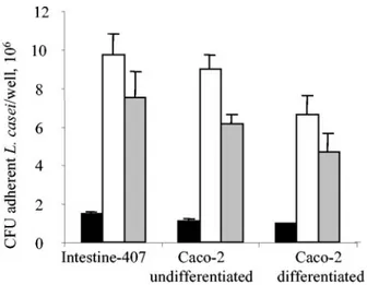
Inhibitory effect of L. casei DN 114 001 on AIEC LF82
adhesion and invasion in preincubation experiments.
The
pro-biotic activity of L. casei DN-114 001 in terms of antiadhesive
and anti-invasive effects on adherent-invasive E. coli
coloniza-tion of the gut was determined in vitro using undifferentiated
and differentiated intestinal epithelial cells.
Preincubation of cultured intestinal epithelial cells was
per-formed with L. casei DN-114 001 prior to infection with AIEC
LF82. As shown in Fig. 3A, L. casei DN-114 001 significantly (P
⬍ 0.05) inhibited the ability of LF82 to adhere to
undifferen-tiated Intestine-407 cells (62%) and to undifferenundifferen-tiated and
differentiated Caco-2 cells (47% and 43%, respectively). The
inhibitory effects on LF82 adhesion were significantly (P
⬍
0.01) increased when the preincubation of L. casei DN-114 001
was performed in the presence of 10% of its spent culture
supernatant. Under such conditions, the LF82 adhesion level
to Intestine-407 cells was reduced by 96%, and we observed
percentages of inhibition of 73% and 70% on LF82 adhesion
to undifferentiated and differentiated Caco-2 cells,
respec-tively.
The inhibitory effect of L. casei DN-114 001 on LF82
inva-sion was only examined with undifferentiated intestinal
epithe-lial cells (Intestine-407 and Caco-2 cells), since low levels of
intracellular LF82 are observed with differentiated intestinal
cells (9). The inhibitory effects of L. casei DN-114 001 on LF82
invasion were slightly higher than those obtained on LF82
adhesion (Fig. 3B). In preincubation experiments of intestinal
epithelial cells with L. casei DN-114 001 alone, inhibitory
ef-fects on LF82 invasion of 90% with Intestine-407 and 56% with
Caco-2 cells were observed. When preincubation with L. casei
DN-114 001 was performed in the presence of 10% of its spent
culture supernatant, inhibition of LF82 invasion was 98.7%
with Intestine-407 and 89% with Caco-2 cells.
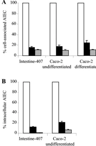
Inhibitory effect of L. casei DN 114 001 on AIEC adhesion
and invasion in coincubation experiments.
Adhesion and
in-vasion levels of AIEC strains with respect to intestinal
epithe-FIG. 1. Adhesion of L. casei DN-114 001 to intestinal epithelial
cells according to multiplicity of infection (A) or to time of incubation
(B). Adhesion of L. casei DN-114 001 was tested with undifferentiated
Intestine-407 or Caco-2 cells cultured for 2 days and with differentiated
Caco-2 cells cultured for 15 days. (A) Cultured cells were incubated at
an MOI of 10 (black bars), 100 (white bars), and 500 (grey bars) for
6 h. (B) Cultured cells were incubated at an MOI of 500 for 1 h (black
bars), 3 h (white bars), and 6 h (grey bars). Adhesion levels are
expressed as the number of CFU per well. Data are given as means
⫾
the standard error of the mean (SEM) of at least three separate
experiments.
FIG. 2. Increased adhesion of L. casei DN-114 001 in the presence
of its spent culture supernatant. Cultured cells were incubated with L.
casei DN-114 001 at an MOI of 500 for 6 h in cell culture medium
alone (black bars), or supplemented with 10% (vol/vol) of its spent
culture supernatant (white bars) and with 10% of neutralized
super-natant (grey bars). Adhesion levels were determined as described in
the legend to Fig. 1.
lial cells were determined by coincubation experiments where
AIEC and L. casei DN-114 001 were added together with
intestinal epithelial cells. In coincubation with L. casei DN-114
001, very marked decreases in AIEC LF82 adhesion levels
were observed (Fig. 4A) with differentiated and
undifferenti-ated cells. The inhibitory effect on LF82 adhesion was highly
significant (P
⬍ 0.01) and percentage of inhibition was 82%
and 84% with undifferentiated Caco-2 and Intestine-407 cells,
respectively. It was 75% with differentiated Caco-2 cells. The
inhibitory effect was even more pronounced when
coincuba-tions of AIEC LF82 and L. casei DN-114 001 were performed
in the presence of 10% of L. casei DN-114 001 spent culture
supernatant. Under these conditions, shown above to increase
the ability of L. casei DN-114 001 to adhere to intestinal
epi-thelial cells, the percentage of inhibition of LF82 adhesion was
89, 90, and 89% with Intestine-407, undifferentiated Caco-2,
and differentiated Caco-2 cells, respectively. When
coincuba-tion of AIEC LF82 and L. casei DN-114 001 was performed in
the presence of 10% of neutralized L. casei spent culture
supernatant, similar inhibitory effects on AIEC LF82 adhesion
to Intestine-407 cells were observed (Table 1).
The inhibitory effect of L. casei DN-114 001 on LF82
inva-sion paralleled inhibition of adheinva-sion (Fig. 4B). In
coincuba-tion with L. casei DN-114 001, the number of intracellular
LF82 bacteria significantly (P
⬍ 0.001) decreased. The
per-centage of inhibition of LF82 invasion was similar to that
observed with LF82 adhesion, 88 and 79% with Intestine-407
and Caco-2 cells, respectively. Similar to results obtained on
LF82 adhesion, the addition of 10% of L. casei DN-114 001
spent culture supernatant in the incubation medium induced a
more pronounced inhibitory effect on LF82 invasion, 99 and
93% with Intestine-407 and Caco-2 cells, respectively. When
the L. casei culture supernatant was neutralized, similar
inhib-FIG. 3. Inhibitory effects of L. casei DN-114 001 on the abilities of
AIEC LF82 to adhere to and to invade intestinal epithelial cells in
pre-incubation experiments. Adhesion (A) and invasion (B) of AIEC LF82
with intestinal epithelial cells preincubated with L. casei DN-114 001
alone (black bars) or supplemented with 10% (vol/vol) of its spent culture
supernatant (grey bars), compared with adhesion and invasion levels of
AIEC LF82 to untreated epithelial cells (white bars), taken as 100%.
Preincubation of cultured cells was performed for 6 h with L. casei
DN-114 001 at an MOI of 500. Infection with AIEC LF82 was performed for
3 h with an MOI of 100. Invasion was determined after gentamicin
treat-ment for an additional hour. Results are expressed as cell-associated
bacteria (adherent plus intracellular bacteria) or intracellular bacteria
relative to those obtained for strain LF82 with untreated cells. Each
value is the mean
⫾ SEM of three to four separate experiments.
FIG. 4. Adhesion (A) and invasion (B) abilities of AIEC LF82 with
respect to intestinal epithelial cells in coincubation experiments with L.
casei DN-114 001 alone (black bars) or supplemented with 10% (vol/
vol) of L. casei DN-114 001 spent culture supernatant (grey bars),
compared with monoinfection experiments with LF82 alone (white
bars). A multiplicity of infection of 500 was used for L. casei DN-114
001 and an MOI of 10 was used for AIEC LF82. Cell-associated
bacteria were quantified after a 6-h incubation period. Invasion was
determined after gentamicin treatment for an additional hour. Results
are expressed as the percentage of cell-associated bacteria (adherent
plus intracellular bacteria) or intracellular bacteria relative to those
obtained in monoinfection with strain LF82, taken as 100%. Each
value is the mean
⫾ SEM of three to five separate experiments.
itory effects on AIEC LF82 invasion of Intestine-407 cells were
observed (Table 1).
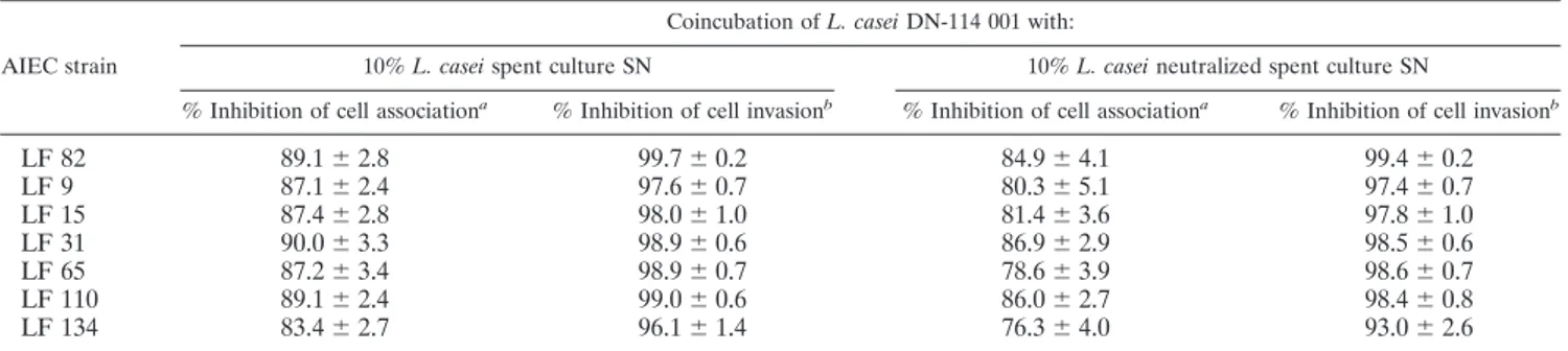
Inhibitory effects of L. casei DN-114 001 on bacterial
adhe-sion and invaadhe-sion of other AIEC strains were investigated with
Intestine-407 cultured cells (Table 1). Strong inhibitory effects
on AIEC adhesion and invasion were observed for all the
AIEC strains tested. Adhesion inhibition levels ranged from 83
to 90%, and invasion inhibition levels ranged from 96 to 99%.
These inhibitory effects continued to be observed when the pH
of L. casei DN-114 001 spent culture supernatant was adjusted
to a neutral value. Slight decreases were observed, but
adhe-sion inhibition levels still ranged from 76 to 86%, and invaadhe-sion
inhibition levels ranged from 93 to 98%.
The inhibitory effect of L. casei DN 114 001 on AIEC LF82
adhesion and invasion is not related to cell cytotoxicity or to
antibacterial activity.
Since the decreased ability of AIEC
LF82 to adhere to and invade intestinal epithelial cells may be
related to cultured cell death induced by L. casei and/or its
spent culture supernatant, we determined the amounts of the
cytoplasmic enzyme LDH released when the integrity of the
cytoplasmic membrane of eukaryotic cells was breached (Table
2). As a positive control inducing LDH release, we used E. coli
strain LF32 producing an
␣-hemolysin (17). Infection of the
different cell models with E. coli LF32 for 6 h induced marked
release of LDH (19.3 to 22.7%). In contrast, LDH release by
intestinal epithelial cells incubated for 6 h with an MOI of 500
of L. casei DN-114 001 alone or in the presence of 10% of its
spent culture supernatant was very low (2.0 to 5.7), and values
were similar to those observed when incubations were
per-formed with the nonpathogenic strain E. coli K-12 C600. This
indicates that L. casei DN-114 001 alone or in association with
its spent culture supernatant did not induce any cell
cytotox-icity even at an MOI of 500 and after 6 h of incubation with
intestinal epithelial cells.
Since inhibition of LF82 adhesion and invasion may be
re-lated to bactericidal effects or inhibition of bacterial growth
induced by L. casei DN-114 001, LF82 bacterial growth was
analyzed by incubation with DMEM with L. casei DN-114 001
alone and in the presence of its spent culture supernatant
(Table 3). Coincubation for 3 h with L. casei DN-114 001 alone
or with 10% of its spent culture supernatant did not induce any
significant (P
⬎ 0.05) decrease in LF82 bacterial growth. After
6 h of coincubation with L. casei DN-114 001, a 26% decrease
in LF82 replication was observed. This decrease reached 38%
TABLE 1. Inhibition of adhesion and invasion of various AIEC strains by probiotic L. casei DN-114 001
cAIEC strain
Coincubation of L. casei DN-114 001 with:
10% L. casei spent culture SN 10% L. casei neutralized spent culture SN
% Inhibition of cell associationa
% Inhibition of cell invasionb
% Inhibition of cell associationa
% Inhibition of cell invasionb
LF 82
89.1
⫾ 2.8
99.7
⫾ 0.2
84.9
⫾ 4.1
99.4
⫾ 0.2
LF 9
87.1
⫾ 2.4
97.6
⫾ 0.7
80.3
⫾ 5.1
97.4
⫾ 0.7
LF 15
87.4
⫾ 2.8
98.0
⫾ 1.0
81.4
⫾ 3.6
97.8
⫾ 1.0
LF 31
90.0
⫾ 3.3
98.9
⫾ 0.6
86.9
⫾ 2.9
98.5
⫾ 0.6
LF 65
87.2
⫾ 3.4
98.9
⫾ 0.7
78.6
⫾ 3.9
98.6
⫾ 0.7
LF 110
89.1
⫾ 2.4
99.0
⫾ 0.6
86.0
⫾ 2.7
98.4
⫾ 0.8
LF 134
83.4
⫾ 2.7
96.1
⫾ 1.4
76.3
⫾ 4.0
93.0
⫾ 2.6
aInhibition (%)⫽ [(cell-associated bacteria alone ⫺ cell-associated bacteria in coincubation experiments with L. casei DN-114 001)/(cell-associated bacteria alone)]
⫻ 100.
bInhibition (%)⫽ [(intracellular bacteria alone ⫺ intracellular bacteria in coincubation experiments with L. casei DN-114 001)/(intracellular bacteria alone)] ⫻ 100. cCoincubation experiments were performed with Intestine-407 cells for 6 h at a multiplicity of infection of 500 for the probiotic strain L. casei DN-114 001 and of
10 for AIEC strains. Results are expressed as a percent inhibition of cell-associated bacteria or intracellular bacteria relative to those obtained in monoinfections with AIEC strains (taken as 100%). Each value is the mean⫾ SEM of three separate experiments.
TABLE 2. Absence of LDH release by intestinal epithelial cells
incubated with L. casei DN-114 001 alone and in the presence of its
spent culture SN
Strain % LDH releasea Intestine-407 Caco-2 Undifferentiated DifferentiatedE. coli LF 32
22.7
⫾ 0.2
19.3
⫾ 0.7
20.2
⫾ 2.0
E. coli K12-C600
2.6
⫾ 0.9
5.0
⫾ 0.4
6.2
⫾ 0.2
L. casei DN-114 001
2.0
⫾ 0.6
5.7
⫾ 0.7
2.3
⫾ 0.1
L. casei DN-114 001
⫹
10% SN
3.1
⫾ 0.9
5.2
⫾ 0.4
4.2
⫾ 0.9
aLDH release was determined in supernatants using NADH as the substrate.
It was measured after 6 h of incubation with a multiplicity of infection of 500 for
L. casei DN-114 001 and of 100 for E. coli strains. Results are expressed as
percent cytotoxycity, calculated as (experimental release⫺ spontaneous LDH release)/(total LDH release⫺ spontaneous release) ⫻ 100. E. coli LF32 pro-ducing an␣-hemolysin was used as a positive control (17). E. coli strain K-12 C600 was used as a negative control.
TABLE 3. Activity of L. casei DN-114 001 alone or in the presence
of 10% of its spent culture supernatant on AIEC LF82
bacterial growth
aBacterial growth in CFU (107)/ml (% of growth inhibition/DMEM)
Growth medium Time of incubation
3 h 6 h
DMEM
2.9
⫾ 0.2
43.5
⫾ 5.2
DMEM
⫹ L. casei DN-114 001
3.0
⫾ 0.4 (0)
32.2
⫾ 3.3 (26)
DMEM
⫹ L. casei DN-114 001 ⫹
10% SN
2.7
⫾ 0.4 (5)
26.7
⫾ 2.1 (38)
DMEM
⫹ L. casei DN-114 001 ⫹
10% SN (pH
⫽ 7)
3.0
⫾ 0.3 (0)
31.8
⫾ 2.1 (27)
aControl bacterial growth of LF82 was determined in cell culture medium
(DMEM). Bacterial growth of LF82 was also determined in coincubation with L.
casei DN-114 001 alone or supplemented with 10% of L. casei DN-114 001 spent
culture supernatant (10% SN) or of neutralized supernatant (10% SN, pH 7.0). Results are expressed as (CFU) after 3 h or 6 h incubation. Each value is the mean⫾ SEM of five separate experiments.
when coincubation of AIEC LF82 was performed with L. casei
DN-114 001 in the presence of 10% of the spent supernatant.
When the pH of the L. casei DN-114 001 supernatant was
adjusted to a neutral value (pH 7.0), the decrease in LF82
bacterial growth was similar to that observed with L. casei
alone. Compared to the inhibitory effects of L. casei on AIEC
LF82 adhesion and invasion (89 to 98% of inhibition), the
influence of L. casei on LF82 bacterial growth could not fully
explain the resulting inhibitory effects of L. casei DN-114 001
on the abilities of strain LF82 to adhere to and invade cultured
intestinal epithelial cells.
DISCUSSION
The onset of inflammation in IBD may be associated with an
imbalance in the intestinal microflora, with a relative
predom-inance of aggressive bacteria and an insufficient amount of
protective species. Since early and chronic ileal lesions of
pa-tients with Crohn’s disease are abnormally colonized by AIEC
strains (16), the eradication of these pathogenic bacteria and
their replacement by probiotic bacteria may provide a new
option for the treatment of CD and for the maintenance of
remission.
To provide clinical benefits, adhesion of probiotic bacteria to
the intestinal mucosa is an interesting trait for antagonistic
activity against pathogens. We showed in the present study that
the probiotic strain L. casei DN-114 001 exhibited a dose- and
incubation time-dependent ability to adhere to
undifferenti-ated Intestine-407 and Caco-2 cells and to differentiundifferenti-ated
Caco-2 cells. This result was expected, since several studies
showed that various Lactobacillus strains adhere to cultured
intestinal epithelial cells (18, 20, 22, 29, 44, 50). Their ability to
adhere is independent of bacterial species, but is strain specific
(10, 49), and is increased in the presence of spent culture
supernatant (3, 10, 12, 44, 50). A markedly increased ability of
L. casei DN-114 001 to adhere to intestinal epithelial cells was
also observed in the presence of 10% of its spent culture
supernatant. This indicates that a secreted factor enhancing
bacterial adhesion or the low pH due to acid lactic production
may interfere with the adhesion of Lactobacilli to intestinal
epithelial cells (3, 10, 12, 15, 22, 25). However, this study shows
that low pH is not required for adhesion of L. casei DN-114
001 strain to adhere. Besides, Greene and Klaenhammer (22)
previously reported that lowering the pH of fresh MRS cannot
account for the increased adherence of Lactobacilli to Caco-2
cells following the addition of the spent culture supernatant.
An important function of probiotic bacteria is to provide
protection of the host gastrointestinal tract from invading
pathogens. In this study, we showed that preincubation of
intestinal epithelial cells with L. casei DN-114 001 prior to
infection with pathogenic AIEC strain LF82 resulted in a
strong decrease in LF82 adhesion (43 to 62%) and invasion (56
to 90%). In coincubation experiments, where L. casei DN-114
001 and AIEC strain LF82 were added together with the
in-testinal epithelial cells, higher inhibitory effects on AIEC
ad-hesion (75 to 84%) and invasion (79 to 88%) were observed.
The inhibitory effects of L. casei DN-114 001 on both LF82
adhesion and invasion are not surprising, since this result is
consistent with previous observations showing that the
adhe-sion step is crucial for AIEC to invade epithelial cells (7).
Several reports have confirmed the ability of probiotic
Lacto-bacilli to inhibit the ability of pathogenic bacteria to adhere to
and to invade intestinal epithelial cells (4, 10, 20, 26, 27). The
inhibition levels observed with L. casei DN-114 001 are similar
to or even higher than those reported for Lactobacilli on the
adhesion of various pathogens (4, 11, 18, 20, 49). We noticed
that L. casei DN-114 001 exerts stronger inhibitory effects on
AIEC invasion than those reported with various Lactobacilli on
invasive enteropathogens (11, 13, 20). Concerning its
inhibi-tory role on AIEC adhesion and invasion, L. casei DN-114 001
is as efficient as E. coli Nissle (8).
The inhibitory activity of L. casei DN-114 001 on LF82
adhesion and invasion was enhanced when 10% of spent L.
casei DN-114 001 culture supernatant was added to
coincuba-tion and preincubacoincuba-tion media. This finding continued to be
observed when the acidic pH of the supernatant was
neutral-ized. The increased inhibitory effects may be related to the
increased adhesion of L. casei DN-114 001 that we observed in
the presence of its spent culture supernatant. Such increased
adhesion could induce greater competition between probiotic
and pathogens for attachment sites by specific blockage or
steric hindrance. This could also be due to the production of
antimicrobial substances active against AIEC. Several studies
have reported the ability of Lactobacilli to secrete
antimicro-bial compounds such as bacteriocins (1, 2, 31, 36, 52, 53).
Lactic acid produced by all Lactobacillus strains was also
shown to inhibit bacterial growth (32–34). Under the
experi-mental conditions of this study, even though the secretion by L.
casei DN-114 001 of an antibacterial substance active on AIEC
LF82 bacterial growth was observed, this substance alone
could not explain the high inhibitory effects of L. casei DN-114
001 on the abilities of strain LF82 to adhere to and to invade
cultured intestinal epithelial cells. Thus, the high inhibitory
effects of L. casei on AIEC adhesion to and invasion of
intes-tinal epithelial cells would result from several cumulative
fac-tors such as the adhesion of L. casei to the intestinal epithelial
cells and the secretion by L. casei of compounds, which induce
a high increase in L. casei adhesion and a moderate decrease
in AIEC bacterial growth.
In conclusion, probiotic strain L. casei DN-114 001 exerts
strong inhibitory effects on both AIEC adhesion to and
inva-sion of intestinal epithelial cells. The present in vitro study
indicates that this probiotic may be efficient for preventive and
curative probiotic therapy, since inhibitory effects were
ob-served when intestinal cells were preincubated with L. casei
DN-114 001 and when the probiotic was used in coincubation
experiments. Thus, the use of this probiotic could be of great
interest, especially in maintaining remission in a subset of CD
patients harboring pathogenic AIEC colonizing early and
chronic ileal lesions.
REFERENCES
1. Allison, G. E., C. Fremaux, and T. R. Klaenhammer. 1994. Expansion of bacteriocin activity and host range upon complementation of two peptides encoded within the lactacin F operon. J. Bacteriol. 176:2235–2241. 2. Axelsson, L., A. Holck, S. E. Birkeland, T. Aukrust, and H. Blom. 1993.
Cloning and nucleotide sequence of a gene from Lactobacillus sake Lb706 necessary for sakacin A production and immunity. Appl. Environ. Microbiol.
59:2868–2875.
3. Bernet, M. F., D. Brassart, J. R. Neeser, and A. L. Servin. 1994. Lactobacillus acidophilus LA 1 binds to cultured human intestinal cell lines and inhibits cell attachment and cell invasion by enterovirulent bacteria. Gut 35:483–489. 4. Bernet-Camard, M. F., V. Lievin, D. Brassart, J. R. Neeser, A. L. Servin, and
S. Hudault.1997. The human Lactobacillus acidophilus strain LA1 secretes a nonbacteriocin antibacterial substance(s) active in vitro and in vivo. Appl. Environ. Microbiol. 63:2747–2753.
5. Borruel, N., M. Carol, F. Casellas, M. Antolin, F. de Lara, E. Espin, J. Naval,
F. Guarner, and J. R. Malagelada.2002. Increased mucosal tumour necrosis factor alpha production in Crohn’s disease can be downregulated ex vivo by probiotic bacteria. Gut 51:659–664.
6. Borruel, N., F. Casellas, M. Antolin, M. Llopis, M. Carol, E. Espiin, J. Naval,
F. Guarner, and J. R. Malagelada.2003. Effects of nonpathogenic bacteria on cytokine secretion by human intestinal mucosa. Am. J. Gastroenterol.
98:865–870.
7. Boudeau, J., N. Barnich, and A. Darfeuille-Michaud. 2001. Type 1 pili-mediated adherence of Escherichia coli strain LF82 isolated from Crohn’s disease is involved in bacterial invasion of intestinal epithelial cells. Mol. Microbiol. 39:1272–1284.
8. Boudeau, J., A. L. Glasser, S. Julien, J. F. Colombel, and A.
Darfeuille-Michaud.2003. Inhibitory effect of probiotic Escherichia coli strain Nissle 1917 on adhesion to and invasion of intestinal epithelial cells by adherent-invasive E. coli strains isolated from patients with Crohn’s disease. Aliment. Pharmacol. Ther. 18:45–56.
9. Boudeau, J., A. L. Glasser, E. Masseret, B. Joly, and A. Darfeuille-Michaud. 1999. Invasive ability of an Escherichia coli strain isolated from the ileal mucosa of a patient with Crohn’s disease. Infect. Immun. 67:4499–4509. 10. Chauviere, G., M. H. Coconnier, S. Kerneis, A. Darfeuille-Michaud, B. Joly,
and A. L. Servin.1992. Competitive exclusion of diarrheagenic Escherichia
coli (ETEC) from human enterocyte-like Caco-2 cells by heat-killed Lacto-bacillus. FEMS Microbiol. Lett. 70:213–217.
11. Coconnier, M. H., M. F. Bernet, S. Kerneis, G. Chauviere, J. Fourniat, and
A. L. Servin.1993. Inhibition of adhesion of enteroinvasive pathogens to human intestinal Caco-2 cells by Lactobacillus acidophilus strain LB de-creases bacterial invasion. FEMS Microbiol. Lett. 110:299–305.
12. Coconnier, M. H., T. R. Klaenhammer, S. Kerneis, M. F. Bernet, and A. L.
Servin. 1992. Protein-mediated adhesion of Lactobacillus acidophilus BG2FO4 on human enterocyte and mucus-secreting cell lines in culture. Appl. Environ. Microbiol. 58:2034–2039.
13. Coconnier, M. H., V. Lievin, M. Lorrot, and A. L. Servin. 2000. Antagonistic activity of Lactobacillus acidophilus LB against intracellular Salmonella
en-terica serovar Typhimurium infecting human enterocyte-like Caco-2/TC-7
cells. Appl. Environ. Microbiol. 66:1152–1157.
14. Colombel, J. F., M. Lemann, M. Cassagnou, Y. Bouhnik, B. Duclos, J. L.
Dupas, B. Notteghem, J. Y. Mary, et al.1999. A controlled trial comparing ciprofloxacin with mesalazine for the treatment of active Crohn’s disease. Am. J. Gastroenterol. 94:674–678.
15. Conway, P. L., and S. Kjelleberg. 1989. Protein-mediated adhesion of Lac-tobacillus fermentum strain 737 to mouse stomach squamous epithelium. J. Gen. Microbiol. 135:1175–1186.
16. Darfeuille-Michaud, A., J. Boudeau, P. Bulois, C. Neut, A. L. Glasser, N.
Barnich, M. A. Bringer, A. Swidsinski, L. Beaugerie, and J. F. Colombel.
2004. High prevalence of adherent-invasive Escherichia coli associated with ileal mucosa in Crohn’s disease. Gastroenterology 127:412–421.
17. Darfeuille-Michaud, A., C. Neut, N. Barnich, E. Lederman, P. Di Martino,
P. Desreumaux, L. Gambiez, B. Joly, A. Cortot, and J. F. Colombel.1998. Presence of adherent Escherichia coli strains in ileal mucosa of patients with Crohn’s disease. Gastroenterology 115:1405–1413.
18. Fernandez, M. F., S. Boris, and C. Barbes. 2003. Probiotic properties of human lactobacilli strains to be used in the gastrointestinal tract. J. Appl. Microbiol. 94:449–455.
19. Glasser, A. L., J. Boudeau, N. Barnich, M. H. Perruchot, J. F. Colombel, and
A. Darfeuille-Michaud.2001. Adherent invasive Escherichia coli strains from patients with Crohn’s disease survive and replicate within macrophages with-out inducing host cell death. Infect. Immun. 69:5529–5537.
20. Gopal, P. K., J. Prasad, J. Smart, and H. S. Gill. 2001. In vitro adherence properties of Lactobacillus rhamnosus DR20 and Bifidobacterium lactis DR10 strains and their antagonistic activity against an enterotoxigenic
Esch-erichia coli. Int. J. Food Microbiol. 67:207–216.
21. Greenbloom, S. L., A. H. Steinhart, and G. R. Greenberg. 1998. Combination ciprofloxacin and metronidazole for active Crohn’s disease. Can. J. Gastro-enterol. 12:53–56.
22. Greene, J. D., and T. R. Klaenhammer. 1994. Factors involved in adherence of lactobacilli to human Caco-2 cells. Appl. Environ. Microbiol. 60:4487– 4494.
23. Guerin-Danan, C., C. Chabanet, C. Pedone, F. Popot, P. Vaissade, C. Bouley,
O. Szylit, and C. Andrieux.1998. Milk fermented with yogurt cultures and
Lactobacillus casei compared with yogurt and gelled milk: influence on
in-testinal microflora in healthy infants. Am. J. Clin. Nutr. 67:111–117. 24. Guslandi, M., G. Mezzi, M. Sorghi, and P. A. Testoni. 2000. Saccharomyces
boulardii in maintenance treatment of Crohn’s disease. Dig. Dis. Sci. 45: 1462–1464.
25. Henriksson, A., R. Szewzyk, and P. L. Conway. 1991. Characteristics of the adhesive determinants of Lactobacillus fermentum 104. Appl. Environ. Mi-crobiol. 57:499–502.
26. Hirano, J., T. Yoshida, T. Sugiyama, N. Koide, I. Mori, and T. Yokochi. 2003.
The effect of Lactobacillus rhamnosus on enterohemorrhagic Escherichia coli infection of human intestinal cells in vitro. Microbiol. Immunol. 47:405–409. 27. Hudault, S., V. Lievin, M. F. Bernet-Camard, and A. L. Servin. 1997. An-tagonistic activity exerted in vitro and in vivo by Lactobacillus casei (strain GG) against Salmonella typhimurium C5 infection. Appl. Environ. Microbiol.
63:513–518.
28. Isolauri, E., P. V. Kirjavainen, and S. Salminen. 2002. Probiotics: a role in the treatment of intestinal infection and inflammation? Gut 50(Suppl. 3): III54–III59.
29. Jacobsen, C. N., V. Rosenfeldt Nielsen, A. E. Hayford, P. L. Moller, K. F.
Michaelsen, A. Paerregaard, B. Sandstrom, M. Tvede, and M. Jakobsen.
1999. Screening of probiotic activities of forty-seven strains of Lactobacillus spp. by in vitro techniques and evaluation of the colonization ability of five selected strains in humans. Appl. Environ. Microbiol. 65:4949–4956. 30. Kerneis, S., S. S. Bilge, V. Fourel, G. Chauviere, M. H. Coconnier, and A. L.
Servin.1991. Use of purified F1845 fimbrial adhesin to study localization and expression of receptors for diffusely adhering Escherichia coli during entero-cytic differentiation of human colon carcinoma cell lines HT-29 and Caco-2 in culture. Infect. Immun. 59:4013–4018.
31. Larsen, A. G., F. K. Vogensen, and J. Josephsen. 1993. Antimicrobial activity of lactic acid bacteria isolated from sour doughs: purification and character-ization of bavaricin A, a bacteriocin produced by Lactobacillus bavaricus MI401. J. Appl. Bacteriol. 75:113–122.
32. Lehto, E. M., and S. J. Salminen. 1997. Inhibition of Salmonella typhimurium adhesion to Caco-2 cell cultures by Lactobacillus strain GG spent culture supernate: only a pH effect? FEMS Immunol. Med. Microbiol. 18:125–132. 33. Midolo, P. D., J. R. Lambert, R. Hull, F. Luo, and M. L. Grayson. 1995. In vitro inhibition of Helicobacter pylori NCTC 11637 by organic acids and lactic acid bacteria. J. Appl. Bacteriol. 79:475–479.
34. Ogawa, M., K. Shimizu, K. Nomoto, R. Tanaka, T. Hamabata, S. Yamasaki,
T. Takeda, and Y. Takeda.2001. Inhibition of in vitro growth of Shiga toxin-producing Escherichia coli O157:H7 by probiotic Lactobacillus strains due to production of lactic acid. Int. J. Food Microbiol. 68:135–140. 35. Oozeer, R., N. Goupil-Feuillerat, C. A. Alpert, M. van de Guchte, J. Anba, J.
Mengaud, and G. Corthier.2002. Lactobacillus casei is able to survive and initiate protein synthesis during its transit in the digestive tract of human flora-associated mice. Appl. Environ. Microbiol. 68:3570–3574.
36. Parente, E., and A. Ricciardi. 1999. Production, recovery and purification of bacteriocins from lactic acid bacteria. Appl. Microbiol. Biotechnol. 52:628– 638.
37. Pedone, C. A., C. C. Arnaud, E. R. Postaire, C. F. Bouley, and P. Reinert. 2000. Multicentric study of the effect of milk fermented by Lactobacillus casei on the incidence of diarrhoea. Int. J. Clin. Pract. 54:568–571.
38. Pedone, C. A., A. O. Bernabeu, E. R. Postaire, C. F. Bouley, and P. Reinert. 1999. The effect of supplementation with milk fermented by Lactobacillus
casei (strain DN-114 001) on acute diarrhoea in children attending day care
centres. Int. J. Clin. Pract. 53:179–184.
39. Perdigon, G., E. Vintini, S. Alvarez, M. Medina, and M. Medici. 1999. Study of the possible mechanisms involved in the mucosal immune system activa-tion by lactic acid bacteria. J. Dairy Sci. 82:1108–1114.
40. Podolsky, D. K. 2002. Inflammatory bowel disease. N. Engl. J. Med. 347: 417–429.
41. Prantera, C., F. Zannoni, M. L. Scribano, E. Berto, A. Andreoli, A. Kohn,
and C. Luzi.1996. An antibiotic regimen for the treatment of active Crohn’s disease: a randomized, controlled clinical trial of metronidazole plus cipro-floxacin. Am. J. Gastroenterol. 91:328–332.
42. Rembacken, B. J., A. M. Snelling, P. M. Hawkey, D. M. Chalmers, and A. T.
Axon.1999. Non-pathogenic Escherichia coli versus mesalazine for the treat-ment of ulcerative colitis: a randomised trial. Lancet 354:635–639. 43. Rutgeerts, P., M. Hiele, K. Geboes, M. Peeters, F. Penninckx, R. Aerts, and
R. Kerremans.1995. Controlled trial of metronidazole treatment for pre-vention of Crohn’s recurrence after ileal resection. Gastroenterology 108: 1617–1621.
44. Sarem, F., L. O. Sarem-Damerdji, and J. P. Nicolas. 1996. Comparison of the adherence of three Lactobacillus strains to Caco-2 and Int-407 human intestinal cell lines. Lett. Appl. Microbiol. 22:439–442.
45. Shanahan, F. 2000. Probiotics and inflammatory bowel disease: is there a scientific rationale? Inflamm. Bowel Dis. 6:107–115.
46. Shanahan, F. 2001. Probiotics in inflamatory bowel disease. Gut 48:609. 47. Sullivan, A., and C. E. Nord. 2002. Probiotics in human infections. J.
Anti-microb Chemother. 50:625–627.
48. Sutherland, L., J. Singleton, J. Sessions, S. Hanauer, E. Krawitt, G. Rankin,
R. Summers, H. Mekhjian, N. Greenberger, M. Kelly, et al.1991. Double blind, placebo controlled trial of metronidazole in Crohn’s disease. Gut
32:1071–1075.
49. Todoriki, K., T. Mukai, S. Sato, and T. Toba. 2001. Inhibition of adhesion of food-borne pathogens to Caco-2 cells by Lactobacillus strains. J. Appl. Mi-crobiol. 91:154–159.
Lactobacillus strains to Caco-2 cell cultures. Int. J. Food Microbiol. 41:45–51. 51. Venturi, A., P. Gionchetti, F. Rizzello, R. Johansson, E. Zucconi, P. Brigidi,
D. Matteuzzi, and M. Campieri.1999. Impact on the composition of the faecal flora by a new probiotic preparation: preliminary data on maintenance treatment of patients with ulcerative colitis. Aliment. Pharmacol. Ther. 13: 1103–1108.
52. Yamato, M., K. Ozaki, and F. Ota. 2003. Partial purification and character-ization of the bacteriocin produced by Lactobacillus acidophilus YIT 0154. Microbiol. Res. 158:169–172.
53. Zhu, W. M., W. Liu, and D. Q. Wu. 2000. Isolation and characterization of a new bacteriocin from Lactobacillus gasseri KT7. J. Appl. Microbiol. 88: 877–886.